Article

Comparative Evaluation of Computational Methods for Validating Housekeeping Gene RT-qPCR Data in 3T3-L1 Cells

1
Department of Pharmacology, Animal Physiology, Biochemistry and Chemistry, Faculty of Veterinary Medicine, Trakia University, 6000 Stara Zagora, Bulgaria
2
Department of Biochemistry, Microbiology and Physics, Faculty of Agriculture, Trakia University, 6000 Stara Zagora, Bulgaria
*
Author to whom correspondence should be addressed.
Biomedicines 2025, 13(8), 2036; https://doi.org/10.3390/biomedicines13082036
Submission received: 30 July 2025 / Revised: 17 August 2025 / Accepted: 19 August 2025 / Published: 21 August 2025
(This article belongs to the Section Molecular Genetics and Genetic Diseases)

Abstract

Background: Postbiotics with anti-adipogenic properties can significantly modify adipocyte metabolism by influencing key cellular pathways involved in lipid accumulation. In preliminary in vitro studies, it is essential to monitor various cellular and subcellular variables, including gene expression and protein synthesis potential, through RT-qPCR analysis. It is also crucial to select internal controls carefully and evaluate their stability for effective normalization and accurate interpretation of the results. Methods: In this study, we assessed the stability of six commonly used housekeeping genes: GAPDH, Actb, HPRT, HMBS, 18S, and 36B4. We analyzed their variability in mature 3T3-L1 adipocytes treated with supernatants from newly isolated Lacticaseibacillus paracasei strains. Our analysis combined classical statistical methods, a ∆Ct analysis, and software algorithms such as geNorm, NormFinder, BestKeeper, and RefFinder. Results: Our stepwise, multiparameter strategy for selecting reference genes led to the exclusion of Actb and 18S as the most variable reference genes. We identified HPRT as the most stable internal control. Additionally, HPRT and HMBS emerged as a stable pair, while the recommended triplet of genes for reliable normalization consists of HPRT, 36B4, and HMBS. Conclusions: The widely used putative genes in similar studies—GAPDH and Actb—did not confirm their presumed stability, which once again emphasizes the need for experimental validation of internal controls to increase the accuracy and reliability of gene expression. Combining a unique biological model—postbiotic-treated adipocytes—with multiple algorithms integrated into a single workflow allows us to provide a methodological template applicable to similar nutritional and metabolic research settings.

1. Introduction

Probiotics play a key role in maintaining overall health. Current studies have focused on the relationship between intestinal microbiota composition and obesity-related metabolic disorders [1,2,3,4]. Members of the genera Lactobacillus and Bifidobacterium additionally can provoke hypolipidemia and hypoglycemia, influencing key physiological processes in white adipose tissue [5,6,7,8]. These effects are mediated by biologically active soluble factors—so-called postbiotics—secreted by live bacteria and absorbed in the intestine. Reaching peripheral tissues, they modulate signaling pathways related to metabolic balance [9,10]. Postbiotics exhibit most of the beneficial effects of probiotics, including their antimicrobial, antioxidant, anti-obesogenic, and immunomodulatory properties, and excluding some of their negative characteristics [9,11,12,13,14]. The existing intestinal flora exercises a minimal influence on them and, once absorbed, they launch a more targeted action in easily controlled doses and remain stable during storage and transportation. Postbiotics are particularly important in people with compromised immunity, highlighting the need for modern nutrigenomic approaches to move towards personalized strategies [15,16,17,18,19,20].
Due to the significant strain specificity in the metabolic profile, there is a real scientific and practical need to identify new strains with substantial health effects. Lacticaseibacillus paracasei (L. paracasei) is a well-known lactic acid bacterium, widely used in the food industry [21,22]. Some strains exhibit antimicrobial and anti-inflammatory and stress-response modulating activity [23,24], with their efficiency being strictly strain-specific. However, insufficiently studied probiotics can lead to adverse effects, such as diarrhea, bacterial translocation, or even bacteremia in risk groups [24,25]. Therefore, it is crucial to identify and characterize new, safe, and functional strains. The autochthonous L. paracasei strains isolated from unique ecological niches have attracted particular scientific interest [22,26]. Their preliminary screening is often based on in vitro molecular analysis of key genes’ regulatory mechanisms and expression patterns. When assessing eventual probiotic and postbiotic modulation of fat metabolism, the 3T3-L1 cell line, derived from mouse preadipocytes, is considered a universal model for monitoring adipocyte differentiation, glucose transport, lipid metabolism, and insulin sensitivity, prior to the shift to in vivo experiments [27,28,29]. These cells have been used for studying anti-adipogenic Lactobacillus-mediated effects in numerous recent investigations [8,30,31,32,33,34].
In these initial studies, molecular technologies, such as the reverse transcription quantitative polymerase chain reaction (RT-qPCR), provide a powerful tool for precise gene expression analysis [35]. Reliabile RT-qPCR results require stable internal standards (reference genes) to correct sample-to-sample RNA and reverse transcription variations [36,37,38]. Postbiotics with antiadipogenic properties, such as L. paracasei metabolites, can alter adipocyte metabolism and affect the expression of widely used housekeeping genes (HKGs) such as glyceraldehyde-3-phosphate dehydrogenase (GAPDH) and actin beta (Actb), which are involved in glycolysis and cytoskeletal structure [39,40].
The Minimum Information for Publication of Quantitative Real-Time PCR Experiments (MIQE) guidelines recommend at least two stable reference genes validated by algorithms such as NormFinder, geNorm, and BestKeeper [41,42,43,44,45]. However, recent publications investigating the impact of L. paracasei on adipocyte metabolism have often relied on a single reference gene without stability assessment or a solid rationale for its selection [10,46,47,48]. This poses a significant risk of acquiring unreliable RT-qPCR results, especially given the complex metabolic changes induced.
Therefore, the current study aimed to evaluate the expression stability of six commonly used HKGs (GAPDH, Actb, hypoxanthine-guanine phosphoribosyltransferase (HPRT), hydroxymethylbilane synthase (HMBS), 18S ribosomal RNA (18S), and ribosomal protein lateral stalk subunit P0 (36B4)) in mature 3T3-L1 adipocytes treated with supernatants from newly isolated L. paracasei strains (M2.1, C8, C15, and P4), applying the widely used mathematical algorithms geNorm, NormFinder, BestKeeper, and RefFinder, as well as classical ∆Ct and statistical data analyses.

2. Materials and Methods

2.1. Propagation and Adipogenic Induction of Preadipocytes

Cryopreserved 3T3-L1 mouse fibroblasts (CRL-3242, ATCC, Washington, DC, USA) were thawed and seeded in T75, following the manufacturer’s instructions for propagation. After two passages to obtain sufficient cell mass, cells were plated at a density of 1 × 10⁴/mL in 12 and 24-well plates for the subsequent experimental procedures. During the proliferation phase, the cells were grown in basal medium (BM) composed of high-glucose Dulbecco’s modified Eagle’s medium (DMEM, 4.5 g/L glucose with L-glutamine), (Sigma-Aldrich Chemie GmbH, St. Louis, MI, USA), 10% (v/v) fetal bovine serum (FBS) (Sigma-Aldrich Chemie GmbH), and 1% antibiotic solution (penicillin G, streptomycin and amphotericin B, Sigma-Aldrich Chemie GmbH). Cultures were maintained in a 95% humidity incubator at 37 °C and 5% CO2. Upon reaching 100% confluency, cells were growth-arrested for 24 h and treated for 48 h with adipogenic induction media (AIM) to induce adipogenesis. AIM was composed of DMEM, 4.5 g/L glucose, 10 µg/mL insulin (cell application, San Diego, CA, USA), 0.05 mM indomethacin (Sigma-Aldrich Chemie GmbH (Merck KGaA, Darmstadt, Germany), 0.1 mM 3-isobutyl-1-methylxanthine (Cayman Chemical, Ann Arbor, ML, USA), and 1 µM dexamethasone (Sigma-Aldrich, St. Louis, MO, USA). For seven days following induction, adipocytes were cultured in maintenance media (AMM) containing BM supplemented with 10 µg/mL insulin until they reached full maturity according to the differentiation protocol.

2.2. L. paracasei Cell-Free Supernatants Preparation and Application on Mature Adipocytes

The isolation of the autochthonous microorganisms L. paracasei strains M2.1, C8, C15, and P4, their identification, subsequent proliferation, and preparation of cell-free supernatants have been described previously [49]. Immediately before application, the pH of all supernatants was neutralized to pH 7 and applied for 48 h to already differentiated 3T3-L1 adipocytes. They were divided into six groups: untreated control (IC), treated with 10% v/v de Man, Rogosa, and Sharpe broth controls (MRS), and treated with 10% v/v supernatants from L. paracasei strains (M2.1, C8, C15, and P4). Each experimental group consisted of six independent culture wells (biological replicates) obtained from the same cell passage and cultured in parallel under identical conditions.

2.3. Intracellular Lipid Deposition Visualization

To confirm successful adipogenesis and visualize potential phenotypic differences between mature adipocytes from the various experimental groups, cells were stained with Oil Red O (Sigma-Aldrich, St. Louis, MO, USA) at the end of the experiment. The supernatant from each well was carefully removed, and the cells were washed three times with phosphate-buffered saline (PBS). The adipocytes were then fixed in 10% (v/v) neutral buffered formalin for 10 min, washed with isopropanol for 5 min, and stained for 30 min with a freshly prepared working solution of Oil Red O. Following staining, the wells were rinsed with PBS, and images were acquired using a Leica DMi1 Inverted Microscope equipped with a 5-megapixel-resolution camera.

2.4. Gene Expression Assays

Total mRNA was isolated from previously lysed adipocyte cells using the RNeasy Mini Lipid Tissue Kit (QIAGEN GmbH, Hilden, Germany), strictly following the manufacturer’s instructions. The purity and integrity of RNA samples were determined spectrophotometrically at 260 and 280 nm wavelengths using a Take3 Microvolume Plate of Synergy™ LX Multi-Mode Microplate Reader (BioTek Instruments, Inc., Santa Clara, CA, USA). The OD ratio in all samples was in the range of 1.9 to 2.1, which confirmed the sound quality of the obtained RNA. Then, cDNA was synthesized using a RevertAid First Strand cDNA Synthesis Kit (Thermo Scientific, Waltham, MA, USA); the RNA concentration was previously equalized across all samples to 1000 ng/µL. The subsequent RT-qPCR analyses were performed with KAPA SYBR® fast qPCR Master Mix kit (QIAGEN Sciences, Inc.; Germantown, MD, USA). A SYBR Green-based two-step real-time PCR method was employed to evaluate gene expression using the PikoReal-Time PCR System (PikoReal, Thermo Scientific, Waltham, MA, USA). The PCR reaction was performed using the temperature program recommended by the manufacturer of the SYBR Green Real-Time PCR Master Mix.

2.5. Primer Design

Table 1 shows the primer sequences for qPCR of the tested HKGs. Most of the primers were designed using open-access data in NCBI software (www.ncbi.nlm.nih.gov, accessed on 20 October 2024) following the manufacturer’s instructions described in SYBR Green Master Mix. Only 18S sequences were previously designed by Arnhold et al. [50]. The specific primer pairs were further confirmed using the additional tools Primer-BLAST (NCBI) https://www.ncbi.nlm.nih.gov/tools/primer-blast (accessed on 21 October 2024), Primer 3 (https://primer3.ut.ee/, accessed on 21 October 2024), Primer 3plus (version: 3.2.5) (https://www.bioinformatics.nl/cgi-bin/primer3plus/primer3plus.cgi, accessed on 21 October 2024), and SerialCloner (version 2.6.1). The web-based software Primer 3 was also used to determine the product length. Then, the melting temperatures and predicted melting curves of the RT-qPCR products were analyzed using uMelt QuartzSM (Version 3.6.2) (https://www.dna-utah.org/umelt/quartz/um.php, accessed on 22 October 2024).
A temperature gradient was performed to determine the optimal annealing temperature for the primers before RT-qPCR analysis. The optimal annealing temperature for each primer pair was found to be very close to the commonly recommended value of 60 °C, with efficient amplification observed within the range of 58 to 61 °C.
The specificity of the amplification products was confirmed by gel electrophoresis and melting curve analysis, and the results can be provided upon request.
Reaction efficiency for each primer pair was calculated using the PicoReal software 2.2 based on standard curves generated from a 6-point 5-fold serial dilution of cDNA. Efficiencies between 90% and 110%, with correlation coefficients (R2) ≥ 0.99, were considered acceptable. The achieved results indicate efficiency values of the tested genes ranging between 98% and 99%.

2.6. Analysis of Expression Stability Among Selected HKGs

Four validation software tools, NormFinder (an Excel add-in, MS Office version 2016) [31], geNorm v3.5 [37], BestKeeper v1 [33], and RefFinder (https://www.ciidirsinaloa.com.mx/RefFinder-master/, accessed on 26 May 2025) [38], were applied to investigate the stability of candidate reference genes.
NormFinder was utilized through its original Excel-based macro. The software is available for download as an Excel add-in (.xla format) from the official NormFinder source. Relative expression quantities were generated using PicoReal Software 2.2 (Thermo Scientific) and imported directly into the algorithm to evaluate gene stability based on intra-group and inter-group variation–stability value (CV). There are no strictly defined maximum acceptable CV values, but a value below 0.15 is often considered indicative of good stability [42].
The relative expression levels of the analyzed HKGs were used to assess their stability using the Excel-based geNorm macro (geNorm v3.5, Windows/VBA, Ghent, Belgium: Center for Medical Genetics, Ghent University, https://genorm.cmgg.be/, accessed 20 May 2025). The algorithm calculated stability values (M) through pairwise comparisons of all genes with a threshold of M < 1 for heterogeneous samples, while M < 0.5 was preferred for homogeneous ones [43,51]. Lower M-values indicated greater stability [52].
The BestKeeper Excel-based tool (Munich, Germany:Technical University of Munich, Institute of Physiology, https://www.gene-quantification.de/bestkeeper.html, accessed on 19 May 2025) was used to analyse raw Ct values of the candidate reference genes and the target gene PPARγ. The software calculated descriptive statistics, including standard deviation (SD), coefficient of variation (CV), arithmetic and geometric means, minimum and maximum Ct values, and fold variation. Pearson correlation coefficients (r) were estimated between each reference gene and the BestKeeper index to assess internal consistency. A regression analysis was also performed to evaluate expression stability further [44]. In addition, a Pearson correlation analysis was performed between each candidate reference gene and the target gene PPARγ to measure potential co-regulation under the experimental conditions. The correlation coefficient (r) and corresponding p-value were extracted and reported from this analysis. The commonly suggested BestKeeper thresholds for reliability of the internal control include a standard deviation (SD) of less than 1, a coefficient of variation (CV) below 2%, and a Pearson correlation coefficient with the BestKeeper index (r) ≥ 0.9 (p < 0.05) [44,53].
RefFinder was used as an online tool (https://www.ciidirsinaloa.com.mx/RefFinder-master/ (accessed on 21 October 2024)) to integrate the results from all algorithms and provide a final ranking of the reference genes based on the geometric mean of their Ct-derived stability ranks [54].
A pairwise ΔCt-based stability analysis was evaluated manually in Microsoft Excel according to the method of Silver et al. [55]. The standard deviation of the ΔCt values across all samples was calculated for each pair of candidate reference genes. Genes with lower average standard deviations were considered more stable.

2.7. Statistical Analysis

Statistical analyses and figures visualization were performed using GraphPad Prism 10.5.0 (774) Software LLC (Boston, MA, USA) and Statistica version 10 (StatSoft Inc., 2011, Tulsa, OK, USA). Descriptive statistics were used to calculate the mean and standard deviation (SD) for ΔCt values, and the standard error of the mean (SEM) for raw Ct values. The results are shown in the figures as means ± SEM. The normality of data distribution was assessed using the Shapiro–Wilk test. For normally distributed data, one-way ANOVA and Student’s t-test were applied to assess intergroup differences in Ct values, while the non-parametric Mann–Whitney test was used for comparative analysis of SD, using the GraphPad Prism tool. A p-value less than 0.05 (p < 0.05) was considered statistically significant.

3. Results

3.1. Adipogenic Induction and LB Treatment

The obtained microscopic images stained with Oil Red O proved the successful adipose differentiation (Figure 1). After 9 days of induction, followed by 48 h of treatment with supernatants of four L. paracasei strains and MRS, the cells displayed distinct morphological and phenotypic features characteristic of differentiated adipocytes, including the accumulation of numerous lipid droplets.

3.2. Expression Stability Assessment via Four Popular Algorithms

3.2.1. NormFinder and geNorm

According to the average M-values evaluated by geNorm software v3.5, presented in Table 2, all of the genes cover the minimal requirements for homogeneous samples except 18S (M = 0.510), which revealed itself as the least stable reference gene. HPRT is outlined as the most reliable HKG for further normalization, confirmed also by NormFinder analysis (Table 2).

3.2.2. BestKeeper and RefFinder

In contrast to the previously discussed algorithms, which rely on relative expression quantities, BestKeeper and RefFinder evaluate gene stability using raw Ct values. According to the descriptive statistical analysis provided by BestKeeper (Table 3), the genes HPRT, HMBS, 36B4, GAPDH, and Actb meet the commonly accepted criteria for reference gene stability, mentioned above. Based on these parameters, the genes were ranked as follows: HPRT was the most stable, followed by HMBS, 36B4, GAPDH, and Actb. The gene 18S slightly exceeded the commonly accepted CV threshold (CV = 2.13%).
Data from the BestKeeper pairwise correlation and regression statistics were further evaluated to assess the candidate reference gene variability (Table 4). Genes with higher Pearson’s correlation coefficient (r) values, closer to 1.0, are regarded as more stably expressed under the experimental conditions [44]. The r values ranged from 0.48 (18S) to 0.80 (36B4), with 36B4 and HPRT showing the highest correlations (r = 0.80 and r = 0.79, respectively).
The equally strong determination coefficients (r2 = 0.63) for both HPRT and 36B4 support their suitability as reliable internal controls. Moreover, 36B4 and HPRT exhibited slope values closest to 1, with values of 1.00 and 0.99, respectively, indicating that their expression levels are proportional to the BestKeeper index. HMBS, 18S, and GAPDH showed moderate deviation, while Actb demonstrated a more significant divergence with a slope of 1.31. The higher slope and standard error for Actb (±0.316) suggest variability in its expression. All p-values were below 0.001, confirming the statistical significance of the observed correlations (Table 4).
Additionally, a pairwise correlation analysis of each HKG vs. a target gene, PPARγ, was performed. A strong correlation indicates that the same biological factors could influence a reference gene as the target gene. Among the genes analyzed, only Actb demonstrated a strong and statistically significant positive correlation with PPARγ, with an r value of 0.759 and a p-value of 0.001 (Table 5).
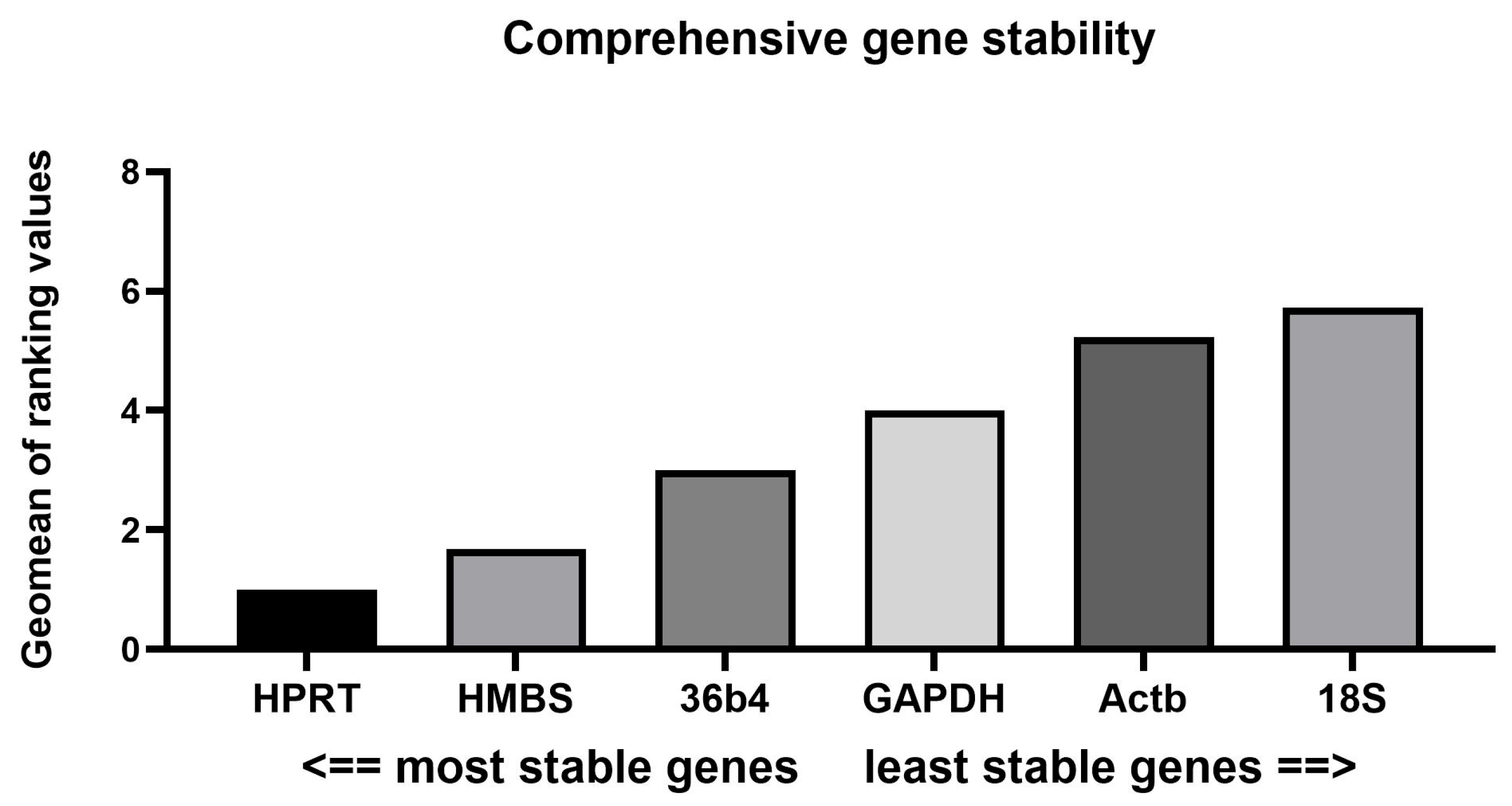
In confirmation of the results obtained so far, the ReFinder tool also highlighted HPRT as the most stable gene (geomean rank = 1), followed by HMBS (1.68) and 36B4 (3). These genes demonstrated consistent performance across the applied evaluation methods and thus could be recommended for reliable normalization (Figure 2).

3.3. Pairwise ΔCt Analysis

The stability of the candidate reference genes—HPRT, 18S, 36B4, GAPDH, HMBS, and Actb—was also evaluated using the pairwise ΔCt method described by Silver et al. [55]. The analysis revealed that several gene combinations involving HPRT, 36B4, and HMBS exhibited the lowest variability, suggesting stable expression across the tested samples. Conversely, 18S and Actb were associated with greater ΔCt variation, indicating their lower reliability as internal controls (Table 6).

3.4. Inter-Group Statistical Analysis of Raw Ct Values for Reference Gene Evaluation

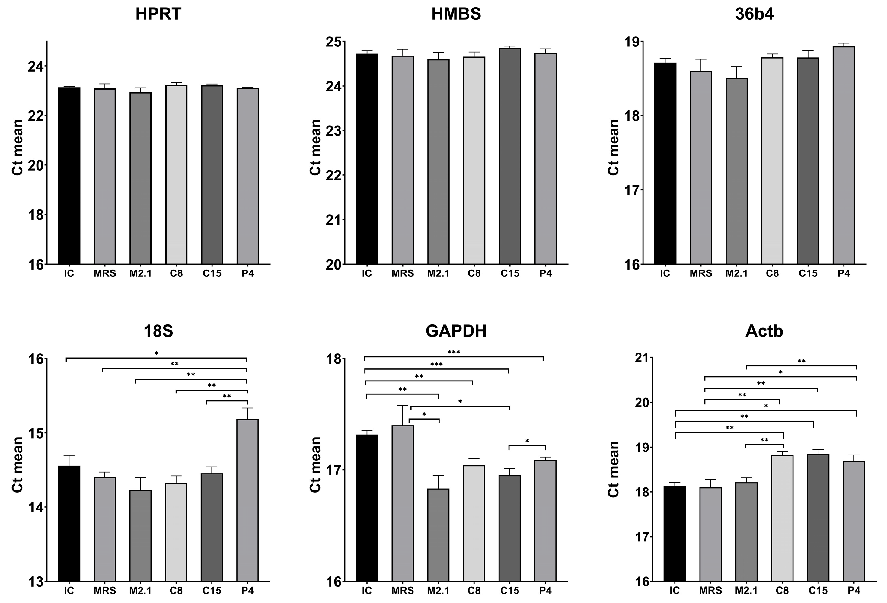
To further support the findings, we compared the mean Ct values of each individual HKG across the experimental groups (Figure 3). A statistical analysis using Tukey’s post hoc test revealed no significant differences and only minimal variation in Ct values for the genes 36B4, HPRT, and HMBS. In contrast, the genes 18S, GAPDH, and particularly Actb showed significantly greater variability between groups. These results confirm the pairwise ΔCt analysis and suggest that 36B4, HPRT, and HMBS are more reliable endogenous controls for normalizing gene expression.

4. Discussion

According to the principles of the three Rs—Replacement, Reduction, and Refinement—established for the ethical use of animals in biomedical and pharmaceutical research, each experiment is expected to minimize the need for animal models [56,57]. Thus, the initial step in studying the effects of a new food supplement, such as the supernatants from potential probiotics, would ideally involve a preliminary in vitro investigation using a suitable cell line aligned with the study’s objectives [58,59].
This approach facilitates the rapid screening of a wide range of molecular markers, allowing researchers to investigate the effects of different doses, administration, and other experimental parameters [60]. The simultaneous monitoring of multiple variables at the cellular and subcellular level, often in a highly dynamic cell system, such as differentiating mature adipocytes, establishing variations in gene expression patterns, and reporting the cell’s potential for protein synthesis. This is achieved through RT-qPCR analysis, which can detect even subtle changes in mRNA levels within a small sample volume over a very short period [61,62].
RT-qPCR is one of the most sensitive methods for measuring gene expression. However, it is prone to errors if approached or analyzed incorrectly. To ensure accurate measurement, it is essential to standardize and adapt several preanalytical steps, including sampling, RNA extraction and evaluation, optimization of reverse transcription, precise selection and testing of primers, as well as fine-tuning of the PCR reaction [63,64]. To unify the criteria and better comparability of the results obtained, in 2009, Bustin et al. [41] introduced standards for design, method of work, and description of the methods used for highly reliable publications, known as the MIQE guidelines, which we have adhered to in the present study.
The high precision and amplification efficiency of RT-qPCR are absolutely necessary but not sufficient to ensure the representativeness and repeatability of the obtained results. Nevertheless, an incorrect approach to analyzing the obtained results may misinterpret the target gene’s expression profile [65].
Quantifying RT-qPCR data is usually performed via absolute and relative methods [66]. In many nutrigenomic studies, it is usual to compare changes in the target gene(s) against an internal control, which is known as the relative method. Several mathematical models are applied for the relative quantification of gene expression, the most widespread being the delta delta Ct method (∆∆Ct) [67,68]. This method compares the differences in Ct values between the target and a reference gene (ΔCt) in both experimental and control groups, or so-called normalization of the target gene. The difference between these ΔCt values (ΔΔCt) is then used to calculate the relative gene expression. Therefore, selecting stable internal controls with an optimal efficiency of the RT-qPCR reaction is crucial for the reliability and relevance of RT-qPCR results [65,69].
In practice, however, such ideally stable HKGs are the exception, especially in cells undergoing dynamic physiological and metabolic changes. Actb or GAPDH is commonly cited in the literature as a single reference gene in in vitro studies investigating the effects of Lactobacillus strains, including L. paracasei, on the 3T3-L1 cells [31,46,47,48]. However, these studies frequently fail to provide experimental validation of the stability, amplification efficiency, or independence of these genes from particular study conditions. This raises concerns about the results’ validity and highlights the need for empirical confirmation of the reference gene choice.
Stability assessment of suitable genes should be conducted using statistical methods and tools such as geNorm, NormFinder, BestKeeper, and RefFinder. Supplementary Table S1 presents a summary of our analyses’ results as ranks. Most of the implemented approaches of stability evaluation pointed out the HPRT as the most stable reference gene under the tested experimental conditions.
However, according to the MIQE guidelines, it is not advisable to normalize data using only one internal control gene, as no gene remains stable under all experimental conditions. It is recommended to use at least two, and optimally three or more validated HKGs, the expression of which has been proven stable in the specific biological model [43,52]. Vandesompele et al. [52] also emphasize the importance of using at least three stable reference genes and introduce an improved ΔΔCt model, in which normalization is performed by calculating the geometric mean Ct values of the selected internal controls.
In our study, the stability values of the tested HKGs were relatively close. At first glance, all fell within the acceptable limits according to the stability guidelines of the respective algorithms. However, referring back to the MIQE requirements, abundant reference genes should be excluded if there is a significant difference in expression levels compared to the target gene. When there is such a significant difference (more than 5 Cts), the reference gene’s amplification enters the exponential phase much earlier than that of the gene of interest, potentially compromising the sensitivity and accuracy of detecting actual expression changes.
To determine whether the internal control genes meet the specified criteria, we selected the highly expressed gene PPARγ as a specific target. PPARγ is a master regulator of adipogenesis and plays a crucial role in regulating lipid metabolism, insulin sensitivity, and the inflammatory status of mature adipocytes. This makes it a classic and widely used marker in studies evaluating potential hypolipidemic supplements’ effects and metabolic modulators [70,71].
The average Ct values are shown in Supplementary Table S1 The 18S gene exhibited the earliest expression, with a difference of more than 7 Ct units when compared to PPARγ. Therefore, 18S should be excluded from the normalization panel to avoid potential sensitivity loss during normalization. Additionally, the results of the BestKeeper analysis indicate that the intra-group variation for 18S exceeds the acceptable threshold of 2%, and the gene does not demonstrate a significant correlation with the BestKeeper index. The BestKeeper software v1 analyzes not only the internal consistency between the candidate reference genes but also the correlation of each of them with the overall index and with the target gene [44], which allows the detection of potential coincidences in expression trends.
It is important to note that the correlation coefficient of 18S with the BestKeeper index is 0.48, indicating that 18S does not correlate with the panel of internal controls. Moreover, it has a similar correlation with the BestKeeper index to that of the target gene, PPARγ (r = 0.44). While the moderate correlation with the BestKeeper index may not be critical on its own, the high inter-group variability among the internal controls increases the risk of misinterpreting the biological effects. This is especially true if any of the HKGs correlate with the expression of the target gene, as this would suggest that they are involved in the cellular response and are not neutral.
Such correlation with PPARγ (r = 0.759, p < 0.001) was observed in Actb gene expression, as shown in a gene-specific BestKeeper analysis. It appears that the effects exerted by the L. paracasei strains we investigated on mature 3T3-L1 adipocytes also involve remodeling of the actin cytoskeleton—a key event in adipogenesis that leads to PPARγ activation through mechanisms involving MKL1 and RhoA–ROCK signaling [72]. This finding contradicts a key criterion for selecting a reference gene: the expression of the reference gene should not correlate with that of the target gene to ensure reliable normalization and doubts about its role as a neutral internal control in this experiment. In accordance, the initial BestKeeper regression analysis of Actb revealed a high slope of 1.31, which further supports this conclusion since a slope greater than 1 indicates that the reference genes do not behave independently of the target gene [44].
To further confirm the selection of internal controls, we analyzed the intergroup variations in the expression of each gene using classical statistical methods. According to the generally accepted standard, ideal HKGs should maintain stable expression independent of experimental conditions [73,74,75]. Therefore, no statistically significant changes in expression between the different groups should be detected. A lack of significant differences between the groups was found only in HPRT, 36B4, and HMBS, highlighting their stability across experimental conditions.
Almeida-Oliveira et al. [76] point out that using multiple software tools simultaneously to validate internal control genes often leads to conflicting results, making it hard to identify a reliable and stable internal control. However, a clear and sensible ranking of these genes can be achieved by applying a systematic, step-by-step approach that considers all key stability criteria and gradually excludes variable genes.
In the current study, we employed a stepwise and multiparameter approach to select reference genes, which led to the well-founded exclusion of Actb and 18S due to their high variability. Our analysis clearly identified HPRT as the most stable reference gene. Additionally, HPRT and HMBS were found to be stable, while the recommended triplet of genes for reliable normalization included HPRT, 36B4, and HMBS. This reference gene set could serve as a useful starting point for validating housekeeping genes in similar in vitro studies based on mature adipocytes.
It is important to emphasize that developing and implementing such a methodological framework for selecting reference genes is consistent with current recommendations for standardization and transparency in gene expression analyses. Such approaches increase the reproducibility and reliability of the results, which are essential for interpreting molecular changes in biomedical models.

5. Conclusions

Our study highlights the need to carefully select internal controls in gene expression analysis in mature 3T3-L1 adipocytes under specific treatment with supernatants from newly isolated L. paracasei strains, a biological context that has not been previously investigated in this type of analysis. Based on the combined results of five independent algorithms and ∆Ct and inter-group statistical analysis of the raw Ct data from the RT_PCR reaction, HPRT was identified as the most stable gene, and the panel of HPRT, 36B4, and HMBS is recommended as a reliable combination for normalization in expression studies of similar nutritional supplements.
The widely used reference genes in similar studies—GAPDH and Actb—did not confirm their presumed stability. This again emphasizes the need for experimental validation of internal controls against specific conditions to increase the accuracy and reliability of RT-qPCR gene expression analyses.
Integration of several stability assessment algorithms into a single coherent workflow and its application to a unique metabolic model provided a ready-to-use validation framework for future nutrigenomic and postbiotic research.

Supplementary Materials

The following supporting information can be downloaded at: https://www.mdpi.com/article/10.3390/biomedicines13082036/s1, Table S1. Summary of stability ranking of candidate reference genes across multiple analytical methods and their average Ct values.

Author Contributions

Conceptualization, Z.I., N.G. and E.V.; Methodology, N.G., G.B., E.V., V.P. and Z.I.; Software, N.G., V.P. and Z.I.; Validation, Z.I., V.P. and N.G.; Formal analysis, Z.I., N.G. and V.P.; Investigation, N.G. and Z.I.; Resources, Z.I., N.G. and E.V.; Data curation, Z.I., N.G. and V.P.; Writing—Original draft preparation, N.G., V.P. and Z.I.; Writing—Review and editing, N.G., Z.I., V.P., E.V. and G.B., Visualization, N.G. and Z.I.; Supervision, E.V. and G.B.; Project administration, Z.I.; Funding acquisition, Z.I. All authors have read and agreed to the published version of the manuscript.

Funding

The current investigation was funded by scientific project № 07/2024 within the Faculty of Veterinary Medicine, Trakia University, Stara Zagora, Bulgaria. Additional support was provided by the Bulgarian Ministry of Education and Science within the framework of the Bulgarian National Recovery and Resilience Plan, Component “Innovative Bulgaria”, Project No. BG-RRP-2.004-0006-C02 “Development of research and innovation at Trakia University in the service of health and sustainable well-being”, which covered the article processing charge.

Institutional Review Board Statement

Not applicable.

Informed Consent Statement

Not applicable.

Data Availability Statement

The datasets generated for this study are available from the corresponding author upon request.

Acknowledgments

The authors gratefully acknowledge Toncho Penev for his kind assistance in facilitating access to funding for the article processing charge. The current experiment and all listed analyses were conducted in the “Laboratory of experimental cellular physiology and therapeutic drug monitoring” of the Faculty of Veterinary Medicine, Trakia University, Stara Zagora, Bulgaria.

Conflicts of Interest

The authors declare no conflicts of interest.

Abbreviations

HKG(s)Housekeeping gene(s)
RT-qPCRReverse transcription quantitative polymerase chain reaction
RNARibonucleic acid
cDNAComplementary deoxyribonucleic acid
36B4Ribosomal protein, large, P0
HPRTHypoxanthine-guanine phosphoribosyl transferase
ActbActin, beta
HMBSHydroxymethylbilane synthase
GAPDHGlyceraldehyde-3-phosphate dehydrogenase
18S18S ribosomal RNA
PPARγPeroxisome proliferator-activated receptor gamma, transcript variant 2
L. paracaseiLacticaseibacillus paracasei
MIQEMinimum Information for Publication of Quantitative Real-Time PCR Experiments
DMEMDulbecco’s modified Eagle’s medium
FBSFetal bovine serum
PBSPhosphate-buffered saline
BMBasal medium
AIMAdipogenic induction media
AMMMaintenance media
MRSde Man, Rogosa, and Sharpe broth-treated induced control group
ICUntreated, induced control group
M2.1, C8, C15, P4Experimental group of induced adipocytes treated with 10% v/v supernatants from the respective L. paracasei strain
SDStandard deviation
CVCoefficient of variation
rPearson correlation coefficient
CtCycle threshold
∆Ctdelta Ct
∆∆Ctdelta delta Ct

References

  1. Vallianou, N.G.; Kounatidis, D.; Tsilingiris, D.; Panagopoulos, F.; Christodoulatos, G.S.; Evangelopoulos, A.; Karampela, I.; Dalamaga, M. The Role of Next-Generation Probiotics in Obesity and Obesity-Associated Disorders: Current Knowledge and Future Perspectives. Int. J. Mol. Sci. 2023, 24, 6755. [Google Scholar] [CrossRef] [PubMed]
  2. Sankararaman, S.; Noriega, K.; Velayuthan, S.; Sferra, T.; Martindale, R. Gut Microbiome and Its Impact on Obesity and Obesity-Related Disorders. Curr. Gastroenterol. Rep. 2023, 25, 31–44. [Google Scholar] [CrossRef] [PubMed]
  3. Oudat, Q.; Okour, A. The Role of Probiotics in Modulating Gut Microbiota and Metabolic Health for Weight Management: A Mini Review. Acta Microbiol. Hell. 2025, 70, 5. [Google Scholar] [CrossRef]
  4. Kamble, N.S.; Thomas, S.; Madaan, T.; Ehsani, N.; Sange, S.; Tucker, K.; Muhumure, A.; Kunkler, S.; Kotagiri, N. Engineered Bacteria as an Orally Administered Anti-Viral Treatment and Immunization System. Gut Microbes 2025, 17, 2500056. [Google Scholar] [CrossRef] [PubMed]
  5. Kim, H.; Jeong, Y.; Kim, J.-E.; Kim, Y.; Paek, N.-S.; Kang, C.-H. Anti-Obesity Potential of Lactobacillus Spp. Isolated from Infant Feces. Biotechnol. Bioproc. E 2021, 26, 575–585. [Google Scholar] [CrossRef]
  6. Li, S.; Liu, Z.; Zhang, Q.; Su, D.; Wang, P.; Li, Y.; Shi, W.; Zhang, Q. The Antidiabetic Potential of Probiotics: A Review. Nutrients 2024, 16, 2494. [Google Scholar] [CrossRef]
  7. Kadeer, G.; Fu, W.; He, Y.; Feng, Y.; Liu, W.-H.; Hung, W.-L.; Feng, H.; Zhao, W. Effect of Different Doses of Lacticaseibacillus Paracasei K56 on Body Fat and Metabolic Parameters in Adult Individuals with Obesity: A Pilot Study. Nutr. Metab. 2023, 20, 16. [Google Scholar] [CrossRef]
  8. Sitdhipol, J.; Niwasabutra, K.; Chaiyawan, N.; Nuankham, K.; Thanagornyothin, T.; Tanasupawat, S.; Chanput, W.P.; Phapugrangkul, P.; Chaipanya, C.; Phuengjayaem, S.; et al. Evaluating the Safety and Efficacy of Lacticaseibacillus Paracasei TISTR 2593 as a Therapeutic Probiotic for Obesity Prevention. Front. Microbiol. 2025, 16, 1501395. [Google Scholar] [CrossRef]
  9. Scott, E.; De Paepe, K.; Van de Wiele, T. Postbiotics and Their Health Modulatory Biomolecules. Biomolecules 2022, 12, 1640. [Google Scholar] [CrossRef]
  10. Lee, H.B.; Kang, S.-S. Inhibitory Effect of Bacterial Lysates Extracted from Pediococcus Acidilactici on the Differentiation of 3T3-L1 Pre-Adipocytes. Int. J. Mol. Sci. 2022, 23, 11614. [Google Scholar] [CrossRef]
  11. Salminen, S.; Collado, M.C.; Endo, A.; Hill, C.; Lebeer, S.; Quigley, E.M.M.; Sanders, M.E.; Shamir, R.; Swann, J.R.; Szajewska, H.; et al. The International Scientific Association of Probiotics and Prebiotics (ISAPP) Consensus Statement on the Definition and Scope of Postbiotics. Nat. Rev. Gastroenterol. Hepatol. 2021, 18, 649–667. [Google Scholar] [CrossRef]
  12. Barros, C.P.; Guimarães, J.T.; Esmerino, E.A.; Duarte, M.C.K.; Silva, M.C.; Silva, R.; Ferreira, B.M.; Sant’Ana, A.S.; Freitas, M.Q.; Cruz, A.G. Paraprobiotics and Postbiotics: Concepts and Potential Applications in Dairy Products. Curr. Opin. Food Sci. 2020, 32, 1–8. [Google Scholar] [CrossRef]
  13. Tomičić, Z.; Šarić, L.; Tomičić, R. Potential Future Applications of Postbiotics in the Context of Ensuring Food Safety and Human Health Improvement. Antibiotics 2025, 14, 674. [Google Scholar] [CrossRef] [PubMed]
  14. Thorakkattu, P.; Khanashyam, A.C.; Shah, K.; Babu, K.S.; Mundanat, A.S.; Deliephan, A.; Deokar, G.S.; Santivarangkna, C.; Nirmal, N.P. Postbiotics: Current Trends in Food and Pharmaceutical Industry. Foods 2022, 11, 3094. [Google Scholar] [CrossRef]
  15. Molina, D.; Marinas, I.C.; Angamarca, E.; Hanganu, A.; Stan, M.; Chifiriuc, M.C.; Tenea, G.N. Postbiotic-Based Extracts from Native Probiotic Strains: A Promising Strategy for Food Preservation and Antimicrobial Defense. Antibiotics 2025, 14, 318. [Google Scholar] [CrossRef]
  16. Harat, S.G.; Pourjafar, H. Health Benefits and Safety of Postbiotics Derived from Different Probiotic Species. Curr. Pharm. Des. 2025, 31, 116–127. [Google Scholar] [CrossRef]
  17. Żółkiewicz, J.; Marzec, A.; Ruszczyński, M.; Feleszko, W. Postbiotics—A Step Beyond Pre- and Probiotics. Nutrients 2020, 12, 2189. [Google Scholar] [CrossRef]
  18. Hamdi, A.; Lloyd, C.; Eri, R.; Van, T.T.H. Postbiotics: A Promising Approach to Combat Age-Related Diseases. Life 2025, 15, 1190. [Google Scholar] [CrossRef]
  19. Zhao, X.; Liu, S.; Li, S.; Jiang, W.; Wang, J.; Xiao, J.; Chen, T.; Ma, J.; Khan, M.Z.; Wang, W.; et al. Unlocking the Power of Postbiotics: A Revolutionary Approach to Nutrition for Humans and Animals. Cell Metab. 2024, 36, 725–744. [Google Scholar] [CrossRef] [PubMed]
  20. Ma, L.; Tu, H.; Chen, T. Postbiotics in Human Health: A Narrative Review. Nutrients 2023, 15, 291. [Google Scholar] [CrossRef]
  21. Colautti, A.; Ginaldi, F.; Camprini, L.; Comi, G.; Reale, A.; Iacumin, L. Investigating Safety and Technological Traits of a Leading Probiotic Species: Lacticaseibacillus Paracasei. Nutrients 2024, 16, 2212. [Google Scholar] [CrossRef]
  22. Shah, A.B.; Baiseitova, A.; Zahoor, M.; Ahmad, I.; Ikram, M.; Bakhsh, A.; Shah, M.A.; Ali, I.; Idress, M.; Ullah, R.; et al. Probiotic significance of Lactobacillus strains: A comprehensive review on health impacts, research gaps, and future prospects. Gut Microbes 2024, 16, 2431643. [Google Scholar] [CrossRef]
  23. Bengoa, A.A.; Dardis, C.; Garrote, G.L.; Abraham, A.G. Health-Promoting Properties of Lacticaseibacillus Paracasei: A Focus on Kefir Isolates and Exopolysaccharide-Producing Strains. Foods 2021, 10, 2239. [Google Scholar] [CrossRef]
  24. Li, C.-H.; Chen, T.-Y.; Wu, C.-C.; Cheng, S.-H.; Chang, M.-Y.; Cheng, W.-H.; Chiu, S.-H.; Chen, C.-C.; Tsai, Y.-C.; Yang, D.-J.; et al. Safety Evaluation and Anti-Inflammatory Efficacy of Lacticaseibacillus Paracasei PS23. Int. J. Mol. Sci. 2023, 24, 724. [Google Scholar] [CrossRef] [PubMed]
  25. Chen, C.-T.; Chao, W.-Y.; Lin, C.-H.; Shih, T.-W.; Pan, T.-M. Comprehensive Safety Assessment of Lacticaseibacillus Paracasei Subsp. Paracasei NTU 101 Through Integrated Genotypic and Phenotypic Analysis. Curr. Issues Mol. Biol. 2024, 46, 12354–12374. [Google Scholar] [CrossRef]
  26. Lee, H.B.; Bang, W.Y.; Shin, G.R.; Jeon, H.J.; Jung, Y.H.; Yang, J. Isolation, Characterization, and Safety Evaluation of the Novel Probiotic Strain Lacticaseibacillus Paracasei IDCC 3401 via Genomic and Phenotypic Approaches. Microorganisms 2024, 12, 85. [Google Scholar] [CrossRef]
  27. Dufau, J.; Shen, J.X.; Couchet, M.; De Castro Barbosa, T.; Mejhert, N.; Massier, L.; Griseti, E.; Mouisel, E.; Amri, E.-Z.; Lauschke, V.M.; et al. In Vitro and Ex Vivo Models of Adipocytes. Am. J. Physiol.-Cell Physiol. 2021, 320, C822–C841. [Google Scholar] [CrossRef] [PubMed]
  28. Park, S.-J.; Sharma, A.; Lee, H.-J. Postbiotics against Obesity: Perception and Overview Based on Pre-Clinical and Clinical Studies. Int. J. Mol. Sci. 2023, 24, 6414. [Google Scholar] [CrossRef] [PubMed]
  29. Kober, A.K.M.H.; Saha, S.; Ayyash, M.; Namai, F.; Nishiyama, K.; Yoda, K.; Villena, J.; Kitazawa, H. Insights into the Anti-Adipogenic and Anti-Inflammatory Potentialities of Probiotics against Obesity. Nutrients 2024, 16, 1373. [Google Scholar] [CrossRef]
  30. Song, S.Y.; Kim, J.-W.; Lee, N.-K.; Paik, H.-D. Lactiplantibacillus Plantarum WB4201, WB4202, and WB4203 Modulated Adipogenesis, Lipogenesis, and Fatty Acid β-Oxidation in 3T3-L1 Cells. Probiotics Antimicro. Prot. 2025, 1–13. [Google Scholar] [CrossRef]
  31. Wang, S.; Li, J.; Liu, W.-H.; Li, N.; Liang, H.; Hung, W.; Jiang, Q.; Cheng, R.; Shen, X.; He, F. Lacticaseibacillus paracasei K56 Inhibits Lipid Accumulation in Adipocytes by Promoting Lipolysis. Food Sci. Hum. Wellness 2024, 13, 3511–3521. [Google Scholar] [CrossRef]
  32. Jeong, Y.; Kim, H.; Lee, J.Y.; Won, G.; Choi, S.-I.; Kim, G.-H.; Kang, C.-H. The Antioxidant, Anti-Diabetic, and Anti-Adipogenesis Potential and Probiotic Properties of Lactic Acid Bacteria Isolated from Human and Fermented Foods. Fermentation 2021, 7, 123. [Google Scholar] [CrossRef]
  33. Hyun, I.K.; Lee, J.S.; Yoon, J.-W.; Kang, S.-S. Skimmed Milk Fermented by Lactic Acid Bacteria Inhibits Adipogenesis in 3T3-L1 Pre-Adipocytes by Downregulating PPARγ via TNF-α Induction in Vitro. Food Funct. 2021, 12, 8605–8614. [Google Scholar] [CrossRef]
  34. Han, K.J.; Lee, N.-K.; Yu, H.-S.; Park, H.; Paik, H.-D. Anti-Adipogenic Effects of the Probiotic Lactiplantibacillus Plantarum KU15117 on 3T3-L1 Adipocytes. Probiotics Antimicro. Prot. 2022, 14, 501–509. [Google Scholar] [CrossRef] [PubMed]
  35. Nolan, T.; Hands, R.E.; Bustin, S.A. Quantification of mRNA Using Real-Time RT-PCR. Nat. Protoc. 2006, 1, 1559–1582. [Google Scholar] [CrossRef]
  36. Grätz, C.; Bui, M.L.U.; Thaqi, G.; Kirchner, B.; Loewe, R.P.; Pfaffl, M.W. Obtaining Reliable RT-qPCR Results in Molecular Diagnostics—MIQE Goals and Pitfalls for Transcriptional Biomarker Discovery. Life 2022, 12, 386. [Google Scholar] [CrossRef]
  37. Tang, Q.; Zhou, G.-C.; Liu, S.-J.; Li, W.; Wang, Y.-L.; Xu, G.-Y.; Li, T.-F.; Meng, G.-Q.; Xue, J.-Y. Selection and Validation of Reference Genes for qRT-PCR Analysis of Gene Expression in Tropaeolum Majus (Nasturtium). Horticulturae 2023, 9, 1176. [Google Scholar] [CrossRef]
  38. Li, S.; Ge, X.; Bai, G.; Chen, C. Selection of Reference Genes for Expression Normalization by RT-qPCR in Dracocephalum moldavica L. Curr. Issues Mol. Biol. 2024, 46, 6284–6299. [Google Scholar] [CrossRef] [PubMed]
  39. Cahyadi, D.D.; Warita, T.; Irie, N.; Mizoguchi, K.; Tashiro, J.; Hosaka, Y.Z.; Warita, K. Housekeeping Gene Expression Variability in Differentiating and Non-Differentiating 3T3-L1 Cells. Adipocyte 2023, 12, 2235081. [Google Scholar] [CrossRef]
  40. Gong, H.; Sun, L.; Chen, B.; Han, Y.; Pang, J.; Wu, W.; Qi, R.; Zhang, T. Evaluation of Candidate Reference Genes for RT-qPCR Studies in Three Metabolism Related Tissues of Mice after Caloric Restriction. Sci. Rep. 2016, 6, 38513. [Google Scholar] [CrossRef]
  41. Bustin, S.A.; Benes, V.; Garson, J.A.; Hellemans, J.; Huggett, J.; Kubista, M.; Mueller, R.; Nolan, T.; Pfaffl, M.W.; Shipley, G.L.; et al. The MIQE Guidelines: Minimum Information for Publication of Quantitative Real-Time PCR Experiments. Clin. Chem. 2009, 55, 611–622. [Google Scholar] [CrossRef]
  42. Andersen, C.L.; Jensen, J.L.; Ørntoft, T.F. Normalization of Real-Time Quantitative Reverse Transcription-PCR Data: A Model-Based Variance Estimation Approach to Identify Genes Suited for Normalization, Applied to Bladder and Colon Cancer Data Sets. Cancer Res. 2004, 64, 5245–5250. [Google Scholar] [CrossRef]
  43. Hellemans, J.; Mortier, G.; De Paepe, A.; Speleman, F.; Vandesompele, J. qBase Relative Quantification Framework and Software for Management and Automated Analysis of Real-Time Quantitative PCR Data. Genome Biol. 2007, 8, R19. [Google Scholar] [CrossRef]
  44. Pfaffl, M.W.; Tichopad, A.; Prgomet, C.; Neuvians, T.P. Determination of Stable Housekeeping Genes, Differentially Regulated Target Genes and Sample Integrity: BestKeeper--Excel-Based Tool Using Pair-Wise Correlations. Biotechnol. Lett. 2004, 26, 509–515. [Google Scholar] [CrossRef]
  45. Lian, C.; Zhang, B.; Yang, J.; Lan, J.; Yang, H.; Guo, K.; Li, J.; Chen, S. Validation of suitable reference genes by various algorithms for gene expression analysis in Isodon rubescens under different abiotic stresses. Sci. Rep. 2022, 12, 19599. [Google Scholar] [CrossRef] [PubMed]
  46. Kim, S.; Choi, S.-I.; Jang, M.; Jeong, Y.; Kang, C.-H.; Kim, G.-H. Anti-Adipogenic Effect of Lactobacillus Fermentum MG4231 and MG4244 through AMPK Pathway in 3T3-L1 Preadipocytes. Food Sci. Biotechnol. 2020, 29, 1541–1551. [Google Scholar] [CrossRef]
  47. Oh, N.; Lee, J.; Kim, H.; Kwon, M.; Seo, J.; Roh, S. Comparison of Cell-Free Extracts from Three Newly Identified Lactobacillus Plantarum Strains on the Inhibitory Effect of Adipogenic Differentiation and Insulin Resistance in 3T3-L1 Adipocytes. BioMed Res. Int. 2021, 2021, 6676502. [Google Scholar] [CrossRef]
  48. Lee, C.S.; Park, M.H.; Kim, S.H. Selection and Characterization of Probiotic Bacteria Exhibiting Antiadipogenic Potential in 3T3-L1 Preadipocytes. Probiotics Antimicrob. Proteins 2022, 14, 72–86. [Google Scholar] [CrossRef] [PubMed]
  49. Grigorova, N.; Ivanova, Z.; Vachkova, E.; Petrova, V.; Beev, G. Antidiabetic and Hypolipidemic Properties of Newly Isolated Wild Lacticaseibacillus Paracasei Strains in Mature Adipocytes. Appl. Sci. 2023, 13, 6489. [Google Scholar] [CrossRef]
  50. Arnhold, S.; Elashry, M.I.; Klymiuk, M.C.; Geburek, F. Investigation of Stemness and Multipotency of Equine Adipose-Derived Mesenchymal Stem Cells (ASCs) from Different Fat Sources in Comparison with Lipoma. Stem Cell Res. Ther. 2019, 10, 309. [Google Scholar] [CrossRef]
  51. Chechi, K.; Gelinas, Y.; Mathieu, P.; Deshaies, Y.; Richard, D. Validation of Reference Genes for the Relative Quantification of Gene Expression in Human Epicardial Adipose Tissue. PLoS ONE 2012, 7, e32265. [Google Scholar] [CrossRef]
  52. Vandesompele, J.; De Preter, K.; Pattyn, F.; Poppe, B.; Van Roy, N.; De Paepe, A.; Speleman, F. Accurate Normalization of Real-Time Quantitative RT-PCR Data by Geometric Averaging of Multiple Internal Control Genes. Genome Biol. 2002, 3, research0034.1. [Google Scholar] [CrossRef]
  53. Arora, R.; Malla, W.A.; Tyagi, A.; Saxena, S.; Mahajan, S.; Sajjanar, B.; Tiwari, A.K. Identification of Suitable Reference Genes for qPCR Analysis of 4T1 Mouse Mammary Tumor Cell Line. Indian J. Anim. Res. 2021, 58, 1872–1877. [Google Scholar] [CrossRef]
  54. Xie, F.; Xiao, P.; Chen, D.; Xu, L.; Zhang, B. miRDeepFinder: A miRNA Analysis Tool for Deep Sequencing of Plant Small RNAs. Plant Mol. Biol. 2012, 80, 75–84. [Google Scholar] [CrossRef]
  55. Silver, N.; Best, S.; Jiang, J.; Thein, S.L. Selection of Housekeeping Genes for Gene Expression Studies in Human Reticulocytes Using Real-Time PCR. BMC Mol. Biol. 2006, 7, 33. [Google Scholar] [CrossRef] [PubMed]
  56. Hubrecht, R.C.; Carter, E. The 3Rs and Humane Experimental Technique: Implementing Change. Animals 2019, 9, 754. [Google Scholar] [CrossRef] [PubMed]
  57. Assessment, U.E.N.C. for E. The Principles of Humane Experimental Technique. Available online: https://hero.epa.gov/hero/index.cfm/reference/details/reference_id/4219117 (accessed on 29 July 2025).
  58. Papadimitriou, K.; Zoumpopoulou, G.; Foligné, B.; Alexandraki, V.; Kazou, M.; Pot, B.; Tsakalidou, E. Discovering Probiotic Microorganisms: In Vitro, in Vivo, Genetic and Omics Approaches. Front. Microbiol. 2015, 6, 58. [Google Scholar] [CrossRef]
  59. Vinderola, G.; Gueimonde, M.; Gomez-Gallego, C.; Delfederico, L.; Salminen, S. Correlation between in Vitro and in Vivo Assays in Selection of Probiotics from Traditional Species of Bacteria. Trends Food Sci. Technol. 2017, 68, 83–90. [Google Scholar] [CrossRef]
  60. Institute of Medicine (US) and National Research Council (US) Committee on the Framework for Evaluating the Safety of Dietary Supplements. Dietary Supplements: A Framework for Evaluating Safety; National Academies Press (US): Washington, DC, USA, 2005. [Google Scholar] [CrossRef]
  61. Hays, H.; Gu, Z.; Mai, K.; Zhang, W. Transcriptome-Based Nutrigenomics Analysis Reveals the Roles of Dietary Taurine in the Muscle Growth of Juvenile Turbot (Scophthalmus maximus). Comp. Biochem. Physiol. Part D Genom. Proteom. 2023, 47, 101120. [Google Scholar] [CrossRef] [PubMed]
  62. Martín-Alonso, S.; Frutos-Beltrán, E.; Menéndez-Arias, L. Reverse Transcriptase: From Transcriptomics to Genome Editing. Trends Biotechnol. 2021, 39, 194–210. [Google Scholar] [CrossRef]
  63. Mehta, N. RT-qPCR Made Simple: A Comprehensive Guide on the Methods, Advantages, Disadvantages, and Everything in Between. Undergrad. Res. Nat. Clin. Sci. Technol. J. 2022, 6, 1–6. [Google Scholar] [CrossRef]
  64. Bong, D.; Sohn, J.; Lee, S.-J.V. Brief Guide to RT-qPCR. Mol. Cells 2024, 47, 100141. [Google Scholar] [CrossRef]
  65. Zhao, F.; Maren, N.A.; Kosentka, P.Z.; Liao, Y.Y.; Lu, H.; Duduit, J.R.; Huang, D.; Ashrafi, H.; Zhao, T.; Huerta, A.I.; et al. An optimized protocol for stepwise optimization of real-time RT-PCR analysis. Hortic. Res. 2021, 8, 179. [Google Scholar] [CrossRef]
  66. Harshitha, R.; Arunraj, D.R. Real-Time Quantitative PCR: A Tool for Absolute and Relative Quantification. Biochem. Mol. Biol. Educ. 2021, 49, 800–812. [Google Scholar] [CrossRef] [PubMed]
  67. Livak, K.J.; Schmittgen, T.D. Analysis of Relative Gene Expression Data Using Real-Time Quantitative PCR and the 2(-Delta Delta C(T)) Method. Methods 2001, 25, 402–408. [Google Scholar] [CrossRef]
  68. Pfaffl, M.W.; Horgan, G.W.; Dempfle, L. Relative Expression Software Tool (REST) for Group-Wise Comparison and Statistical Analysis of Relative Expression Results in Real-Time PCR. Nucleic Acids Res. 2002, 30, e36. [Google Scholar] [CrossRef] [PubMed]
  69. Chen, X.; Mao, Y.; Huang, S.; Ni, J.; Lu, W.; Hou, J.; Wang, Y.; Zhao, W.; Li, M.; Wang, Q.; et al. Selection of Suitable Reference Genes for Quantitative Real-Time PCR in Sapium Sebiferum. Front. Plant Sci. 2017, 8, 637. [Google Scholar] [CrossRef]
  70. Tontonoz, P.; Spiegelman, B.M. Fat and Beyond: The Diverse Biology of PPARγ. Annu. Rev. Biochem. 2008, 77, 289–312. [Google Scholar] [CrossRef] [PubMed]
  71. Lehrke, M.; Lazar, M.A. The Many Faces of PPARγ. Cell 2005, 123, 993–999. [Google Scholar] [CrossRef]
  72. Nobusue, H.; Onishi, N.; Shimizu, T.; Sugihara, E.; Oki, Y.; Sumikawa, Y.; Chiyoda, T.; Akashi, K.; Saya, H.; Kano, K. Regulation of MKL1 via Actin Cytoskeleton Dynamics Drives Adipocyte Differentiation. Nat. Commun. 2014, 5, 3368. [Google Scholar] [CrossRef]
  73. Eisenberg, E.; Levanon, E.Y. Human Housekeeping Genes, Revisited. Trends Genet. 2013, 29, 569–574. [Google Scholar] [CrossRef] [PubMed]
  74. Chapman, J.R.; Waldenström, J. With Reference to Reference Genes: A Systematic Review of Endogenous Controls in Gene Expression Studies. PLoS ONE 2015, 10, e0141853. [Google Scholar] [CrossRef]
  75. You, S.; Cao, K.; Chen, C.; Li, Y.; Wu, J.; Zhu, G.; Fang, W.; Wang, X.; Wang, L. Selection and Validation Reference Genes for qRT-PCR Normalization in Different Cultivars during Fruit Ripening and Softening of Peach (Prunus Persica). Sci. Rep. 2021, 11, 7302. [Google Scholar] [CrossRef] [PubMed]
  76. Almeida-Oliveira, F.; Leandro, J.G.B.; Ausina, P.; Sola-Penna, M.; Majerowicz, D. Reference Genes for Quantitative PCR in the Adipose Tissue of Mice with Metabolic Disease. Biomed. Pharmacother. 2017, 88, 948–955. [Google Scholar] [CrossRef] [PubMed]
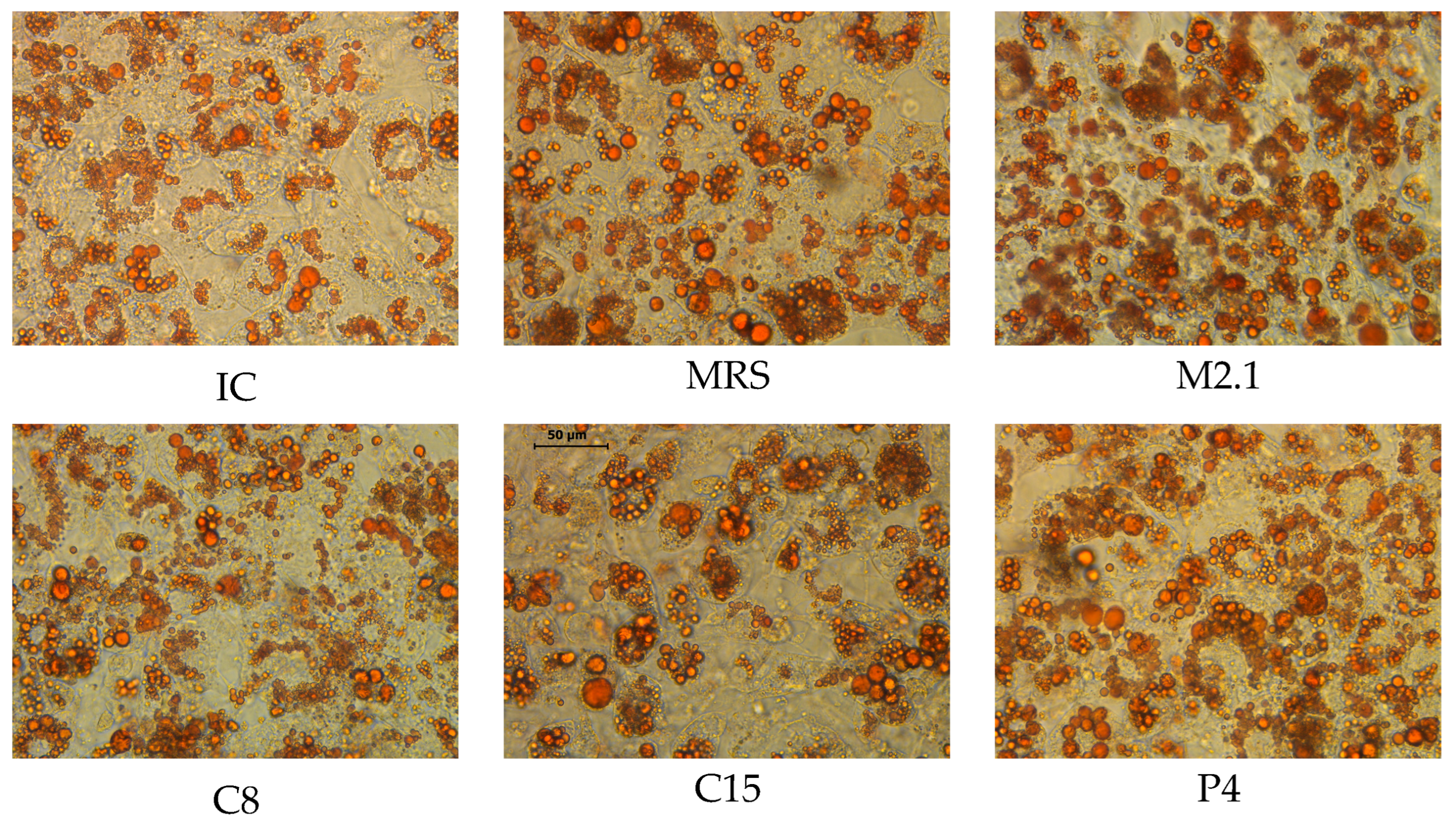
Figure 1. Morphology and intracellular neutral lipid accumulation in mature 3T3-L1 cells, stained with Oil Red O, after 48 h of 10% (v/v) cell-free supernatant treatments (20× magnification, bar = 50 µm). Images were acquired using a Leica DMi1 Inverted Microscope equipped with a 5-megapixel-resolution camera and the Leica Application Suite Core software 4.12.0 (Heerbrugg, Switzerland). Abbreviations: IC—mature non-treated cells; MRS—mature cells, treated with MRS broth; M2.1, C8, C15, and P4—mature cells, treated with supernatants from M2.1, C8, C15, or P4 strains.
Biomedicines 13 02036 g001
Figure 2. Comprehensive ranking of candidate reference genes calculated via the RefFinder tool. Data were obtained from six experimental groups with six biological replicates each (n = 36 per gene). The geometric mean of ranking values integrates stability scores from geNorm, NormFinder, BestKeeper, and the ΔCt method, with lower values indicating higher expression stability. Visualization was performed in GraphPad Prism (v.10).
Biomedicines 13 02036 g002
Figure 3. Inter-group statistical comparison of raw Ct values for selected reference genes (HPRT, HMBS, 36B4, 18S, GAPDH, and Actb) in mature 3T3-L1 adipocytes treated with 10% (v/v) cell-free supernatants from L. paracasei strains. Abbreviations: IC—untreated, induced control; MRS—induced control, treated with MRS; M2.1, C8, C15, P4—experimental groups, induced and treated with supernatants for 48 h. Data are presented as mean ± SEM of six biological replicates per group (n = 6). Statistical analyses and visualization were performed in GraphPad Prism (v.10). Inter-group variability was first assessed by one-way ANOVA, and genes showing statistically significant differences were further analyzed using Student’s t-test to confirm the significance level, as follows: * p < 0.05; ** p < 0.01; *** p < 0.001.
Biomedicines 13 02036 g003
Table 1. Primer sequences and description of mouse housekeeping and target genes used in RT-qPCR.
AbbreviationFull NameForwardReverseProduct Length
36B4Ribosomal protein, large, P0TTATAACCCTGAAGTGCTCGACCGCTTGTACCCATTGATGATG147
HPRTHypoxanthine-guanine phosphoribosyl transferaseACAGGCCAGACTTTGTTGGAACTTGCGCTCATCTTAGGCT150
ActbActin, betaCCTCTATGCCAACACAGTGCGTACTCCTGCTTGCTGATCC211
HMBSHydroxymethylbilane synthaseCCTGAAGGATGTGCCTACCACCACTCGAATCACCCTCATCT175
GAPDHGlyceraldehyde-3-phosphate dehydrogenaseAAATGGTGAAGGTCGGTGTGTGAATTTGCCGTGAGTGGAG583
18S18S ribosomal RNAATGCGGCGGCGTTATTCCGCTATCAATCTGTCAATCCTGTC204
PPARγ Peroxisome proliferator-activated receptor gamma, transcript var. 2AGGGCGATCTTGACAGGAAACGAAACTGGCACCCTTGAAA164
Table 2. Stability values and ranking of reference genes by geNorm and NormFinder.
Gene Abbr.Stability M-Value (geNorm)Ranking Stability Value (NormFinder)Ranking
36B40.32530.0822
HPRT0.29910.0711
Actb0.45250.2025
HMBS0.32420.1093
GAPDH0.40240.1814
18S0.51060.2156
Table 3. Descriptive statistics of the candidate reference genes obtained through the BestKeeper algorithm.
18S36B4GAPDHHMBSHPRTActb
n363636363636
geo Mean [CP]14.5218.7217.1024.7123.1418.46
ar Mean [CP]14.5218.7217.1124.7123.1418.47
min [CP]13.5818.0316.3423.9122.1117.30
max [CP]15.5619.2118.0125.1123.74519.34
std dev [±CP]0.310.200.210.190.160.33
CV [% CP]2.131.081.250.770.701.80
min [x-fold]−73.63−24.12−33.87−38.69−113.1−211.3
max [x-fold]119.89.3962.826.2316.4454.91
std dev [±x-fold]4.132.532.672.402.114.56
Table 4. BestKeeper pairwise correlation and regression analysis of candidate reference genes.
Regression Analysis:
HKG vs. BestKeeper
18S36B4GAPDHHMBSHPRTActb
coeff. of corr. [r]0.480.800.570.740.790.67
coeff. of det. [r2]0.230.630.330.540.630.45
intercept [CP]−3.64−0.412.067.984.16−6.64
slope [CP]0.951.000.790.870.991.31
SE [CP]±0.374±0.165±0.246±0.175±0.167±0.316
p-value0.000.000.000.000.000.00
Power [x-fold]78.0099.0136.8955.2894.92411.45
Table 5. Pairwise Pearson correlation coefficients (r) and p-values between candidate reference genes (18S, HPRT, HMBS, 36B4, GAPDH, Actb) and PPARγ based on BestKeeper analysis.
18SHPRT HMBS36B4GAPDHActb
PPARγ coeff. of correlation. [r] −0.1300.4630.3420.512−0.0020.759
PPARγ p-value 0.4520.0040.0410.0010.9920.001
Table 6. Pairwise ΔCt-based stability analysis of candidate HKGs according to the method of Silver et al. [55].
Gene NamesMean Δ CtStd.Dev.Mean STD. Dev. *
HPRT/18S8.610.50
HPRT/36B44.420.21
HPRT/GAPDH6.030.24
HPRT/HMBS1.570.17
HPRT/Actb4.670.340.29 a
18S/36B44.200.48
18S/GAPDH2.580.51
18S/HMBS10.180.47
18S/Actb3.940.58
18S/HPRT8.610.500.51 b
36B4/GAPDH1.610.29
36B4/HMBS5.990.24
36B4/Actb0.320.24
36B4/HPRT4.420.21
36B4/18S4.200.480.29 a
GAPDH/HMBS7.600.28
GAPDH/Actb1.360.51
GAPDH/HPRT6.030.24
GAPDH/18S2.580.51
GAPDH/36B41.610.290.37 ab
HMBS/Actb6.240.38
HMBS/HPRT1.570.17
HMBS/18S10.180.47
HMBS/36B45.990.24
HMBS/GAPDH7.600.280.31 a
Actb/HPRT4.670.34
Actb/18S/3.940.58
Actb/36B40.320.24
Actb/GAPDH1.360.51
Actb/HMBS6.240.380.41 ab
* Alphabetic indices indicate statistically significant differences in mean SD value between Housekeeping genes, evaluated by Mann–Whitney nonparametric test; p < 0.05.
Disclaimer/Publisher’s Note: The statements, opinions and data contained in all publications are solely those of the individual author(s) and contributor(s) and not of MDPI and/or the editor(s). MDPI and/or the editor(s) disclaim responsibility for any injury to people or property resulting from any ideas, methods, instructions or products referred to in the content.