Review

A Comprehensive Review of Somatic and Germline Biomarkers Associated with Childhood B-Cell Precursor Acute Lymphoblastic Leukemia: From Biological Significance to Precision Medicine Opportunities

by
Daniel Martínez Anaya
1,2,
Johana Itzel Rodriguez Ruiz
1,3,
María del Pilar Navarrete-Meneses
1 and
Patricia Pérez-Vera
1,*
1
Laboratorio de Genética y Cáncer, Instituto Nacional de Pediatría, Mexico City 04530, Mexico
2
Posgrado en Ciencias Biológicas, Universidad Nacional Autónoma de México, Mexico City 04510, Mexico
3
Becaria de la Dirección General de Calidad y Educación en Salud, Secretaría de Salud, Mexico City 11400, Mexico
*
Author to whom correspondence should be addressed.
Biomedicines 2025, 13(7), 1626; https://doi.org/10.3390/biomedicines13071626
Submission received: 13 May 2025 / Revised: 17 June 2025 / Accepted: 28 June 2025 / Published: 2 July 2025

Abstract

B-cell precursor acute lymphoblastic leukemia (B-ALL) is characterized by a constellation of somatic pathogenic variants associated with malignant transformation. These variants have implications for clinical management by providing clinical biomarkers. Most B-ALL cases have a sporadic presentation. However, some patients may present the disease as the neoplastic manifestation of cancer predisposition syndromes caused by germline pathogenic variants. In these cases, genetic counseling and personalized oncologic management is mandatory, considering the patient’s sensitivity to conventional therapies. In this review, we have summarized current knowledge on the biological role and clinical relevance of somatic and germline pathogenic variants associated with B-ALL, and discuss three aspects of their application as biomarkers: (1) their usefulness to determine specific molecular subtypes, predicting prognosis and response to specific therapies, (2) their influence in genetic counseling and therapy adaptation for B-ALL in the context of underlying cancer predisposition syndromes, and (3) their detection and interpretation through methodologies. We also included a brief discussion on the need to reclassify variants of uncertain significance to clarify their clinical relevance. Finally, we discuss cases illustrating the impact of somatic and germline pathogenic variants in personalized medicine.

1. Introduction

Acute lymphoblastic leukemia (ALL) is an uncontrolled proliferation of abnormal lymphoid cells and their precursors that invade the bone marrow and extramedullary tissues [1]. In 2019, approximately 5930 individuals were diagnosed with ALL in the US, and nearly 1500 individuals died because of the disease [1]. This neoplasm is the most common childhood cancer, and approximately ~85% of cases involve the B-lymphoid lineage (B-ALL) [2]. Over the past 60 years, the overall survival rate for pediatric ALL in high-income countries has increased from ~10% in the 1960s to >90% at present, making this disease one of the most curable malignancies [3]. This success is attributed to factors such as the monitoring of minimal residual disease (MRD) after treatment. Additionally, the progress achieved in the discovery of the genomic landscape of B-ALL has substantially improved the survival rate for this disease. This progress has enabled the development of personalized therapeutic strategies that consider genetic biomarkers in diagnosis, prognosis, and therapeutic decisions [3,4,5].
Diagnosis of B-ALL traditionally begins with bone marrow cytomorphologic analysis and determination of cell lineage markers by immunophenotyping. Subsequent cytogenetic and molecular analysis allows the identification of prognostic biomarkers [6,7]. At the time of diagnosis, patients are stratified into clinical risk groups, which allows prediction of outcome and tailoring of treatment intensity [6,7].
According to the NCI criteria, high-risk (HR) patients are infants, children, or adolescents, of Hispanic, Native American, or Black ethnicities, presenting a white blood cell (WBC) count >50 × 109/L, and harboring genetic biomarkers associated with poor prognosis. In contrast, standard risk (SR) patients are children aged 1–9 years of Caucasian or Asian ethnicity who typically have a WBC count <50 × 109/L, and have genetic biomarkers associated with favorable prognosis [6,7].
Chemotherapy regimens typically comprise 2 to 2.5 years and include three phases: A 4–6-week remission induction therapy aimed to destroy as many leukemic blasts as possible to restore normal hematopoiesis; this phase includes the use of glucocorticoids, vincristine, asparaginase, and intrathecal therapy. In general, this is the baseline chemotherapy for SR patients, while HR patients require higher doses of antileukemic drugs. The consolidation phase (6–8 months) includes high doses of methotrexate to prevent relapses. Finally, the maintenance phase (18–30 months) includes the use of antimetabolites [6,7].
The assessment of early response to treatment by measuring MRD allows prediction of relapse and adjustment of the chemotherapy intensity. A high MRD level at the end of the remission induction phase is a criterion for increasing the intensity of treatment or even considering hematopoietic stem cell transplantation (HSCT). MRD levels can be measured with high sensitivity using next-generation sequencing (NGS), which allows the detection of the disease by revealing the presence of genetic variants associated with leukemic cell surveillance [4,7].

2. The Role of Genetic Variants in the Development of B-ALL

B-ALL is driven by a constellation of genetic lesions, which are mainly somatic changes that can be divided into gross chromosomal alterations and cryptic variants [5]. Chromosomal alterations are recurrent in B-ALL and can be detected by conventional analysis techniques such as karyotype, FISH or RT-PCR due to their size and low variability. These alterations include gains (hyperdiploidy) or losses (hypodiploidy) of whole chromosomes and translocations that form fusion genes [5,8,9]. In contrast, cryptic variants (<5 Mb) include copy number variants (CNVs), which are duplications or deletions of large DNA segments, copy neutral loss of heterozygosity (CN-LOH) regions, gene fusions, DNA sequence insertions or deletions (Indels), and single nucleotide variants (SNVs) (Figure 1A). Due to their size and molecular heterogeneity, cryptic variants require the use of genomic techniques for their detection [4,10,11]. The genetic variants are classified according to their clinical significance, with pathogenic variants being those that show the strongest association with tumor initiation, progression, and survival [11].
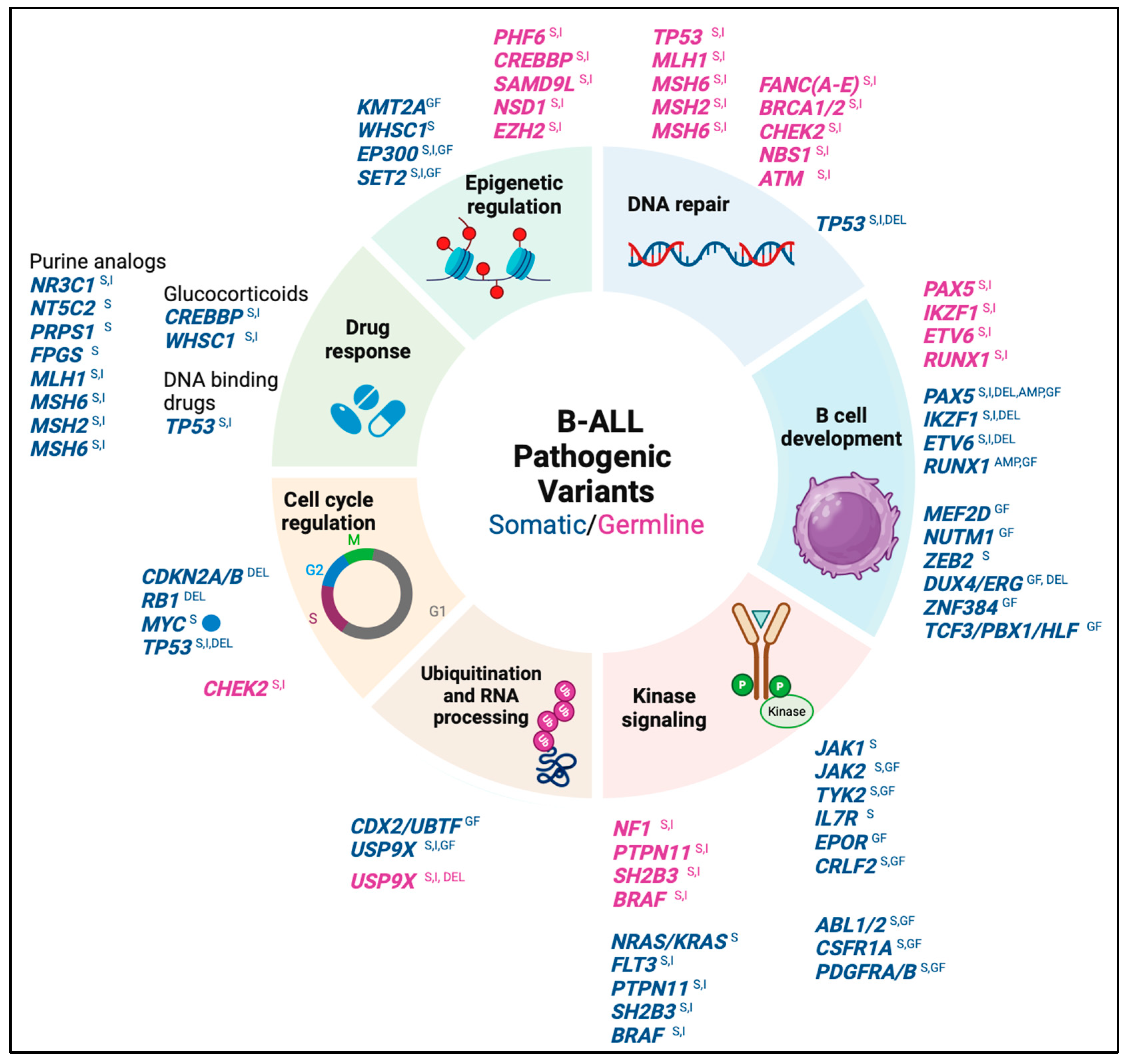
Most B-ALL cases arise sporadically due to somatic chromosomal alterations that may occur during the early stages of embryonic life due to aberrant recombinase enzyme activity (triggered by environmental stimuli), promoting the development of a pre-leukemic clone that already has an initial predisposition to malignant transformation (Figure 1B) [12,13,14]. Chromosomal alterations are considered initial events in leukemogenesis, whereas pathogenic somatic variants are considered cooperative events that allow disease progression through the involvement of oncogenes and tumor suppressor genes relevant for lymphoid development, cell cycle regulation, kinase signaling, chromatin structure regulation, and drug response (Figure 2) [5,15,16].
Some cases of B-ALL are caused by genetic predisposition, such as some constitutional chromosomal abnormalities, e.g., trisomy 21, which is associated with a high risk of B-ALL. In contrast, common low-penetrance germline variants typically affect non-coding regions of genes involved in the normal function of lymphoid progenitors. Individually, these variants have a modest effect, conferring a one- to two-fold increase in the relative risk of developing the disease, although their co-occurrence in an individual can increase the risk of leukemia by up to nine-fold [12,17]. In contrast, rare pathogenic germline variants are highly penetrant and confer a >10-fold increased risk of developing leukemia. These variants commonly involve coding regions of genes encoding transcription factors involved in lymphoid differentiation, proteins associated with DNA damage response, and regulators of signaling pathways involved in cell proliferation (Figure 2) [12,18,19].
There is a complex interplay between somatic and germline genetic variants in the biology of B-ALL. Germline pathogenic variants predispose to the development of B-ALL by promoting subnormal lymphopoiesis in which lymphoid progenitors are susceptible to the acquisition of somatic pathogenic variants (Figure 1B). In another scenario, germline pathogenic variants may lead to epigenomic disruptions that increase the likelihood of the acquisition of cooperative variants, driving the transformation of pre-leukemic clones [19,20].

3. The Somatic Pathogenic Variants as Clinical Biomarkers for B-ALL

Based on the AMP/ASCO/CAP and ACMG/CGC 2017–2019 guidelines, pathogenic variants are classified into two levels according to their ability to serve as biomarkers for diagnosis, prognosis, or response to specific therapy [10,11,16,21].
  • Tier I (with strong clinical significance): These variants constitute the molecular taxonomy of B-ALL and produce a biochemical or immunological phenotype associated with specific gene expression profiles, thus, allowing the identification of molecular subtypes with prognostic value [2,22]. Some of these variants have therapeutic value because they allow the prediction of FDA-approved therapies in pediatric patients with B-ALL. Based on the level of evidence, tier 1A variants are described in the World Health Organization (WHO) 2022 classification of lymphoid tumors and/or in professional clinical management guidelines for B-ALL (e.g., COG guidelines), while level 1B variants are recognized by consensus of disease experts based on multiple studies from different groups [10,11].
  • Tier II variants (with potential clinical significance): These variants are recurrent in different hematological and non-hematological neoplasms, and some of them may integrate a profile of alterations that, in coexistence, have prognostic value. The presence of tier 2 variants may explain the heterogeneity in clinical response among patients with the same molecular subtype of B-ALL [10,11,16,22].
The prevalence of genetic somatic biomarkers with prognostic value is influenced by factors such as age, ethnicity, and clinical risk group [2]. In general, 70% of B-ALL cases harbor some of the sentinel chromosomal alterations defining the classical molecular subtypes, the remaining cases are identified as B-other and are characterized by a diversity of pathogenic variants defining new molecular subtypes or novel provisional entities. [23,24].
To increase the accuracy of clinical risk stratification, the identification of biomarkers with prognostic value needs to be performed in the early stages of the disease, because patients with unfavorable or intermediate prognostic biomarkers could benefit from increased chemotherapy intensity and/or treatment with drugs against targetable pathogenic variants. Conversely, patients with favorable prognostic biomarkers could benefit from reduced chemotherapy intensity if they show an early response with negative MRD levels [2,4,16,22,25].

3.1. Biomarkers Associated with Poor Prognosis

In this section, we describe some features of known and newly described somatic biomarkers associated with poor prognosis in B-ALL (Figure 3 and Table 1):
Table 1. Somatic Pathogenic Variants Recognized as Clinical Biomarkers of B-ALL.
B-ALL Molecular Subtypes/ProfilesAMP
Classification †
General FrequencyDetection
Technique
Common Underlying
Pathogenic Variants
Therapeutic
Approaches
References
POOR PROGNOSIS
Hypodiploidy
(Patterns of multiple chromosome losses)
Tier 1A

Dx/Px
1–2%Karyotype, FISH,
CMA, WGS
RNA-seqGEP
TP53
germline variants
Intensive chemotherapy and HSCT[2,25,26,27]
TCF3::HLF
fusion
Tier 1A

Dx/Px
<1%Karyotype, FISH
RT-PCR, WGS,
RNA-seq GF/GEP
PAX5, BTG1, CDKN2A/B deletions and RAS signaling activating variantsVenetoclax
§ NCT05292664

Intensive chemotherapy
[27,28,29]
MYC rearrangement

MYC juxtaposed with IGH, IGK, IGL
Tier 2<1%Karyotype
FISH
WGS
RNA- seqGF/GEP
CDKN2A/B, RB1 deletions

RAS signaling activating variants
Mature B-cell leukemia/lymphoma chemotherapeutic regimen[27,30]
BCR::ABL1
fusion
Tier 1A

Dx/Px/Tx
2–4%Karyotype, FISH,
RT-PCR
WGS,
RNA-seqGF/GEP
IKZF1 deletionIntensive chemotherapy and Imatinib mesylate[26,27]
Ph-likeSNVs, CNVs and GFs involving kinase genes 10–15%CMA
WGS
RNA-seq
IKZF1 deletionsIntensive chemotherapy and tyrosine kinase inhibitors[22,27]
Ph-like
positive to CRLF2 rearrangement or
mutation
(See supplementary Table S1)
Tier 1A

Dx/Px/Tx
~50%
Ph-like
FISHrearrangement
CMA P2RY8::CRLF2
WGSrearrangement, PM
RNA-seq GF/PM/GEP.
IKZF1 deletions and RAS/JAK-STAT signaling activating variantsIntensive chemotherapy and Ruxolitinib
(JAK inhibitor)

§NCT02723994

§NCT04996160

§NCT03117751
[3,27,28,31]
Ph-like
positive to JAK-STAT class fusions
(See supplementary Table S1)
Tier 1A

Dx/Px/Tx
~7–10%
Ph-like
WGSrearrangements
RNA- seqGF
FISHrearrangements
Deletions in lymphoid differentiation genes.

RAS signaling activating variants.
Intensive chemotherapy and Ruxolitinib

§NCT02723994

§NCT04996160

§NCT03117751
[3,22,28,31]
Ph-like
positive to ABL class fusions
(See supplementary Table S1)
Tier 1A

Dx/Px/Tx
~12%
Ph-like
WGSrearrangements
FISHrearrangements
CMA NUP214::ABL1
RNA- seqGF
Deletions in lymphoid differentiation genes

RAS signaling activating variants
Intensive chemotherapy and Dasatinib
(Abl/Src inhibitor)

§NCT02883049
§NCT03117751
§NCT03020030
[3,22,28,31]
Ph-like
positive to JAK-STAT and RAS activating point mutations
Tier 2~6–11%
Ph-like
WGSPM
WESPM
RNA- seqPM
Deletions in lymphoid differentiation genes Intensive chemotherapy and Ruxolitinib

§ NCT02723994
§ NCT04996160
§ NCT03117751
[3,22,28,31]
Ph-like
positive to other kinase fusions
(See supplementary Table S1)
Tier 1A

Dx/Px/
Tx
(NTRK3 fusion)
~1%
Ph-like
WGS,
FISH
RNA- seqGF
Deletions in lymphoid differentiation genes

RAS signaling activating variants
Intensive chemotherapy and NTRK3 inhibitors

§NCT03066661 Entrectinib
§ NCT03834961 Larotrectinib
[3,22,28,31]
iAMP21
(21q22.12 chromotripsis with multiple RUNX1 copies)
Tier 1A

Dx/Px
~2%Karyotype, FISH
CMA, WGS,
RNA-seqGEP
SH2B3 loss of function variantsIntensive chemotherapy[27,32]
KMT2A
rearrangements
Tier 1A

Dx/Px
1–2%Karyotype, FISH, RT-PCR, WGS,
RNA-seqGF/GEP
Subclonal RAS signaling activating variantsAzacitidine and
Histone Methyltransferase inhibitors
§ NCT03724084
[27,33]
MEF2D
rearrangement
(See supplementary Table S2)
Tier 1B

Dx/Px
~4–5%FISH
WGS
RNA- seq GF/GEP
Loss of function variants in PHF6HDACs
Vorinostat
Quisinostat
(preclinical evidence)
[27,34,35]
CDX2/UBTF
rearrangements
Tier 1B

Dx/Px
<1%WGS
FISH
RNA- seqGF/GEP
ETV6 monoallelic deletion
PAX5::ZCCHC7 estructural variant
Intensive chemotherapy[27,36,37,38]
IKZF1
c.475A>T
p. N159Y


IKZF1
Deletion


IKZF1plus
Profile
Tier 1B

Dx/Px


Tier 1B
Px
(IKZF1 deletions)

Tier 2
Px
(IKZF1 plus Concomitant deletions)
<1%


~15%


~6%
WGS,
RNA- seqPM/GEP


WGS
CMA
FISH
Unknown


IKZF1 whole or intragenic deletion are underlying lesions in various B-ALL molecular subtypes


Concomintant deletions in PAX5, CDKN2A,
CDKN2B (homozygous) or PAR1 in absence of ERG deletion
Focal adhesion kinase inhibitors and rethinoids
(preclinical evidence)
[27,39]
GOOD PROGNOSIS
Hyperdiploidy
(Patterns of multiple chromosome gains)
Tier 1A

Dx/Px
20–30%Karyotype
FISH
CMA
WGS
RNA-seq
GEP
RAS signaling activating variantsAntimetabolite based chemotherapy[9,22,40]
TCF3::PBX1
fusion
Tier 1A

Dx/Px
2–6%Karyotype
FISH
RT-PCR
WGS
RNA-seq
GF/GEP
PAX5, CTCF and SET2 amplifications.
CDKN2A deletions
Intensive chemotherapy[27,41]
ETV6::RUNX1
fusion
Tier 1A

Dx/Px
15–25% Karyotype
FISH
RT-PCR
WGS
RNA-seq
GF/GEP
PAX5 deletions and WHSC1 PMStandard chemotherapy[27,40]
DUX4/ERG
rearrangements
Tier 1B

Dx/Px
4–6%WGS
FISH
RNA-seq
GEP
ERG and IKZF1 deletionsIntensive chemotherapy[22,27,42,43,44]
NUTM1
rearrangement
(See supplementary Table S2)
Tier 1B
Dx/Px
<2%WGS
FISH
RNA-seq
GF/GEP
Deletions in lymphoid differentiation genes Standard chemotherapy[27,45]
INTERMEDIATE PROGNOSIS
PAX5 altered

PAX5 fusions
(See supplementary Table S2)

PAX5 intragenic amplification (iAMP)

PAX5 c.239C>G
p.Pro80Arg
Tier 1B

Dx/Px
3.3–9.3%
for PAX5 fusions or iAMP

0.4–1.9% for P80R mutation
WGS
CMA
FISH
RNA-seq
GF/PM/GEP
CDKN2A/B deletions
RAS/JAK-STAT signaling activating variants.
Intensive chemotherapy[15]
ETV6::RUNX1 like
(See supplementary Table S2)
Tier 1B

Dx/Px
<5%WGS
FISH
RNA-seqGEP/GF
ETV6, IKZF1 and ARPP21 deletions

ETV6 germline variants
Intensive chemotherapy[27,42]
ZNF384
rearrangement
(See supplementary Table S2)
Tier 1B

Dx/Px
1–6%WGS
WES
FISH
RNA-seqGEP/GF
Loss of function variants in CREBBP, EP300, KDM6A, CHD4 and CHD8 epigenetic regulators

RAS signaling activating variants
Intensive chemotherapy[27,46,47,48]
ZEB2 mutated and IGH::CEBPE fusionTier 1B

Dx/Px
<1%WGS, WES,
RNA-seq GEP/PM
RAS signaling activating variants Undefined[27]
† Classification based on AMP/ASCO/CAP 2019 guidelines. Dx: Diagnostic value. Px: Prognostic value. Tx: Therapeutic value. HSCT: Hematopoietic stem cell transplantation. WGS: Whole genome sequencing. WES: Whole exome sequencing. CMA: Chromosome microarray analysis. FISH: Fluorescence in situ Hybridization. RNA seq: RNA sequencing. GF: Gene fusion. PM: Point mutation. GEP: Gene expression profile. § Clinical trial ID available on https://clinicaltrials.ucsf.edu or https://clinicaltrials.gov accessed on 18 February 2025.
Hypodiploidy: At the cytogenetic level, this subtype is characterized by a karyotype with fewer than 44 chromosomes, with patients with near-haploid karyotypes (24–31 chromosomes) and low hypodiploidy (32–39 chromosomes) having the worst prognosis. At the molecular level, most cases harbor pathogenic variants in TP53 that are also present in non-tumor cells, suggesting a germline etiology and the manifestation of B-ALL as part of the neoplastic spectrum of Li-Fraumeni syndrome [2,25,26]. This subtype is associated with a poor prognosis and low event-free survival (EFS) rates (<38.3%), particularly in patients with a positive MRD at the end of induction. Most patients are treated with HSCT once remission is achieved, although the efficacy of this intervention and its potential impact on patient prognosis remains to be elucidated.
t(17;19)(q22;p13): This translocation is very rare in pediatric B-ALL, and is associated with a poor prognosis with EFS <2 years. At the molecular level, the translocation generates the TCF3::HLF gene fusion on derivative chromosome 19, which is an initial event in leukemogenesis and is often accompanied by CNVs in lymphoid differentiation genes and activating variants of RAS signaling present in subclones. Cells carrying the TCF3::HLF gene are resistant to conventional chemotherapy but show sensitivity to Venetoclax, an inhibitor of the apoptosis regulator BCL-2 [9,29].
MYC rearrangements: This is a very rare subtype in pediatric B-ALL and is associated with an unfavorable prognosis. It is characterized by chromosomal translocations that cause arrest of lymphoid differentiation by promoting the overexpression of the MYC gene through its repositioning toward immunoglobulin chain gene clusters. MYC rearranged lymphoblasts show FAB L2 or L3 cytomorphology without surface expression of immunoglobulin light chains and can be confused with mature B-cell leukemia/lymphoma; nevertheless, the absence of somatic hypermutation (VDJ rearrangement of the IGVH genes) in pediatric B-ALL is the principal difference [27,30].
t(9;22) (q34;12) and Ph-like subtypes: The t(9;22) originates the Philadelphia chromosome (derivative chromosome 22), which carries the BCR::ABL1 oncogene, encoding a chimeric kinase that deregulates ABL-class signaling. This subtype confers a poor prognosis, but the addition of Imatinib mesylate to intensive chemotherapy significantly improves prognosis [25,26]. Similarly, Ph-like patients have high-risk clinical features such as elevated WBC counts, higher prevalence in adolescents and young adults, suboptimal response to remission induction chemotherapy, high relapse rates, and poor overall survival [25,26].
At the molecular level, the Ph-like subtype possesses a transcriptome similar to the Philadelphia-positive subtype but lacks the t(9;22) chromosome translocation and shows a plethora of pathogenic variants that deregulate the activity of lymphoid developmental transcription factors (mainly deletions of IKZF1), cytokine receptors, and tyrosine kinase signaling pathways. In general, CRLF2 rearrangements are the most common abnormality, occurring in approximately 50% of Ph-like cases. The CRLF2 gene encodes a transmembrane receptor that forms a heterodimer with the alpha chain of the interleukin-7 receptor in the presence of thymic stromal lymphopoietin ligand. This gene can be altered by deletions, translocations, and point mutations that deregulate JAK-STAT signaling (Supplementary Figure S1A), half of these cases harbor activating pathogenic variants in the JAK1 and JAK2 kinases, which are important mediators of hematopoietic signaling. In this context, it is important to identify the JAK2 R683G mutation in patients with CRLF2 abnormalities because it confers resistance to a panel of JAK inhibitors, including Ruxolitinib [49]. Other relevant pathogenic variants include ABL or JAK-STAT class gene fusions (Supplementary Figure S2A), activating variants of the RAS signaling pathway, and gene fusions leading to deregulation of other kinase signaling pathways (Supplementary Figure S3A) [25,31,50].
Kinase dysregulation can be successfully treated with tyrosine kinase inhibitors (TKIs) such as Imatinib and Dasatinib, which abolish the ABL-class signaling, or with Ruxolitinib, which inhibits the JAK-STAT class signaling. The efficacy of TKIs has been demonstrated in cell lines and xenograft models of B-ALL. Currently, there are reports of Ph-like patients showing good responses to the use of TKIs in combination with conventional chemotherapy. These clinical and experimental observations have prompted the development of clinical protocols focused on determining the efficacy and safety of TKIs for the treatment of pediatric Ph-like B-ALL [31,50].
Intrachromosomal amplification of chromosome 21 (iAMP21): This subtype is characterized by chromothripsis (multiple cycles of amplification through DNA cleavage and splicing in contiguous regions) of chromosome 21. Cells carrying iAMP21 have more than 3 copies of the RUNX1 gene per cell [51]. These patients typically present low to moderate WBC counts and a high relapse rate following standard risk-adjusted chemotherapy. Nevertheless, the risk of relapse can be reduced to less than 20% with high-risk adapted chemotherapy [51,52].
KMT2A rearrangement: At the molecular level, this subtype is defined by the presence of translocations at 11q23 loci, that fuse the 5′ region of the KMT2A gene (which contains the methyltransferase domain) with the 3′ region of a variety of gene partners. The leukemic cells exhibit a mixed lineage immunophenotype with the co-expression of lymphoid (CD34 and CD19) and myeloid (CD15 and CD65) markers. KMT2A rearranged B-ALL is characterized by a high incidence in children under 1 year of age and a poor prognosis with high WBC counts and leukemic extramedullary infiltration. Treatment consists of intensive chemotherapy and allogeneic HSCT; although most children achieve remission (82.5%), this is usually short-lived, with rapid relapse and poor survival. Other therapeutic options based on histone deacetylase inhibitors (HDACi) in combination with standard chemotherapy are under preclinical investigation [33].
MEF2D rearrangement: The MEF2D gene encodes a transcription factor required for lymphoid development and can form gene fusions with several partners, including BCL9, which is the most common. All fusions retain the DNA-binding domain, resulting in deregulation of MEF2D target genes (except for MEFD2::CSF1R, which belongs to the ABL-class Ph-like subtype). Cells positive to MEF2D fusion have an abnormal CD10-, CD38+ immunophenotype and are characterized by highly vacuolated basophilic nuclei like the mature B-cell leukemia morphology. This B-ALL molecular subtype has few cooperative lesions, and the most common are pathogenic SNVs in the transcriptional regulator PHF6 (50% of cases). Clinically, it is a biomarker of poor prognosis with low EFS rates, but similar to KMT2A rearranged B-ALL, it also shows overexpression of the HDAC9 gene and, thus, is responsive to HDACi therapy [27,34].
CDX2/UBTF rearrangement: This recently discovered B-ALL subtype is characterized by the co-occurrence of two alterations, a ~280 Kb microdeletion that relocates the CDX2 gene next to the FLT3/PAN3 gene enhancer, causing its overexpression, and a ~10 Kb microdeletion at 17q21.31 that results in the formation of UBTF::ATXN7L3 gene fusion (Supplementary Figure S2A). CDX2 regulates homeotic gene expression during embryonic hematopoiesis, and UBTF is a nuclear protein that regulates rRNA transcription. The biological significance of these alterations is still unknown; however, both produce a CD10-, IgM+ immunophenotype and a specific gene expression profile. This is a rare subtype in pediatric B-ALL, with most cases occurring in adolescents and young adults with a female preponderance. It confers an unfavorable prognosis with a high relapse rate and treatment refractory with high MRD levels. Treatment with intensive chemotherapy improves the prognosis in adults, although the response in pediatric patients remains suboptimal [36].
Somatic pathogenic variants in IKZF1: IKZF1 is located at 7p12.2 and encodes the Ikaros protein, a transcription factor that plays a key role in lymphoid differentiation. Ikaros has six zinc fingers, four of which are in the N-terminal domain and allow its binding to DNA, and two of which are in the C-terminal domain and allow its homo- or heterodimerization (Supplementary Figure S2B). The most common alterations of IKZF1 in B-ALL are monoallelic deletions of the whole gene, with pathogenic effect by haploinsufficiency. Another common alteration is the intragenic deletion from exons 4 to 7, resulting in the Ik6 isoform. This isoform loses the DNA binding domains and retains the dimerization domains, allowing it to exert a negative dominant effect on wild-type transcripts, reducing the function of genes that regulate the self-renewal capacity of lymphoid progenitors [53,54].
IKZF1 deletions (IKZF1del) confer an unfavorable prognosis, according to the DFCI ALL consortium study 05-001, IKZF1del [53]. IKZF1del is commonly an underlying lesion in poor-prognosis B-ALL molecular subtypes, such as the Ph+ and Ph-like cases, in which it is highly prevalent (70% and 68%, respectively). In contrast, favorable prognostic subtypes such as hyperdiploidy and t(12;21) show a lower prevalence (15% and 3%, respectively), although even in t(12;21) cases, IKZF1del reduces 5-year EFS, reinforcing its negative impact [54].
The prognostic value of IKZF1del is modified by the epistatic effect of CNVs in other lymphoid development genes such as ERG. The coexistence of IKZF1 and ERG deletions reduces the negative prognostic impact. In contrast, the absence of the ERG deletion together with positive deletions in the CDKN2A/B, PAX5, and PAR1 genes constitutes the IKZF1plus profile, which amplifies the negative impact of IKZF1del in patients with MRD > 104 at the end of induction therapy, resulting in a 5-year EFS of 53%, 79%, and 89% in the IKZF1plus, IKZF1del, and normal IKZF1 patients, respectively [54,55].
The heterozygous variant IKZF1p.Asn159Tyr defines a subtype of B-ALL with a specific gene expression profile. This subtype is rare, and its molecular features and prognostic value are currently unknown. The Asn159 residue, which is in the DNA binding domain (Supplementary Figure S2B), disrupts the function of IKZF1, promoting its aberrant nuclear localization and increasing cellular adhesion. In contrast to other subtypes with underlying IKZF1 alterations, the N159Y variant increases the overexpression of chromatin remodelers and regulators of kinase signaling that are not deregulated in IKZF1del subtypes [48].

3.2. Biomarkers Associated with Good Prognosis

In this section, we describe some features of known and newly described somatic biomarkers associated with good prognosis in B-ALL (Figure 3 and Table 1):
Hyperdiploidy: This is the most common alteration in B-ALL and is characterized by a karyotype with 51–67 chromosomes. Trisomies of chromosomes X, 4, 6, 10, 14, 17, 18, X and tetrasomy 21 are the most common gains. Variants in genes involved in the RAS pathway and epigenetic regulation are common cooperating lesions in this subtype, but patients with hyperdiploidy generally have a favorable prognosis and are candidates for reduction of chemotherapy intensity [2,40].
t(1;19) (q23;p13.3): This translocation produces a derivative chromosome 19 carrying the oncogenic fusion TCF3::PBX1, which encodes a transcription factor that confers self-renewal capacity and disrupts lymphoid differentiation. Initially, this subtype was considered to have an intermediate prognosis, attributable to its propensity for central nervous system relapse. However, subsequent research has demonstrated that intensive chemotherapy treatments can improve the prognosis and, thus, it is now considered to have a favorable prognosis [9,26].
t(12;21) (p13;q22): This translocation produces a derivative chromosome 12 carrying the oncogenic fusion ETV6::RUNX1, which requires cooperating alterations to induce leukemogenesis. It is generally associated with a favorable prognosis with good response to low-intensity chemotherapy [2,26].
DUX4/ERG rearrangement: This subtype is defined by a rearrangement that affects the DUX4 gene, which encodes a homeodomain transcription factor involved in lymphoid differentiation and is not expressed in most somatic tissues. This gene is located within the D4Z4 repeat region in the subtelomeric region of chromosomes 4q and 10q, and its insertion into the IGH locus results in its overexpression. It can also be inserted into intron 6 of the ERG transcription factor, and this rearrangement also results in the expression of a non-canonical ERG transcript that contains a truncated C-terminal domain but retains its DNA-binding domains. This transcript has an aberrant activity that promotes deletion of the second allele of ERG, driving leukemic transformation (Supplementary Figure S2C). ERG and IKZF1 deletions are frequent secondary events in patients with DUX4 rearrangement (50–63% and 40%, respectively). This subtype is more prevalent in adolescents and young adults. In pediatric patients, it presents at a median age of 6.5 years with low WBC counts and elevated MRD levels. There is a slow response to remission induction with standard therapy. However, the prognosis undergoes a substantial transformation, shifting from a poor prognosis to a favorable one, particularly in cases where intensive chemotherapy is applied [22,43,44].
NUTM1 rearrangement: This is a rare entity that mainly affects infants and is associated with a good response to standard chemotherapy. The presence of NUTM1 fusion with numerous partners is characteristic of this subtype, with BRD9 being the most prevalent. All rearrangements retain almost the entire coding region of NUTM1 and result in overexpression of this gene, although the associated biological effect is still unknown [45].

3.3. Biomarkers Associated with Intermediate Prognosis

In this section, we describe some features of known and newly described somatic biomarkers associated with intermediate prognosis in B-ALL (Figure 3 and Table 1):
PAX5 abnormalities: PAX5 is a tumor suppressor gene involved in the process of lymphoid differentiation. Two distinct somatic pathogenic variants in PAX5 have been identified, each exhibiting significant prognostic value. These genetic abnormalities have been shown to promote leukemogenesis through the inhibition of transcriptional activity associated with PAX5 haploinsufficiency. These variants are described as follows: (1) Rearrangements of PAX5. Include in-frame or out-of-frame gene fusions resulting in loss or disruption of the transactivation domain. The most common gene partners are ETV6, NOL4L, and CBFA2T3 (fusions with ZCCHC7 and JAK2 belong to other subtypes). Another structural variant is the intragenic amplification of PAX5 exons 2–5, which encode the DNA-binding domain and the octapeptide domain. Finally, mainly heterozygous and missense SNVs at residues Arg38 and Arg140 of the DNA binding domain (Supplementary Figure S2B), nonsense splice site variants, and reading frameshift Indels clustered in the distal region of the protein have been identified [15,22]. (2) PAX5 Pro80Arg: Unlike other mutations in PAX5, the p.Pro80Arg variant produces a specific gene expression profile, is present in the homozygous state due to the inactivation of the second allele (through deletion, loss of heterozygosity, or mutation) and impairs the activity of the DNA binding domain, causing arrest of lymphoid differentiation at the pre-proB stage [15,22].
ETV6::RUNX1-like: A molecular subtype defined by a CD27+ CD44 dim/negative immunophenotype and a gene expression profile similar to that of leukemic cells harboring the t(12;21), but without the ETV6:: RUNX1 fusion. Instead, fusions of ETV6 with partners other than RUNX1 are observed, as well as concomitant deletions in the other allele of ETV6 and IKZF1 deletions. In addition, some cases carry germline pathogenic variants in ETV6. As ETV6 and IKZF1 are key genes for lymphoid differentiation, it is assumed that the combination of these alterations may disrupt the transcriptome in the same way as the ETV6::RUNX1 fusion. This subtype is rare and restricted to pediatric patients with low WBC counts who achieve remission with intensive and standard-risk chemotherapy. Nonetheless, there have been reports of relapses, and research examining cohorts of patients who have received uniform treatment is still lacking. As a result, the prognosis remains uncertain [42,46,56].
ZNF384 rearrangement: The ZNF384 gene encodes a C2H2-type zinc finger protein that binds to the promoters of several genes involved in extracellular matrix production, JAK-STAT pathway activation, and lymphoid differentiation. This gene fuses with multiple partners (TCF3, TAF15, and EP300 are the most common) and produces a specific gene expression profile and immunophenotype. The immunophenotype is characterized by low expression of CD10 and the presence of the myeloid markers CD13 and CD33. Loss-of-function variants in epigenetic regulatory genes and gain-of-function variants in RAS pathway genes are common co-occurring lesions in this subtype. Clinical features depend on the gene fusion, with the EP300::ZNF384 and TCF3::ZNF384 fusions commonly associated with high WBC counts, relapse, and presentation at a median age of 5 years, whereas the remaining fusions are more common in adolescents and are associated with favorable and high-risk clinical features. Therefore, the prognosis associated with the specific subtype remains indeterminate [42,46,57].
ZEB2 mutated/IGH::CEBPE: This subtype is defined by recurrent SNVs at residues His1038 and Gln1072 located in the DNA binding domain of the ZEB2 (Supplementary Figure S2B), which is a zinc finger homeotic protein involved in lymphoid differentiation. When this mutation co-occurs with the IGH::CEBPE rearrangement, a characteristic gene expression profile is produced. However, the role of both alterations in leukemogenesis is uncertain. ZEB2 mutations may underlie other lesions, e.g., DUX4 and ZNF384 fusions. At the clinical level, ZEB2 mutations occur in pediatric patients with a mean age of 8 years and standard WBC counts, these patients have a high relapse rate (50%) when treated with standard risk chemotherapy, which decreases in patients treated with high-risk chemotherapy (8%), suggesting that the prognosis depends significantly on the intensity of chemotherapy. Nevertheless, given their low frequency and association with other subtypes, the clinical relevance of ZEB2 mutations remains to be elucidated [25,47,48].

3.4. Biomarkers Associated with Chemoresistance

Clinical risk stratification and appropriate intensification of therapy have led to a significant improvement in the prognosis of pediatric patients with B-ALL, although the risk of relapse remains high for some patients [3,40,57]. Response to chemotherapy depends on three main factors: (1) non-genetic variation (age, drug interactions, adherence). (2) The presence of common germline variants of low penetrance that modify the pharmacokinetics of antileukemic drugs, although their clinical value is controversial due to their low penetrance. (3) The presence of somatic pathogenic variants that may be present at diagnosis or acquired during chemotherapy treatment in specific leukemic clones that emerge during relapse and have a direct effect on the mechanism of action of antileukemic drugs [58,59].
Genomic analysis in patients before and after therapy has revealed the genetic landscape of relapsed B-ALL. Approximately 75% of pathogenic variants present at relapse are derived from pre-existing subclones that survive therapy and acquire cooperative mutations to become a relapse promoter clone (Figure 1B). These variants primarily involve RAS and JAK-STAT signaling genes that emerge at diagnosis as underlying pathogenic variants in multiple molecular subtypes of B-ALL. Some variants are specific or enriched in relapse clones, most commonly involving genes that regulate response to glucocorticoids (NR3C1, CREBBP, WHSC1), purine analogues and methotrexate (MSH6, MSH2, PMS2, NT5C2, PRPS1 and FPGS), and DNA-binding antileukemic drugs (TP53) (Supplementary Figure S3) [60,61,62].
Variants in genes that regulate the response to glucocorticoids have primarily been found at relapse, but also at diagnosis. NR3C1 mutations have been found in 0.9% of patients with newly diagnosed ALL and in 11.1–13% of patients with relapsed ALL. Loss-of-function mutations in NR3C1 have been shown to influence glucocorticoid resistance [63]. CREBBP mutations have been detected in 18.3% of relapsed ALL cases. In contrast, mutations in this gene are uncommon in non-relapsed ALL [60]. CREBBP mutations are notably prevalent in patients with high hyperdiploid ALL [64]. WHSC1 mutation frequency is 10% in standard-risk and high-risk ALL patients [63]. A study of 99 B-ALL cell lines harboring diverse gene fusions revealed that mutations in NR3C1 tend to be more prevalent in cell lines with MEF2D fusions and TCF3-HLF. In cell lines harboring CREBBP mutations, MEF2D fusions were more prevalent, while KMT2A fusion was less prevalent. Regarding WHSC1 mutations, TCF3-PBX1 and ETV6-RUNX1 incidences tended to be higher. Unfortunately, this study did not analyze hyperdiploid cell lines [65].
Variants in genes that respond to purine analogues are usually not detected at diagnosis and, thus, they are considered relapse-associated mutations that are possibly acquired during treatment within a minor subclone present at diagnosis [61]. NT5C2 is particularly important as it is associated with mercaptopurine resistance and has an overall occurrence rate of 10% [66]. In general, the prevalence of mutations resulting from chemotherapy exposure in this group of genes is estimated to be 17% in patients with very early relapse (<9 months since diagnosis), 65% in early relapse groups (9–36 months) and 32% in late relapse groups (>36 months). Based on this, monitoring of chemoresistance variants during remission using high-throughput deep sequencing has been proposed [61].
Conversely, immunotherapy has transformed the treatment of relapsed B-ALL, exhibiting the advantage of not targeting the genetic alterations of the disease and the ability to overcome chemoresistance variants present in relapsed clones [67]. Blinatumomab is a monoclonal antibody that facilitates binding between T cells (CD3+) and B cells (CD19+), thereby promoting the destruction of malignant B cells. It has demonstrated improved efficacy compared to standard chemotherapy as a remission induction agent in patients with relapsed B-ALL, with moderate toxicity and an MRD-free response rate [67].
Therapy based on the use of chimeric antigen receptor T cells (CAR-T) has achieved 65–90% responses in clinical trials for the treatment of relapsed B-ALL. Phase I and II clinical trials have reported response rates of 50% at one-year post-treatment. This therapy represents the major therapeutic advance for B-ALL and may even replace HSCT. Based on the good response, this therapy has been approved by the FDA as Tisagenlecleucel [68,69,70].

4. Germline Pathogenic Variants as Biomarkers of Susceptibility to B-ALL

Germline predisposition to cancer has been recognized for decades, mainly in solid tumors [19]. Although most cases of pediatric B-ALL are sporadic, detailed genomic analyses in cohorts of patients with this disease have revealed that between 8.3 and 11% of cases carry SNV/Indel-type germline pathogenic variants in cancer predisposition genes [71,72,73]. Most of these variants follow an autosomal dominant (AD) inheritance pattern in which a single copy of the altered gene is sufficient to confer cancer risk, and the neoplastic phenotypes correlate with the gene expression pattern of the genes involved. Genetic variants expressed during the process of hematopoiesis have been identified as a contributing factor to the development of B-ALL, whereas variants in genes expressed in different cell lineages cause syndromes that predispose to solid tumors and sporadically to B-ALL [19,20].

4.1. Biomarkers Conferring High Susceptibility to B-ALL

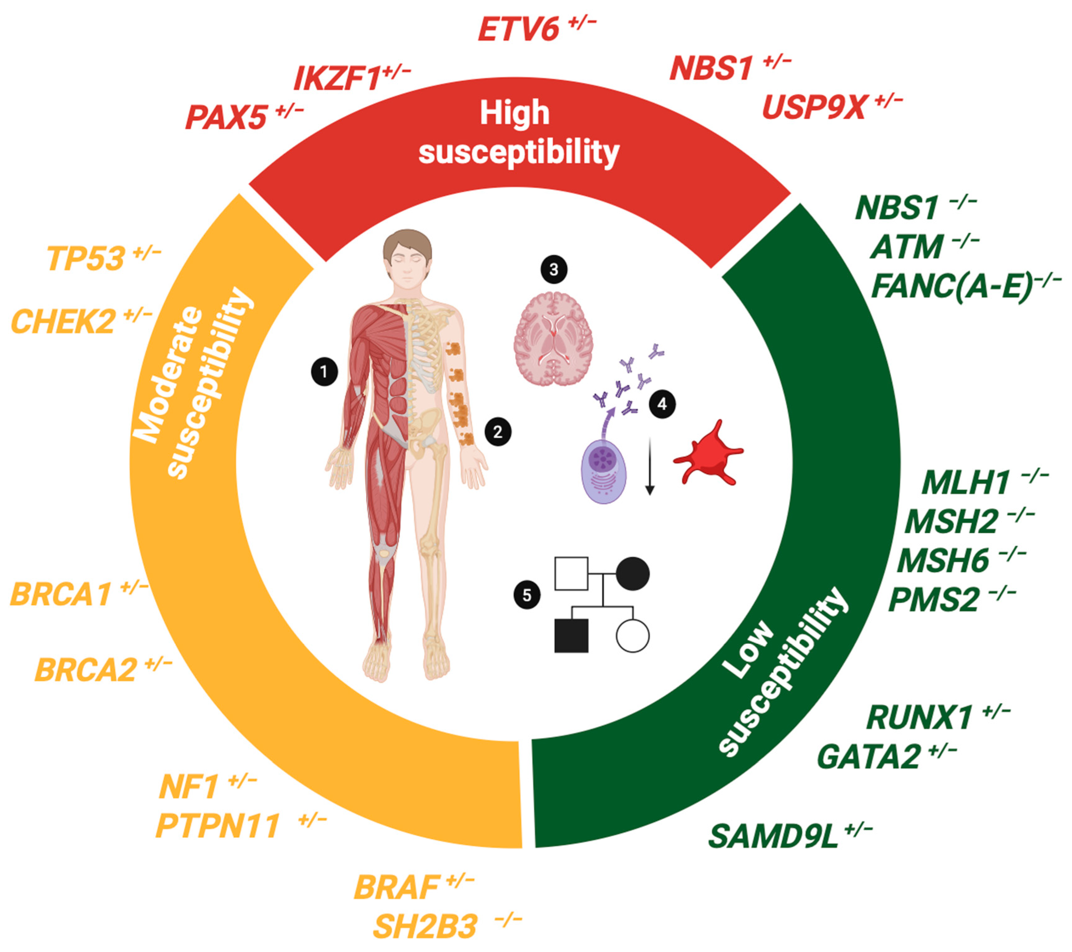
In this section, we describe constitutional entities that manifest B-ALL as the main neoplastic phenotype (Figure 4 and Table 2):
Table 2. Genetic Syndromes Associated with B-ALL Susceptibility.
Genetic Syndrome
(OMIM #ID)
Gene
(Loci)
InheritanceNeoplastic
Phenotype
Main Non-Neoplastic
Phenotypic Characteristics
References
HIGH SUSCEPTIBILITY SYNDROMES
PAX5
Deficiency

(615545)
PAX5
(9p13.2)
ADB-ALLImmunodeficiency with
low levels of B cells and immunoglobulins
[16,74,75]
ETV6
Deficiency

(616216)
ETV6
(12p13.2)
ADHyperdiploid or ETV6::RUNX1 like B-ALLThrombocytopenia[56,76,77]
IKZF1
Deficiency

(613065)
IKZF1
(7p12.2)
ADB-ALLLymphopenia[78]
USP9X
Deficiency
(300968)
USP9X
(Xp11.4)
XLDB-ALL
restricted to female patients
Psychomotor delay and intellectual disability
Congenital abnormalities
[79]
MODERATE SUSCEPTIBILITY SYNDROMES
Li-Fraumeni syndrome
(151623)
TP53
(17p13.1)
ADBreast cancer, sarcoma, glioblastoma, adrenal gland cancer

Hypodiploid B-ALL
NA[80,81,82]
Neurofibromatosis type 1 (162200)NF1
(17q11.2)
ADNeurofibromas
Gliomas, JMML
B-ALL
CALMs
Skeletal disorders
Cognitive deficits
[83,84]
Noonan/Leopard
syndrome
(163950/15110)


Cardiofaciocutaneous syndrome
(115150)


SH2B3
Deficiency
(NA)
PTPN11
(12q24.13)
(~50% cases)
Other RAS genes
(SOS, BRAF1, RAF1, MAPK1, KRAS, NRAS)

BRAF
(7q34)


SH2B3
(12q24.12)
AD


AD


AR
JMML
Neuroblastoma
Rhabdomyosarcoma
Hyperdiploid B-ALL


Non-Hodgkin’s lymphoma
Hepatoblastoma
Rhabdomyosarcoma
B-ALL

MPS/JMML
B-ALL
Craniofacial Anomalies, Congenital Heart Anomalies
CALMs
Short stature
Thrombocytopenia


Hepatosplenomegaly
Autoimmunity


Hepatosplenomegaly
Autoimmunity
[85,86,87]
LOW SUSCEPTIBILITY SYNDROMES
Nijmegen syndrome
(615545)
NBN (NBS1)
(8q21.3)
ART-cell lymphoproliferative disorders
B-ALL *
Craniofacial dysmorphias
Developmental delay
Immunodeficiency
[88,89]
Constitutional mismatch repair deficiency syndrome.
(CMMRD)
(619069)
MLH1
(3p22.2)
MSH2
(2p21-p16.3)
MSH6
(2p16.3)
PMS2
(7p22.1)
ARColorectal cancer
Glioblastoma
T-cell lymphoproliferative disorders
B-ALL *
CALMs
Immunodeficiency
[90,91]
Fanconi anemia
(Various genes)
Genes FANC(A-E)
(Múltiple loci)
AR, XLRAML
Head and neck cancer
B-ALL *
Dysmorphological syndrome
CALMs
Musculoskeletal disorders
Intellectual disability
[92,93]
Ataxia telangiectasia
syndrome
(208900)
ATM
(11q22.3)
ART-cell lymphoproliferative disorders
B-ALL *
Neurological disorders
Telangiectatic disorders
Immunodeficiency
[94,95]
RUNX1
Deficiency
(601399)
RUNX1
(21q22.12)
ADMDS and T-ALL
B-ALL *
Thrombocytopenia[96]
GATA2
Deficiency
(614172)
GATA2
(3q21.3)
ADMDS, AML, JMML, T-ALL
B-ALL *
Immunodeficiency[97]
Ataxia pancytopenia
Syndrome

(159550)
SAMD9L
(7q21.2)
ADMDS, AML,
B-ALL *
Neurological disorders
Cytopenia
Immunodeficiency
[98,99]
AD: Autosomal dominant. AR: Autosomal recessive, XLR: X-linked recessive. MPS: Myeloproliferative syndrome. MDS: Myelodysplastic syndrome. JMML: Juvenile myelomonocytic leukemia. NA: not available. CALMs: Café au lait macules. AML: Acute myeloid leukemia. * Some rare cases of B-ALL have been reported in patients with these genetic syndromes.
PAX5 deficiency: The germline variants in PAX5 show an AD inheritance pattern with incomplete penetrance; consequently, patients may be asymptomatic, develop immunodeficiency, or B-ALL. All reported variants are unique, except for PAX5 c.113G > A p. Glu183Ser, which has been described in individuals from three unrelated families and involves a highly conserved residue of the octapeptide domain essential for normal gene function (Supplementary Figure S4). Complete loss of PAX5 function is required for leukemogenesis, as all heterozygous carriers of germline variants in PAX5 who develop B-ALL show somatic inactivation of the second allele [16,74]. According to the findings of functional studies, germline variants in PAX5 have been demonstrated to exert a “hypomorphic” effect, characterized by a modest but significant reduction in transcriptional activity when compared to the normal protein. The conserved activity is sufficient to promote B-lymphocyte development but not to prevent leukemic transformation [74,75].
ETV6 deficiency: This is an incompletely penetrant AD disorder characterized by thrombocytopenia and predisposition to ALL. It is caused by germline variants in ETV6, which encodes a transcriptional repressor essential for hematopoiesis. These variants have a frequency of <1% in patients with B-ALL, and approximately 23–30% of carriers develop the disease, mainly the hyperdiploid and ETV6::RUNX1-like subtype. Ninety-six individuals from 23 different families have been reported with recurrent variants involving conserved residues of the DNA-binding domain (Supplementary Figure S4); their functional effects include destabilization of DNA binding and protein delocalization with a dominant negative effect on the protein encoded by the normal allele [56,76,77].
IKZF1 deficiency: Germline variants in IKZF1 have an AD inheritance pattern with incomplete penetrance and can result in low levels of B cells or the development of B-ALL. Germline variants in IKZF1 have a prevalence of 0.9% in pediatric patients with B-ALL and can occur in sporadic and familial cases. Unlike the IKZF1 germline variants that cause the common variable immunodeficiency syndrome, the IKZF1 B-ALL predisposition variants are located along the coding region of the gene (Supplementary Figure S4) and have a moderate to strong functional impact on the processes of dimerization, DNA binding, subcellular localization, cell adhesion, and drug response [78].
USP9X deficiency: Located on Xp11.4, USP9X is a tumor suppressor and oncogene that regulates the ubiquitylation of various cellular substrates. At the somatic level, it is altered by loss-of-function variants in 1.1% of B-ALL cases. This gene escapes X-chromosome inactivation and, when affected by germline pathogenic variants, causes embryonic lethality in males and X-linked mental retardation syndrome restricted to females. This is a rare dysmorphological syndrome with only 42 patients described in the literature, of which 7.1% developed pediatric B-ALL [79].
NBS1 heterozygous variants: Carriers of homozygous pathogenic variants in the NBS1 gene develop Nijmegen syndrome (NJS), an autosomal recessive disorder characterized by predisposition to lymphoid neoplasms, mainly of the T-cell type. In contrast, carriers of heterozygous variants in NBS1 do not manifest NJS but are at risk of developing B-ALL [89]. NBS1 encodes nibrin, which is a sensor of double-strand breaks in DNA and an adaptor for the activation of repair pathways for these lesions. The germline heterozygous NBN (c.657_661del5)p.K219fs variant was identified in 0.8% of a cohort of 4183 pediatric patients with B-ALL, a frequency similar to that of germline pathogenic variants in PAX5, IKZF1, and ETV6. Although most of these variants are of uncertain significance, functional studies have demonstrated a pathogenic effect mainly associated with alterations in protein stability. These variants show “hot spots” in the N-terminal domain at highly conserved residues (Supplementary Figure S4) and result in hypomorphic DNA damage response activity. However, the role of these alterations in the transformation of B-lymphoid precursors is still unclear [89,100,101].

4.2. Biomarkers Conferring Moderate Susceptibility to B-ALL

In this section, we describe constitutional entities that manifest solid tumors or myeloid malignancies as the predominant neoplastic phenotype, but occasionally, some carriers develop B-ALL (Figure 4 and Table 2):
TP53 deficiency: TP53 controls the response mechanisms to DNA damage. Its loss of function due to germline pathogenic variants results in Li-Fraumeni syndrome (LFS), which has an AD inheritance pattern and confers an almost 100% risk of developing cancer. The neoplastic spectrum of LFS is broad, with brain tumors being the most common and leukemias less frequent. Less than 1% of B-ALL cases are associated with the LFS syndrome, and most of these (65.4%) are characterized by a hypodiploid karyotype [81,82]. The germline pathogenic variants in TP53 detected in LFS B-ALL patients are mainly clustered in the DNA binding domain (Supplementary Figure S4), and involve the Arg 248 residue in approximately 20% of cases [80,102,103]. Furthermore, some patients with pathogenic variants in genes that regulate p53 activity (e.g., CHEK2, BRCA1) show clinical criteria compatible with LFS [104].
Constitutional deficiency in RAS pathway genes: Germline loss-of-function variants in the NF1 and PTPN11 genes cause neurofibromatosis type 1 (NFS) and Noonan/Leopard syndrome (NLS), respectively. Both syndromes have AD inheritance and belong to a group of syndromes with constitutive dysregulation of RAS signaling known as RASopathies. They produce a phenotype characterized by body and skin malformations and a predisposition to brain tumors, myeloid leukemia, and occasionally B-ALL. The estimated frequency of NFS and NLS is ~0.12% and 0.02–0.04%, respectively, calculated in 4939 pediatric patients, predominantly with B-ALL and hyperdiploid karyotypes [80,105].

4.3. Biomarkers Conferring Low Susceptibility to B-ALL

In Table 2, we summarize cancer predisposition syndromes that mainly predispose to the development of solid tumors and myeloid and T-cell neoplasms in pediatric patients. These syndromes are associated with germline pathogenic variants in genes that occasionally cooperate in leukemogenesis; therefore, some rare cases of B-ALL have been reported [106].

4.4. Clinical Management of Patients with B-ALL Associated with Germline Predisposition

Pediatric patients with B-ALL should be screened for predisposing germline variants if they meet the following criteria (Figure 4) [106,107,108]:
  • Consanguineous parents
  • Presence of cytopenia in the patient and/or family members
  • Dysmorphologic syndrome (musculoskeletal disorders)
  • Pigmented lesions or other symptoms associated with RASopathies
  • B-ALL of the hypodiploid or ETV6::RUNX1-like subtypes
  • Family history of cancers included in the neoplastic spectrum of LFS
  • Occurrence of a second primary neoplasm
  • Siblings with childhood cancer or close relatives diagnosed with LFS spectrum cancers at age ≤ 45 years
Identification of the genetic syndrome underlying B-ALL is essential for the accurate clinical management of affected individuals and their families [107]. This requires a clinical evaluation by medical oncologists and geneticists that includes analysis of clinical and family history, physical examination for signs and symptoms associated with the genetic syndrome in question, and cytopathologic analysis for lymphopenia or thrombocytopenia. Once B-ALL susceptibility syndrome has been identified, it is strongly recommended that the patient be referred to a center with expertise in the management of hereditary cancer syndromes [107]. Clinical management of hereditary B-ALL should include identification of relatives who are carriers of the germline pathogenic variant and genetic counseling, selection of appropriate treatment considering the underlying genetic syndrome, and regular clinical surveillance to prevent the occurrence of second malignancies [107].
Treatment of patients with B-ALL associated with syndromes deficient in DNA repair mechanisms requires a delicate balance between therapeutic efficacy and toxicity (Table 2). Patients with LFS are highly sensitive to agents that directly damage DNA. Patients with NJS, ataxia-telangectasia and Fanconi anemia are particularly sensitive to radiation-based treatments. Patients with constitutional mismatch repair deficiency syndrome (CMMRD) are particularly sensitive to treatment with purine analogues. Currently, there is insufficient evidence to adjust chemotherapy in these patients; however, there are reports of patients who have benefited from low-sensitivity treatment [80].
Asymptomatic patients who carry germline pathogenic variants that predispose them to B-ALL should be monitored with complete blood counts at regular intervals, typically every 6–12 months. If indications of B-ALL emerge, the frequency of these counts should be repeated more frequently depending on the severity of the findings [106].
Cytogenetic and molecular analysis of the bone marrow should be considered to look for early signs of clonal evolution that may progress to B-ALL. The identification of dysplasia, increased lymphoblasts, cytogenetic and/or molecular abnormalities in the bone marrow requires consideration of preventive HSCT. Nevertheless, for patients who will receive allogeneic HSCT, the identification of relatives who are carriers of the germline pathogenic variant is critical to avoid selecting a donor who is a carrier of this variant [106].

5. Detection and Interpretation of Genetic Variants in B-ALL Patients

Genomic Techniques Applied to B-ALL Analysis

Next-generation sequencing (NGS) refers to a set of advanced technologies that allow the analysis of the whole genome (WGS), exome (WES), or transcriptome (RNA-seq) in multiple samples simultaneously. These technologies have different applications in the characterization of variants in B-ALL. Depending on their depth and coverage, WES is useful for the detection of SNVs/Indels present in subclones, whereas WGS allows the detection of structural variants (translocations, inversions, insertions). RNA-seq enables the analysis of gene expression profiles and the detection of fusion genes [109,110,111]. NGS-based technologies have important limitations for the detection of CNVs. For WGS, uneven coverage in different regions of the genome makes it difficult to obtain accurate breakpoints, and the lack of a reference normal sample makes data normalization difficult, which is particularly challenging in aneuploid genomes. With respect to RNA-seq, the relationship between copy number and gene expression levels is influenced by epigenetic regulation. Therefore, the detection of CNVs is mainly performed by chromosomal microarray analysis (CMA), which can include comparative genomic hybridization microarray or SNP microarray, which also allow the detection of CN-LOHs [109,110,111].
It is important to consider that no single method can detect different types of genetic variation with equal efficacy. Moreover, the broad coverage of WGS, WES, and RNA-seq increases the cost and the need for bioinformatics resources, limiting their clinical application. In contrast, targeted NGS panels represent a relatively inexpensive alternative that allows targeted analysis of clinically relevant regions of the genome with optimal depth and coverage, and the data obtained is manageable with fewer bioinformatics resources. Unfortunately, most of the currently commercially available panels are not fully applicable to the study of B-ALL, as they are designed for the study of solid tumors or adult hematological malignancies. Therefore, some research groups have opted to use engineered NGS panels or the combination of different genomic methods to maximize the detection of the different types of genetic variations associated with the disease [112,113,114,115].

6. Clinical Significance and Etiology of Variants

There are several bioinformatic algorithms for variant detection from genomic data, with varying degrees of sensitivity. A consensus on the application of these algorithms is yet to be achieved. A comprehensive data curation process is necessary to exclude numerous variants that represent false positives, common variants, and variants of unknown clinical significance. This process involves the use of prior knowledge of variants associated with B-ALL to identify clinically relevant alterations [10,11,21]. This process should be performed by qualified personnel and should follow the recommendations of the guidelines for variant interpretation in cancer. Automated search engines are available to consult the information available in various public databases, that include the population frequency of variants in groups of individuals with specific characteristics such as ethnicity, age, cancer type (e.g., GenomAD Genomes/Exomes, ClinVar, COSMIC), and the available evidence on the diagnostic, prognostic, or therapeutic value associated with the variants (OncoKB) [10,11,21]. This information facilitates the identification of pathogenic variants that are enriched in patients with the disease, exhibit high penetrance, and are of clinical significance in B-ALL or cancer in general. In contrast, benign or probably benign variants, which are prevalent in the general population, have low penetrance and are typically not associated with the disease. A prevalent finding is variants of uncertain significance (VUS), which do not satisfy the requisite criteria to be classified as benign variants. However, there is also an absence of substantial evidence to support their association with cancer [10,11,21].
VUS represent a significant challenge in clinical practice due to their inherent uncertainty, which can complicate decision-making processes and result in the application of inappropriate clinical treatments. Therefore, it is necessary to apply analytical resources focused on generating the necessary evidence to determine the biological and, ultimately, clinical role of these variants. The reclassification of these variants as benign or pathogenic is dependent upon this evidence. To differentiate between the potentially deleterious or neutral effects of VUS, an integrative analysis must be performed. This analysis should incorporate various characteristics of the variant, including its population frequency, the type of variant, its location in the functional product, its effect on function or stability, its degree of conservation across species, its presence in morphologically abnormal tissues, and its segregation among relatives. Unfortunately, all of this information is not always available, and no single type of evidence is sufficient to confidently predict or rule out the pathogenicity of a VUS [116,117].
Leukemic cells contain somatic and germline variants, and distinguishing the etiology of the variants can be a complex process. Somatic variants have been observed to manifest as leukemia-specific or to arise from clonal hematopoiesis (somatic mosaicism restricted to hematopoietic lineages). In contrast, germline variants are present in all cells of the organism and are heritable [118]. A variant should be suspected to be germline if it affects a proportion of cells greater than the leukemic blast population as determined by cytomorphology and/or flow cytometry. The proportion of the variant in the tumor can be inferred by examining the value of the allelic fraction (VAF) (in the case of NGS) or the Log2R and BAF values (in the case of CMA) [118]. Ideally, germline variants will have a VAF ∼0.5 if heterozygous, ∼1.0 if homozygous, or show concordant patterns between Log2R/BAF plots if present in all cells analyzed. However, when the biological sample is a pure tumor specimen, it becomes difficult to distinguish between the germline and somatic etiology of the variant, and the VAF may be altered by loss of heterozygosity, the presence of CNVs, and structural rearrangements, thus, altering its association with the zygosity of the variant. Therefore, a VAF > 0.3 may indicate the presence of a germline variant, whereas a VAF < 0.3 may indicate a somatic variant present in tumor subclones, a product of clonal hematopoiesis, or an artifact of NGS [118].
Suspicion of germline etiology is increased when the variant involves a cancer predisposition gene in a patient with clinical criteria for a hereditary cancer syndrome; in such cases, it is necessary to confirm the etiology of the variant in germline DNA [118]. In healthy individuals, germline DNA is commonly obtained from venous blood, buccal swabs, or hair bulbs; however, in B-ALL patients, such samples may be contaminated with tumor cells or have low yields due to common oncologic treatments. Best practices include the use of skin fibroblasts, although it should be noted that culturing these cells takes several weeks and can be technically demanding. The use of buccal mucosa-derived epithelial cells is also a reliable source of germline DNA when procedures to minimize leukocyte contamination are required [118,119].

7. Genetic Variants Applied to Clinical Practice

This section reviews three case reports of pediatric patients with B-ALL carrying somatic and germline pathogenic variants, in which variant characterization influenced disease management, provided an explanation for the observed phenotype, and/or prevented disease progression.
Case #1: Vatsayan et al., (2023) [120] reported a 12-year-old patient diagnosed with CRLF2::P2RY8 B-ALL in 93% of cells, who was treated with the high-risk chemotherapy regimen AALL1131 (vincristine- and daunorubicin-based) [121]. Seven months later, he developed thyroid cancer. The presence of multiple neoplasms in less than one year suggested the possibility of a cancer predisposition syndrome. Family history revealed thyroid cancer in a maternal uncle. An NGS panel study of the patient revealed the pathogenic variant CHEK2 p.T367Mfs*15. Fifteen months after diagnosis of B-ALL (maintenance), the patient relapsed in the central nervous system, and the bone marrow aspirate showed 15% blasts positive for CRLF2::P2RY8. The patient received intrathecal chemotherapy with vincristine and dexamethasone for several weeks until remission was achieved and was then lymphodepleted in preparation for CAR-T therapy. Bone marrow evaluation 30 days later was negative for MRD, and the patient remained in remission at subsequent evaluations every 3 to 6 months.
Comment: The review of the patient’s family history from the time of diagnosis of B-ALL could have anticipated the detection of the pathogenic variant in CHEK2. With this information, it may have been possible to adapt the therapeutic regimen or add Ruxolitinib, which has been used as a treatment for B-ALL positive for patients with CRLF2::P2RY8 rearrangement [22,28]. The administration of a DNA-binding drug-based chemotherapy regimen likely contributed to the development of the patient’s second cancer and relapse of B-ALL. The efficacy of CAR-T can be attributed to its immunotherapeutic mechanism, which functions independently of the patient’s genetic characteristics, thereby ensuring broad applicability and significant therapeutic potential.
Case #2: Duployez et al., (2021) [74] reported a 17-year-old patient (proband) with an 11-year-old sister (II.1) and a 25-year-old brother (II.3), children of asymptomatic parents. All three siblings were diagnosed with B-ALL, negative for sentinel chromosomal abnormalities and positive for CDKN2A deletions and activating variants in RAS genes. After receiving chemotherapy, siblings II.1 and II.3 remained in remission, but the proband relapsed and was treated with allogeneic HSCT from sibling II.3 (in remission). The proband experienced a second relapse 20 years after HSCT and subsequently died. The sister II.1 remained in remission until 30 years after diagnosis and never received HSCT, and the brother II.3 relapsed and died 14 years post donation. WES was performed on bone marrow DNA from different disease time points of all 3 siblings, as well as on peripheral blood from both parents. The likely pathogenic variant PAX5p.R38H, with VAF 50%, was detected in all the bone marrow samples from the patients and in the peripheral blood of one parent [74].
Comment: The following three factors provide substantial evidence for the hypothesis that hereditary B-ALL is associated with PAX5 deficiency: (1) the presence of a likely pathogenic variant in PAX5 (a B-ALL high-susceptibility gene), (2) a 50% VAF observed in samples from most of the family members, strongly suggesting a germline etiology, and (3) the diagnosis of B-ALL in several family members. According to the literature, treatment with intensive chemotherapy improves EFS in patients with PAX5-altered B-ALL, as evidenced in the sister II.1. For the proband, it is not specified whether the donor’s germline status was determined prior to HSCT; however, it is essential to know this information to avoid failure of HSCT therapy.
Case #3: Lin et al. (2019) [122] reported a patient with B-ALL positive for somatic deletions in the IKZF1 and CDKN2A genes detected by CMA. This patient also inherited two germline variants from both healthy parents: the probably pathogenic JAK2 p.Gly571Ser variant, from heterozygous carrier father, and VUS STAT3 p.Lys370Arg, from heterozygous carrier mother. There was no family history of cancer, and both variants have a population frequency of less than 0.01. JAK2 and STAT3 encode proteins of the JAK-STAT pathway. Both variants are located in the catalytic domains of the corresponding proteins. Functional assays using in vitro lymphoid models revealed that the variants do not possess oncogenic properties when present in isolation, but they promote independent proliferation stimuli when coexisting.
Comment: The patient’s somatic deletions are compatible with the IKZF1plus profile, which confers an unfavorable prognosis. It is recommended to consider intensive chemotherapy and monitoring for minimal residual disease. The addition of Ruxolitinib to chemotherapy could be effective against hyperactivation of the JAK-STAT pathway caused by the presence of germline variants in genes of this pathway. The study of the properties of VUS revealed that this variant acts synergistically with the pathogenic variant in JAK2 to promote leukemogenesis. This case exemplifies the importance of analyzing VUS to determine its biological and clinical relevance.

8. Future Directions

The implementation of pathogenic variants in the clinical management of B-ALL requires the collaborative efforts of medical oncologists, geneticists, and specialists in the interpretation of genomic data. Despite the noteworthy advancements in the field of somatic and germline characterization, further exploration into genetic variation remains imperative. This encompasses the use of different genomic approaches, which allow a complete characterization of the diverse molecular classes of pathogenic variants. However, the application of these approaches is limited by their relatively high cost and the difficulty of interpreting bioinformatic data. Additionally, comprehensive characterization of VUS is necessary to improve our understanding of the pathological processes associated with B-ALL. This requires a high-cost effort for sustained reclassification. Addressing these research gaps can facilitate the implementation of precision medicine for patients with this disease.

Supplementary Materials

The following supporting information can be downloaded at: https://www.mdpi.com/article/10.3390/biomedicines13071626/s1, Table S1: Common kinase gene rearrangements associated with Ph-like B-ALL. Table S2: Common gene fusions involving transcription factor genes associated with specific B-ALL molecular subtypes. Figure S1: (A). Schematic representation of the functional effect of CRLF2 rearrangements. (B). Schematic representation of JAK/ABL class gene fusions. (C). Principal Ph-like activating point mutations in CRLF2, JAK2, NRAS and KRAS genes. Supplementary [123]. Figure S2: (A). The UBTF::ATXNTL3 and CDX2 rearrangements. (B). Somatic point mutations in transcription factor genes associated with B-ALL molecular subtypes. (C). The DUX4/ERG rearrangements. Figure S3. Somatic pathogenic variants commonly associated with chemoresistance in B-ALL. Figure S4. Characteristic germline pathogenic variants associated with B-ALL susceptibility.

Author Contributions

Conceptualization, data search, data analysis, data summary, data curation, and writing—original draft preparation, D.M.A.; original draft preparation, J.I.R.R.; writing—review and editing, M.d.P.N.-M. writing—review and editing, manuscript supervision, and funding acquisition, P.P.-V. All authors have read and agreed to the published version of the manuscript.

Funding

This research received no external funding and was supported by the Convocatoria Recursos Fiscales Instituto Nacional de Pediatría 2025 (INP 032/2023). D.M.A. is supported by a SECIHTI graduate scholarship (816116). J.I.R.R. is supported by an SSA graduate scholarship.

Institutional Review Board Statement

Not applicable.

Informed Consent Statement

Not applicable.

Data Availability Statement

Not applicable.

Acknowledgments

This work is part of the production obtained for the Doctoral degree of D.M.A. at the Posgrado en Ciencias Biológicas, UNAM.

Conflicts of Interest

The authors declare no conflicts of interest.

Abbreviations

The following abbreviations are used in this manuscript:
ACMGAmerican college of genetics and genomic medicine
ADAutosomal dominant inheritance
ALLAcute lymphoblastic leukemia
AMPAssociation of molecular pathology
ASCOAmerican society of clinical oncology
BAFB allele frequency
B-ALLB-cell precursor acute lymphoblastic leukemia
CAR-TChimeric antigen receptor therapy
CAPCollege of American pathologist
CGCCancer genomic consortium
CNVCopy number variant
CN-LOHCopy neutral loss of heterozygosity
CMAChromosomal microarray analysis
CMMRDConstitutional mismatch repair deficiency syndrome
COGChildren oncology group
EFSEvent-free survival
FDAFood and drug administration
FISHFluorescence in situ hybridization
HRHigh-risk
HSCTHematopoietic stem cell transplant
LFSLi Fraumeni syndrome
MRDMinimal residual disease
NCINational cancer institute
NFSNeurofibromatosis syndrome
NLSNoonan/Leopard syndrome
NGSNext-generation sequencing
NJSNijmegen syndrome
RT-PCRReverse transcript polymerase chain reaction
SNVSingle nucleotide variant
SRStandard risk
TKITyrosine kinase inhibitor
VAFVariant allele fraction
VUSVariant of uncertain significance
WESWhole exome sequencing
WBCWhite blood cell
WHOWorld health organization
WGSWhole genome sequencing

References

  1. Malard, F.; Mohty, M. Acute lymphoblastic leukaemia. Lancet Lond. Engl. 2020, 395, 1146–1162. [Google Scholar] [CrossRef] [PubMed]
  2. Tasian, S.K.; Hunger, S.P. Genomic characterization of paediatric acute lymphoblastic leukaemia: An opportunity for precision medicine therapeutics. Br. J. Haematol. 2017, 176, 867–882. [Google Scholar] [CrossRef]
  3. Tran, T.H.; Hunger, S.P. The genomic landscape of pediatric acute lymphoblastic leukemia and precision medicine opportunities. Semin. Cancer Biol. 2022, 84, 144–152. [Google Scholar] [CrossRef]
  4. Heikamp, E.B.; Pui, C.-H. Next-Generation Evaluation and Treatment of Pediatric Acute Lymphoblastic Leukemia. J. Pediatr. 2018, 203, 14–24.e2. [Google Scholar] [CrossRef] [PubMed]
  5. Mullighan, C.G. Genomic characterization of childhood acute lymphoblastic leukemia. Semin. Hematol. 2013, 50, 314–324. [Google Scholar] [CrossRef]
  6. Inaba, H.; Greaves, M.; Mullighan, C.G. Acute lymphoblastic leukaemia. Lancet Lond. Engl. 2013, 381, 1943–1955. [Google Scholar] [CrossRef]
  7. Hunger, S.P.; Mullighan, C.G. Acute Lymphoblastic Leukemia in Children. N. Engl. J. Med. 2015, 373, 1541–1552. [Google Scholar] [CrossRef] [PubMed]
  8. Zaliova, M.; Stuchly, J.; Winkowska, L.; Musilova, A.; Fiser, K.; Slamova, M.; Starkova, J.; Vaskova, M.; Hrusak, O.; Sramkova, L.; et al. Genomic landscape of pediatric B-other acute lymphoblastic leukemia in a consecutive European cohort. Haematologica 2019, 104, 1396–1406. [Google Scholar] [CrossRef]
  9. Roberts, K.G.; Mullighan, C.G. Genomics in acute lymphoblastic leukaemia: Insights and treatment implications. Nat. Rev. Clin. Oncol. 2015, 12, 344–357. [Google Scholar] [CrossRef]
  10. Li, M.M.; Datto, M.; Duncavage, E.J.; Kulkarni, S.; Lindeman, N.I.; Roy, S.; Tsimberidou, A.M.; Vnencak-Jones, C.L.; Wolff, D.J.; Younes, A.; et al. Standards and Guidelines for the Interpretation and Reporting of Sequence Variants in Cancer: A Joint Consensus Recommendation of the Association for Molecular Pathology, American Society of Clinical Oncology, and College of American Pathologists. J. Mol. Diagn. JMD 2017, 19, 4–23. [Google Scholar] [CrossRef]
  11. Mikhail, F.M.; Biegel, J.A.; Cooley, L.D.; Dubuc, A.M.; Hirsch, B.; Horner, V.L.; Newman, S.; Shao, L.; Wolff, D.J.; Raca, G. Technical laboratory standards for interpretation and reporting of acquired copy-number abnormalities and copy-neutral loss of heterozygosity in neoplastic disorders: A joint consensus recommendation from the American College of Medical Genetics and Genomics (ACMG) and the Cancer Genomics Consortium (CGC). Genet. Med. Off. J. Am. Coll. Med. Genet. 2019, 21, 1903–1916. [Google Scholar] [CrossRef]
  12. Cobaleda, C.; Vicente-Dueñas, C.; Nichols, K.E.; Sanchez-Garcia, I. Childhood B cell leukemia: Intercepting the paths to progression. BioEssays News Rev. Mol. Cell. Dev. Biol. 2024, 46, e2400033. [Google Scholar] [CrossRef]
  13. de Smith, A.J.; Spector, L.G. In Utero Origins of Acute Leukemia in Children. Biomedicines 2024, 12, 236. [Google Scholar] [CrossRef] [PubMed]
  14. Chi, X.; Li, Y.; Qiu, X. V(D)J recombination, somatic hypermutation and class switch recombination of immunoglobulins: Mechanism and regulation. Immunology 2020, 160, 233–247. [Google Scholar] [CrossRef] [PubMed]
  15. Gu, Z.; Churchman, M.L.; Roberts, K.G.; Moore, I.; Zhou, X.; Nakitandwe, J.; Hagiwara, K.; Pelletier, S.; Gingras, S.; Berns, H.; et al. PAX5-driven subtypes of B-progenitor acute lymphoblastic leukemia. Nat. Genet. 2019, 51, 296–307. [Google Scholar] [CrossRef]
  16. Pui, C.-H.; Nichols, K.E.; Yang, J.J. Somatic and germline genomics in paediatric acute lymphoblastic leukaemia. Nat. Rev. Clin. Oncol. 2019, 16, 227–240. [Google Scholar] [CrossRef]
  17. Xu, H.; Zhang, H.; Yang, W.; Yadav, R.; Morrison, A.C.; Qian, M.; Devidas, M.; Liu, Y.; Perez-Andreu, V.; Zhao, X.; et al. Inherited coding variants at the CDKN2A locus influence susceptibility to acute lymphoblastic leukaemia in children. Nat. Commun. 2015, 6, 7553. [Google Scholar] [CrossRef]
  18. Kratz, C.P.; Stanulla, M.; Cavé, H. Genetic predisposition to acute lymphoblastic leukemia: Overview on behalf of the I-BFM ALL Host Genetic Variation Working Group. Eur. J. Med. Genet. 2016, 59, 111–115. [Google Scholar] [CrossRef]
  19. Cobaleda, C.; Godley, L.A.; Nichols, K.E.; Wlodarski, M.W.; Sanchez-Garcia, I. Insights into the Molecular Mechanisms of Genetic Predisposition to Hematopoietic Malignancies: The Importance of Gene-Environment Interactions. Cancer Discov. 2024, 14, 396–405. [Google Scholar] [CrossRef]
  20. Gocho, Y.; Yang, J.J. Genetic defects in hematopoietic transcription factors and predisposition to acute lymphoblastic leukemia. Blood 2019, 134, 793–797. [Google Scholar] [CrossRef]
  21. Strom, S.P. Current practices and guidelines for clinical next-generation sequencing oncology testing. Cancer Biol. Med. 2016, 13, 3–11. [Google Scholar] [CrossRef] [PubMed]
  22. Iacobucci, I.; Mullighan, C.G. Genetic Basis of Acute Lymphoblastic Leukemia. J. Clin. Oncol. Off. J. Am. Soc. Clin. Oncol. 2017, 35, 975–983. [Google Scholar] [CrossRef] [PubMed]
  23. Schwab, C.; Harrison, C.J. Advances in B-cell Precursor Acute Lymphoblastic Leukemia Genomics. HemaSphere 2018, 2, e53. [Google Scholar] [CrossRef]
  24. Schwab, C.J.; Murdy, D.; Butler, E.; Enshaei, A.; Winterman, E.; Cranston, R.E.; Ryan, S.; Barretta, E.; Hawking, Z.; Murray, J.; et al. Genetic characterisation of childhood B-other-acute lymphoblastic leukaemia in UK patients by fluorescence in situ hybridisation and Multiplex Ligation-dependent Probe Amplification. Br. J. Haematol. 2022, 196, 753–763. [Google Scholar] [CrossRef]
  25. Pui, C.-H.; Yang, J.J.; Hunger, S.P.; Pieters, R.; Schrappe, M.; Biondi, A.; Vora, A.; Baruchel, A.; Silverman, L.B.; Schmiegelow, K.; et al. Childhood Acute Lymphoblastic Leukemia: Progress Through Collaboration. J. Clin. Oncol. Off. J. Am. Soc. Clin. Oncol. 2015, 33, 2938–2948. [Google Scholar] [CrossRef]
  26. Shago, M. Recurrent Cytogenetic Abnormalities in Acute Lymphoblastic Leukemia. Methods Mol. Biol. Clifton NJ 2017, 1541, 257–278. [Google Scholar] [CrossRef]
  27. Duffield, A.S.; Mullighan, C.G.; Borowitz, M.J. International Consensus Classification of acute lymphoblastic leukemia/lymphoma. Virchows Arch. Int. J. Pathol. 2023, 482, 11–26. [Google Scholar] [CrossRef] [PubMed]
  28. Roberts, K.G.; Li, Y.; Payne-Turner, D.; Harvey, R.C.; Yang, Y.-L.; Pei, D.; McCastlain, K.; Ding, L.; Lu, C.; Song, G.; et al. Targetable kinase-activating lesions in Ph-like acute lymphoblastic leukemia. N. Engl. J. Med. 2014, 371, 1005–1015. [Google Scholar] [CrossRef]
  29. Fischer, U.; Forster, M.; Rinaldi, A.; Risch, T.; Sungalee, S.; Warnatz, H.-J.; Bornhauser, B.; Gombert, M.; Kratsch, C.; Stütz, A.M.; et al. Genomics and drug profiling of fatal TCF3-HLF-positive acute lymphoblastic leukemia identifies recurrent mutation patterns and therapeutic options. Nat. Genet. 2015, 47, 1020–1029. [Google Scholar] [CrossRef]
  30. Bomken, S.; Enshaei, A.; Schwalbe, E.C.; Mikulasova, A.; Dai, Y.; Zaka, M.; Fung, K.T.M.; Bashton, M.; Lim, H.; Jones, L.; et al. Molecular characterization and clinical outcome of B-cell precursor acute lymphoblastic leukemia with IG-MYC rearrangement. Haematologica 2023, 108, 717–731. [Google Scholar] [CrossRef]
  31. Yokota, T.; Kanakura, Y. Genetic abnormalities associated with acute lymphoblastic leukemia. Cancer Sci. 2016, 107, 721–725. [Google Scholar] [CrossRef] [PubMed]
  32. Baughn, L.B.; Meredith, M.M.; Oseth, L.; Smolarek, T.A.; Hirsch, B. SH2B3 aberrations enriched in iAMP21 B lymphoblastic leukemia. Cancer Genet. 2018, 226–227, 30–35. [Google Scholar] [CrossRef]
  33. Forgione, M.O.; McClure, B.J.; Eadie, L.N.; Yeung, D.T.; White, D.L. KMT2A rearranged acute lymphoblastic leukaemia: Unravelling the genomic complexity and heterogeneity of this high-risk disease. Cancer Lett. 2020, 469, 410–418. [Google Scholar] [CrossRef]
  34. Ohki, K.; Kiyokawa, N.; Saito, Y.; Hirabayashi, S.; Nakabayashi, K.; Ichikawa, H.; Momozawa, Y.; Okamura, K.; Yoshimi, A.; Ogata-Kawata, H.; et al. Clinical and molecular characteristics of MEF2D fusion-positive B-cell precursor acute lymphoblastic leukemia in childhood, including a novel translocation resulting in MEF2D-HNRNPH1 gene fusion. Haematologica 2019, 104, 128–137. [Google Scholar] [CrossRef] [PubMed]
  35. Suzuki, K.; Okuno, Y.; Kawashima, N.; Muramatsu, H.; Okuno, T.; Wang, X.; Kataoka, S.; Sekiya, Y.; Hamada, M.; Murakami, N.; et al. MEF2D-BCL9 Fusion Gene Is Associated With High-Risk Acute B-Cell Precursor Lymphoblastic Leukemia in Adolescents. J. Clin. Oncol. Off. J. Am. Soc. Clin. Oncol. 2016, 34, 3451–3459. [Google Scholar] [CrossRef] [PubMed]
  36. Passet, M.; Kim, R.; Gachet, S.; Sigaux, F.; Chaumeil, J.; Galland, A.; Sexton, T.; Quentin, S.; Hernandez, L.; Larcher, L.; et al. Concurrent CDX2 cis-deregulation and UBTF::ATXN7L3 fusion define a novel high-risk subtype of B-cell ALL. Blood 2022, 139, 3505–3518. [Google Scholar] [CrossRef]
  37. Kimura, S.; Montefiori, L.; Iacobucci, I.; Zhao, Y.; Gao, Q.; Paietta, E.M.; Haferlach, C.; Laird, A.D.; Mead, P.E.; Gu, Z.; et al. Enhancer retargeting of CDX2 and UBTF::ATXN7L3 define a subtype of high-risk B-progenitor acute lymphoblastic leukemia. Blood 2022, 139, 3519–3531. [Google Scholar] [CrossRef]
  38. Leeman-Neill, R.J.; Song, D.; Bizarro, J.; Wacheul, L.; Rothschild, G.; Singh, S.; Yang, Y.; Sarode, A.Y.; Gollapalli, K.; Wu, L.; et al. Noncoding mutations cause super-enhancer retargeting resulting in protein synthesis dysregulation during B cell lymphoma progression. Nat. Genet. 2023, 55, 2160–2174. [Google Scholar] [CrossRef]
  39. Churchman, M.L.; Evans, K.; Richmond, J.; Robbins, A.; Jones, L.; Shapiro, I.M.; Pachter, J.A.; Weaver, D.T.; Houghton, P.J.; Smith, M.A.; et al. Synergism of FAK and tyrosine kinase inhibition in Ph+ B-ALL. JCI Insight 2016, 1, e86082. [Google Scholar] [CrossRef]
  40. Roberts, K.G.; Mullighan, C.G. The Biology of B-Progenitor Acute Lymphoblastic Leukemia. Cold Spring Harb. Perspect. Med. 2020, 10, a034835. [Google Scholar] [CrossRef]
  41. Zhou, B.; Chu, X.; Tian, H.; Liu, T.; Liu, H.; Gao, W.; Chen, S.; Hu, S.; Wu, D.; Xu, Y. The clinical outcomes and genomic landscapes of acute lymphoblastic leukemia patients with E2A-PBX1: A 10-year retrospective study. Am. J. Hematol. 2021, 96, 1461–1471. [Google Scholar] [CrossRef] [PubMed]
  42. Lilljebjörn, H.; Henningsson, R.; Hyrenius-Wittsten, A.; Olsson, L.; Orsmark-Pietras, C.; von Palffy, S.; Askmyr, M.; Rissler, M.; Schrappe, M.; Cario, G.; et al. Identification of ETV6-RUNX1-like and DUX4-rearranged subtypes in paediatric B-cell precursor acute lymphoblastic leukaemia. Nat. Commun. 2016, 7, 11790. [Google Scholar] [CrossRef]
  43. Li, Z.; Lee, S.H.R.; Chin, W.H.N.; Lu, Y.; Jiang, N.; Lim, E.H.; Coustan-Smith, E.; Chiew, K.H.; Oh, B.L.Z.; Koh, G.S.; et al. Distinct clinical characteristics of DUX4- and PAX5-altered childhood B-lymphoblastic leukemia. Blood Adv. 2021, 5, 5226–5238. [Google Scholar] [CrossRef]
  44. Rehn, J.A.; O’Connor, M.J.; White, D.L.; Yeung, D.T. DUX Hunting-Clinical Features and Diagnostic Challenges Associated with DUX4-Rearranged Leukaemia. Cancers 2020, 12, 2815. [Google Scholar] [CrossRef]
  45. Hormann, F.M.; Hoogkamer, A.Q.; Beverloo, H.B.; Boeree, A.; Dingjan, I.; Wattel, M.M.; Stam, R.W.; Escherich, G.; Pieters, R.; den Boer, M.L.; et al. NUTM1 is a recurrent fusion gene partner in B-cell precursor acute lymphoblastic leukemia associated with increased expression of genes on chromosome band 10p12.31-12.2. Haematologica 2019, 104, e455–e459. [Google Scholar] [CrossRef] [PubMed]
  46. Lilljebjörn, H.; Fioretos, T. New oncogenic subtypes in pediatric B-cell precursor acute lymphoblastic leukemia. Blood 2017, 130, 1395–1401. [Google Scholar] [CrossRef]
  47. Li, J.-F.; Dai, Y.-T.; Lilljebjörn, H.; Shen, S.-H.; Cui, B.-W.; Bai, L.; Liu, Y.-F.; Qian, M.-X.; Kubota, Y.; Kiyoi, H.; et al. Transcriptional landscape of B cell precursor acute lymphoblastic leukemia based on an international study of 1,223 cases. Proc. Natl. Acad. Sci. USA 2018, 115, E11711–E11720. [Google Scholar] [CrossRef]
  48. Zaliova, M.; Potuckova, E.; Lukes, J.; Winkowska, L.; Starkova, J.; Janotova, I.; Sramkova, L.; Stary, J.; Zuna, J.; Stanulla, M.; et al. Frequency and prognostic impact of ZEB2 H1038 and Q1072 mutations in childhood B-other acute lymphoblastic leukemia. Haematologica 2021, 106, 886–890. [Google Scholar] [CrossRef] [PubMed]
  49. Weigert, O.; Lane, A.A.; Bird, L.; Kopp, N.; Chapuy, B.; Van Bodegom, D.; Toms, A.V.; Marubayashi, S.; Christie, A.L.; McKeown, M.; et al. Genetic resistance to JAK2 enzymatic inhibitors is overcome by HSP90 inhibition. J. Exp. Med. 2012, 209, 259–273. [Google Scholar] [CrossRef]
  50. Hunger, S.P.; Mullighan, C.G. Redefining ALL classification: Toward detecting high-risk ALL and implementing precision medicine. Blood 2015, 125, 3977–3987. [Google Scholar] [CrossRef]
  51. Gao, Q.; Ryan, S.L.; Iacobucci, I.; Ghate, P.S.; Cranston, R.E.; Schwab, C.; Elsayed, A.H.; Shi, L.; Pounds, S.; Lei, S.; et al. The genomic landscape of acute lymphoblastic leukemia with intrachromosomal amplification of chromosome 21. Blood 2023, 142, 711–723. [Google Scholar] [CrossRef]
  52. Moorman, A.V. New and emerging prognostic and predictive genetic biomarkers in B-cell precursor acute lymphoblastic leukemia. Haematologica 2016, 101, 407–416. [Google Scholar] [CrossRef] [PubMed]
  53. Paolino, J.; Tsai, H.K.; Harris, M.H.; Pikman, Y. IKZF1 Alterations and Therapeutic Targeting in B-Cell Acute Lymphoblastic Leukemia. Biomedicines 2024, 12, 89. [Google Scholar] [CrossRef]
  54. Stanulla, M.; Cavé, H.; Moorman, A.V. IKZF1 deletions in pediatric acute lymphoblastic leukemia: Still a poor prognostic marker? Blood 2020, 135, 252–260. [Google Scholar] [CrossRef] [PubMed]
  55. Stanulla, M.; Dagdan, E.; Zaliova, M.; Möricke, A.; Palmi, C.; Cazzaniga, G.; Eckert, C.; Te Kronnie, G.; Bourquin, J.-P.; Bornhauser, B.; et al. IKZF1plus Defines a New Minimal Residual Disease-Dependent Very-Poor Prognostic Profile in Pediatric B-Cell Precursor Acute Lymphoblastic Leukemia. J. Clin. Oncol. Off. J. Am. Soc. Clin. Oncol. 2018, 36, 1240–1249. [Google Scholar] [CrossRef]
  56. Nishii, R.; Baskin-Doerfler, R.; Yang, W.; Oak, N.; Zhao, X.; Yang, W.; Hoshitsuki, K.; Bloom, M.; Verbist, K.; Burns, M.; et al. Molecular basis of ETV6-mediated predisposition to childhood acute lymphoblastic leukemia. Blood 2021, 137, 364–373. [Google Scholar] [CrossRef] [PubMed]
  57. Yasuda, T.; Sanada, M.; Tsuzuki, S.; Hayakawa, F. Oncogenic lesions and molecular subtypes in adults with B-cell acute lymphoblastic leukemia. Cancer Sci. 2023, 114, 8–15. [Google Scholar] [CrossRef]
  58. Erichsen, H.C.; Chanock, S.J. SNPs in cancer research and treatment. Br. J. Cancer 2004, 90, 747–751. [Google Scholar] [CrossRef]
  59. Relling, M.V.; Ramsey, L.B. Pharmacogenomics of acute lymphoid leukemia: New insights into treatment toxicity and efficacy. Hematol. Am. Soc. Hematol. Educ. Program 2013, 2013, 126–130. [Google Scholar] [CrossRef]
  60. Mullighan, C.G.; Zhang, J.; Kasper, L.H.; Lerach, S.; Payne-Turner, D.; Phillips, L.A.; Heatley, S.L.; Holmfeldt, L.; Collins-Underwood, J.R.; Ma, J.; et al. CREBBP mutations in relapsed acute lymphoblastic leukaemia. Nature 2011, 471, 235–239. [Google Scholar] [CrossRef]
  61. Li, B.; Brady, S.W.; Ma, X.; Shen, S.; Zhang, Y.; Li, Y.; Szlachta, K.; Dong, L.; Liu, Y.; Yang, F.; et al. Therapy-induced mutations drive the genomic landscape of relapsed acute lymphoblastic leukemia. Blood 2020, 135, 41–55. [Google Scholar] [CrossRef] [PubMed]
  62. Pierro, J.; Saliba, J.; Narang, S.; Sethia, G.; Saint Fleur-Lominy, S.; Chowdhury, A.; Qualls, A.; Fay, H.; Kilberg, H.L.; Moriyama, T.; et al. The NSD2 p.E1099K Mutation Is Enriched at Relapse and Confers Drug Resistance in a Cell Context-Dependent Manner in Pediatric Acute Lymphoblastic Leukemia. Mol. Cancer Res. MCR 2020, 18, 1153–1165. [Google Scholar] [CrossRef] [PubMed]
  63. Zhang, J.; Loh, M.L.; Ma, X.; Rusch, M.; Wu, G.; Harvey, R.C.; Wheeler, D.A.; Hampton, O.A.; Carroll, W.L.; Chen, I.-M.; et al. Comparison Of Mutational Profiles Of Diagnosis and Relapsed Pediatric B-Acute Lymphoblastic Leukemia: A Report From The COG ALL Target Project. Blood 2013, 122, 824. [Google Scholar] [CrossRef]
  64. Malinowska-Ozdowy, K.; Frech, C.; Schönegger, A.; Eckert, C.; Cazzaniga, G.; Stanulla, M.; Zur Stadt, U.; Mecklenbräuker, A.; Schuster, M.; Kneidinger, D.; et al. KRAS and CREBBP mutations: A relapse-linked malicious liaison in childhood high hyperdiploid acute lymphoblastic leukemia. Leukemia 2015, 29, 1656–1667. [Google Scholar] [CrossRef]
  65. Tamai, M.; Kasai, S.; Akahane, K.; Thu, T.N.; Kagami, K.; Komatsu, C.; Abe, M.; Watanabe, A.; Goi, K.; Miyake, K.; et al. Glucocorticoid receptor gene mutations confer glucocorticoid resistance in B-cell precursor acute lymphoblastic leukemia. J. Steroid Biochem. Mol. Biol. 2022, 218, 106068. [Google Scholar] [CrossRef] [PubMed]
  66. Meyer, J.A.; Wang, J.; Hogan, L.E.; Yang, J.J.; Dandekar, S.; Patel, J.P.; Tang, Z.; Zumbo, P.; Li, S.; Zavadil, J.; et al. Relapse-specific mutations in NT5C2 in childhood acute lymphoblastic leukemia. Nat. Genet. 2013, 45, 290–294. [Google Scholar] [CrossRef]
  67. Brown, P.A.; Ji, L.; Xu, X.; Devidas, M.; Hogan, L.E.; Borowitz, M.J.; Raetz, E.A.; Zugmaier, G.; Sharon, E.; Bernhardt, M.B.; et al. Effect of Postreinduction Therapy Consolidation With Blinatumomab vs Chemotherapy on Disease-Free Survival in Children, Adolescents, and Young Adults With First Relapse of B-Cell Acute Lymphoblastic Leukemia: A Randomized Clinical Trial. JAMA 2021, 325, 833–842. [Google Scholar] [CrossRef]
  68. DeAngelo, D.J.; Jabbour, E.; Advani, A. Recent Advances in Managing Acute Lymphoblastic Leukemia. Am. Soc. Clin. Oncol. Educ. Book Am. Soc. Clin. Oncol. Annu. Meet. 2020, 40, 330–342. [Google Scholar] [CrossRef]
  69. Maude, S.L.; Frey, N.; Shaw, P.A.; Aplenc, R.; Barrett, D.M.; Bunin, N.J.; Chew, A.; Gonzalez, V.E.; Zheng, Z.; Lacey, S.F.; et al. Chimeric antigen receptor T cells for sustained remissions in leukemia. N. Engl. J. Med. 2014, 371, 1507–1517. [Google Scholar] [CrossRef] [PubMed]
  70. Schultz, L. Chimeric Antigen Receptor T Cell Therapy for Pediatric B-ALL: Narrowing the Gap Between Early and Long-Term Outcomes. Front. Immunol. 2020, 11, 1985. [Google Scholar] [CrossRef]
  71. Mendes-de-Almeida, D.P.; Andrade, F.G.; Sampaio Carvalho, M.d.P.S.; Córdoba, J.C.; Souza, M.D.S.; Neto, P.C.; Spector, L.G.; Pombo-de-Oliveira, M.S. Identifying childhood leukemia with an excess of hematological malignancies in first-degree relatives in Brazil. Front. Oncol. 2023, 13, 1207695. [Google Scholar] [CrossRef]
  72. Douglas, S.P.M.; Lahtinen, A.K.; Koski, J.R.; Leimi, L.; Keränen, M.A.I.; Koskenvuo, M.; Heckman, C.A.; Jahnukainen, K.; Pitkänen, E.; Wartiovaara-Kautto, U.; et al. Enrichment of cancer-predisposing germline variants in adult and pediatric patients with acute lymphoblastic leukemia. Sci. Rep. 2022, 12, 10670. [Google Scholar] [CrossRef] [PubMed]
  73. Shin, S.-Y.; Lee, H.; Lee, S.-T.; Choi, J.R.; Jung, C.W.; Koo, H.H.; Kim, S.-H. Recurrent somatic mutations and low germline predisposition mutations in Korean ALL patients. Sci. Rep. 2021, 11, 8893. [Google Scholar] [CrossRef] [PubMed]
  74. Duployez, N.; Jamrog, L.A.; Fregona, V.; Hamelle, C.; Fenwarth, L.; Lejeune, S.; Helevaut, N.; Geffroy, S.; Caillault, A.; Marceau-Renaut, A.; et al. Germline PAX5 mutation predisposes to familial B-cell precursor acute lymphoblastic leukemia. Blood 2021, 137, 1424–1428. [Google Scholar] [CrossRef]
  75. Hyde, R.K.; Liu, P.P. Germline PAX5 mutations and B cell leukemia. Nat. Genet. 2013, 45, 1104–1105. [Google Scholar] [CrossRef] [PubMed]
  76. Di Paola, J.; Porter, C.C. ETV6-related thrombocytopenia and leukemia predisposition. Blood 2019, 134, 663–667. [Google Scholar] [CrossRef]
  77. Feurstein, S.; Godley, L.A. Germline ETV6 mutations and predisposition to hematological malignancies. Int. J. Hematol. 2017, 106, 189–195. [Google Scholar] [CrossRef]
  78. Churchman, M.L.; Qian, M.; Te Kronnie, G.; Zhang, R.; Yang, W.; Zhang, H.; Lana, T.; Tedrick, P.; Baskin, R.; Verbist, K.; et al. Germline Genetic IKZF1 Variation and Predisposition to Childhood Acute Lymphoblastic Leukemia. Cancer Cell 2018, 33, 937–948.e8. [Google Scholar] [CrossRef]
  79. Sisoudiya, S.D.; Mishra, P.; Li, H.; Schraw, J.M.; Scheurer, M.E.; Salvi, S.; Doddapaneni, H.; Muzny, D.; Mitchell, D.; Taylor, O.; et al. Identification of USP9X as a leukemia susceptibility gene. Blood Adv. 2023, 7, 4563–4575. [Google Scholar] [CrossRef]
  80. Schmiegelow, K. Treatment-related toxicities in children with acute lymphoblastic leukaemia predisposition syndromes. Eur. J. Med. Genet. 2016, 59, 654–660. [Google Scholar] [CrossRef]
  81. Itov, A.; Ilyasova, K.; Soldatkina, O.; Kazakova, A.; Kozeev, V.; Semchenkova, A.; Osipova, E.; Boichenko, E.; Volchkov, E.; Popov, A.; et al. TP53 variants underlying pediatric low-hypodiploidy B-cell acute lymphoblastic leukemia demonstrate diverse origins and may persist as a hematopoietic clone in remission. EJHaem 2024, 5, 1010–1013. [Google Scholar] [CrossRef] [PubMed]
  82. Qian, M.; Cao, X.; Devidas, M.; Yang, W.; Cheng, C.; Dai, Y.; Carroll, A.; Heerema, N.A.; Zhang, H.; Moriyama, T.; et al. TP53 Germline Variations Influence the Predisposition and Prognosis of B-Cell Acute Lymphoblastic Leukemia in Children. J. Clin. Oncol. Off. J. Am. Soc. Clin. Oncol. 2018, 36, 591–599. [Google Scholar] [CrossRef]
  83. Heatley, S.L.; Page, E.C.; Eadie, L.N.; McClure, B.J.; Rehn, J.; Yeung, D.T.; Osborn, M.; Revesz, T.; Kirby, M.; White, D.L. Case Report: Precision Medicine Target Revealed by In Vitro Modeling of Relapsed, Refractory Acute Lymphoblastic Leukemia From a Child With Neurofibromatosis. Front. Oncol. 2022, 12, 851572. [Google Scholar] [CrossRef] [PubMed]
  84. Gutmann, D.H.; Ferner, R.E.; Listernick, R.H.; Korf, B.R.; Wolters, P.L.; Johnson, K.J. Neurofibromatosis type 1. Nat. Rev. Dis. Primer 2017, 3, 17004. [Google Scholar] [CrossRef]
  85. Carcavilla, A.; Suárez-Ortega, L.; Rodríguez Sánchez, A.; Gonzalez-Casado, I.; Ramón-Krauel, M.; Labarta, J.I.; Quinteiro Gonzalez, S.; Riaño Galán, I.; Ezquieta Zubicaray, B.; López-Siguero, J.P. [Noonan syndrome: Genetic and clinical update and treatment options]. An. Pediatr. 2020, 93, 61.e1–61.e14. [Google Scholar] [CrossRef] [PubMed]
  86. Bess, J.; Brown, T.; Bhala, S.; Faizer, A.; Ahmadzada, M.; Livinski, A.A.; Pemov, A.; Kim, J.; Rosenberg, P.S.; Ney, G.M.; et al. A Literature Review and Pooled Case Analysis of Cardiofaciocutaneous Syndrome to Estimate Cancer Risk. MedRxiv Prepr. Serv. Health Sci. 2024, 27, 101423. [Google Scholar] [CrossRef]
  87. Niemeyer, C.M.; Erlacher, M. SH2B3 alterations in a novel genetic condition, juvenile myelomonocytic leukemia, and myeloproliferative neoplasia. Haematologica 2024, 109, 2391–2394. [Google Scholar] [CrossRef]
  88. Boyarchuk, O.; Kostyuchenko, L.; Akopyan, H.; Bondarenko, A.; Volokha, A.; Hilfanova, A.; Savchak, I.; Nazarenko, L.; Yarema, N.; Urbas, O.; et al. Nijmegen breakage syndrome: 25-year experience of diagnosis and treatment in Ukraine. Front. Immunol. 2024, 15, 1428724. [Google Scholar] [CrossRef]
  89. Escherich, C.S.; Chen, W.; Li, Y.; Yang, W.; Nishii, R.; Li, Z.; Raetz, E.A.; Devidas, M.; Wu, G.; Nichols, K.E.; et al. Germ line genetic NBN variation and predisposition to B-cell acute lymphoblastic leukemia in children. Blood 2024, 143, 2270–2283. [Google Scholar] [CrossRef]
  90. Wimmer, K.; Kratz, C.P.; Vasen, H.F.A.; Caron, O.; Colas, C.; Entz-Werle, N.; Gerdes, A.-M.; Goldberg, Y.; Ilencikova, D.; Muleris, M.; et al. Diagnostic criteria for constitutional mismatch repair deficiency syndrome: Suggestions of the European consortium “care for CMMRD” (C4CMMRD). J. Med. Genet. 2014, 51, 355–365. [Google Scholar] [CrossRef]
  91. Oshrine, B.; Grana, N.; Moore, C.; Nguyen, J.; Crenshaw, M.; Edwards, M.; Sudhaman, S.; Forster, V.J.; Tabori, U. B-cell acute lymphoblastic leukemia with high mutation burden presenting in a child with constitutional mismatch repair deficiency. Blood Adv. 2019, 3, 1795–1798. [Google Scholar] [CrossRef]
  92. Fiesco-Roa, M.Ó.; García-de Teresa, B.; Leal-Anaya, P.; van ’t Hek, R.; Wegman-Ostrosky, T.; Frías, S.; Rodríguez, A. Fanconi anemia and dyskeratosis congenita/telomere biology disorders: Two inherited bone marrow failure syndromes with genomic instability. Front. Oncol. 2022, 12, 949435. [Google Scholar] [CrossRef] [PubMed]
  93. Mushtaq, N.; Wali, R.; Fadoo, Z.; Saleem, A.F. Acute lymphoblastic leukemia in a child with Fanconi’s anaemia. J. Coll. Physicians Surg.-Pak. JCPSP 2012, 22, 458–460. [Google Scholar]
  94. Rothblum-Oviatt, C.; Wright, J.; Lefton-Greif, M.A.; McGrath-Morrow, S.A.; Crawford, T.O.; Lederman, H.M. Ataxia telangiectasia: A review. Orphanet J. Rare Dis. 2016, 11, 159. [Google Scholar] [CrossRef] [PubMed]
  95. Elitzur, S.; Shiloh, R.; Loeffen, J.; Pastorczak, A.; Takagi, M.; Bomken, S.; Baruchel, A.; Ducassou, S.; Mahlaoui, N.; Strullu, M.; et al. ATM Germline Pathogenic Variants Affect Treatment Outcomes in Children with Acute Lymphoblastic Leukemia/Lymphoma and Ataxia Telangiectasia. Blood 2023, 142 (Suppl. 1), 520. [Google Scholar] [CrossRef]
  96. Schlegelberger, B.; Heller, P.G. RUNX1 deficiency (familial platelet disorder with predisposition to myeloid leukemia, FPDMM). Semin. Hematol. 2017, 54, 75–80. [Google Scholar] [CrossRef]
  97. Calvo, K.R.; Hickstein, D.D. The spectrum of GATA2 deficiency syndrome. Blood 2023, 141, 1524–1532. [Google Scholar] [CrossRef] [PubMed]
  98. Tesi, B.; Davidsson, J.; Voss, M.; Rahikkala, E.; Holmes, T.D.; Chiang, S.C.C.; Komulainen-Ebrahim, J.; Gorcenco, S.; Rundberg Nilsson, A.; Ripperger, T.; et al. Gain-of-function SAMD9L mutations cause a syndrome of cytopenia, immunodeficiency, MDS, and neurological symptoms. Blood 2017, 129, 2266–2279. [Google Scholar] [CrossRef]
  99. Cheah, J.J.C.; Brown, A.L.; Schreiber, A.W.; Feng, J.; Babic, M.; Moore, S.; Young, C.-C.; Fine, M.; Phillips, K.; Guandalini, M.; et al. A novel germline SAMD9L mutation in a family with ataxia-pancytopenia syndrome and pediatric acute lymphoblastic leukemia. Haematologica 2019, 104, e318–e321. [Google Scholar] [CrossRef]
  100. Tomasik, B.; Pastorczak, A.; Fendler, W.; Bartłomiejczyk, M.; Braun, M.; Mycko, M.; Madzio, J.; Polakowska, E.; Ulińska, E.; Matysiak, M.; et al. Heterozygous carriers of germline c.657_661del5 founder mutation in NBN gene are at risk of central nervous system relapse of B-cell precursor acute lymphoblastic leukemia. Haematologica 2018, 103, e200–e203. [Google Scholar] [CrossRef]
  101. de Smith, A.J. NBN: Protein instability, ALL susceptibility. Blood 2024, 143, 2221–2222. [Google Scholar] [CrossRef] [PubMed]
  102. Winter, G.; Kirschner-Schwabe, R.; Groeneveld-Krentz, S.; Escherich, G.; Möricke, A.; von Stackelberg, A.; Stanulla, M.; Bailey, S.; Richter, L.; Steinemann, D.; et al. Clinical and genetic characteristics of children with acute lymphoblastic leukemia and Li-Fraumeni syndrome. Leukemia 2021, 35, 1475–1479. [Google Scholar] [CrossRef] [PubMed]
  103. Bougeard, G.; Renaux-Petel, M.; Flaman, J.-M.; Charbonnier, C.; Fermey, P.; Belotti, M.; Gauthier-Villars, M.; Stoppa-Lyonnet, D.; Consolino, E.; Brugières, L.; et al. Revisiting Li-Fraumeni Syndrome From TP53 Mutation Carriers. J. Clin. Oncol. Off. J. Am. Soc. Clin. Oncol. 2015, 33, 2345–2352. [Google Scholar] [CrossRef] [PubMed]
  104. Silva, A.G.; Ewald, I.P.; Sapienza, M.; Pinheiro, M.; Peixoto, A.; de Nóbrega, A.F.; Carraro, D.M.; Teixeira, M.R.; Ashton-Prolla, P.; Achatz, M.I.W.; et al. Li-Fraumeni-like syndrome associated with a large BRCA1 intragenic deletion. BMC Cancer 2012, 12, 237. [Google Scholar] [CrossRef]
  105. Schütte, P.; Möricke, A.; Zimmermann, M.; Bleckmann, K.; Reismüller, B.; Attarbaschi, A.; Mann, G.; Bodmer, N.; Niggli, F.; Schrappe, M.; et al. Preexisting conditions in pediatric ALL patients: Spectrum, frequency and clinical impact. Eur. J. Med. Genet. 2016, 59, 143–151. [Google Scholar] [CrossRef]
  106. Bloom, M.; Maciaszek, J.L.; Clark, M.E.; Pui, C.-H.; Nichols, K.E. Recent advances in genetic predisposition to pediatric acute lymphoblastic leukemia. Expert Rev. Hematol. 2020, 13, 55–70. [Google Scholar] [CrossRef]
  107. McReynolds, L.J.; Savage, S.A. Pediatric leukemia susceptibility disorders: Manifestations and management. Hematol. Am. Soc. Hematol. Educ. Program 2017, 2017, 242–250. [Google Scholar] [CrossRef]
  108. Hampel, H.; Bennett, R.L.; Buchanan, A.; Pearlman, R.; Wiesner, G.L. Guideline Development Group, American College of Medical Genetics and Genomics Professional Practice and Guidelines Committee and National Society of Genetic Counselors Practice Guidelines Committee A practice guideline from the American College of Medical Genetics and Genomics and the National Society of Genetic Counselors: Referral indications for cancer predisposition assessment. Genet. Med. Off. J. Am. Coll. Med. Genet. 2015, 17, 70–87. [Google Scholar] [CrossRef]
  109. Coccaro, N.; Anelli, L.; Zagaria, A.; Specchia, G.; Albano, F. Next-Generation Sequencing in Acute Lymphoblastic Leukemia. Int. J. Mol. Sci. 2019, 20, 2929. [Google Scholar] [CrossRef]
  110. Ni Chin, W.H.; Li, Z.; Jiang, N.; Lim, E.H.; Suang Lim, J.Y.; Lu, Y.; Chiew, K.H.; Yin Kham, S.K.; Zhi Oh, B.L.; Tan, A.M.; et al. Practical Considerations for Using RNA Sequencing in Management of B-Lymphoblastic Leukemia: Malaysia-Singapore Acute Lymphoblastic Leukemia 2020 Implementation Strategy. J. Mol. Diagn. JMD 2021, 23, 1359–1372. [Google Scholar] [CrossRef]
  111. Bařinka, J.; Hu, Z.; Wang, L.; Wheeler, D.A.; Rahbarinia, D.; McLeod, C.; Gu, Z.; Mullighan, C.G. RNAseqCNV: Analysis of large-scale copy number variations from RNA-seq data. Leukemia 2022, 36, 1492–1498. [Google Scholar] [CrossRef] [PubMed]
  112. Gil, J.V.; Such, E.; Sargas, C.; Simarro, J.; Miralles, A.; Pérez, G.; de Juan, I.; Palanca, S.; Avetisyan, G.; Santiago, M.; et al. Design and Validation of a Custom Next-Generation Sequencing Panel in Pediatric Acute Lymphoblastic Leukemia. Int. J. Mol. Sci. 2023, 24, 4440. [Google Scholar] [CrossRef] [PubMed]
  113. Montaño, A.; Hernández-Sánchez, J.; Forero-Castro, M.; Matorra-Miguel, M.; Lumbreras, E.; Miguel, C.; Santos, S.; Ramírez-Maldonado, V.; Fuster, J.L.; de Las Heras, N.; et al. Comprehensive Custom NGS Panel Validation for the Improvement of the Stratification of B-Acute Lymphoblastic Leukemia Patients. J. Pers. Med. 2020, 10, 137. [Google Scholar] [CrossRef]
  114. Surrey, L.F.; MacFarland, S.P.; Chang, F.; Cao, K.; Rathi, K.S.; Akgumus, G.T.; Gallo, D.; Lin, F.; Gleason, A.; Raman, P.; et al. Clinical utility of custom-designed NGS panel testing in pediatric tumors. Genome Med. 2019, 11, 32. [Google Scholar] [CrossRef]
  115. Schieck, M.; Lentes, J.; Thomay, K.; Hofmann, W.; Behrens, Y.L.; Hagedorn, M.; Ebersold, J.; Davenport, C.F.; Fazio, G.; Möricke, A.; et al. Implementation of RNA sequencing and array CGH in the diagnostic workflow of the AIEOP-BFM ALL 2017 trial on acute lymphoblastic leukemia. Ann. Hematol. 2020, 99, 809–818. [Google Scholar] [CrossRef]
  116. Burke, W.; Parens, E.; Chung, W.K.; Berger, S.M.; Appelbaum, P.S. The Challenge of Genetic Variants of Uncertain Clinical Significance: A Narrative Review. Ann. Intern. Med. 2022, 175, 994–1000. [Google Scholar] [CrossRef]
  117. Calò, V.; Bruno, L.; La Paglia, L.; Perez, M.; Margarese, N.; Di Gaudio, F.; Russo, A. The Clinical Significance of Unknown Sequence Variants in BRCA Genes. Cancers 2010, 2, 1644–1660. [Google Scholar] [CrossRef]
  118. Kraft, I.L.; Godley, L.A. Identifying potential germline variants from sequencing hematopoietic malignancies. Hematol. Am. Soc. Hematol. Educ. Program 2020, 2020, 219–227. [Google Scholar] [CrossRef]
  119. Padron, E.; Ball, M.C.; Teer, J.K.; Painter, J.S.; Yoder, S.J.; Zhang, C.; Zhang, L.; Moscinski, L.C.; Rollison, D.E.; Gore, S.D.; et al. Germ line tissues for optimal detection of somatic variants in myelodysplastic syndromes. Blood 2018, 131, 2402–2405. [Google Scholar] [CrossRef] [PubMed]
  120. Ipe, A.; Angiolillo, A.; Jacobsohn, D.; Cheng, J.; Bornhorst, M.; Turner, J.; Vatsayan, A. Case report: Tisagenlecleucel for treatment of relapsed B- acute lymphoblastic leukemia in a patient with CHEK2 mutation. Front. Pediatr. 2023, 11, 1067131. [Google Scholar] [CrossRef]
  121. Salzer, W.L.; Burke, M.J.; Devidas, M.; Dai, Y.; Hardy, K.K.; Kairalla, J.A.; Gore, L.; Hilden, J.M.; Larsen, E.; Rabin, K.R.; et al. Impact of Intrathecal Triple Therapy Versus Intrathecal Methotrexate on Disease-Free Survival for High-Risk B-Lymphoblastic Leukemia: Children’s Oncology Group Study AALL1131. J. Clin. Oncol. Off. J. Am. Soc. Clin. Oncol. 2020, 38, 2628–2638. [Google Scholar] [CrossRef] [PubMed]
  122. Lin, M.; Nebral, K.; Gertzen, C.G.W.; Ganmore, I.; Haas, O.A.; Bhatia, S.; Fischer, U.; Kuhlen, M.; Gohlke, H.; Izraeli, S.; et al. JAK2 p.G571S in B-cell precursor acute lymphoblastic leukemia: A synergizing germline susceptibility. Leukemia 2019, 33, 2331–2335. [Google Scholar] [CrossRef] [PubMed]
  123. McLeod, C.; Gout, A.M.; Zhou, X.; Thrasher, A.; Rahbarinia, D.; Brady, S.W.; Macias, M.; Birch, K.; Finkelstein, D.; Sunny, J.; et al. St. Jude Cloud: A Pediatric Cancer Genomic Data-Sharing Ecosystem. Cancer Discov. 2021, 11, 1082–1099. [Google Scholar] [CrossRef] [PubMed]
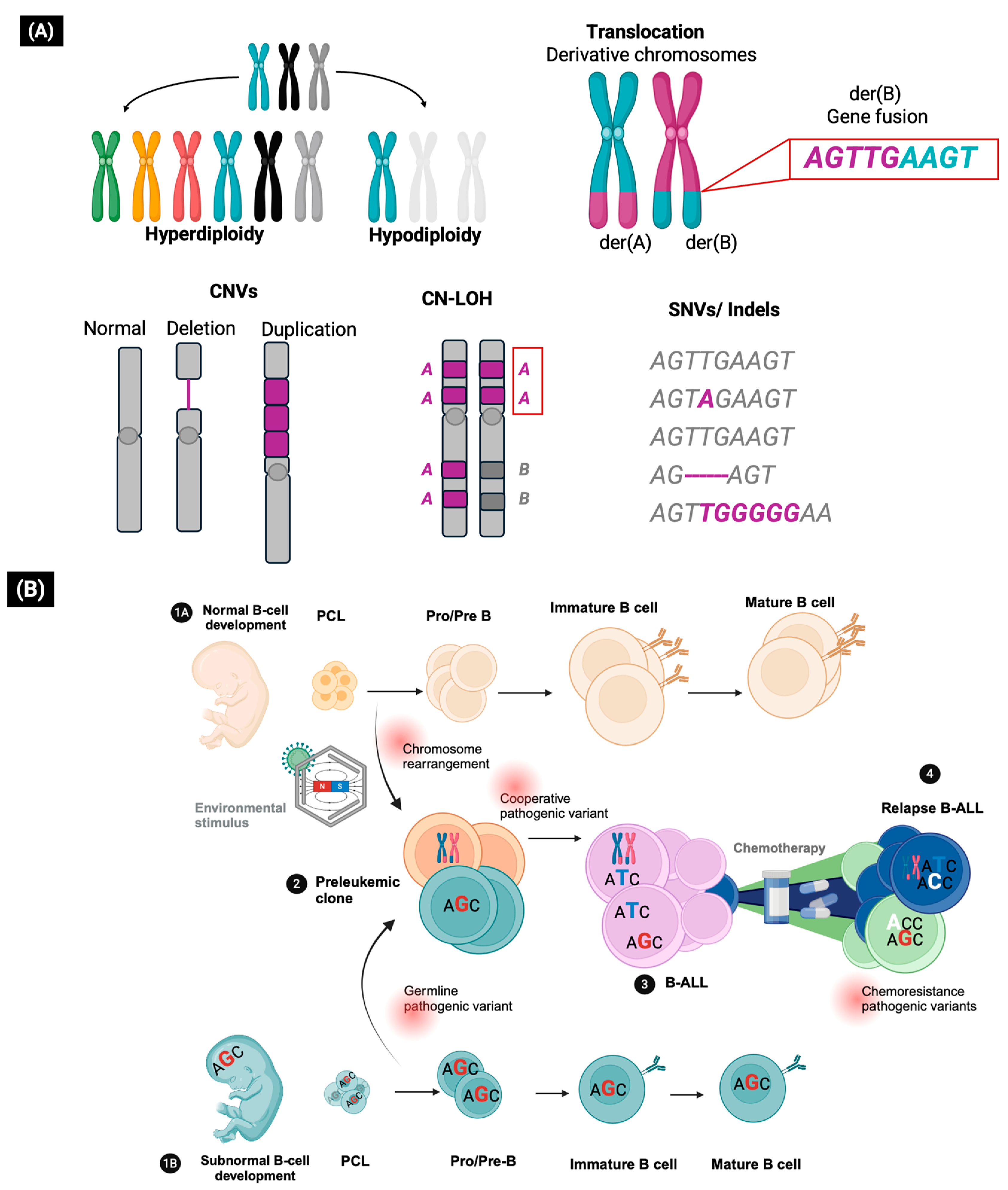
Figure 1. Genetic alterations involved in the pathogenesis of B-ALL. (A) Types of genetic alterations. Gain and loss of whole chromosomes, translocations producing derivative chromosomes (der) carrying gene fusions (red box). Copy number alterations (CNVs), copy neutral loss of homozygosity regions (CN-LOH) (red box), single nucleotide variants (SNVs), and insertions and deletions at the sequence level (Indels). (B) Development of B-ALL. 1A: Normal lymphopoiesis. 1B: Germline predisposition can lead to subnormal lymphopoiesis with reduced progenitor cell populations. 2: The preleukemic clone has chromosomal rearrangements induced by environmental stimuli or by the effect of germline pathogenic variants. 3: The preleukemic clone is susceptible to acquiring cooperative variants that drive its transformation to B-ALL. 4: Chemotherapy may promote the emergence of chemoresistance variants in new or pre-existing clones that drive relapse.
Biomedicines 13 01626 g001
Figure 2. Genes altered by somatic and germline pathogenic variants and their molecular function. S: SNV variant. I: Indel variant. DEL: CNV deletion. AMP: Amplification. GF: Gene fusion.
Biomedicines 13 01626 g002
Figure 3. Somatic biomarkers with clinical value in B-ALL. They are grouped according to their prognostic value. C: Classic biomarkers detected by conventional analysis techniques. B: Biomarkers within the B-other group and detected only by genomic analysis techniques.
Biomedicines 13 01626 g003
Figure 4. Genes affected by germline pathogenic variants grouped by B-ALL susceptibility. The clinical signs associated with genetic syndromes predisposing to B-ALL are shown. 1. Musculoskeletal disorders. 2. CALMS: Café au lait macules. 3. Neurological disorders. 4. Lymphopenia or thrombocytopenia. 5. Family history of neoplasms. Heterozygous genotype: +/−. Homozygous recessive genotype: −/−.
Biomedicines 13 01626 g004
Disclaimer/Publisher’s Note: The statements, opinions and data contained in all publications are solely those of the individual author(s) and contributor(s) and not of MDPI and/or the editor(s). MDPI and/or the editor(s) disclaim responsibility for any injury to people or property resulting from any ideas, methods, instructions or products referred to in the content.