Review

Genetic Animal Models of Cardiovascular Pathologies

1
Institute of Medicine, RUDN University, 6 Miklukho-Maklaya St, Moscow 117198, Russia
2
Laboratory of Molecular Technologies, Shemyakin-Ovchinnikov Institute of Bioorganic Chemistry, Russian Academy of Sciences, Moscow 117997, Russia
3
Institute of Translational Medicine, Pirogov Russian National Research Medical University, Moscow 117997, Russia
4
Institute of Gene Biology, Russian Academy of Sciences, Moscow 119334, Russia
5
Institute for Regenerative Medicine, Sechenov University, Moscow 119991, Russia
6
Department of Sensory Organs, Faculty of Medicine and Dentistry, Sapienza University of Rome, University Hospital Policlinico Umberto I, I 00161 Rome, Italy
7
International Polyamines Foundation, ETS ONLUS, I 00159 Rome, Italy
8
Federal Center of Brain Research and Neurotechnologies, Federal Medical Biological Agency, Moscow 117513, Russia
9
Life Improvement by Future Technologies (LIFT) Center, Moscow 143025, Russia
10
Joint Department with RAS Shemyakin-Ovchinnikov Institute of Bioorganic Chemistry, Faculty of Biology and Biotechnology, National Research University Higher School of Economics, Moscow 105066, Russia
*
Author to whom correspondence should be addressed.
Biomedicines 2025, 13(7), 1518; https://doi.org/10.3390/biomedicines13071518 (registering DOI)
Submission received: 28 April 2025 / Revised: 8 June 2025 / Accepted: 17 June 2025 / Published: 21 June 2025
(This article belongs to the Section Molecular Genetics and Genetic Diseases)

Abstract

:
This review critically examines the evolving landscape of genetic animal models for investigating cardiovascular diseases (CVDs). We analyze established models, including spontaneously hypertensive rats, Watanabe hyperlipidemic rabbits, etc., and transgenic models that have advanced our understanding of essential and secondary hypertension, atherosclerosis, and non-ischemic diseases of the heart. This review systematically evaluates the translational strengths and physiological limitations of these approaches across species barriers. Particular attention is paid to emerging technologies—AAV-mediated gene delivery, CRISPR-Cas9 editing, and chemogenetic tools—that enable unprecedented precision in manipulating cardiac-specific gene expression to study pathophysiological mechanisms. We address persistent challenges including off-target effects and transgene expression variability, while highlighting innovations in synthetic vectors and tissue-specific targeting strategies. This synthesis underscores how evolving genetic technologies are revolutionizing cardiovascular research paradigms, offering refined disease models and optimized therapeutic interventions that pave the way toward personalized medicine approaches for the world’s leading cause of mortality.

1. Introduction

Animal models for investigating the mechanisms of pathogenesis underlying cardiovascular diseases can be categorized into three distinct groups: (1) models carried out using surgical intervention; (2) models in which a pathological process is initiated by introducing chemical or infectious agents into the animal body; (3) genetic models. Definitely, any combination of these methods for modeling a certain pathology are also possible. Each of these model groups has its advantages and limitations. A critical criterion for their selection is the degree to which the physiology of an animal’s circulatory system aligns with human physiology [1]. Unfortunately, none of the currently used models of cardiac diseases can entirely replicate the pathogenesis of the corresponding human pathology, although they do possess certain pros and cons [2,3,4,5,6]. It is also very important to use animal models providing a reliable means of evaluating the efficacy of pharmaceuticals and other methods of intervention in pathological processes in basic and preclinical investigations [2,7]. Recently, the horizons of cardiac pathology modeling have been significantly expanded through the employment of genetic instruments. The advent of extensive genetic resources has also opened new avenues for studying the activity of signaling pathways and modifying the functions of genes across various cellular types [8]. Genetic models are widely used in the investigations of different forms of cardiomyopathy, as they effectively reproduce the intricate processes underlying the disease, which is predominantly inherited. In particular, there are animal models of idiopathic cardiomyopathy, including dilated [9,10,11], hypertrophic [12,13], restrictive [14,15], and arrhythmogenic right ventricular dysplasia (cardiomyopathy) [16,17].
When discussing disorders of the cardiovascular system that can only be attributed to hereditary predispositions, such as hypertension, coronary artery disease (CAD), or various types of cardiac arrhythmia, genetic models provide a valuable tool for exploring the underlying mechanisms of these conditions.
At present, small mammal studies in the field of cardiac pathophysiology, such as mice and rats, are particularly prevalent. The advantages of using rodents in research include a short breeding cycle [18], the ability to perform genetic manipulations, and a wide range of available antibodies specific to mice, which can be used for both basic and pre-clinical in vivo studies [19]. The limitations concerning experiments on small animals include the technical difficulties associated with measuring various indicators using instrumental methods. It is also rather challenging to obtain enough biological material from such animals for research purposes (especially fluids, such as urine and blood samples). Nonetheless, advanced surgical and imaging techniques have alleviated at least some of these challenges [20].
The entire variety of genetic models of heart pathology that exist today can be subdivided into the following groups: (1) genetic strains selected to maintain phenotypic characteristics resulting from spontaneous genetic mutations and naturally occurring pathology; (2) genetically modified animals with certain genes knocked out; (3) transgenic organisms with the genome modified by the insertion of genes from unrelated species [21]; (4) models developed based on genetically encoded tools (vectors) [22,23,24,25].
When developing an experimental design, researchers commonly face the challenge of selecting an animal model that most closely resembles the actual pathological process observed in humans. In our review, we analyze genetic models of the most common cardiovascular diseases, focusing on their advantages, limitations, and the suitability of their application depending on the specific research task.
The process of preparing this review involved employing several methodical approaches in the search and selection of literature sources. The search was performed using databases, including, first of all, PubMed, Google Scholar, and Research Gate. When searching for publications, the following keywords were used (both separately and in combination) in general: genetic models; animal models, mice, murine, rat, rabbit, cat, feline, dog, zebrafish, transgenic; knockout; hypertension; cardiomyopathy; atherosclerosis; arrhythmias; adeno-associated dependoparvovirus A; CRISPR-associated protein 9. Depending on the particular section, we employed the following terms for our search: Section 2 —genetic models, animal models, inbred strains, heart, cardiac, hypertension, cardiomyopathy, atherosclerosis, coronary heart disease; Section 3—genetic models, animal models, transgenic, knockout, heart, cardiac, CRISPR/Cas9; cardiac arrhythmias, channelopathies, cardiomyopathy, atherosclerosis, myocardial hypertrophy; Section 4—genetic models, heart, cardiac, genetically encoded tolls, adeno-associated dependoparvovirus A, transduction, knockout, Cre-LoxP system, plasmid. Priority was given to publications from the last 5–10 years. In total, more than 900 sources were analyzed, and 244 of them were included in the final list of references for this review. The inclusion criteria were as follows: publications strictly related to the topic of this review (genetic models of cardiovascular disease), sufficient breadth and depth of coverage, relevance and reliability of sources (only articles from scientific journals and monographs were considered), as well as accessibility to the full-text version. And we also applied the following exclusion criteria: irrelevant sources, publications with methodological flaws, and sources that did not correspond to the methodology we were analyzing (we were interested in experimental research-based publications).

2. Genetic Strains Selected to Maintain Phenotypic Characteristics Resulting from Spontaneous Genetic Mutations and Naturally Occurring Pathology

Among the genetic animal models of cardiovascular pathology, inbred strains created by breeding animals with spontaneous mutations were developed earlier than other models—in the 1950s. These models, widely used in experimental work, have demonstrated their efficacy, as the corresponding phenotypic traits are largely reproduced in the majority of progeny.
The genetic models demonstrating cardiovascular pathology phenotype, the mechanisms of its formation, possible applications, their advantages and limitations, physiological relevance, genetic manipulability, reproducibility, and translational relevance are summarized in Table 1.
Animal models of hypertension: Today, the most common object for the study of hypertension is represented by rats. Most rats genetically predisposed to hypertension were derived from outbred Wistar and Sprague Dawley (SD) stocks [19]. Inbred strains that are known to reproduce this pathology are as follows: SHRs (spontaneously hypertensive rats), DSS (Dahl salt-sensitive) rats, FHH (fawn-hooded hypertensive) rats, the Milan hypertensive strain, the Lyon hypertensive rat, the Sabra hypertensive rat, Buffalo (BUF) rats, Goto-Kakizaki (GK) rats, Munich Wistar Frömter (MWF) rats, the New Zeland Genetically Hypertensive (NZGH) strain, and some more rare strains.
The SHR strain is the most used rat model that demonstrates a hypertensive phenotype, and it is widely used to study essential hypertension. This strain was developed by crossing outbred Wistar rats that had spontaneously increased blood pressure (BP) [26]. It is considered that the primary pathogenic mechanism underlying the onset of arterial hypertension (AH) in this strain of animals is neurogenic [27,28]. SHR animals also exhibit overexpression of the renin gene [29] and the presence of four mutations in the first 1100 base pairs of the first intron of the renin gene [30]. Thus, this model closely resembles the pathogenesis of human essential hypertension [28]. In adult male SHRs, SBP is typically above 180 mmHg and DBP is above 130 mmHg at rest, as established by the method of telemetric monitoring that eliminates the influence of stress or any other external factors [31,32].
In most cases, SHRs develop cerebral stroke, so this strain or its substrains (SHR-SP—spontaneously hypertensive stroke-prone rats) are widely used as a model for studying cerebral circulatory disorders [33,34]. This model can also serve as a valuable tool for investigating vascular and renal abnormalities in hypertension, as well as genetic mechanisms underlying its development. Moreover, SHRs can also be used to study the transition from compensatory left ventricular hypertrophy to heart failure [35,36].
The DSS (Dahl salt-sensitive) strain was developed by Lewis Dahl in the 1950s, and it represents inbred rodents that are extremely sensitive to high-salt diets [37,38]. Initially, the animals were fed on this diet, and it was found that some of them developed AH, while the others maintained a normal BP pressure level. After three generations, a strain of rats sensitive to high-salt diets was selected [39]. Interestingly, this study, on the one hand, confirmed the role of excessive salt intake as a risk factor for hypertension. On the other hand, it was shown that the development of hypertension due to salt intake is influenced by genetic predisposition. Subsequently, it was found that salt-sensitive rats have the allele of the gene encoding 11B-hydroxylase (CYP11B1), an enzyme necessary for the synthesis of a steroid that stimulates salt retention, which is responsible for the development of hypertension [40]. This model is particularly valuable for studying the role of sodium in the development of hypertension.
The FHH (fawn-hooded hypertensive) is an inbred strain that was selected by crossing outbred “Fawn-hooded” animals with chronic renal failure accompanied by a mild increase in BP, compared to Wistar rats [41]. In fact, the FHH strain is a genetic model of chronic renal disease, characterized by the development of glomerulosclerosis and proteinuria [42] followed by the development of hypertension [43]. It is still difficult to determine with certainty whether hypertension or nephropathy is the primary condition in this case. However, it was shown that in FHH K572Q rats, the ADD3 gene contributes to modifying the myogenic response and the autoregulation of renal and cerebral blood flow, which results in increased susceptibility to kidney disease caused by hypertension [44,45].
The FH strain finds its application in the study of hypertensive nephropathy, as well as the genetic basis of hypertension.
The MHS (Milan hypertensive strain) was developed as a result of breeding Wistar rats that exhibited increased BP [46]. The main mechanism underlying the development of hypertension in animals from this strain is a genetically determined kidney dysfunction, characterized by an imbalance between the glomerular filtration and the tubular reabsorption of sodium and water [27]. The hypertension in this rat strain is linked to a mutation in the ADD gene, encoding the protein adducin (actin-binding protein) [44]. The mutation in this gene is also associated with renal dysfunction and cognitive disorders [47]. This model can be used to study primary hypertension and the genetic mechanisms of its development.
The Lyon hypertensive rat is an inbred strain of animals characterized by elevated BP levels. This strain was developed in the 1970s by a group of French researchers by repeated crossbreeding of Sprague Dawley rats with different BP levels, which resulted in the creation of three animal strains: those with normal BP (LN), those with elevated BP (LH), and those with lowered BP (LL) [48,49]. Interestingly, this model is not only characterized by signs of spontaneous hypertension but also demonstrates manifestations of metabolic syndrome [50], salt-sensitive hypertension [51], impaired renal function and proteinuria [52], and increased insulin blood levels and insulin-to-glucose ratio (insulin resistance) [50]. The results of genetic mapping in Lyon hypertensive rats showed that chromosome 17 contains two QTL clusters: one associated with body weight and organ hypertrophy, and the other associated with BP level, plasma lipids, and insulin levels [53]. It has recently been shown that a mutation in the gene Ercc6 l2 appears to be the best candidate determining the phenotype of obesity and metabolism in a novel congenic strain LH17LNa [54]. For today, the Lyon hypertensive rat is the gold standard model for studying hypertension as part of metabolic syndrome.
Sabra hypertensive rats are a less commonly used genetic model for hypertension. Nowadays, the PubMed database contains a modest number of a bit over 90 articles. Two inbred substrains, Sabra hypertensive-prone (SHP) and Sabra hypertensive-resistant (SHR) rats, were selected: the former one exhibited increased sensitivity to salt with the development of hypertension [55]. Animals of both genetic substrains receiving an ordinary diet are characterized by normal BP [56]. The main mechanism of hypertension in this model is genetically determined salt susceptibility [57]. It is noted that chromosome 1 loci are associated with increased BP in this rat line [58,59]. Moreover, three potential gene loci of chromosomes 1 and 17, which play an important role in salt sensitivity and/or resistance, have been identified [59]. SBH animals also have proteinuria and nephrosclerosis [60]. The SBH strain is a good model for the study of salt-induced hypertension, as well as hypertensive nephropathy.
The New Zealand Genetically Hypertensive (NZGH) rat strain was developed in the 1970s by crossing outbred animals with a hypertensive phenotype [61]. It was shown that the renin–angiotensin–aldosterone and sympathoadrenal systems do not play a primary role in the development of hypertension in this animal model. However, they are characterized by increased reactivity of peripheral vessels to various vasoconstrictor agents, which is due to the involvement of neurogenic and myogenic factors, as well as structural factors. It is also notable that animals of this strain do not develop hypertension even when kept on a high-salt diet [61].
Some reports suggest the possibility of hypertension in Munich Wistar Frömter (MWF) rats, which are a model of chronic kidney disease associated with a decrease in the number of superficial glomeruli [62,63]. At the same time, there is little evidence in the literature indicating the development of persistent hypertension in this animal strain. There are also limited data concerning the possible genetic basis for its formation. In this regard, it is challenging to recommend this strain as a hypertension model.
Genetic models of hypertension in large animals: Research on hypertension employing large animal models is currently less prevalent. However, in some cases, it is necessary. Large animals are preferable for experiments using various surgical techniques, as well as intraventricular manometry. Spontaneous increases in BP were found in turkeys [64], rabbits [65], pigs [66], and dogs [67,68]. Strains of New Zealand and Dutch white rabbits with spontaneous hypertension were developed using the method of selective breeding [19].
Animal models of idiopathic cardiomyopathy: Idiopathic cardiomyopathies include dilated cardiomyopathy (DCMP), hypertrophic cardiomyopathy (HCMP), restrictive cardiomyopathy (RCMP), arrhythmogenic right ventricular dysplasia (cardiomyopathy), and unclassified cardiomyopathy. Currently, there are animal genetic models for every type of cardiomyopathy that reproduce the pathogenesis of these diseases.
Genetic models of dilated cardiomyopathy (DCMP): The most prevalent DCMP model involves Syrian golden hamsters derived from various genetic substrains of the BIO strain (BIO 1.50, BIO T0-2 and others) (Figure 1) [69,70].
This strain was first described in 1962. It was the result of breeding a naturally occurring mutant strain of Syrian hamsters, with an autosomal recessive mode of inheritance, with 100% penetrance. This strain exhibited signs characteristic of both cardiomyopathy and muscular dystrophy [69]. The main cause of structural and functional changes in the hearts of these hamsters is the deletion of the gene encoding delta-sarcoglycan. This protein is part of a complex associated with dystrophin. This leads to a loss in delta-sarcoglycan in the CMC [71]. The animal model of the BIO T0-2 substrain shows a complex of morphological changes, in particular, enlargement of the heart chambers and inflammatory infiltration of the myocardium [72]. Electron microscopy reveals shortening of sarcomeres in 7 out of 10 cases [70]. Moreover, in experiments on the same substrain, BIO T0-2, a complex of intra-cardiac and systemic hemodynamic abnormalities observed in human DCMP was identified: low cardiac output, increased preload, and decreased renal blood flow [73], as well as pronounced changes in the Frank–Starling mechanism [70]. All these facts allow us to recommend Syrian golden hamsters from the BIO strain as an optimal model for studying the pathogenesis and genetic basis of DCMP, as well as its gene therapy.
DCMP also occurs in wild and domestic turkeys and dogs (Figure 2) [74].
In domestic turkeys, in 2–5% of cases, DCMP develops within the first 4 weeks of life and occurs in the absence of other cardiac abnormalities. This condition is characterized by dilated cardiomyopathy, leading to heart failure and high mortality rates in affected turkeys. The underlying causes are not fully understood but may involve genetic predisposition and environmental factors [75]. In turkeys with DCMP, low-molecular cardiac troponin T (cTnT) is expressed in myofibrils due to the absence of exon 8. This is accompanied by a change in the conformation and binding affinity for TnI и TP [76]. We should note that the presence of DCMP in turkeys and dogs has not resulted in the development of genetic strains with the corresponding pathological phenotype. In this regard, we are not discussing the existence of a specific animal model. At the same time, mutations in the troponin T gene in these animals allow us to study the genetic mechanisms of DCMP, as it is known that troponin T mutations are found in 3–6% of human DCMP patients [77].
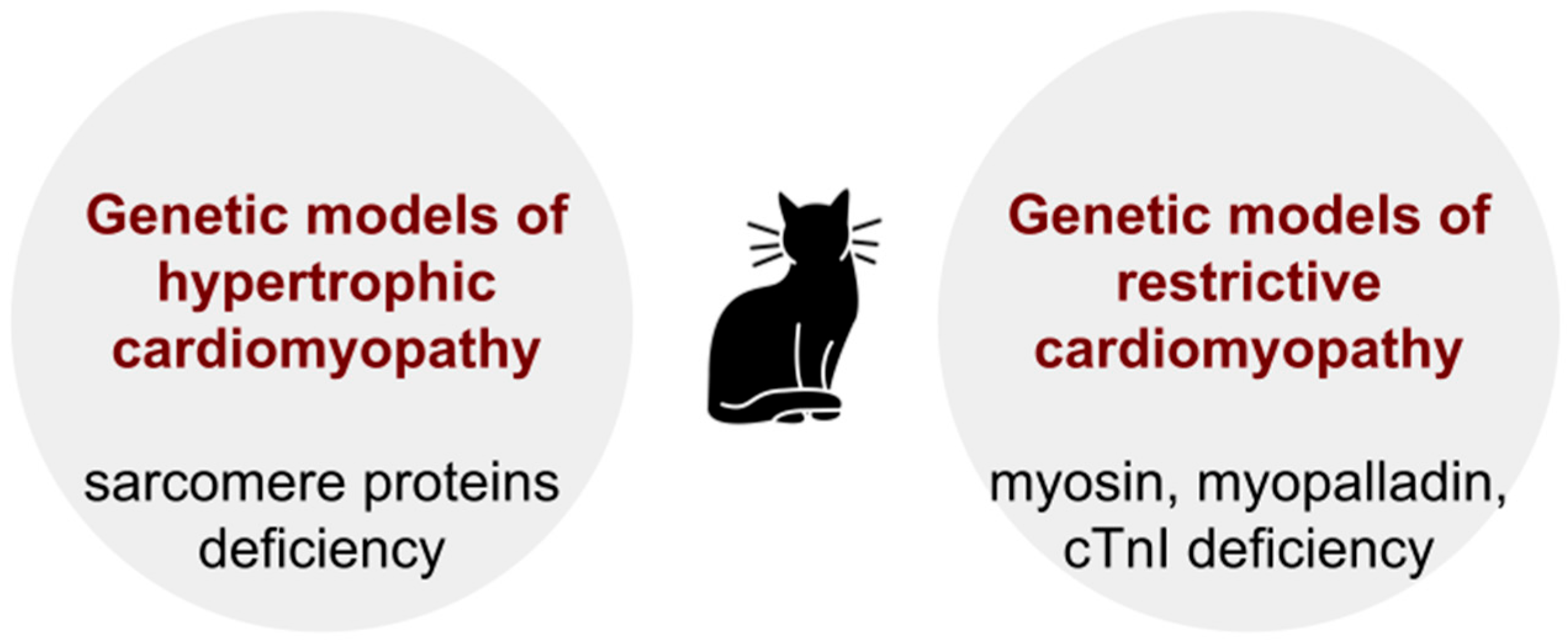
Genetic animal models of hypertrophic cardiomyopathy (HCMP). The estimated incidence of hypertrophic cardiomyopathy (HCMP) in the general population is approximately 1:500 [78]. In contrast to DCMP, familial cases of HCMP are 30–50% [79,80], while only 30–40% of them can be attributed to a genetic cause [79,81]. HCMP has a well-established genetic basis (mutation) [82,83,84]. Most patients have mutations in the gene MYH7 (OMIM: 160760) encoding myosin heavy chains and the gene MYBPC3 (OMIM: 600958) encoding myosin binding protein, as well as cardiac troponin C (TNNC1; OMIM: 191040), cardiac troponin I (TNNI3; OMIM:191044), cardiac troponin T (TNNT2; OMIM:191045), cardiac actin (ACTC1; OMIM: 102540), α-tropomyosin (TPM1; OMIM:191010), regulatory myosin light chain, essential myosin light chain, and titin/connectin. In more than 50% of cases, the disease is caused by mutations in the MYH7 or MYBPC3 genes [85]. In 5–10% of cases, the disease is caused by a mutation in the genes encoding non-sarcomeric proteins, which are associated with neuromuscular diseases [86].
Naturally occurring HCMP is often found in pet cats, rendering them as an acceptable model of this disease [87]. The prevalence of HCMP in the general cat population is 10 to 15% [88]. An even higher risk is typical for some certain breeds of felines, including the Maine Coon cat, Persian, Ragdoll, and Sphynx [87]. It is important that feline HCMP is characterized by the same morphological, pathogenetic, and clinical features as human HCMP [89,90]. As in humans, the pathogenesis of feline HCMP is based on mutations in the genes that encode sarcomere proteins [87]. Maine Coon cats have mutation A31P in the gene MYOC, which is found in 22–42% of individuals. In Ragdoll animals, the mutation R820W of the same gene is observed in 27% of cases [91]. The R820W mutation of the MYBPC3 gene has also been found in human patients with HCMP, which allows us to consider this genetic abnormality as a common pathogenic basis for HCMP in both cats and humans [92]. Nonetheless, not all Maine Coon cats with advanced HCMP have the A31P mutation in the gene MYBPC3 [87]. Despite the existence of animals with naturally occurring HCMP, there is currently a wide number of transgenic models. The disadvantage of rodent transgenic models is the fact that, even with the same genetic mutations as in humans, the phenotypic manifestations can be different. Mice with mutations in the gene encoding myosin-binding protein (MYBPC) do not develop myocardial hypertrophy [87].
Genetic models of restrictive cardiomyopathy (RCMP): In approximately 30% of cases, idiopathic RCMP has a familial character. In particular, in patients with familial RCMP, mutations associated with this disease were found in the following genes: TNNT2 (cardiac troponin T; OMIM: 191045), TNNI3 (cardiac troponin I; OMIM: 191044), MYBPC3 (myosin-binding protein C), MYH7 (myosin heavy chain 7; OMIM: 160760), MYL2 (myosin light chain 2; OMIM: 160781), MYL3 (myosin light chain 3; OMIM: 160790), DES (desmin; OMIM: 125660), MYPN (myopalladin; OMIM: 608517), TTN (titin; OMIM:188840), BAG3 (Bcl-2-associated athanogene 3; OMIM: 603883), DCBLD2 (discoidin, CUB and LCCL domain containing 2; OMIM: 608698), LMNA (lamin A/C; OMIM: 150330), and FLNC (filamin C; OMIM: 102565) [93]. And as in the case of HCMP, these are mutations in genes encoding structural proteins of sarcomeres or proteins associated with sarcomeres [94]. Some clinical and histopathological features of RCMP are like those of HCMP. Moreover, mutations were found in some of the same genes (TNNT, TNNI3, MYBPC3, MYH7). And we may suggest that DCMP and HCMP share a few common genetic and pathogenic mechanisms [95]. Naturally occurring genetically determined RCMP was found in cats. Genetic animal models with similar mutations in the genes of myosin, TNNI3, and MYPN demonstrate the diastolic dysfunction phenotype, which is a major clinical manifestation of human RCMP [15]. Particularly, under natural RCMP in cats, there is a pronounced thickening of the endocardium, as well as replacement fibrosis and deformation of the heart chambers [96,97].
The use of animal models for HCMP and RCMP is complicated by the fact that inbred strains have not been developed. Since, as mentioned previously (Figure 3), the pathogenic, morphological, and clinical patterns of these diseases more closely resemble human cardiomyopathy than transgenic models, it is a more suitable approach for translational studies. In this regard, it seems reasonable to use an approach that involves collaboration with veterinary clinics and relevant services that are ready to provide autopsy tissue samples. Such an approach was used in the study performed by Kimura Y. et al. [97].
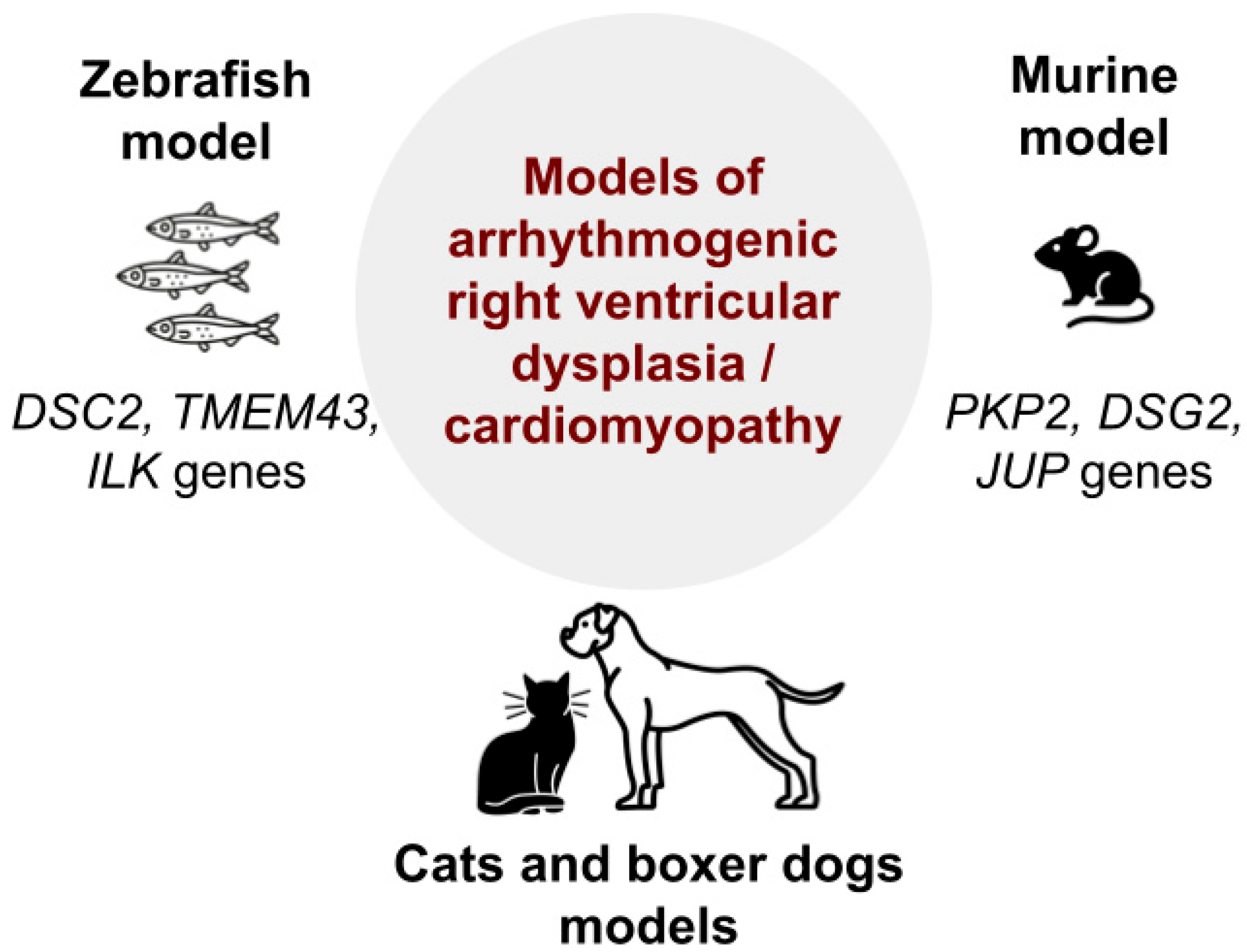
Animal models of arrhythmogenic right ventricular dysplasia/cardiomyopathy (ARVC): ARVC is a rare genetically determined disease that is inherited in an autosomal dominant pattern. It is characterized by the replacement of myocardial elements with fibrous and fatty tissue, which can lead to heart failure and sudden cardiac death. This disease is associated with mutations in the genes encoding proteins of the desmosome complex [98], in particular, DSP (desmoplakin; OMIM: 125647) [99], PKP2 (plakophilin 2; OMIM: 602861) [100], DSG2 (desmoglein 2, desmocollin 2; OMIM: 125671) [101], JUP (plakoglobin (related to γ-catenin; OMIM: 173325) [102,103], TMEM43 (transmembrane protein 43; OMIM: 612048), and ILK (integrin-linked kinase; OMIM: 602366) [104,105].
Mutations in the gene PKP2 are the most common cause of familial ARVC [106]. Today, there are no available animal models of ARVC developing because of mutations in the gene DSP [98]. However, there are knockout murine models of deficiency of the genes PKP2 [107], DSG2 [108], and JUP [109], as well as DSC2-related ARVC in zebrafish [110]. Moreover, it was also found that overexpression of wild-type and p.P111L-mutant TMEM43 in a transgenic zebrafish model was associated with cardiac enlargement and cardiomyocyte hypertrophy, which confirms the role of transcriptomic alterations in the pathogenesis of TMEM43-associated cardiomyopathy [111]. It was also shown that two missense variants (p.H33N and p.H77Y) in the ILK gene are associated with arrhythmogenic cardiomyopathy. Additionally, expression of human wild-type and mutant ILK in zebrafish suggests that p.H77Y and p.P70L may be responsible for cardiac dysfunction and death within 2–3 weeks after birth [105]. Transgenic models will be discussed in more detail in the following section of this review. As regards natural genetic models, spontaneous ARVC is observed in dogs and cats. In particular, spontaneous genetically inherited ARVC closely resembling this disease in humans was described in boxer dogs. It is characterized by ventricular arrhythmia, syncope, and sudden cardiac arrest. Morphological examination reveals a reduction of the number of cardiomyocytes and their replacement by fatty or fibrous–fatty elements [112]. Spontaneous ARVC in pet cats was described as a model for this pathology in several studies.
Fox P.R. et al. examined 12 cats with this disease, and in 8 animals, they diagnosed right ventricular congestive heart failure with supraventricular and ventricular tachyarrhythmias. Morphological analysis demonstrated a marked enlargement of the right ventricle and thinning of its wall in all 12 animals, atrophy and death of CMCs, replacement fibrosis, and fatty degeneration [113] (Figure 4).
Genetic animal models of atherosclerosis and coronary heart disease: Currently, surgical occlusion (ligation) of one of the branches of the coronary artery is still successfully used as a model for acute myocardial infarction. Obviously, this approach does not reproduce the natural pathogenesis of coronary heart disease, which is typically characterized by a slow progressive atherosclerotic lesion of muscular–elastic arteries. The classical model of atherosclerosis, suggested by N.N. Anichkov, which has been used for many decades, also has certain limitations. In the 1980s, Watanabe Y. et al. used selective breeding to develop a genetic strain of rabbits with congenital hypercholesterolemia, Watanabe heritable hyperlipidemic (WHHL) rabbits, as a model for atherosclerosis [114]. These animals have a genetic abnormality resulting in a disorder of the expression of cellular receptors to LDL, similar to human familial hypercholesterolemia [115]. Rabbits of the WHHL strain are characterized by increased LDL levels, followed by the development of severe atherosclerosis. Atherosclerotic arterial damage undergoes several stages in its development. As the intimal damage progresses, deposits of lipids and foam cells appear on the inner surface of the vessel, leading to the formation of plaques and an increase in lipid content in the tunica media. Afterwards, calcification often takes place [115,116]. The atherosclerotic changes in arteries in this case are more similar to the morphogenesis of atherosclerosis in humans compared to the cholesterol-fed rabbit model [117].
As concerns the use of WHHL strain rabbits as a model for coronary heart disease, including myocardial infarction, there is still no clear consensus on this matter. Some studies provide data on the development of coronary syndrome and spontaneous myocardial infarction following severe atherosclerosis in animals of this strain [118,119]. However, we could not find confirmation of these observations in the works of other authors. At the same time, the WHHL rabbit model is widely used to study the cholesterol-lowering effects of statins [120,121,122,123] and non-statin compounds [124,125] in atherosclerosis. Animals from the WHHL family have also been successfully used in the development of gene therapy for atherosclerosis [126]. Particularly, the transfection of the LDLR (low-density lipoprotein receptor; OMIM: 606945) gene into the liver cells of WHHL rabbits using a lentiviral vector resulted in a significant decrease in serum cholesterol levels [127]. There are also transgenic models of hypercholesterolemia and atherosclerosis developed in mice and rats. However, it is rabbits that have several metabolic features close to humans [128]. Rabbits have high activity of the cholesterol ester transporter protein (CETP) in blood plasma [129].
Summarizing the above, we can conclude that the WHHL strain is the optimal model of atherosclerosis that can be successfully used to investigate its pathogenetic features, as well as to develop new methods of pharmacotherapy and gene therapy. However, to induce ischemic and ischemic–reperfusion injury to the myocardium, models based on the mechanical occlusion of coronary arteries are more appropriate.

3. Transgenic and Knockout Animal Models

Methods of genome modification in animal models of cardiovascular pathology. Several genome modification methods are commonly used to create animal models. The oldest one is transgenesis by random integration. This method is based on the ability of DNA molecules delivered into the zygote pronucleus to integrate into the genome [130]. The design of genetic constructs for random integration necessarily includes a gene of interest, a promoter, and regulatory elements to mitigate potential position effects. The integration of delivered constructs occurs randomly, making it impossible to predict the insertion site, the number of integrated transgene copies, or the level of their expression in advance. This phenomenon is known as the “position effect of the transgene” [131,132]. Therefore, when creating transgenic animal strains using the random integration method, multiple strains with the same transgene insertion are typically generated. In this case, a strain with the desired level of transgene expression is subsequently selected. For example, a mouse model of short QT syndrome was generated using the random transgenic insertion method [133]. Similarly, a model of myocardial hypertrophy induced by the overexpression of platelet-derived growth factors (PDGFs) has also been developed using this approach [134]. Moreover, transgenic mice with overexpression of desmocollin-2 serve as a model for cardiomyopathy [135].
The development of the CRISPR/Cas9 genome editing system has significantly increased the efficiency of gene modification. This method is widely used for generating knockout models, particularly in research focused on evaluating the role of genes in cardiac function [136,137].
Another commonly used approach for obtaining genetically modified animal strains with desired mutations is the generation of chimeric embryos via embryonic stem cell (ESC) microinjection. In this method, the genome of ESCs is modified, and clones with the desired genetic modifications are selected and used to create chimeric embryos. When such chimeric mice (F0) are born, genetically modified non-chimeric offspring (F1) must be obtained from them. To produce homozygous animals, it is necessary to breed heterozygous F1 individuals. This process requires a significant period, but it enables the creation of animals with highly precise genetic mutations. The ESC method for generating genetically modified animals is widely used in various fields, particularly in cardiovascular research [138,139].
Recent advances in genome sequencing have enabled the identification of the genetic basis of many myocardial pathologies, including channelopathies and cardiomyopathies [140,141,142,143,144]. At the same time, genome editing technologies empower the development of near-personalized models replicating mutations observed in specific patients (or patient groups) [145,146]. These models facilitate the investigation of various factors, such as the role of pro-inflammatory responses in cardiovascular diseases [147]. This raises the following question [148,149,150]: are certain laboratory animals appropriate as model organisms for specific studies?
Common genetically modified model animals of cardiovascular pathologies: Research has extensively explored animal models of heart conditions, and mice remain the predominant choice for modeling these diseases [1,148,149,150,151,152,153]. Their advantages include a well-characterized genome homologous to the human one, established genetic modification techniques, and relatively low operational costs. Genetically modified mice have been developed to mimic various human cardiac conditions, such as cardiomyopathies, e.g., transgenic mouse models of dilated cardiomyopathy carrying the TTNtv c.13254T > G mutation [154] or Lmna c.1621C > T mutation [155], transgenic murine models of hypertrophic cardiomyopathy carrying the c.1826dupA mutation [156] or p.G823E mutation [157]; arrhythmias, e.g., mice models of Mog1 knockout [158], Kcne3 knockout [159], or the p.Asn1768Asp mutation [160]; and other inherited heart diseases, e.g., mice models of congenital heart defects, including truncating FOXJ1 variant (c.784_799dup; p.Glu267Glyfs*12) [161], Fgf10 and Fgfr2-IIIb [162], and Hnrnpa1 mutations [163]. However, there are some challenges in using mice as biomedical models. In some cases, mutations that are homologous to the ones found in humans are not realized as a pathological phenotype in mice or result in a different phenotype [164]. Moreover, the investigation of cardiac development during embryogenesis is complicated due to difficulties in dynamic imaging, despite existing ex utero culturing techniques [165,166].
Another popular model organism is the zebrafish (D. rerio) [17,167,168]. Homozygous D. rerio with a mutation in the bag3e2/e2 gene serve as a model for dilated cardiomyopathy [169]. Knockout of gtpbp3 leads to hypertrophic cardiomyopathy [170]. Overexpression of a transgenic construct containing the atrial-specific myosin light chain 4 gene MYL4 (OMIM: 160770) with the p.Glu11Lys mutation results in the development of arrhythmia [171]. One of its key advantages is the relatively high genomic homology with humans [172]. Additionally, D. rerio exhibits rapid embryonic development: its heart begins to beat just one day after fertilization [173]. The transparency of zebrafish embryos and their external development facilitate easy observation of embryonic stages. Another significant advantage of D. rerio as a model organism is the low cost of genetic modification [174] and the general simplicity of maintaining these animals. However, despite its many advantages, the major limitation of D. rerio for the research of cardiac disorders is its two-chambered heart. For this reason, certain processes characteristic of the human heart cannot be fully modeled using D. rerio.
Rabbits are an excellent model organism for numerous biomedical studies [175]. This is primarily due to the similarity between rabbit and human lipid metabolism, which enables the development of appropriate models of lipid metabolism disorders, including, first of all, atherosclerosis [176,177,178]. Moreover, the efficiency of the CRISPR/Cas9 genome editing system in rabbit embryos is exceptionally high, significantly accelerating and simplifying the creation of knockout models [176,179,180,181]. Despite the existence of rabbit models for heart diseases [182], the use of this species in biomedical research remains limited. This is primarily due to the relatively high cost of their maintenance and their relatively long reproductive cycle. Transgenic rabbits serve as models for monogenic human diseases. For example, the expression of a mutant variant of β-MHC-Q403 leads to myocardial hypertrophy [183]. Additionally, the expression of a mutant variant KCNQ1/KvLQT1-Y315S (a dominant-negative mutation in the K+ channel α-subunit [KvLQT1]) in models prolonged QT/APD [184].
The comparative advantages and disadvantages of popular animal species used for modeling heart diseases are presented in Table 2.
Since there is currently a vast variety of transgenic animal models available for studying cardiovascular pathology, it would be challenging to thoroughly analyze all of them in the frame of single review. In this regard, we only present general principles and approaches that are used in this area, as well as certain examples of transgenic models of diseases of the cardiovascular system.
Transgenic animal models of genetic channelopathies and some types of cardiac arrhythmias:
Channelopathies arise from mutations in ion channel genes or their regulatory components, increasing the risk of arrhythmias in structurally normal hearts. Various animal models have been developed to investigate the pathophysiology of these disorders.
For example, mutations leading to spontaneous Ca2⁺ release from RYR2 (ryanodine receptor 2; OMIM: 180902) sarcoplasmic reticulum (SR) Ca2⁺ release channels result in catecholaminergic polymorphic ventricular tachycardia (CPVT) and atrial fibrillation (AF). The RyR2+/ mouse model exhibits arrhythmogenic phenotypes like the ones that are observed in CPVT patients [185].
Cardiac calsequestrin 2 (CASQ2; OMIM: 114251) mutations, transmitted by an autosomal recessive type of inheritance, lead to a loss-of-function phenotype. CASQ2 acts as a high-capacity Ca2⁺ buffer in the SR and regulates RYR2 gating. Deletion of CASQ2 in mice (Casq2/) results in severe exercise- and catecholamine-induced arrhythmias, mimicking the human disease phenotype [186].
Congenital long QT syndrome (LQTS) is often a multi-organ disorder caused by mutations in genes essential for cardiac repolarization, leading to the following symptoms: seizures, syncope, arrhythmia, and sudden cardiac death. The majority of LQTS cases arise from mutations in KCNQ1 (potassium channel voltage-gated KQT-like subfamily member 1; OMIM: 607542), KCNH2 (potassium channel voltage-gated subfamily H member 2; OMIM: 152427), or SCN5A (sodium voltage-gated channel alpha subunit 5; OMIM: 600163) [187]. The Kcnq1/ mouse model exhibits characteristic features of LQTS, including abnormal T- and P-wave morphologies and prolonged QT and JT intervals when assessed in vivo [188]. AF can also be triggered by mutations in KCNE1 (potassium channel voltage-gated Isk-related subfamily member 1; OMIM: 176261) and SCN5A with corresponding mouse models replicating AF susceptibility [189,190].
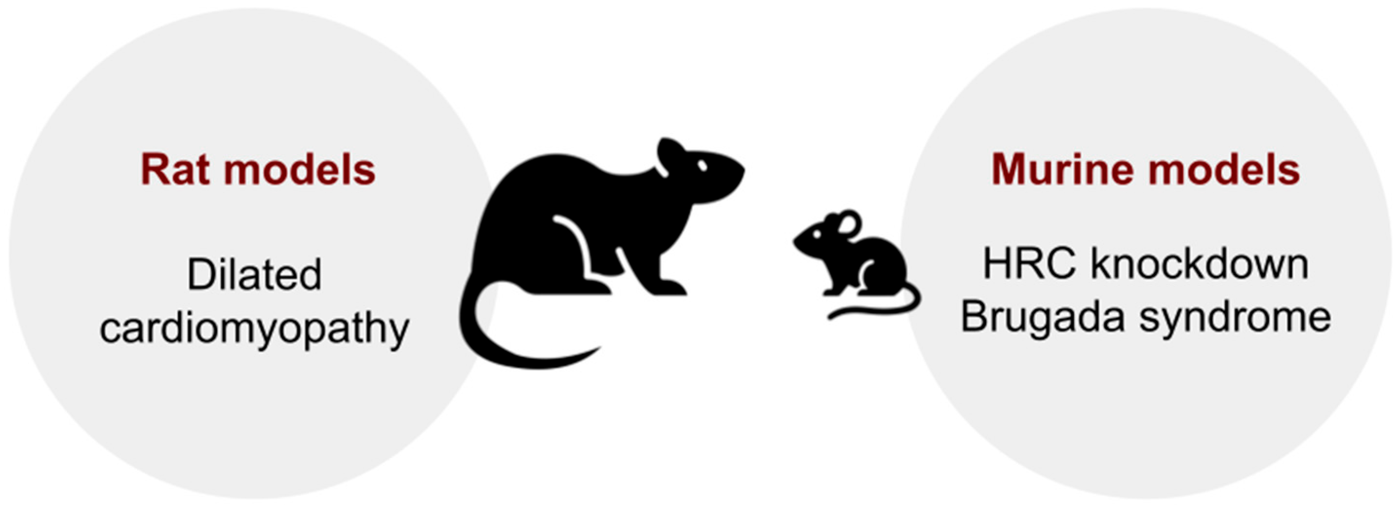
Brugada syndrome, characterized by ST-segment elevation, partial bundle branch block, arrhythmia, and sudden cardiac death, is most commonly associated with SCN5A mutations. The Scn5a+/ mouse model displays conduction disease phenotypes that parallel human Brugada syndrome [191].
Beyond these models, numerous studies have explored the effects of mutations, deletions, or dysregulation of ion channels, transcription factors, developmental pathways, and hormones to better understand the mechanisms underlying cardiac arrhythmias [1]. These animal models remain essential for elucidating disease mechanisms and evaluating potential therapeutic strategies.
Transgenic animal models of cardiomyopathy:
Dilated cardiomyopathy (DCMP): CRISPR/Cas9 genome editing of the dystrophin gene was used to create a Duchenne dystrophy animal model characterized by the development of heart pathology resembling DCMP [192]. Mutant rats developed left and right ventricular systolic dysfunction and myocardial fibrosis. The animals also exhibited systemic muscle atrophy accompanied by high mortality [193]. Several other genetic models of idiopathic DCMP have been created in mice. Homozygous mice with desmin knockout developed heart damage characteristic of DCMP, including thinning of the left ventricle, fibrosis, and systolic dysfunction observed in autosomal recessive desminopathies [194]. Progressive DCMP may be caused by a disruption of the nuclear envelope structure of cardiomyocytes, which may be due to a mutation in the LEMD2 (LEM domain-containing protein 2; OMIM: 616312) gene [195]. The editing of desmosomal genes (desmocollin-2 and desmoglein-2) leads to the development of biventricular cardiomyopathy associated with inflammation, necrotic death of cardiomyocytes, and replacement sclerosis. This complex of changes closely resembles the pathogenesis of DCMP and can serve as its genetic model [135].
Hypertrophic cardiomyopathy (HCMP): For today, more than 1400 autosomal mutations transmitted by the autosomal dominant type of inheritance have been identified in 11 genes encoding proteins of the thick and thin strands of the sarcomere or components of neighboring Z-discs. Transgenic animal models include the following animal species: rodents, rabbits, and zebrafish [196]. The first animal model developed to study the pathogenesis of HCMP was a murine strain carrying a missense mutation in codon 403 in the MYH6 gene corresponding to the R403Q mutation in MYH7 (myosin heavy chain 7 cardiac muscle beta; OMIM: 160760) in humans. The R403Q model exhibited various pathological features, including myocellular disorder, fibrosis, and diastolic dysfunction, reproducing the pathology observed in human tissues [197]. Myosin-binding protein-C (MyBP-C) is the most common gene involved in the pathogenesis of HCMP in humans. A series of murine strains carrying modified MyBP-C with deleted myosin and titin-binding domains were generated and studied [198]: these transgenic mice developed mild hypertrophy, impaired sarcomere structure, and an altered ability to exercise [199]. Further investigations provided a wide range of transgenic models, predominantly in mice and other rodents, through the manipulation of specific involved genes.
Restrictive cardiomyopathy (RCMP): As it was previously noted, the pathogenesis of RCMP is linked to mutations in the myosin genes TNNI3 and MYPN. In the early 2000s, a model of transgenic mice (cTnI193His) simulating cTnI R192H mutation (cTnI R193H in the mouse sequence) in the human heart was created. Another strain of transgenic mice with the RCM cTnI K178E mutation was also developed [200]. In cTnI K179E transgenic mice, a sharp enlargement of both atria was observed in the absence of ventricular hypertrophy and dilation. They exhibited hemodynamic features similar to cTnI193His mice, which confirms the development of RCMP as a result of the TNNI3 mutation: the animals had severe cardiac dysfunction [201]. There was also a marked hypersensitivity of cardiomyocytes to Ca2+ [202].
Arrhythmogenic right ventricular dysplasia/cardiomyopathy (ARVC): Since most patients suffering from ARVC carry one or more genetic variations of desmosomal genes, the number of cardiac desmosomes is significantly reduced in embryos with Iup deficiency. Consequently, it was also shown that the knockout of Dsx, Ds2, and Psp2 leads to embryonic death in mice due to severe heart failure. [99,101,107]. Later, based on advances of animal genetic engineering, various transgenic and knockout animal models were created, including the strains with both desmosomal and non-desmosomal protein knockout [16].
Transgenic animal models of atherosclerosis:
Transgenic animal models have played a crucial role in elucidating the mechanisms of atherosclerosis and evaluating the efficiency of therapeutic interventions. Among rodents, mice remain the most widely used objects for modeling due to their genetic controllability, cost-effectiveness, and short disease induction period. Apolipoprotein E-deficient (apoE−/−) and low-density lipoprotein receptor-deficient (Ldlr−/−) mice are the cornerstone models, accounting for over 95% of atherosclerosis studies [203]. ApoE−/− mice develop spontaneous hypercholesterolemia and aortic lesions on a chow diet, with accelerated plaque progression under high-fat diets, while Ldlr−/− mice require dietary cholesterol to elevate LDL and form lesions [204,205,206]. However, murine models differ from humans in lipoprotein metabolism—mice lack cholesteryl ester transfer protein (CETP) and exhibit HDL-dominant profiles—limiting translational extrapolation.
To address these disparities, novel models have been developed: apoE3-Leiden. CETP mice incorporate human CETP (cholesterol ester transfer protein plasma; OMIM: 118470), enabling LDL-driven atherosclerosis and human-like drug responses [207]. Moreover, PCSK9 gain-of-function adeno-associated virus (AAV) models induce hypercholesterolemia without germline editing, offering flexibility for studying plaque calcification and regression [208]. Advanced models like SR-BI KO/ApoeR61h/h mice replicate coronary atherosclerosis and spontaneous myocardial infarction, bridging the gap between murine physiology and human clinical outcomes [209].
Rabbit models, particularly Watanabe heritable hyperlipidemic (WHHL) and transgenic strains, offer unique advantages due to their human-like lipoprotein metabolism, including LDL dominance and CETP activity. WHHL rabbits, deficient in LDL receptors, develop spontaneous hypercholesterolemia and complex lesions resembling human plaques, making them valuable for studying plaque rupture and statin efficacy [210]. Transgenic rabbits expressing human lipoprotein(a) or apoB-100 further enable research on lipid-driven atherosclerosis and vascular calcification, complementing rodent models in translational studies [211].
Besides mice, hamsters and guinea pigs offer human-like lipoprotein profiles, including CETP activity and LDL dominance. CRISPR-generated Ldlr−/− hamsters develop severe atherosclerosis with coronary lesions and thrombotic events under high-fat diets, mirroring human pathophysiology [212]. Rats, despite their natural resistance, show modest lesion formation in apoE−/− or Ldlr−/− models after prolonged dietary induction, but their utility remains limited compared to other rodents [213,214].
In conclusion, while apoE−/− and Ldlr−/− mice dominate in atherosclerosis research, creating models like CETP-transgenic mice, PCSK9-AAV systems, and CRISPR-engineered hamsters enhances translational relevance by recapitulating human lipid metabolism and complex plaque phenotypes. Species selection should align with experimental goals, balancing genetic manipulability, lipoprotein physiology, and clinical mimicry [211,212].
Transgenic animal models of myocardial hypertrophy:
Transgenic animal models are indispensable tools for studying the molecular mechanisms underlying myocardial hypertrophy, one of the frequently occurring features of cardiovascular diseases.
Mice overexpressing calcineurin develop robust hypertrophy and heart failure, mimicking pressure-overload phenotypes. This model has been pivotal in establishing the role of calcineurin–nuclear factor of activated T-cells (NFAT) signaling in pathological remodeling, with sustained activation leading to fibrosis, apoptosis, and adverse cardiac outcomes [215].
Conversely, transgenic overexpression of constitutively active Akt induces physiological hypertrophy, characterized by increased cardiomyocyte size without systolic dysfunction, highlighting the distinct pathways governing adaptive versus maladaptive hypertrophy. Investigation of cardiac-specific Akt overexpression in mice has demonstrated a remarkable increase in cardiac contractility and concentric left ventricular hypertrophy, with activation of the glycogen synthase kinase3-β/GATA4 pathway rather than MAPK signaling [216].
Recent advancements in genome-editing technologies, particularly CRISPR/Cas9, have revolutionized this field by enabling precise modeling of human hypertrophic cardiomyopathy (HCM)-associated mutations. For example, knock-in models of MYH7 (β-myosin heavy chain) and MYBPC3 (myosin-binding protein C3) mutations have provided critical insights into sarcomere dysfunction, impaired calcium handling, and metabolic dysregulation, closely reflecting human disease progression. Current research has expanded to explore targeted drugs like myosin ATPase inhibitors for treating these genetic variants in HCM [217].
Particularly, MYBPC3 truncations impair diastolic function, while MYH7 mutations promote hypercontractility and energy depletion, offering mechanistic clarity on genotype-phenotype correlations. Some studies have shown that stepwise loss of cMyBPC results in reciprocal augmentation of myosin contractility, with important implications for therapeutic strategies targeting myosin activity [217].
Transgenic models with mutations in the human ventricular myosin essential light chain (ELC) are especially informative for differentiation between pathological (as in Tg-A57G mice) and physiological hypertrophy (as in Tg-Δ43 mice) [218].
Despite their utility, transgenic models face certain limitations, including interspecies differences in calcium handling (e.g., reduced sarcoplasmic reticulum Ca2⁺-ATPase [SERCA2a] activity in rodents) and β-adrenergic signaling, which complicate translational research. Interestingly, transgenic expression of SERCA2a in mice exposed to aortic stenosis has shown the enhancement of survival rate and maintenance of contractile function during the transition from adaptive hypertrophy to early heart failure, highlighting the critical role of defective SR Ca2⁺ function in pressure overload-induced heart failure [219].
To address the multifactorial nature of hypertrophy, hybrid models combining transgenics with pressure-overload (e.g., transverse aortic constriction) or metabolic stress (e.g., high-fat diet) are increasingly utilized [220].

4. Genetically Encoded Tools in Cardiovascular Disease Modeling

Advantages of using animal models of the cardiovascular system with genetically encoded tools: Currently, the use of genetically encoded tools is actively considered in the context of gene therapy for various cardiovascular diseases, such as hereditary and acquired cardiomyopathies, atherosclerosis, arterial hypertension, heart failure, and others [221]. However, in biomedical research, there are also some approaches to create animal models with specific genetic modifications that cause pathological features of certain cardiac diseases.
There are many animal models of cardiovascular diseases produced by microinjection of plasmid and transposon sequences into the zygote to breed transgenic strains that are characterized by stable, intergenerational changes in the genotype throughout their ontogeny (knockout strains, strains overexpressing the target gene, etc.) [222,223,224]. To study socially significant acquired cardiovascular diseases associated with lifestyle, environmental conditions, age, drugs, and other exogenous factors, such models can be considered non-valid, since the specified changes in the genome of animals can affect the embryogenesis of many systems and organs, as well as the expression of other non-target genes during postembryonic development of the organism. They may also cause pathological changes in the heart and blood vessels without controlled experimental conditions [220].
For example, transgenic mice (DCM-TG9) used to model dilated cardiomyopathy (DCMP) with reduced or increased cardiac-specific activity of the enzyme phosphoinositide-3-kinase (PI3K) have already demonstrated changes in cardiac systolic function, remodeling of ventricular configuration, and premature death at a young age, which may complicate the study of acquired forms of cardiomyopathy in such models [225]. Mice with knockout of apolipoprotein E and scavenger receptor class B type I genes used to model atherosclerosis are characterized by frequent undesirable complications in the form of spontaneous plaque ruptures with subsequent infarction in target organs [220].
Some transgenic models require the administration of additional substances for induction or knockout of the target gene, so in mice with TRE-PKM1 and α-MHC-tTA transgenic constructs, it was required to provide animals with regular administration of doxycycline with drinking water, including during the breeding, gestation, and feeding periods, to achieve cardiac-specific induction of the muscle isoenzyme pyruvate kinase 1 (PKM1) at the time required by the investigators; this approach may also cause systemic, undesirable, and non-selective effects on functional, molecular, and morphological parameters in the study of cardiovascular disease pathogenesis [226].
Another popular approach in the creation of transgenic disease models is the use of the Cre-LoxP system, such as lifetime gene knockout induced by tamoxifen administration, which enables activation of target pathways in the adult animals, bringing researchers closer to real conditions for modeling common cardiovascular diseases [227,228]. However, numerous non-specific toxic effects of the Cre-LoxP system are known, complicating the study of specific etiological factors in the development of particular cardiovascular diseases: toxicity to cardiomyocytes, endotheliocytes, blood cells, caused by DNA damage, chromosomal abnormalities, disruption of cell proliferation cycles, and the mechanism of cell death [229].
In summary, the use of genetically encoded tools delivered to wild-type (WT) animals at a certain age, combined with other experimental conditions—surgical models, diets, drug administration, and others—could be considered to model the most common cardiovascular diseases associated with patients’ lifestyle characteristics.
Specific examples of animal models of cardiovascular disease using genetically encoded tools: To create representative animal models of cardiovascular pathologies using genetically encoded tools (e.g., plasmids), it is necessary to build a genetic construct functioning under an organ-specific promoter, select the right serotype of adeno-associated viral particles (AAV) for cardiac-specific plasmid delivery, and choose an adequate method to introduce AAV into the animal in vivo. Thus, depending on the purpose of the study, the biological species of the animal, and the selected virus serotype, there are many different surgical methods for in vivo administration of viral particles containing the target genetically encoded tool for delivery to the heart: intravenous injection, intracoronary introduction by selective retroinfusion, intramyocardial (intramyocardial), intra-aortic injection, and others [22,230]. To enhance cardiomyocyte transduction in vivo, depending on the selected AAV serotype, there are some approaches that enable to modify animal models, such as induction of tissue-specific overexpression of the multi-serotype AAVR receptor or additional administration of recombinant human vascular endothelial growth factor (VEGF) [22,231].
Increased expression of calcium-binding protein (HRC) in the sarcoplasmic reticulum is one of the causes of myocardial hypertrophy development, with subsequent progression of chronic heart failure due to disruption of the cytosolic Ca2+ transport cycle in cardiomyocytes. In a recent study, an attempt to eliminate HRC overexpression, an AAV9-mediated genetically encoded system for HRC gene knockdown (HRC-KD) was proposed. AAV9 was delivered by injection into the WT tail vein of 10-month-old C57BL/6 mice with transverse aortic constriction surgery to simulate heart failure. The control group comprised the operated mice that were not subjected to HRC knockdown. Lifetime inhibition of HRC gene expression resulted in an enhanced sarcoplasmic Ca2+ leak into the cytoplasm of cardiomyocytes by increasing phosphorylated forms of ryanodine receptor 2 (RYR2) and phospholamban (PLB) compared with controls. In turn, the increased cytosolic Ca2+ concentration activated the mitochondrial pathway of apoptosis through phosphorylation of Ca2+/calmodulin-dependent protein kinase II (CaMKII) and p38 mitogen-activated protein kinase (p38 MAPK) proteins, and the level of phosphorylated CaMKII was 2.8-fold higher compared to the control group. The increased cleaved form of caspase-3 and Bax/Bcl-2 ratio analyzed by TUNEL assay on myocardial histological sections also confirmed the enhanced development of heart failure in HRC-KD mice with transverse aortic constriction. According to the results of echocardiography in HRC-KD mice 11 weeks after the operative period, there were signs of heart failure aggravation, including decreased thickness of the posterior wall of the ventricles and interventricular septum combined with increased ventricular dilatation. Moreover, histological examination revealed pronounced fibrosis. So, the described transverse aortic constriction surgery combined with HRC gene knockdown performed with genetically encoded tools may be used as a model of chronic heart failure progression following acquired cardiovascular diseases [232].
To study the pathogenesis of autosomal dominant Brugada syndrome characterized by the R104W mutation in the SCN5A gene, a model using genetically encoded tools in C57BL/6 mice was developed. This mutation causes dysfunction of the alpha-subunit of the cardiac Na+-channel, Nav1.5; the mutant form of Nav1.5-R104W can interact with WT channels, causing a decrease in Na+ current in the sarcoplasmic reticulum of cardiomyocytes, which is responsible for the development of ventricular fibrillation followed by possible sudden cardiac death. To generate such models, newborn mice were cotransduced via the jugular vein of AAV9 with the plasmids AAV-cTnT-5′hNav1.5-R104W (control mice were injected with AAV-cTnT-5′hNav1.5-WT) and AAV-3′hNav1.5-eGFP, which provided cardiac-specific overexpression of the dominant-negative form of hNav1.5-R104W. At 8 weeks of age, echocardiography showed that mice with hNav1.5-R104W had an increased left ventricular diameter (systolic and diastolic), end-diastolic volume, and stroke volume and a decreased left ventricular ejection fraction and fractional shortening compared with control mice. Overexpression of a dominant-negative form of hNav1.5-R104W was responsible for the decreased heart rate and increased P wave duration on the electrocardiogram. Western blotting demonstrated reduced levels of total Nav1.5 protein in mice overexpressing hNav1.5-R104W compared with controls. Patch-clamping of isolated adult cardiomyocytes revealed that hNav1.5-R104W overexpression reduced Na+ current by 15% at −30 mV compared with control cardiomyocytes. Thus, it can be assumed that the mutant form of hNav1.5-R104W causes decreased expression of the endogenous WT Na+-current channel and, as a consequence, there is a decrease in the Na+ current in the sarcoplasmic reticulum of cardiomyocytes—this fact may indicate the validity of the described animal model in the context of autosomal dominant Brugada syndrome in humans [233].
A model of acquired DCMP developing under oxidative stress in Wistar rats using a genetically encoded tool is described in detail. The plasmid cTnT-HyPer7-DAAO was used, providing cardiac-specific expression of a fusion protein complex—biosensor Hyper7 and yeast D-amino acid oxidase (DAAO). These genetic constructs as part of AAV9 were injected intravenously into 3–4-month-old male rats, followed by the addition of 1 M D-alanine to their drinking water for 4–6 weeks. D-alanine under the action of DAAO is oxidized to alpha-keto acid with the formation of the by-product H2O2. The Hyper7 biosensor, in turn, changes fluorescence characteristics in response to changes in the intracellular concentration of H2O2, enabling characterization of the extent of developing oxidative stress in the heart [234,235,236]. Rats with activated oxidative stress 4–6 weeks after transduction developed symptoms of DCMP compared with control animals: an increase in end-diastolic volume combined with a decrease in ejection fraction. Analysis of isolated papillary muscles with cTnT-HyPer7-DAAO showed reduced systolic strain in response to diastolic exercise, as well as a low physiological reaction to stimulation with beta-adrenergic receptor agonists. In the myocardium of DCMP rats, there was a decrease in the level of the phosphorylated form of PLB, decreased expression of the alpha isoform of myosin heavy chain (MYH6), and increased expression of brain and atrial natriuretic peptide (BNP and ANP) accompanied by increased activity of nuclear factor Nrf2. Animals with chronic DAAO activity in the heart showed increased plasma ANP and cardiac troponin I cTnI (cTnI) compared with controls. In the hearts of DAAO-expressing rats, a decrease in reduced glutathione (GSH) of 2 and increased total glutathione were detected compared with control animals. These changes may indicate a pathological level of oxidative stress development in model rats, which is characteristic of other cardiovascular diseases with the development of heart failure. Interestingly, the described model did not reproduce severe heart failure with the development of fibrotic remodeling of the ventricular configuration, which was confirmed by histological and molecular methods of investigation [235,236]. The rat model of heart failure following DCMP using the cTnT-HyPer7-DAAO plasmid was successfully used to study the therapeutic effect of the drugs sacubitril and valsartan [237].
Sometimes, genetically encoded tools are used to complement other existing methods of modeling cardiovascular disease, such as transgenic and surgical models (Figure 5). For example, the plasmid AAV9-ANF-Cre, which enables specific expression of Cre-recombinase in atrial cardiomyocytes through the atrial natriuretic hormone promoter, was used to activate JPH2 (junctophilin-2; OMIM: 605267) gene knockdown via siRNA-mediated mechanism in transgenic mouse atria. After administration of the mentioned plasmids in mice, we observed disruption of normal sarcoplasmic Ca2+ turnover and more frequent Ca2+ sparking exclusively in mouse atria; the described changes in Ca2+ turnover did not affect ventricular cardiomyocytes [24].
Problems with delivery: Despite the above-mentioned advantages of animal models derived from genetically encoded constructs, this method is not yet widely used in biomedical and preclinical research. This fact can be explained by several reasons. Firstly, in experimental biomedicine and biotechnology, there is a substantial number of studies that can be performed on classical animal models, including phenotypically selected strains and transgenic animals. Consequently, genetically encoded tools for disease modeling are used only if classical methods do not provide the desired results. Secondly, genetically encoded tools are limited in their ability to be delivered into adult animals. Further in this section, the existing delivery challenges and their potential solutions will be discussed.
The most prevalent method for in vivo delivery of genetically encoded constructs involves packaging them into AAV (adeno-associated virus) vectors and subsequently injecting them into the systemic bloodstream. However, a challenge associated with viral delivery is the variability in tropism exhibited by viruses with distinct capsids for cardiomyocytes in the heart. The only AAV 1 vector that has undergone clinical trials has not demonstrated efficacy in patient treatment [238].
To date, AAV9 has been identified as the most effective viral serotype for vector delivery into rodent cardiomyocytes [239,240], compared to AAV1 and AAV6 serotypes. However, this type of virus accumulates in the liver of the model organism, demonstrating off-target effects [240]. The mitigation of these off-target effects can be achieved by selecting the correct cardiac-specific promoter, which will enable expression exclusively in the desired tissue type. Therefore, it is feasible to select promoters that will be active exclusively in atrial or ventricular cardiomyocytes, as the pattern of active genes in these cell types differs. Two promoters are shared by all cardiac cardiomyocyte types: cTnT and α-MHC [241]. The first one is the most prevalent for targeted expression in murine cardiomyocytes. The application of the cTnT promoter to the target genetic construct results in the expression of the product exclusively in the heart, as demonstrated by Prasad et al. [242]. This methodology enables the suppression of off-target transcription in other organs of the model animal.
Nonetheless, when AAV serotype 9 is utilized for gene delivery in conjunction with a cardiac-specific promoter to prevent off-target effects, a significant challenge persists. To achieve the targeted effect at the organ level, the genetic construct must enter and be expressed in every cell of the organ. Failure to achieve this effect leads to the development of a mosaic type of disease, wherein the organ consists of cells with the disease phenotype and wild-type cells. The issue of biodistribution of genetic constructs to different organs is a concern [243]. To increase the number of successfully transduced cells in the heart, it is possible to increase the number of viral particles delivered through the systemic bloodstream. However, this would result in both an increase in off-target effects in the animal body and an increase in the cost of such an animal model of the disease. Conversely, the development of synthetic viral serotypes exhibiting enhanced tropism for cardiomyocytes is a promising approach, as it is predicted to lead to more effective transfection of target cells. For instance, two novel viruses, AAV2-THGTPAD and AAV2- NLPGSGD, have been recently engineered based on AAV serotype 2, demonstrating superiority over both the paternal serotype and the most prevalent AAV 9 [244]. Thus, in comparison to AAV9, AAV2-THGTPAD and AAV2-NLPGSGD exhibit an approximately four-fold enhancement in tropism toward cardiomyocytes, as determined by the ratio of expression in the heart to expression in the liver.
Consequently, the creation of animal models of cardiovascular disease based on genetically encoded tools still faces certain challenges, which hinders their widespread use in biomedical research to some extent. Among the encountered issues, we would first of all mention off-target effects and the distribution of transduced cells throughout the whole organ. These problems are now being addressed by synthetic biology and the creation of viruses with increased tropism to tissue, as well as the use of tissue-specific promoters.

5. Conclusions and Perspectives

The examination of genetic cardiovascular models presented in this review highlights their essential role in advancing our understanding of cardiovascular pathophysiology and disease mechanisms. From spontaneous mutation models to precision genetic engineering approaches, these models have progressively enhanced our ability to investigate complex cardiovascular disorders with increasing translational relevance.
We would like to emphasize again that none of the current animal models of cardiovascular diseases can fully replicate the pathogenesis of these conditions in humans. Each model has its certain limitations. The main objective of our review was to provide researchers with some sort of a guide that could help them to select a genetic animal model that aligns with their research goals most closely.
The proposed algorithm for selecting genetic animal models for cardiovascular disease research: When selecting a specific model for studying any multifactorial disease, it is important to consider several aspects: physiological relevance, genetic manipulability, reproducibility, and translational potential (Figure 5).
Inbred models are most suitable for physiologically relevant modeling of multifactorial cardiovascular diseases (e.g., hypertension and non-hereditary forms of atherosclerosis), since the symptoms of the disease are determined by a combination of multiple mutant forms of various tissue-specific genes, which determines the completeness of the clinical picture of the pathology. For example, the development of arterial hypertension in SHR rats is caused by complex changes in the genetic apparatus of the cells of the heart, kidneys, blood vessels, and the vasomotor center of the medulla oblongata. Thus, such models have good translational potential for a more detailed and multifaceted study of the pathogenesis of vascular and heart diseases, as well as for the discovery of new target molecules and candidate genes for further possible therapeutic interventions. However, inbred models have a number of disadvantages—there are difficulties in terms of genetic manipulation and reproducibility, since close inbreeding can result in new undesirable mutations that are difficult for researchers to control and may cause uncontrollable pathological symptoms.
To enhance the possibility of genetic manipulation and controlled reproducibility of a specific factor under study in the pathogenesis of cardiovascular diseases, various transgenic models or models using genetically encoded tools in combination with various delivery systems are being actively developed. Compared to inbred models, when creating constitutive transgenic animals, researchers can accurately induce modifications of specific genes (their knockout, knockdown, overexpression, etc.) responsible for the development of a specific pathological feature of the disease in specific organs, tissues, and cells and also achieve reproducibility of these modifications across several generations of animals with great success. In addition, the use of, for example, genetically encoded tools with their delivery systems or the application of the Cre-LoxP system in models allows for fairly precise control of gene modifications in space and time in model animals. However, such models have limited physiological relevance and translational potential, since the modification of one or more genes does not allow the resulting transgenic model to be equated with many common multifactorial cardiovascular diseases, which are caused by changes in a whole set of genes in humans.
Evolution and contributions of genetic cardiovascular models: Classical genetic strains derived from spontaneous mutations have established foundational principles regarding genetic contributions to cardiovascular pathologies. Hypertensive rat models including SHR, DSS, FHH, and MHS have demonstrated how distinct genetic alterations can converge on similar phenotypes through diverse pathophysiological mechanisms: neurogenic pathways in SHR rats, salt sensitivity in DSS rats, and renal dysfunction in FHH and MHS rats. This pathophysiological heterogeneity mirrors the complex etiology of human cardiovascular diseases. Transgenic and knockout technologies have further refined cardiovascular modeling by enabling targeted investigation of individual genes and regulatory pathways implicated in cardiac pathologies. These approaches have proven valuable in elucidating the molecular mechanisms underlying monogenic disorders such as familial cardiomyopathies and channelopathies. The emergence of genetically encoded tools represents a transformative advancement in cardiovascular research. Viral vector systems, particularly adeno-associated virus (AAV) vectors, have enabled cardiac-specific gene delivery with remarkable efficiency. Similarly, CRISPR-Cas9 technology has revolutionized the field by enabling precise genome editing, facilitating the creation of models that more faithfully recapitulate human cardiovascular conditions.
Current challenges in genetic cardiovascular modeling: Despite significant advances, critical challenges persist in genetic modeling of cardiovascular pathology. The translational gap between animal models and human disease remains substantial, particularly for complex, multifactorial conditions. Species-specific differences in cardiovascular physiology necessitate cautious interpretation when extrapolating findings to human pathology. Moreover, the focus on single-gene modifications often fails to capture the polygenic architecture of most cardiovascular disorders. Methodological limitations also warrant consideration. Off-target effects in gene-editing approaches, variability in transgene expression levels, and incomplete penetrance of phenotypes can confound experimental results. Another significant challenge involves accurately modeling the temporal aspects of cardiovascular disease progression, as human pathologies typically develop over the influence of multiple environmental factors and comorbidities.
Future directions and technological innovations: The future of genetic modeling of cardiovascular diseases lies in integrative approaches and technological innovation. Multi-omics analyses combined with genetic models will enable comprehensive mapping of disease networks, advancing toward systems-level understanding. The integration of transcriptomics, proteomics, and metabolomics with precise genetic modifications will provide unprecedented insights into the molecular signatures of cardiovascular pathologies. The development of humanized cardiovascular models represents another promising frontier. By incorporating human genes, cells, or tissues into animal models, researchers can bridge critical species gaps and create more translation experimental systems. Advances in genome editing precision will fundamentally enhance model fidelity. Next-generation CRISPR systems with reduced off-target effects, base editing capabilities, and prime editing technologies will enable increasingly sophisticated genetic modifications. The integration of genetic modeling with complementary technologies such as optogenetics and genetically encoded biosensors presents extraordinary opportunities for cardiovascular research by enabling dynamic monitoring of cellular processes in living animals.
Translational impact and clinical applications: The ultimate objective of genetic cardiovascular modeling is to advance translational medicine and improve human health outcomes. Refined genetic models will facilitate more accurate identification of therapeutic targets, enable robust preclinical evaluation of drug efficacy, and support the development of precision medicine approaches for cardiovascular diseases. Particularly promising is the emerging interface between genetic animal models and human stem cell biology. Patient-specific induced pluripotent stem cells (iPSCs) harboring disease-causing mutations, coupled with directed differentiation into cardiovascular lineages, offer complementary approaches to traditional animal models. The application of genetic models in drug development has already yielded significant advances in cardiovascular therapeutics. From PCSK9 inhibitors for hypercholesterolemia to novel anti-arrhythmic approaches targeting specific ion channels, insights from genetic models have directly translated into clinical interventions.
Conclusion: Genetic animal models have been instrumental in deciphering the complex pathophysiology of cardiovascular diseases and will continue to evolve alongside technological advances. While acknowledging their inherent limitations, these models remain essential tools for basic and translational cardiovascular research. The future lies in creating increasingly sophisticated models that better reflect human cardiovascular pathology, thereby enhancing their translational value. The integration of genetic models with complementary approaches, including computational modeling and human tissue systems, will be critical for comprehensive understanding and effective intervention in cardiovascular pathologies. The continued refinement of genetic cardiovascular models, coupled with advances in phenotyping technologies, promises to accelerate discovery in cardiovascular medicine and ultimately improve outcomes for patients.

Author Contributions

Conceptualization, writing—original draft preparation, M.B. and A.R.; writing—review and editing, R.K., V.O., M.F., Y.S. and A.M.; editing, S.S., E.A. and V.B.; visualization, A.R. and V.O.; project administration, M.B.; funding acquisition, A.M. and Y.S. All authors have read and agreed to the published version of the manuscript.

Funding

This research was funded by the Russian Science Foundation (RSF), grant number 23-75-10111 (chapters “Genetic strains selected to maintain phenotypic characteristics resulting from spontaneous genetic mutations and naturally occurring pathology”, “Genetically encoded tools in cardiovascular disease modelling”), and by the Russian Science Foundation (RSF), grant number 19-74-30026-P (chapter “Transgenic and knockout animal models”).

Institutional Review Board Statement

Not applicable.

Informed Consent Statement

Not applicable.

Data Availability Statement

Not applicable.

Acknowledgments

The authors have reviewed and edited the output and take full responsibility for the content of this publication. Graphical abstract and Figure 5 were created with BioRender.com. Figure 1, Figure 2, Figure 3 and Figure 4 were created with freepik.com.

Conflicts of Interest

The authors declare no conflicts of interest.

Abbreviations

The following abbreviations are used in this manuscript:
CVDsCardiovascular diseases
AAVAdeno-associated virus
CRISPR-Cas9Cas9, CRISPR-associated protein 9
CADCoronary artery disease
SDSprague Dawley
SHRSpontaneously hypertensive rat
DSSDahl salt-sensitive
FHHFawn-hooded hypertensive
BUFBuffalo
GKGoto-Kakizaki
MWFMunich Wistar Frömter
NZGHNew Zealand genetically hypertensive
BPBlood pressure
AHArterial hypertension
SBPSystolic blood pressure
DBPDiastolic blood pressure
SHR-SPSpontaneously hypertensive stroke-prone rats
MNSMilan normotensive strain
MHSMilan hypertensive strain
QTLQuantitative trait locus
SHPSabra hypertensive-prone
DCMPDilated cardiomyopathy
HCMPHypertrophic cardiomyopathy
RCMPRestrictive cardiomyopathy
cTnTLow-molecular cardiac troponin T
ACTCCardiac actin
TPM1α-tropomyosin
MYBPCMyosin-binding protein
ARVCArrhythmogenic right ventricular dysplasia/cardiomyopathy
WHHLWatanabe heritable hyperlipidaemic
LDLLow-density lipoprotein
CETPCholesterol ester transporter protein
PDGFsPlatelet-derived growth factors
ESCEmbryonic stem cell
APDAutomated peritoneal dialysis
SRSarcoplasmic reticulum
CPVTCatecholaminergic polymorphic ventricular tachycardia
AFAtrial fibrillation
LQTSLong QT syndrome
MyBP-CMyosin-binding protein-C
CETPCholesteryl ester transfer protein
NFATNuclear factor of activated T-cells
Akt (PKB)Protein kinase B
MAPKMitogen-activated protein kinases
MYH7β-myosin heavy chain
MYBPC3Myosin-binding protein C3
ELCEssential light chain
PI3KPhosphoinositide-3-kinase
PKM1Muscle isoenzyme pyruvate kinase 1
AAVRAdeno-associated virus receptor
VEGFVascular endothelial growth factor
HRCCalcium-binding protein
HRC-KDCalcium-binding protein gene knockdown
DAAOD-amino acid oxidase
BNPBrain natriuretic peptide
ANPAtrial natriuretic peptide
Nrf2Nuclear factor
GSHReduced glutathione

References

  1. Blackwell, D.J.; Schmeckpeper, J.; Knollmann, B.C. Animal models to study cardiac arrhythmias. Circ. Res. 2022, 130, 1926–1964. [Google Scholar] [CrossRef] [PubMed]
  2. Tang, Y.-P.; Liu, Y.; Fan, Y.-J.; Zhao, Y.-Y.; Feng, J.-Q.; Liu, Y. To develop a novel animal model of myocardial infarction: A research imperative. Anim. Models. Exp. Med. 2018, 1, 36–39. [Google Scholar] [CrossRef] [PubMed]
  3. Rahman, A.; Li, Y.; Chan, T.K.; Zhao, H.; Xiang, Y.; Chang, X.; Zhou, H.; Xu, D.; Ong, S.B. Large animal models of cardiac ischemia-reperfusion injury: Where are we now? Zool. Res. 2023, 44, 591–603. [Google Scholar] [CrossRef]
  4. Getz, G.S.; Reardon, C.A. Animal models of atherosclerosis. Arterioscler. Thromb. Vasc. Biol. 2012, 32, 1104–1115. [Google Scholar] [CrossRef]
  5. Emini Veseli, B.; Perrotta, P.; De Meyer, G.R.A.; Roth, L.; Van der Donckt, C.; Martinet, W.; De Meyer, G.R.Y. Animal models of atherosclerosis. Eur. J. Pharmacol. 2017, 816, 3–13. [Google Scholar] [CrossRef] [PubMed]
  6. Milani-Nejad, N.; Janssen, P.M.L. Small and large animal models in cardiac contraction research: Advantages and disadvantages. Pharmacol. Ther. 2014, 141, 235–249. [Google Scholar] [CrossRef]
  7. Saljic, A.; Jespersen, T.; Buhl, R. Anti-arrhythmic investigations in large animal models of atrial fibrillation. Br. J. Pharmacol. 2022, 179, 838–858. [Google Scholar] [CrossRef]
  8. Garcia-Gonzalez, I.; Mühleder, S.; Fernández-Chacón, M.; Benedito, R. Genetic tools to study cardiovascular biology. Front. Physiol. 2020, 11, 1084. [Google Scholar] [CrossRef]
  9. Recchia, F.A.; Lionetti, V. Animal models of dilated cardiomyopathy for translational research. Vet. Res. Commun. 2007, 31 (Suppl. 1), 35–41. [Google Scholar] [CrossRef]
  10. Kaur, N.; Sharma, R.K.; Singh Kushwah, A.; Singh, N.; Thakur, S. A Comprehensive review of dilated cardiomyopathy in pre-clinical animal models in addition to herbal treatment options and multi-modality imaging strategies. Cardiovasc. Hematol. Disord. Drug Targets 2023, 22, 207–225. [Google Scholar]
  11. Ikeda, Y.; Ross, J.J. Models of dilated cardiomyopathy in the mouse and the hamster. Curr. Opin. Cardiol. 2000, 15, 197. [Google Scholar] [CrossRef] [PubMed]
  12. Gannon, M.P.; Link, M.S. Phenotypic variation and targeted therapy of hypertrophic cardiomyopathy using genetic animal models. Trends Cardiovasc. Med. 2021, 31, 20–31. [Google Scholar] [CrossRef] [PubMed]
  13. Maass, A.; Leinwand, L.A. Animal models of hypertrophic cardiomyopathy. Curr. Opin. Cardiol. 2000, 15, 189–196. [Google Scholar] [CrossRef] [PubMed]
  14. Yadav, S.; Sitbon, Y.H.; Kazmierczak, K.; Szczesna-Cordary, D. Hereditary heart disease: Pathophysiology, clinical presentation, and animal models of HCM, RCM and DCM associated with mutations in cardiac myosin light chains. Pflugers Arch. 2019, 471, 683–699. [Google Scholar] [CrossRef]
  15. Orgil, B.-O.; Purevjav, E. Molecular pathways and animal models of cardiomyopathies. Adv. Exp. Med. Biol. 2024, 1441, 991–1019. [Google Scholar]
  16. Gerull, B.; Brodehl, A. Genetic animal models for arrhythmogenic cardiomyopathy. Front. Physiol. 2020, 11, 624. [Google Scholar] [CrossRef]
  17. Niu, Y.; Sun, Y.; Liu, Y.; Du, K.; Xu, X.; Ding, Y. Using Zebrafish animal model to study the genetic underpinning and mechanism of arrhythmogenic cardiomyopathy. Int. J. Mol. Sci. 2023, 24, 4106. [Google Scholar] [CrossRef]
  18. Bacmeister, L.; Schwarzl, M.; Warnke, S.; Stoffers, B.; Blankenberg, S.; Westermann, D.; Lindner, D. Inflammation and fibrosis in murine models of heart failure. Basic Res. Cardiol. 2019, 114, 19. [Google Scholar] [CrossRef]
  19. Lerman, L.O.; Kurtz, T.W.; Touyz, R.M.; Ellison, D.H.; Chade, A.R.; Crowley, S.D.; Mattson, D.L.; Mullins, J.J.; Osborn, J.; Eirin, A.; et al. Animal models of hypertension: A scientific statement from the American heart association. Hypertension 2019, 73, e87–e120. [Google Scholar] [CrossRef]
  20. Tsui, B.M.W.; Kraitchman, D.L. Recent advances in small-animal cardiovascular imaging. J. Nucl. Med. 2009, 50, 667–670. [Google Scholar] [CrossRef]
  21. Hickman, D.L.; Johnson, J.; Vemulapalli, T.H.; Crisler, J.R.; Shepherd, R. Chapter 7-Commonly used animal models. In Principles of Animal Research for Graduate and Undergraduate Students; Suckow, M.A., Stewart, K.L., Eds.; Academic Press: Boston, MA, USA, 2017; pp. 117–175. [Google Scholar]
  22. Raake, P.W.; Hinkel, R.; Müller, S.; Delker, S.; Kreuzpointner, R.; Kupatt, C.; Katus, H.A.; Kleinschmidt, J.A.; Boekstegers, P.; Müller, O.J. Cardio-specific long-term gene expression in a porcine model after selective pressure-regulated retroinfusion of adeno-associated viral (AAV) vectors. Gene Ther. 2008, 15, 12–17. [Google Scholar] [CrossRef]
  23. Katz, M.G.; Fargnoli, A.S.; Weber, T.; Hajjar, R.J.; Bridges, C.R. Use of adeno-associated virus vector for cardiac gene delivery in large-animal surgical models of heart failure. Hum. Gene Ther. Clin. Dev. 2017, 28, 157–164. [Google Scholar] [CrossRef] [PubMed]
  24. Ni, L.; Scott, L., Jr.; Campbell, H.M.; Pan, X.; Alsina, K.M.; Reynolds, J.; Philippen, L.E.; Hulsurkar, M.; Lagor, W.R.; Li, N.; et al. Atrial-specific gene delivery using an adeno-associated viral vector. Circ. Res. 2019, 124, 256–262. [Google Scholar] [CrossRef]
  25. Ziegler, T.; Bozoglu, T.; Kupatt, C. AAV-mediated somatic gene editing for cardiac and skeletal muscle in a large animal model. Methods Mol. Biol. 2022, 2573, 63–74. [Google Scholar] [PubMed]
  26. Okamoto, K.; Aoki, K. Development of a strain of spontaneously hypertensive rats. Jpn. Circ. J. 1963, 27, 282–293. [Google Scholar] [CrossRef]
  27. Hallbäck, M.; Weiss, L. Mechanisms of spontaneous hypertension in rats. Med. Clin. N. Am. 1977, 61, 593–609. [Google Scholar] [CrossRef] [PubMed]
  28. Yamori, Y. Pathogenesis of Spontaneous Hypertension as a Model for Essential Hypertension: Symposium on pathogenesis of essential hypertension. Jpn. Circ. J. 1977, 41, 259–266. [Google Scholar] [CrossRef]
  29. Di Nicolantonio, R.; Lan, L.; Wilks, A. Nucleotide variations in intron 1 of the renin gene of the spontaneously hypertensive rat. Clin. Exp. Hypertens. 1998, 20, 27–40. [Google Scholar] [CrossRef]
  30. Yu, H.; Di Nicolantonio, R.; Lan, L.; Wilks, A. Mutations in the first intron of the SHR renin gene disrupt putative regulatory elements. Clin. Exp. Pharmacol. Physiol. 1995, 22, 450–451. [Google Scholar] [CrossRef]
  31. Blagonravov, M.L.; Bryk, A.A.; Goryachev, V.A.; Medvedeva, E.V.; Demurov, E.A.; Korshunova, A.Y. Bright light therapy increases blood pressure and changes the structure of circadian rhythm of melatonin secretion in spontaneously hypertensive rats. Bull. Exp. Biol. Med. 2019, 168, 214–218. [Google Scholar] [CrossRef]
  32. Bryk, A.A.; Blagonravov, M.L.; Goryachev, V.A.; Chibisov, S.M.; Azova, M.M.; Syatkin, S.P. Daytime exposure to blue light alters cardiovascular circadian rhythms, electrolyte excretion and melatonin production. Pathophysiology 2022, 29, 118–133. [Google Scholar] [CrossRef] [PubMed]
  33. Okamoto, K.; Hazama, F.; Yamori, Y.; Haebara, H.; Nagaoka, A. Pathogenesis and prevention of stroke in spontaneously hypertensive rats. Clin. Sci. Mol. Med. 1975, 48 (Suppl. S2), 161s–163s. [Google Scholar] [CrossRef]
  34. Nabika, T.; Ohara, H.; Kato, N.; Isomura, M. The stroke-prone spontaneously hypertensive rat: Still a useful model for post-GWAS genetic studies? Hypertens. Res. 2012, 35, 477–484. [Google Scholar] [CrossRef]
  35. Bing, O.H.; Brooks, W.W.; Robinson, K.G.; Slawsky, M.T.; Hayes, J.A.; Litwin, S.E.; Sen, S.; Conrad, C.H. The spontaneously hypertensive rat as a model of the transition from compensated left ventricular hypertrophy to failure. J. Mol. Cell. Cardiol. 1995, 27, 383–396. [Google Scholar] [CrossRef]
  36. Bing, O.H.L.; Conrad, C.H.; Boluyt, M.O.; Robinson, K.G.; Brooks, W.W. Studies of prevention, treatment and mechanisms of heart failure in the aging spontaneously hypertensive rat. Heart Fail. Rev. 2002, 7, 71–88. [Google Scholar] [CrossRef]
  37. Bashyam, H. Lewis Dahl and the genetics of salt-induced hypertension. J. Exp. Med. 2007, 204, 1507. [Google Scholar] [CrossRef] [PubMed]
  38. Joe, B. Dr. Lewis Kitchener Dahl, the Dahl Rats and the ‘Inconvenient truth’ about the Genetics of Hypertension. Hypertension 2015, 65, 963–969. [Google Scholar] [CrossRef] [PubMed]
  39. Dahl, L.K.; Heine, M.; Tassinari, L. Effects of chronia excess salt ingestion. Evidence that genetic factors play an important role in susceptibility to experimental hypertension. J. Exp. Med. 1962, 115, 1173–1190. [Google Scholar] [CrossRef]
  40. Garrett, M.R.; Rapp, J.P. Defining the blood pressure QTL on chromosome 7 in Dahl rats by a 177-kb congenic segment containing Cyp11b1. Mamm. Genome 2003, 14, 268–273. [Google Scholar] [CrossRef]
  41. Provoost, A.P.; De Keijzer, M.H. The Fawn-Hooded rat: A model for chronic renal failure. In Experimental and Genetic Rat Models of Chronic Renal Failure; Gretz, N., Strauch, M., Eds.; Karger Publishers: Basel, Switzerland, 1993. [Google Scholar]
  42. Kreisberg, J.I.; Karnovsky, M.J. Focal glomerular sclerosis in the fawn-hooded rat. Am. J. Pathol. 1978, 92, 637–652. [Google Scholar]
  43. Mattson, D.L.; Dwinell, M.R.; Greene, A.S.; Kwitek, A.E.; Roman, R.J.; Cowley, A.W.; Jacob, H.J. Chromosomal mapping of the genetic basis of hypertension and renal disease in FHH rats. Am. J. Physiol.-Ren. Physiol. 2007, 293, F1905–F1914. [Google Scholar] [CrossRef] [PubMed]
  44. Gonzalez-Fernandez, E.; Fan, L.; Wang, S.; Liu, Y.; Gao, W.; Thomas, K.N.; Fan, F.; Roman, R.J. The adducin saga: Pleiotropic genomic targets for precision medicine in human hypertension—Vascular, renal, and cognitive diseases. Physiol. Genom. 2022, 54, 58–70. [Google Scholar] [CrossRef] [PubMed]
  45. Fan, F.; Geurts, A.M.; Pabbidi, M.R.; Ge, Y.; Zhang, C.; Wang, S.; Liu, Y.; Gao, W.; Guo, Y.; Li, L.; et al. A mutation in γ-adducin impairs autoregulation of renal blood flow and promotes the development of kidney disease. J. Am. Soc. Nephrol. 2020, 31, 687–700. [Google Scholar] [CrossRef] [PubMed]
  46. Bianchi, G.; Fox, U.; Imbasciati, E. The development of a new strain of spontaneously hypertensive rats. Life Sci. 1974, 14, 339–347. [Google Scholar] [CrossRef]
  47. Gao, W.; Liu, Y.; Fan, L.; Zheng, B.; Jefferson, J.R.; Wang, S.; Zhang, H.; Fang, X.; Nguyen, B.V.; Zhu, T.; et al. Role of γ-adducin in actin cytoskeleton rearrangements in podocyte pathophysiology. Am. J. Physiol-Ren. Physiol. 2021, 320, F97–F113. [Google Scholar] [CrossRef]
  48. Dupont, J.; Dupont, J.C.; Froment, A.; Milon, H.; Vincent, M. Selection of three strains of rats with spontaneously different levels of blood pressure. Biomedicine 1973, 19, 36–41. [Google Scholar]
  49. Vincent, M.; Sassard, J. The Lyonnese strains of genetically hypertensive, normotensive, and hypotensive rats. Paroi Arterielle 1980, 6, 139–142. [Google Scholar]
  50. Vincent, M.; Boussaïri, E.H.; Cartier, R.; Lo, M.; Sassolas, A.; Cerutti, C.; Barrès, C.; Gustin, M.P.; Cuisinaud, G.; Samani, N.J.; et al. High blood pressure and metabolic disorders are associated in the Lyon hypertensive rat. J. Hypertens. 1993, 11, 1179–1185. [Google Scholar] [CrossRef]
  51. Florin, M.; Lo, M.; Liu, K.L.; Sassard, J. Salt sensitivity in genetically hypertensive rats of the Lyon strain. Kidney Int. 2001, 59, 1865–1872. [Google Scholar] [CrossRef]
  52. Vincent, M.; Cartier, R.; Privat, P.; Benzoni, D.; Samani, N.J.; Sassard, J. Major cardiovascular risk factors in Lyon hypertensive rats. A correlation analysis in a segregating population. J. Hypertens. 1996, 14, 469–474. [Google Scholar] [CrossRef]
  53. Bilusic, M.; Bataillard, A.; Tschannen, M.R.; Gao, L.; Barreto, N.E.; Vincent, M.; Wang, T.; Jacob, H.J.; Sassard, J.; Kwitek, A.E. Mapping the genetic determinants of hypertension, metabolic diseases, and related phenotypes in the Lyon hypertensive rat. Hypertension 2004, 44, 695–701. [Google Scholar] [CrossRef] [PubMed]
  54. Clark, K.C.; Wagner, V.A.; Holl, K.L.; Reho, J.J.; Tutaj, M.; Smith, J.R.; Dwinell, M.R.; Grobe, J.L.; Kwitek, A.E. Body composition and metabolic changes in a Lyon hypertensive congenic rat and identification of Ercc6l2 as a positional candidate gene. Front. Genet. 2022, 13, 903971. [Google Scholar] [CrossRef]
  55. Ben-Ishay, D.; Saliternik, R.; Welner, A. Separation of two strains of rats with inbred dissimilar sensitivity to Doca-salt hypertension. Experientia 1972, 28, 1321–1322. [Google Scholar] [CrossRef]
  56. Köhler, R.; Kreutz, R.; Grundig, A.; Rothermund, L.; Yagil, C.; Yagil, Y.; Pries, A.R.; Hoyer, J. Impaired function of endothelial pressure-activated cation channel in salt-sensitive genetic hypertension. J. Am. Soc. Nephrol. 2001, 12, 1624–1629. [Google Scholar] [PubMed]
  57. Yagil, Y.; Yagil, C. Genetic basis of salt-susceptibility in the Sabra rat model of hypertension. Kidney Int. 1998, 53, 1493–1500. [Google Scholar] [CrossRef] [PubMed]
  58. Rapp, J.P. Genetic Analysis of inherited hypertension in the rat. Physiol. Rev. 2000, 80, 135–172. [Google Scholar] [CrossRef]
  59. Yagil, C.; Sapojnikov, M.; Kreutz, R.; Katni, G.; Lindpaintner, K.; Ganten, D.; Yagil, Y. Salt susceptibility maps to chromosomes 1 and 17 with sex specificity in the Sabra rat model of hypertension. Hypertension 1998, 31, 119–124. [Google Scholar] [CrossRef]
  60. Yagil, C.; Sapojnikov, M.; Katni, G.; Ilan, Z.; Zangen, S.W.; Rosenmann, E.; Yagil, Y. Proteinuria and glomerulosclerosis in the Sabra genetic rat model of salt susceptibility. Physiol. Genom. 2002, 9, 167–178. [Google Scholar] [CrossRef]
  61. Simpson, F.O.; Phelan, E.L.; Clark, D.W.J.; Jones, D.R.; Gresson, C.R.; Lee, D.R.; Bird, D.L. Studies on the New Zealand strain of genetically hypertensive rats. Clin. Sci. Mol. Med. 1973, 45, 15s–21s. [Google Scholar] [CrossRef]
  62. Hackbarth, H.; Gwinner, W.; Alt, J.M.; Hagemann, I.; Thiemann, A.; Finke, B. The Munich Wistar Frömter rat: Proteinuria and blood pressure in correlation to the number of superficial glomeruli. Ren. Physiol. Biochem. 1991, 14, 246–252. [Google Scholar] [CrossRef]
  63. van Es, N.; Schulz, A.; Ijpelaar, D.; van der Wal, A.; Kuhn, K.; Schütten, S.; Kossmehl, P.; Nyengaard, J.R.; de Heer, E.; Kreutz, R. Elimination of severe albuminuria in aging hypertensive rats by exchange of 2 chromosomes in double-consomic rats. Hypertension 2011, 58, 219–224. [Google Scholar] [CrossRef] [PubMed]
  64. Schlager, G. Spontaneous hypertension in laboratory animals: A review of the genetic implications. J. Hered. 1972, 63, 35–38. [Google Scholar] [CrossRef] [PubMed]
  65. Alexander, N.; Hinshaw, L.B.; Drury, D.R. Development of a strain of spontaneously hypertensive rabbits. Proc. Soc. Exp. Biol. Med. Soc. Exp. Biol. Med. 1954, 86, 855–858. [Google Scholar] [CrossRef]
  66. Li, D.; Wang, Q.; Zhang, Y.; Li, D.; Yang, D.; Wei, S.; Su, L.; Ye, T.; Zheng, X.; Peng, K.; et al. A novel swine model of spontaneous hypertension with sympathetic hyperactivity responds well to renal denervation. Am. J. Hypertens. 2016, 29, 63–72. [Google Scholar] [CrossRef] [PubMed]
  67. Tippett, F.E.; Padgett, G.A.; Eyster, G.; Blanchard, G.; Bell, T. Primary hypertension in a colony of dogs. Hypertension 1987, 9, 49–58. [Google Scholar] [CrossRef]
  68. Littman, M.P.; Robertson, J.L.; Bovée, K.C. Spontaneous systemic hypertension in dogs: Five cases (1981–1983). J. Am. Vet. Med. Assoc. 1988, 193, 486–494. [Google Scholar] [CrossRef]
  69. Sakamoto, A. Electrical and ionic abnormalities in the heart of cardiomyopathic hamsters: In quest of a new paradigm for cardiac failure and lethal arrhythmia. Mol. Cell. Biochem. 2004, 259, 183–187. [Google Scholar] [CrossRef]
  70. Mancinelli, R.; Vargiu, R.; Cappai, A.; Floris, G.; Fraschini, M.; Faa, G. A metabolic approach to the treatment of dilated cardiomyopathy in BIO T0-2 cardiomyopathic Syrian hamsters. BioFactors Oxf. Engl. 2005, 25, 127–135. [Google Scholar] [CrossRef]
  71. Sakamoto, A.; Ono, K.; Abe, M.; Jasmin, G.; Eki, T.; Murakami, Y.; Masaki, T.; Toyo-oka, T.; Hanaoka, F. Both hypertrophic and dilated cardiomyopathies are caused by mutation of the same gene, delta-sarcoglycan, in hamster: An animal model of disrupted dystrophin-associated glycoprotein complex. Proc. Natl. Acad. Sci. USA 1997, 94, 13873–13878. [Google Scholar] [CrossRef]
  72. Sakamoto, A. Molecular pathogenesis of severe cardiomyopathy in the TO-2 hamster. Exp. Clin. Cardiol. 2003, 8, 143–146. [Google Scholar]
  73. Panchal, B.C.; Trippodo, N.C. Systemic and regional haemodynamics in conscious BIO T0-2 cardiomyopathic hamsters. Cardiovasc. Res. 1993, 27, 2264–2269. [Google Scholar] [CrossRef] [PubMed]
  74. England, J.; Loughna, S.; Rutland, C.S. Multiple species comparison of cardiac troponin T and dystrophin: Unravelling the DNA behind dilated cardiomyopathy. J. Cardiovasc. Dev. Dis. 2017, 4, 8. [Google Scholar] [CrossRef]
  75. Biesiadecki, B.J.; Elder, B.D.; Yu, Z.-B.; Jin, J.-P. Cardiac troponin T variants produced by aberrant splicing of multiple exons in animals with high instances of dilated cardiomyopathy. J. Biol. Chem. 2002, 277, 50275–50285. [Google Scholar] [CrossRef]
  76. Biesiadecki, B.J.; Jin, J.-P. Exon skipping in cardiac troponin T of turkeys with inherited dilated cardiomyopathy. J. Biol. Chem. 2002, 277, 18459–18468. [Google Scholar] [CrossRef] [PubMed]
  77. Chang, A.N.; Parvatiyar, M.S.; Potter, J.D. Troponin and cardiomyopathy. Biochem. Biophys. Res. Commun. 2008, 369, 74–81. [Google Scholar] [CrossRef]
  78. Marsiglia, J.D.C.; Pereira, A.C. Hypertrophic Cardiomyopathy: How do mutations lead to disease? Arq. Bras. Cardiol. 2014, 102, 295–304. [Google Scholar] [CrossRef]
  79. Sweet, M.; Taylor, M.R.G.; Mestroni, L. Diagnosis, prevalence, and screening of familial dilated cardiomyopathy. Expert. Opin. Orphan. Drugs 2015, 3, 869–876. [Google Scholar] [CrossRef] [PubMed]
  80. Grünig, E.; Tasman, J.A.; Kücherer, H.; Franz, W.; Kübler, W.; Katus, H.A. Frequency and phenotypes of familial dilated cardiomyopathy. J. Am. Coll. Cardiol. 1998, 31, 186–194. [Google Scholar] [CrossRef] [PubMed]
  81. Ganesh, S.K.; Arnett, D.K.; Assimes, T.L.; Basson, C.T.; Chakravarti, A.; Ellinor, P.T.; Engler, M.B.; Goldmuntz, E.; Herrington, D.M.; Hershberger, R.E.; et al. Genetics and genomics for the prevention and treatment of cardiovascular disease: Update: A scientific statement from the American Heart Association. Circulation 2013, 128, 2813–2851. [Google Scholar] [CrossRef]
  82. Lopes, L.R.; Ho, C.Y.; Elliott, P.M. Genetics of hypertrophic cardiomyopathy: Established and emerging implications for clinical practice. Eur. Heart J. 2024, 45, 2727–2734. [Google Scholar] [CrossRef]
  83. Teekakirikul, P.; Zhu, W.; Huang, H.C.; Fung, E. Hypertrophic cardiomyopathy: An overview of genetics and management. Biomolecules 2019, 9, 878. [Google Scholar] [CrossRef] [PubMed]
  84. Topriceanu, C.-C.; Pereira, A.C.; Moon, J.C.; Captur, G.; Ho, C.Y. Meta-analysis of penetrance and systematic review on transition to disease in genetic hypertrophic cardiomyopathy. Circulation 2024, 149, 107–123. [Google Scholar] [CrossRef] [PubMed]
  85. Bos, J.M.; Towbin, J.A.; Ackerman, M.J. Diagnostic, prognostic, and therapeutic implications of genetic testing for hypertrophic cardiomyopathy. J. Am. Coll. Cardiol. 2009, 54, 201–211. [Google Scholar] [CrossRef]
  86. Authors/Task Force members; Elliott, P.M.; Anastasakis, A.; Borger, M.A.; Borggrefe, M.; Cecchi, F.; Charron, P.; Hagege, A.A.; Lafont, A.; Limongelli, G.; et al. 2014 ESC Guidelines on diagnosis and management of hypertrophic cardiomyopathy: The task force for the diagnosis and management of hypertrophic cardiomyopathy of the European Society of Cardiology (ESC). Eur. Heart J. 2014, 35, 2733–2779. [Google Scholar]
  87. Freeman, L.M.; Rush, J.E.; Stern, J.A.; Huggins, G.S.; Maron, M.S. Feline hypertrophic cardiomyopathy: A spontaneous large animal model of human HCM. Cardiol. Res. 2017, 8, 139. [Google Scholar] [CrossRef] [PubMed]
  88. Payne, J.R.; Brodbelt, D.C.; Luis Fuentes, V. Cardiomyopathy prevalence in 780 apparently healthy cats in rehoming centres (the CatScan study). J. Vet. Cardiol. 2015, 17, S244–S257. [Google Scholar] [CrossRef]
  89. Kittleson, M.D.; Meurs, K.M.; Munro, M.J.; Kittleson, J.A.; Liu, S.K.; Pion, P.D.; Towbin, J.A. Familial hypertrophic cardiomyopathy in maine coon cats: An animal model of human disease. Circulation 1999, 99, 3172–3180. [Google Scholar] [CrossRef]
  90. Fox, P.R. Hypertrophic cardiomyopathy. Clinical and pathologic correlates. J. Vet. Cardiol. 2003, 5, 39–45. [Google Scholar] [CrossRef]
  91. Kittleson, M.D.; Meurs, K.M.; Harris, S.P. The genetic basis of hypertrophic cardiomyopathy in cats and humans. J. Vet. Cardiol. 2015, 17, S53–S73. [Google Scholar] [CrossRef]
  92. Ripoll Vera, T.; Monserrat Iglesias, L.; Hermida Prieto, M.; Ortiz, M.; Rodriguez Garcia, I.; Govea Callizo, N.; Gómez Navarro, C.; Rosell Andreo, J.; Gámez Martínez, J.M.; Pons Lladó, G.; et al. The R820W mutation in the MYBPC3 gene, associated with hypertrophic cardiomyopathy in cats, causes hypertrophic cardiomyopathy and left ventricular non-compaction in humans. Int. J. Cardiol. 2010, 145, 405–407. [Google Scholar] [CrossRef]
  93. Chintanaphol, M.; Orgil, B.-O.; Alberson, N.R.; Towbin, J.A.; Purevjav, E. Restrictive cardiomyopathy: From genetics and clinical overview to animal modeling. Rev. Cardiovasc. Med. 2022, 23, 108. [Google Scholar] [CrossRef] [PubMed]
  94. Towbin, J.A. Inherited cardiomyopathies. Circ. J. 2014, 78, 2347–2356. [Google Scholar] [CrossRef]
  95. Parvatiyar, M.S.; Pinto, J.R.; Dweck, D.; Potter, J.D. Cardiac troponin mutations and restrictive cardiomyopathy. J. Biomed. Biotechnol. 2010, 2010, 350706. [Google Scholar] [CrossRef]
  96. Fox, P.R.; Basso, C.; Thiene, G.; Maron, B.J. Spontaneously occurring restrictive nonhypertrophied cardiomyopathy in domestic cats: A new animal model of human disease. Cardiovasc. Pathol. 2014, 23, 28–34. [Google Scholar] [CrossRef] [PubMed]
  97. Kimura, Y.; Karakama, S.; Hirakawa, A.; Tsuchiaka, S.; Kobayashi, M.; Machida, N. Pathological features and pathogenesis of the endomyocardial form of restrictive cardiomyopathy in cats. J. Comp. Pathol. 2016, 155, 190–198. [Google Scholar] [CrossRef] [PubMed]
  98. Padrón-Barthe, L.; Domínguez, F.; Garcia-Pavia, P.; Lara-Pezzi, E. Animal models of arrhythmogenic right ventricular cardiomyopathy: What have we learned and where do we go? Insight for therapeutics. Basic Res. Cardiol. 2017, 112, 50. [Google Scholar] [CrossRef]
  99. Gallicano, G.I.; Kouklis, P.; Bauer, C.; Yin, M.; Vasioukhin, V.; Degenstein, L.; Fuchs, E. Desmoplakin is required early in development for assembly of desmosomes and cytoskeletal linkage. J. Cell Biol. 1998, 143, 2009–2022. [Google Scholar] [CrossRef]
  100. Garrod, D.; Chidgey, M. Desmosome structure, composition and function. Biochim. Biophys. Acta 2008, 1778, 572–587. [Google Scholar] [CrossRef]
  101. Eshkind, L.; Tian, Q.; Schmidt, A.; Franke, W.W.; Windoffer, R.; Leube, R.E. Loss of desmoglein 2 suggests essential functions for early embryonic development and proliferation of embryonal stem cells. Eur. J. Cell Biol. 2002, 81, 592–598. [Google Scholar] [CrossRef]
  102. Bierkamp, C.; Mclaughlin, K.J.; Schwarz, H.; Huber, O.; Kemler, R. Embryonic heart and skin defects in mice lacking plakoglobin. Dev. Biol. 1996, 180, 780–785. [Google Scholar] [CrossRef]
  103. Li, J.; Swope, D.; Raess, N.; Cheng, L.; Muller, E.J.; Radice, G.L. Cardiac tissue-restricted deletion of plakoglobin results in progressive cardiomyopathy and activation of {beta}-catenin signaling. Mol. Cell Biol. 2011, 31, 1134–1144. [Google Scholar] [CrossRef]
  104. Merner, N.D.; Hodgkinson, K.A.; Haywood, A.F.; Connors, S.; French, V.M.; Drenckhahn, J.D.; Kupprion, C.; Ramadanova, K.; Thierfelder, L.; McKenna, W.; et al. Arrhythmogenic right ventricular cardiomyopathy type 5 is a fully penetrant, lethal arrhythmic disorder caused by a missense mutation in the TMEM43 gene. Am. J. Hum. Genet. 2008, 82, 809–821. [Google Scholar] [CrossRef] [PubMed]
  105. Brodehl, A.; Rezazadeh, S.; Williams, T.; Munsie, N.M.; Liedtke, D.; Oh, T.; Ferrier, R.; Shen, Y.; Jones, S.J.M.; Stiegler, A.L.; et al. Mutations in ILK, encoding integrin-linked kinase, are associated with arrhythmogenic cardiomyopathy. Transl. Res. 2019, 208, 15–29. [Google Scholar] [CrossRef]
  106. van Tintelen, J.P.; Entius, M.M.; Bhuiyan, Z.A.; Jongbloed, R.; Wiesfeld, A.C.; Wilde, A.A.; van der Smagt, J.; Boven, L.G.; Mannens, M.M.; van Langen, I.M.; et al. Plakophilin-2 mutations are the major determinant of familial arrhythmogenic right ventricular dysplasia/cardiomyopathy. Circulation 2006, 113, 1650–1658. [Google Scholar] [CrossRef]
  107. Grossmann, K.S.; Grund, C.; Huelsken, J.; Behrend, M.; Erdmann, B.; Franke, W.W.; Birchmeier, W. Requirement of plakophilin 2 for heart morphogenesis and cardiac junction formation. J. Cell Biol. 2004, 167, 149–160. [Google Scholar] [CrossRef]
  108. Pilichou, K.; Remme, C.A.; Basso, C.; Campian, M.E.; Rizzo, S.; Barnett, P.; Scicluna, B.P.; Bauce, B.; van den Hoff, M.J.; de Bakker, J.M.; et al. Myocyte necrosis underlies progressive myocardial dystrophy in mouse dsg2-related arrhythmogenic right ventricular cardiomyopathy. J. Exp. Med. 2009, 206, 1787–1802. [Google Scholar] [CrossRef] [PubMed]
  109. Chen, S.N.; Gurha, P.; Lombardi, R.; Ruggiero, A.; Willerson, J.T.; Marian, A.J. The hippo pathway is activated and is a causal mechanism for adipogenesis in arrhythmogenic cardiomyopathy. Circ. Res. 2014, 114, 454–468. [Google Scholar] [CrossRef]
  110. Heuser, A.; Plovie, E.R.; Ellinor, P.T.; Grossmann, K.S.; Shin, J.T.; Wichter, T.; Basson, C.T.; Lerman, B.B.; Sasse-Klaassen, S.; Thierfelder, L.; et al. Mutant desmocollin-2 causes arrhythmogenic right ventricular cardiomyopathy. Am. J. Hum. Genet. 2006, 79, 1081–1088. [Google Scholar] [CrossRef] [PubMed]
  111. Zink, M.; Seewald, A.; Rohrbach, M.; Brodehl, A.; Liedtke, D.; Williams, T.; Childs, S.J.; Gerull, B. Altered expression of TMEM43 causes abnormal cardiac structure and function in Zebrafish. Int. J. Mol. Sci. 2022, 23, 9530. [Google Scholar] [CrossRef]
  112. Basso, C.; Fox, P.R.; Meurs, K.M.; Towbin, J.A.; Spier, A.W.; Calabrese, F.; Maron, B.J.; Thiene, G. Arrhythmogenic right ventricular cardiomyopathy causing sudden cardiac death in boxer dogs: A new animal model of human disease. Circulation 2004, 109, 1180–1185. [Google Scholar] [CrossRef]
  113. Fox, P.R.; Maron, B.J.; Basso, C.; Liu, S.K.; Thiene, G. Spontaneously occurring arrhythmogenic right ventricular cardiomyopathy in the domestic cat: A new animal model similar to the human disease. Circulation 2000, 102, 1863–1870. [Google Scholar] [CrossRef]
  114. Buja, L.M.; Kita, T.; Goldstein, J.L.; Watanabe, Y.; Brown, M.S. Cellular pathology of progressive atherosclerosis in the WHHL rabbit. An animal model of familial hypercholesterolemia. Arteriosclerosis 1983, 3, 87–101. [Google Scholar] [CrossRef]
  115. Buja, L.M.; Clubb, F.J.; Bilheimer, D.W.; Willerson, J.T. Pathobiology of human familial hypercholesterolaemia and a related animal model, the Watanabe heritable hyperlipidaemic rabbit. Eur. Heart J. 1990, 11 (Suppl. E), 41–52. [Google Scholar] [CrossRef] [PubMed]
  116. Aliev, G.; Burnstock, G. Watanabe rabbits with heritable hypercholesterolaemia: A model of atherosclerosis. Histol. Histopathol. 1998, 13, 797–817. [Google Scholar]
  117. Nolte, C.J.; Tercyak, A.M.; Wu, H.M.; Small, D.M. Chemical and physiochemical comparison of advanced atherosclerotic lesions of similar size and cholesterol content in cholesterol-fed New Zealand white and Watanabe heritable hyperlipidemic rabbits. Lab. Investig. J. Tech. Methods Pathol. 1990, 62, 213–222. [Google Scholar]
  118. Shiomi, M.; Ito, T.; Yamada, S.; Kawashima, S.; Fan, J. Development of an animal model for spontaneous myocardial infarction (WHHLMI rabbit). Arterioscler. Thromb. Vasc. Biol. 2003, 23, 1239–1244. [Google Scholar] [CrossRef]
  119. Shiomi, M.; Fan, J. Unstable coronary plaques and cardiac events in myocardial infarction-prone Watanabe heritable hyperlipidemic rabbits: Questions and quandaries. Curr. Opin. Lipidol. 2008, 19, 631–636. [Google Scholar] [CrossRef] [PubMed]
  120. Shiomi, M.; Yamada, S.; Ito, T. Atheroma stabilizing effects of simvastatin due to depression of macrophages or lipid accumulation in the atheromatous plaques of coronary plaque-prone WHHL rabbits. Atherosclerosis 2005, 178, 287–294. [Google Scholar] [CrossRef]
  121. Suzuki, H.; Kobayashi, H.; Sato, F.; Yonemitsu, Y.; Nakashima, Y.; Sueishi, K. Plaque-stabilizing effect of pitavastatin in Watanabe heritable hyperlipidemic (WHHL) rabbits. J. Atheroscler. Thromb. 2003, 10, 109–116. [Google Scholar] [CrossRef]
  122. Li, S.; Liang, J.; Niimi, M.; Bilal Waqar, A.; Kang, D.; Koike, T.; Wang, Y.; Shiomi, M.; Fan, J. Probucol suppresses macrophage infiltration and MMP expression in atherosclerotic plaques of WHHL rabbits. J. Atheroscler. Thromb. 2014, 21, 648–658. [Google Scholar] [CrossRef]
  123. Kurokawa, J.; Hayashi, K.; Toyota, Y.; Shingu, T.; Shiomi, M.; Kajiyama, G. High dose of fluvastatin sodium (XU62-320), a new inhibitor of 3-hydroxy-3-methylglutaryl coenzyme A reductase, lowers plasma cholesterol levels in homozygous Watanabe-heritable hyperlipidemic rabbits. Biochim. Biophys. Acta 1995, 1259, 99–104. [Google Scholar] [CrossRef] [PubMed]
  124. Shiomi, M.; Yamada, S.; Amano, Y.; Nishimoto, T.; Ito, T. Lapaquistat acetate, a squalene synthase inhibitor, changes macrophage/lipid-rich coronary plaques of hypercholesterolaemic rabbits into fibrous lesions. Br. J. Pharmacol. 2008, 154, 949–957. [Google Scholar] [CrossRef] [PubMed]
  125. Imanishi, T.; Kuroi, A.; Ikejima, H.; Kobayashi, K.; Muragaki, Y.; Mochizuki, S.; Goto, M.; Yoshida, K.; Akasaka, T. Effects of angiotensin converting enzyme inhibitor and angiotensin II receptor antagonist combination on nitric oxide bioavailability and atherosclerotic change in Watanabe heritable hyperlipidemic rabbits. Hypertens. Res. 2008, 31, 575–584. [Google Scholar] [CrossRef] [PubMed]
  126. Shiomi, M. The history of the WHHL rabbit, an animal model of familial hypercholesterolemia (II). J. Atheroscler. Thromb. 2020, 27, 119–131. [Google Scholar] [CrossRef]
  127. Chowdhury, J.R.; Grossman, M.; Gupta, S.; Chowdhury, N.R.; Baker, J.R.; Wilson, J.M. Long-term improvement of hypercholesterolemia after ex vivo gene therapy in LDLR-deficient rabbits. Science 1991, 254, 1802–1805. [Google Scholar] [CrossRef]
  128. Fan, J.; Chen, Y.; Yan, H.; Liu, B.; Wang, Y.; Zhang, J.; Chen, Y.E.; Liu, E.; Liang, J. Genomic and transcriptomic analysis of hypercholesterolemic rabbits: Progress and perspectives. Int. J. Mol. Sci. 2018, 19, 3512. [Google Scholar] [CrossRef]
  129. Zhang, J.; Niimi, M.; Yang, D.; Liang, J.; Xu, J.; Kimura, T.; Mathew, A.V.; Guo, Y.; Fan, Y.; Zhu, T.; et al. Deficiency of cholesteryl ester transfer protein protects against atherosclerosis in rabbits. Arterioscler. Thromb. Vasc. Biol. 2017, 37, 1068–1075. [Google Scholar] [CrossRef]
  130. Bruter, A.V.; Varlamova, E.A.; Okulova, Y.D.; Tatarskiy, V.V.; Silaeva, Y.Y.; Filatov, M.A. Genetically modified mice as a tool for the study of human diseases. Mol. Biol. Rep. 2014, 51, 135. [Google Scholar] [CrossRef]
  131. Guy, L.-G.; Kothary, R.; Wall, L. Position effects in mice carrying a lacZ transgene in cis with the β-globin LCR can be explained by a graded model. Nucleic. Acids Res. 1997, 25, 4400–4407. [Google Scholar] [CrossRef]
  132. Grigliatti, T.; Mottus, R.C. Position effects. In Brenner’s Encyclopedia of Genetics, 2nd ed.; Maloy, S., Hughes, K., Eds.; Academic Press: San Diego, CA, USA, 2013; pp. 418–420. [Google Scholar]
  133. Jänsch, M.; Lubomirov, L.T.; Trum, M.; Williams, T.; Schmitt, J.; Schuh, K.; Qadri, F.; Maier, L.S.; Bader, M.; Ritter, O. Inducible over-expression of cardiac Nos1ap causes short QT syndrome in transgenic mice. FEBS Open Bio 2023, 13, 118–132. [Google Scholar] [CrossRef]
  134. Gallini, R.; Lindblom, P.; Bondjers, C.; Betsholtz, C.; Andrae, J. PDGF-A and PDGF-B induces cardiac fibrosis in transgenic mice. Exp. Cell Res. 2016, 349, 282–290. [Google Scholar] [CrossRef] [PubMed]
  135. Brodehl, A.; Belke, D.D.; Garnett, L.; Martens, K.; Abdelfatah, N.; Rodriguez, M.; Diao, C.; Chen, Y.X.; Gordon, P.M.; Nygren, A.; et al. Transgenic mice overexpressing desmocollin-2 (DSC2) develop cardiomyopathy associated with myocardial inflammation and fibrotic remodeling. PLoS ONE 2017, 12, e0174019. [Google Scholar] [CrossRef]
  136. Carroll, K.J.; Makarewich, C.A.; McAnally, J.; Anderson, D.M.; Zentilin, L.; Liu, N.; Giacca, M.; Bassel-Duby, R.; Olson, E.N. A mouse model for adult cardiac-specific gene deletion with CRISPR/Cas9. Proc. Natl. Acad. Sci. USA 2016, 113, 338–343. [Google Scholar] [CrossRef]
  137. Singh Angom, R.; Wang, Y.; Wang, E.; Dutta, S.K.; Mukhopadhyay, D. Conditional, tissue-specific CRISPR/Cas9 vector system in Zebrafish reveals the role of Nrp1b in heart regeneration. Arterioscler. Thromb. Vasc. Biol. 2023, 43, 1921–1934. [Google Scholar] [CrossRef]
  138. Hoofnagle, M.H.; Neppl, R.L.; Berzin, E.L.; Teg Pipes, G.C.; Olson, E.N.; Wamhoff, B.W.; Somlyo, A.V.; Owens, G.K. Myocardin is differentially required for the development of smooth muscle cells and cardiomyocytes. Am. J. Physiol. Heart Circ. Physiol. 2011, 300, H1707–H1721. [Google Scholar] [CrossRef] [PubMed]
  139. Adastra, K.L.; Frolova, A.I.; Chi, M.M.; Cusumano, D.; Bade, M.; Carayannopoulos, M.O.; Moley, K.H. Slc2a8 deficiency in mice results in reproductive and growth impairments. Biol. Reprod. 2012, 87, 49. [Google Scholar] [CrossRef] [PubMed]
  140. Crotti, L.; Johnson, C.N.; Graf, E.; De Ferrari, G.M.; Cuneo, B.F.; Ovadia, M.; Papagiannis, J.; Feldkamp, M.D.; Rathi, S.G.; Kunic, J.D.; et al. Calmodulin mutations associated with recurrent cardiac arrest in infants. Circulation 2013, 127, 1009–1017. [Google Scholar] [CrossRef]
  141. Verdonschot, J.A.J.; Vanhoutte, E.K.; Claes, G.R.F.; Helderman-van den Enden, A.T.J.M.; Hoeijmakers, J.G.J.; Hellebrekers, D.M.E.I.; de Haan, A.; Christiaans, I.; Lekanne Deprez, R.H.; Boen, H.M.; et al. A mutation update for the FLNC gene in myopathies and cardiomyopathies. Hum. Mutat. 2020, 41, 1091–1111. [Google Scholar] [CrossRef]
  142. Badura, K.; Buławska, D.; Dąbek, B.; Witkowska, A.; Lisińska, W.; Radzioch, E.; Skwira, S.; Młynarska, E.; Rysz, J.; Franczyk, B. Primary electrical heart disease—Principles of pathophysiology and genetics. Int. J. Mol. Sci. 2024, 25, 1826. [Google Scholar] [CrossRef]
  143. Rivolta, I.; Binda, A.; Masi, A.; DiFrancesco, J.C. Cardiac and neuronal HCN channelopathies. Pflugers Arch. 2020, 472, 931–951. [Google Scholar] [CrossRef]
  144. Crotti, L.; Brugada, P.; Calkins, H.; Chevalier, P.; Conte, G.; Finocchiaro, G.; Postema, P.G.; Probst, V.; Schwartz, P.J.; Behr, E.R. From gene-discovery to gene-tailored clinical management: 25 years of research in channelopathies and cardiomyopathies. Europace 2023, 25, euad180. [Google Scholar] [CrossRef] [PubMed]
  145. Charpentier, F.; Bourgé, A.; Mérot, J. Mouse models of SCN5A-related cardiac arrhythmias. Prog. Biophys. Mol. Biol. 2008, 98, 230–237. [Google Scholar] [CrossRef] [PubMed]
  146. Marchal, G.A.; Rivaud, M.R.; Wolswinkel, R.; Basso, C.; van Veen, T.A.B.; Bezzina, C.R.; Remme, C.A. Genetic background determines the severity of age-dependent cardiac structural abnormalities and arrhythmia susceptibility in Scn5a-1798insD mice. Europace 2024, 26, euae153. [Google Scholar] [CrossRef]
  147. Higashikuni, Y.; Liu, W.; Numata, G.; Tanaka, K.; Fukuda, D.; Tanaka, Y.; Hirata, Y.; Imamura, T.; Takimoto, E.; Komuro, I.; et al. NLRP3 inflammasome activation through heart-brain interaction initiates cardiac inflammation and hypertrophy during pressure overload. Circulation 2023, 147, 338–355. [Google Scholar] [CrossRef] [PubMed]
  148. Lebek, S.; Caravia, X.M.; Straub, L.G.; Alzhanov, D.; Tan, W.; Li, H.; McAnally, J.R.; Chen, K.; Xu, L.; Scherer, P.E.; et al. CRISPR-Cas9 base editing of pathogenic CaMKIIδ improves cardiac function in a humanized mouse model. J. Clin. Investig. 2024, 134, e175164. [Google Scholar] [CrossRef]
  149. Ma, Z.G.; Yuan, Y.P.; Fan, D.; Zhang, X.; Teng, T.; Song, P.; Kong, C.Y.; Hu, C.; Wei, W.Y.; Tang, Q.Z. IRX2 regulates angiotensin II-induced cardiac fibrosis by transcriptionally activating EGR1 in male mice. Nat. Commun. 2023, 14, 4967. [Google Scholar] [CrossRef]
  150. Choy, L.; Yeo, J.M.; Tse, V.; Chan, S.P.; Tse, G. Cardiac disease and arrhythmogenesis: Mechanistic insights from mouse models. Int. J. Cardiol. Heart Vasc. 2016, 12, 1–10. [Google Scholar] [CrossRef]
  151. Tamargo, J.; Caballero, R.; Núñez, L.; Gómez, R.; Vaquero, M.; Delpón, E. Genetically engineered mice as a model for studying cardiac arrhythmias. Front. Biosci. J. Virtual. Libr. 2007, 12, 22–38. [Google Scholar] [CrossRef]
  152. Dobrev, D.; Wehrens, X.H.T. Mouse models of cardiac arrhythmias. Circ. Res. 2018, 123, 332–334. [Google Scholar] [CrossRef]
  153. Sharma, A.K.; Singh, S.; Bhat, M.; Gill, K.; Zaid, M.; Kumar, S.; Shakya, A.; Tantray, J.; Jose, D.; Gupta, R.; et al. New drug discovery of cardiac anti-arrhythmic drugs: Insights in animal models. Sci. Rep. 2023, 13, 16420. [Google Scholar] [CrossRef]
  154. Sun, W.; Liu, X.; Song, L.; Tao, L.; Lai, K.; Jiang, H.; Xiao, H. The TTN p. Tyr4418Ter mutation causes cardiomyopathy in human and mice. PLoS ONE 2024, 19, e0296802. [Google Scholar] [CrossRef] [PubMed]
  155. Yang, L.; Liu, Z.; Sun, J.; Chen, Z.; Gao, F.; Guo, Y. Adenine base editor-based correction of the cardiac pathogenic Lmna c.1621C > T mutation in murine hearts. J. Cell Mol. Med. 2024, 28, e18145. [Google Scholar] [CrossRef]
  156. Huang, J.Y.; Kan, S.H.; Sandfeld, E.K.; Dalton, N.D.; Rangel, A.D.; Chan, Y.; Davis-Turak, J.; Neumann, J.; Wang, R.Y. CRISPR-Cas9 generated pompe knock-in murine model exhibits early-onset hypertrophic cardiomyopathy and skeletal muscle weakness. Sci. Rep. 2020, 10, 10321. [Google Scholar] [CrossRef]
  157. Xia, Y.; Hu, J.; Li, X.; Zheng, S.; Wang, G.; Tan, S.; Zou, Z.; Ling, Q.; Yang, F.; Fan, X. Investigating the pathogenesis of MYH7 mutation Gly823Glu in familial hypertrophic cardiomyopathy using a mouse model. J. Vis. Exp. 2022, 186. [Google Scholar] [CrossRef] [PubMed]
  158. Zhao, M.; Han, M.; Liang, L.; Song, Q.; Li, X.; Du, Y.; Hu, D.; Cheng, Y.; Wang, Q.K.; Ke, T. Mog1 deficiency promotes cardiac contractile dysfunction and isoproterenol-induced arrhythmias associated with cardiac fibrosis and Cx43 remodeling. Biochim. Biophys. Acta Mol. Basis Dis. 2022, 1868, 166429. [Google Scholar] [CrossRef]
  159. Hu, Z.; Crump, S.M.; Anand, M.; Kant, R.; Levi, R.; Abbott, G.W. Kcne3 deletion initiates extracardiac arrhythmogenesis in mice. FASEB J. 2014, 28, 935–945. [Google Scholar] [CrossRef]
  160. Frasier, C.R.; Wagnon, J.L.; Bao, Y.O.; McVeigh, L.G.; Lopez-Santiago, L.F.; Meisler, M.H.; Isom, L.L. Cardiac arrhythmia in a mouse model of sodium channel SCN8A epileptic encephalopathy. Proc. Natl. Acad. Sci. USA 2016, 113, 12838–12843. [Google Scholar] [CrossRef] [PubMed]
  161. Padua, M.B.; Helm, B.M.; Wells, J.R.; Smith, A.M.; Bellchambers, H.M.; Sridhar, A.; Ware, S.M. Congenital heart defects caused by FOXJ1. Hum. Mol. Genet. 2023, 32, 2335–2346. [Google Scholar] [CrossRef] [PubMed]
  162. Marguerie, A.; Bajolle, F.; Zaffran, S.; Brown, N.A.; Dickson, C.; Buckingham, M.E.; Kelly, R.G. Congenital heart defects in Fgfr2-IIIb and Fgf10 mutant mice. Cardiovasc. Res. 2006, 71, 50–60. [Google Scholar] [CrossRef]
  163. Yu, Z.; Tang, P.L.; Wang, J.; Bao, S.; Shieh, J.T.; Leung, A.W.; Zhang, Z.; Gao, F.; Wong, S.Y.; Hui, A.L.; et al. Mutations in Hnrnpa1 cause congenital heart defects. JCI Insight 2018, 3, e98555. [Google Scholar] [CrossRef]
  164. Ilchuk, L.A.; Kochegarova, K.K.; Baikova, I.P.; Safonova, P.D.; Bruter, A.V.; Kubekina, M.V.; Okulova, Y.D.; Minkovskaya, T.E.; Kuznetsova, N.A.; Dolmatova, D.M.; et al. Mutations in filamin C associated with both alleles do not affect the functioning of mice cardiac muscles. Int. J. Mol. Sci. 2025, 26, 1409. [Google Scholar] [CrossRef] [PubMed]
  165. Filatov, M.A.; Ilchuk, L.A.; Baikova, I.P. E12.5 whole mouse embryo culture. Reprod. Biol. 2025, 25, 100995. [Google Scholar] [CrossRef] [PubMed]
  166. Yue, Y.; Zong, W.; Li, X.; Li, J.; Zhang, Y.; Wu, R.; Liu, Y.; Cui, J.; Wang, Q.; Bian, Y.; et al. Long-term, in toto live imaging of cardiomyocyte behaviour during mouse ventricle chamber formation at single-cell resolution. Nat. Cell Biol. 2020, 22, 332–340. [Google Scholar] [CrossRef] [PubMed]
  167. Angom, R.S.; Nakka, N.M.R. Zebrafish as a model for cardiovascular and metabolic disease: The future of precision medicine. Biomedicines 2024, 12, 693. [Google Scholar] [CrossRef]
  168. Verkerk, L.; Verkerk, A.O.; Wilders, R. Zebrafish as a model system for Brugada syndrome. Rev. Cardiovasc. Med. 2024, 25, 313. [Google Scholar] [CrossRef]
  169. Ding, Y.; Dvornikov, A.V.; Ma, X.; Zhang, H.; Wang, Y.; Lowerison, M.; Packard, R.R.; Wang, L.; Chen, J.; Zhang, Y.; et al. Haploinsufficiency of mechanistic target of rapamycin ameliorates bag3 cardiomyopathy in adult zebrafish. Dis. Model. Mech. 2019, 12, dmm040154. [Google Scholar] [CrossRef]
  170. Chen, D.; Zhang, Z.; Chen, C.; Yao, S.; Yang, Q.; Li, F.; He, X.; Ai, C.; Wang, M.; Guan, M.X. Deletion of Gtpbp3 in zebrafish revealed the hypertrophic cardiomyopathy manifested by aberrant mitochondrial tRNA metabolism. Nucleic. Acids Res. 2019, 47, 5341–5355. [Google Scholar] [CrossRef]
  171. Orr, N.; Arnaout, R.; Gula, L.J.; Spears, D.A.; Leong-Sit, P.; Li, Q.; Tarhuni, W.; Reischauer, S.; Chauhan, V.S.; Borkovich, M.; et al. A mutation in the atrial-specific myosin light chain gene (MYL4) causes familial atrial fibrillation. Nat. Commun. 2016, 7, 11303. [Google Scholar] [CrossRef]
  172. Shih, Y.-H.; Zhang, Y.; Ding, Y.; Ross, C.A.; Li, H.; Olson, T.M.; Xu, X. Cardiac transcriptome and dilated cardiomyopathy genes in zebrafish. Circ. Cardiovasc. Genet. 2015, 8, 261–269. [Google Scholar] [CrossRef]
  173. Kimmel, C.B.; Ballard, W.W.; Kimmel, S.R.; Ullmann, B.; Schilling, T.F. Stages of embryonic development of the zebrafish. Dev. Dyn. 1995, 203, 253–310. [Google Scholar] [CrossRef]
  174. Varshney, G.K.; Sood, R.; Burgess, S.M. Understanding and editing the Zebrafish genome. Adv. Genet. 2015, 92, 1–52. [Google Scholar]
  175. Xu, J.; Zhang, J.; Yang, D.; Song, J.; Pallas, B.; Zhang, C.; Hu, J.; Peng, X.; Christensen, N.D.; Han, R.; et al. Gene editing in rabbits: Unique opportunities for translational biomedical research. Front. Genet. 2021, 12, 642444. [Google Scholar] [CrossRef] [PubMed]
  176. Silaeva, Y.Y.; Safonova, P.D.; Popov, D.V.; Filatov, M.A.; Okulova, Y.D.; Shafei, R.A.; Skobel, O.I.; Vysotskii, D.E.; Gubarev, Y.D.; Glazko, V.I.; et al. Generation of LEPR knockout rabbits with CRISPR/CAS9 system. Dokl. Biol. Sci. 2024, 518, 248–255. [Google Scholar] [CrossRef]
  177. Fan, J.; Niimi, M.; Chen, Y.; Suzuki, R.; Liu, E. Use of rabbit models to study atherosclerosis. Methods Mol. Biol. 2022, 2419, 413–431. [Google Scholar] [PubMed]
  178. Fan, J.; Kitajima, S.; Watanabe, T.; Xu, J.; Zhang, J.; Liu, E.; Chen, Y.E. Rabbit models for the study of human atherosclerosis: From pathophysiological mechanisms to translational medicine. Pharmacol. Ther. 2015, 146, 104–119. [Google Scholar] [CrossRef]
  179. Yan, Q.; Zhang, Q.; Yang, H.; Zou, Q.; Tang, C.; Fan, N.; Lai, L. Generation of multi-gene knockout rabbits using the Cas9/gRNA system. Cell Regen. 2014, 3, 12. [Google Scholar] [CrossRef] [PubMed]
  180. Lv, Q.; Yuan, L.; Deng, J.; Chen, M.; Wang, Y.; Zeng, J.; Li, Z.; Lai, L. Efficient generation of myostatin gene mutated rabbit by CRISPR/Cas9. Sci. Rep. 2016, 6, 25029. [Google Scholar] [CrossRef]
  181. Hashikawa, Y.; Hayashi, R.; Tajima, M.; Okubo, T.; Azuma, S.; Kuwamura, M.; Takai, N.; Osada, Y.; Kunihiro, Y.; Mashimo, T.; et al. Generation of knockout rabbits with X-linked severe combined immunodeficiency (X-SCID) using CRISPR/Cas9. Sci. Rep. 2020, 10, 9957. [Google Scholar] [CrossRef]
  182. Lang, C.N.; Koren, G.; Odening, K.E. Transgenic rabbit models to investigate the cardiac ion channel disease long QT syndrome. Prog. Biophys. Mol. Biol. 2016, 121, 142–156. [Google Scholar] [CrossRef]
  183. Lombardi, R.; Rodriguez, G.; Chen, S.N.; Ripplinger, C.M.; Li, W.; Chen, J.; Willerson, J.T.; Betocchi, S.; Wickline, S.A.; Efimov, I.R.; et al. Resolution of established cardiac hypertrophy and fibrosis and prevention of systolic dysfunction in a transgenic rabbit model of human cardiomyopathy through thiol-sensitive mechanisms. Circulation 2009, 119, 1398–1407. [Google Scholar] [CrossRef]
  184. Biermann, J.; Wu, K.; Odening, K.E.; Asbach, S.; Koren, G.; Peng, X.; Zehender, M.; Bode, C.; Brunner, M. Nicorandil normalizes prolonged repolarisation in the first transgenic rabbit model with long-QT syndrome 1 both in vitro and in vivo. Eur. J. Pharmacol. 2011, 650, 309–316. [Google Scholar] [CrossRef]
  185. Shan, J.; Xie, W.; Betzenhauser, M.; Reiken, S.; Chen, B.-X.; Wronska, A.; Marks, A.R. Calcium leak through ryanodine receptors leads to atrial fibrillation in three mouse models of catecholaminergic polymorphic ventricular tachycardia. Circ. Res. 2012, 111, 708–717. [Google Scholar] [CrossRef] [PubMed]
  186. Blackwell, D.J.; Faggioni, M.; Wleklinski, M.J.; Gomez-Hurtado, N.; Venkataraman, R.; Gibbs, C.E.; Baudenbacher, F.J.; Gong, S.; Fishman, G.I.; Boyle, P.M.; et al. The Purkinje–myocardial junction is the anatomic origin of ventricular arrhythmia in CPVT. JCI Insight 2022, 7, e151893. [Google Scholar] [CrossRef] [PubMed]
  187. Shimizum, W.; Horie, M. Phenotypic manifestations of mutations in genes encoding subunits of cardiac potassium channels. Circ. Res. 2011, 109, 97–109. [Google Scholar] [CrossRef]
  188. Casimiro, M.C.; Knollmann, B.C.; Ebert, S.N.; Vary, J.C., Jr.; Greene, A.E.; Franz, M.R.; Grinberg, A.; Huang, S.P.; Pfeifer, K. T Targeted disruption of the Kcnq1 gene produces a mouse model of Jervell and Lange–Nielsen Syndrome. Proc. Natl. Acad. Sci. USA 2001, 98, 2526–2531. [Google Scholar] [CrossRef]
  189. Wan, E.; Abrams, J.; Weinberg, R.L.; Katchman, A.N.; Bayne, J.; Zakharov, S.I.; Yang, L.; Morrow, J.P.; Garan, H.; Marx, S.O. Aberrant sodium influx causes cardiomyopathy and atrial fibrillation in mice. J. Clin. Investig. 2016, 126, 112–122. [Google Scholar] [CrossRef]
  190. Temple, J.; Frias, P.; Rottman, J.; Yang, T.; Wu, Y.; Verheijck, E.E.; Zhang, W.; Siprachanh, C.; Kanki, H.; Atkinson, J.B.; et al. Atrial fibrillation in KCNE1-null mice. Circ. Res. 2005, 97, 62–69. [Google Scholar] [CrossRef] [PubMed]
  191. Leoni, A.L.; Gavillet, B.; Rougier, J.S.; Marionneau, C.; Probst, V.; Le Scouarnec, S.; Schott, J.J.; Demolombe, S.; Bruneval, P.; Huang, C.L.; et al. Variable Nav1.5 protein expression from the wild-type allele correlates with the penetrance of cardiac conduction disease in the Scn5a +/− mouse model. PLoS ONE 2010, 5, e9298. [Google Scholar] [CrossRef]
  192. Sugihara, H.; Kimura, K.; Yamanouchi, K.; Teramoto, N.; Okano, T.; Daimon, M.; Morita, H.; Takenaka, K.; Shiga, T.; Tanihata, J.; et al. Age-dependent echocardiographic and pathologic findings in a rat model with Duchenne muscular dystrophy generated by CRISPR/Cas9 genome editing. Int. Heart J. 2020, 61, 1279–1284. [Google Scholar] [CrossRef]
  193. Ponzoni, M.; Coles, J.G.; Maynes, J.T. Rodent models of dilated cardiomyopathy and heart failure for translational investigations and therapeutic discovery. Int. J. Mol. Sci. 2023, 24, 3162. [Google Scholar] [CrossRef]
  194. Ruppert, T.; Heckmann, M.B.; Rapti, K.; Schultheis, D.; Jungmann, A.; Katus, H.A.; Winter, L.; Frey, N.; Clemen, C.S.; Schröder, R.; et al. AAV-mediated cardiac gene transfer of wild-type desmin in mouse models for recessive desminopathies. Gene Ther. 2019, 27, 516–524. [Google Scholar] [CrossRef] [PubMed]
  195. Caravia, X.M.; Ramirez-Martinez, A.; Gan, P.; Wang, F.; McAnally, J.R.; Xu, L.; Bassel-Duby, R.; Liu, N.; Olson, E.N. Loss of function of the nuclear envelope protein LEMD2 causes DNA damage-dependent cardiomyopathy. J. Clin. Investig. 2022, 132, e158897. [Google Scholar] [CrossRef]
  196. Santini, L.; Palandri, C.; Nediani, C.; Cerbai, E.; Coppini, R. Modelling genetic diseases for drug development: Hypertrophic cardiomyopathy. Pharmacol. Res. 2020, 160, 105176. [Google Scholar] [CrossRef] [PubMed]
  197. Geisterfer-Lowrance, A.A.; Christe, M.; Conner, D.A.; Ingwall, J.S.; Schoen, F.J.; Seidman, C.E.; Seidman, J.G. A mouse model of familial hypertrophic cardiomyopathy. Science 1996, 272, 731–734. [Google Scholar] [CrossRef] [PubMed]
  198. Yang, Q.; Sanbe, A.; Osinska, H.; Hewett, T.E.; Klevitsky, R.; Robbins, J. A mouse model of myosin binding protein C human familial hypertrophic cardiomyopathy. J. Clin. Investig. 1998, 102, 1292–1300. [Google Scholar] [CrossRef]
  199. Yang, Q.; Sanbe, A.; Osinska, H.; Hewett, T.E.; Klevitsky, R.; Robbins, J. In vivo modeling of myosin binding protein C familial hypertrophic cardiomyopathy. Circ Res. 1999, 85, 841–847. [Google Scholar] [CrossRef]
  200. Mogensen, J.; Kubo, T.; Duque, M.; Uribe, W.; Shaw, A.; Murphy, R.; Gimeno, J.R.; Elliott, P.; McKenna, W.J. Idiopathic restrictive cardiomyopathy is part of the clinical expression of cardiac troponin I mutations. J. Clin. Investig. 2003, 111, 209–216. [Google Scholar] [CrossRef]
  201. Liu, X.; Zhang, L.; Pacciulli, D.; Zhao, J.; Nan, C.; Shen, W.; Quan, J.; Tian, J.; Huang, X. Restrictive cardiomyopathy caused by troponin mutations: Application of disease animal models in translational studies. Front. Physiol. 2016, 7, 629. [Google Scholar] [CrossRef]
  202. Yumoto, F.; Lu, Q.W.; Morimoto, S.; Tanaka, H.; Kono, N.; Nagata, K.; Ojima, T.; Takahashi-Yanaga, F.; Miwa, Y.; Sasaguri, T.; et al. Drastic Ca2+ sensitization of myofilament associated with a small structural change in troponin I in inherited restrictive cardiomyopathy. Biochem. Biophys. Res. Commun. 2005, 338, 1519–1526. [Google Scholar] [CrossRef]
  203. von Scheidt, M.; Zhao, Y.; Kurt, Z.; Pan, C.; Zeng, L.; Yang, X.; Schunkert, H.; Lusis, A.J. Applications and limitations of mouse models for understanding human atherosclerosis. Cell Metab. 2017, 25, 248–261. [Google Scholar] [CrossRef]
  204. Plump, A.S.; Smith, J.D.; Hayek, T.; Aalto-Setälä, K.; Walsh, A.; Verstuyft, J.G.; Rubin, E.M.; Breslow, J.L. Severe hypercholesterolemia and atherosclerosis in apolipoprotein E-deficient mice created by homologous recombination in ES cells. Cell 1992, 71, 343–353. [Google Scholar] [CrossRef] [PubMed]
  205. Zhang, S.H.; Reddick, R.L.; Piedrahita, J.A.; Maeda, N. Spontaneous hypercholesterolemia and arterial lesions in mice lacking apolipoprotein E. Science 1992, 258, 468–471. [Google Scholar] [CrossRef] [PubMed]
  206. Ishibashi, S.; Goldstein, J.L.; Brown, M.S.; Herz, J.; Burns, D.K. Massive xanthomatosis and atherosclerosis in cholesterol-fed low density lipoprotein receptor-negative mice. J. Clin. Investig. 1994, 93, 1885–1893. [Google Scholar] [CrossRef] [PubMed]
  207. Westerterp, M.; van der Hoogt, C.C.; de Haan, W.; Offerman, E.H.; Dallinga-Thie, G.M.; Jukema, J.W.; Havekes, L.M.; Rensen, P.C. Cholesteryl ester transfer protein decreases high-density lipoprotein and severely aggravates atherosclerosis in APOE*3-Leiden mice. Arterioscler. Thromb. Vasc. Biol. 2006, 26, 2552–2559. [Google Scholar] [CrossRef]
  208. Goettsch, C.; Hutcheson, J.D.; Hagita, S.; Rogers, M.A.; Creager, M.D.; Pham, T.; Choi, J.; Mlynarchik, A.K.; Pieper, B.; Kjolby, M.; et al. A single injection of gain-of-function mutant PCSK9 adeno-associated virus vector induces cardiovascular calcification in mice with no genetic modification. Atherosclerosis 2016, 251, 109–118. [Google Scholar] [CrossRef]
  209. Zhang, S.; Picard, M.H.; Vasile, E.; Zhu, Y.; Raffai, R.L.; Weisgraber, K.H.; Krieger, M. Diet-induced occlusive coronary atherosclerosis, myocardial infarction, cardiac dysfunction, and premature death in scavenger receptor class B type I-deficient, hypomorphic apolipoprotein ER61 mice. Circulation 2005, 111, 3457–3464. [Google Scholar] [CrossRef]
  210. Fan, J.; Chen, Y.; Yan, H.; Niimi, M.; Wang, Y.; Liang, J. Principles and applications of rabbit models for atherosclerosis research. J. Atheroscler. Thromb. 2018, 25, 213–220. [Google Scholar] [CrossRef]
  211. Zhao, Y.; Qu, H.; Wang, Y.; Xiao, W.; Zhang, Y.; Shi, D. Small rodent models of atherosclerosis. Biomed. Pharmacother. 2020, 129, 110426. [Google Scholar] [CrossRef]
  212. Wu, Y.; Xu, M.J.; Cao, Z.; Yang, C.; Wang, J.; Wang, B.; Liu, J.; Wang, Y.; Xian, X.; Zhang, F.; et al. Heterozygous Ldlr-deficient hamster as a model to evaluate the efficacy of PCSK9 antibody in hyperlipidemia and atherosclerosis. Int. J. Mol. Sci. 2019, 20, 5936. [Google Scholar] [CrossRef]
  213. Guyard-Dangremont, V.; Desrumaux, C.; Gambert, P.; Lallemant, C.; Lagrost, L. Phospholipid and cholesteryl ester transfer activities in plasma from 14 vertebrate species. Relation to atherogenesis susceptibility. Comp. Biochem. Physiol. B Biochem. Mol. Biol. 1998, 120, 517–525. [Google Scholar] [CrossRef]
  214. Xiangdong, L.; Yuanwu, L.; Hua, Z.; Liming, R.; Qiuyan, L.; Ning, L. Animal models for the atherosclerosis research: A review. Protein Cell 2011, 2, 189–201. [Google Scholar] [CrossRef]
  215. Wilkins, B.J.; Dai, Y.S.; Bueno, O.F.; Parsons, S.A.; Xu, J.; Plank, D.M.; Jones, F.; Kimball, T.R.; Molkentin, J.D. Calcineurin/NFAT coupling participates in pathological, but not physiological, cardiac hypertrophy. Circ. Res. 2004, 94, 110–118. [Google Scholar] [CrossRef] [PubMed]
  216. Condorelli, G.; Drusco, A.; Stassi, G.; Bellacosa, A.; Roncarati, R.; Iaccarino, G.; Russo, M.A.; Gu, Y.; Dalton, N.; Chung, C.; et al. Akt induces enhanced myocardial contractility and cell size in vivo in transgenic mice. Proc. Natl. Acad. Sci. USA 2002, 99, 12333–12338. [Google Scholar] [CrossRef]
  217. Toepfer, C.N.; Wakimoto, H.; Garfinkel, A.C.; McDonough, B.; Liao, D.; Jiang, J.; Tai, A.C.; Gorham, J.M.; Lunde, I.G.; Lun, M.; et al. Hypertrophic cardiomyopathy mutations in MYBPC3 dysregulate myosin. Sci. Transl. Med. 2019, 11, eaat1199. [Google Scholar] [CrossRef]
  218. Kazmierczak, K.; Yuan, C.-C.; Liang, J.; Huang, W.; Rojas, A.I.; Szczesna-Cordary, D. Remodeling of the heart in hypertrophy in animal models with myosin essential light chain mutations. Front. Physiol. 2014, 5, 353. [Google Scholar] [CrossRef] [PubMed]
  219. Ito, K.; Yan, X.; Feng, X.; Manning, W.J.; Dillmann, W.H.; Lorell, B.H. Transgenic expression of sarcoplasmic reticulum Ca(2+) atpase modifies the transition from hypertrophy to early heart failure. Circ. Res. 2001, 89, 422–429. [Google Scholar] [CrossRef]
  220. van der Velden, J.; Asselbergs, F.W.; Bakkers, J.; Batkai, S.; Bertrand, L.; Bezzina, C.R.; Bot, I.; Brundel, B.J.J.M.; Carrier, L.; Chamuleau, S.; et al. Animal models and animal-free innovations for cardiovascular research: Current status and routes to be explored. Consensus document of the ESC working group on myocardial function and the ESC working group on cellular biology of the heart. Cardiovasc. Res. 2002, 118, 3016–3051. [Google Scholar] [CrossRef] [PubMed]
  221. Kim, Y.; Landstrom, A.P.; Shah, S.H.; Wu, J.C.; Seidman, C.E.; American Heart Association. Gene therapy in cardiovascular disease: Recent advances and future directions in science: A science advisory from the American Heart Association. Circulation 2024, 150, e471–e480. [Google Scholar] [CrossRef]
  222. Oláh, A.; Ruppert, M.; Orbán, T.I.; Apáti, Á.; Sarkadi, B.; Merkely, B.; Radovits, T. Hemodynamic characterization of a transgenic rat strain stably expressing the calcium sensor protein GCaMP2. Am. J. Physiol.-Heart Circ. Physiol. 2019, 316, H1224–H1228. [Google Scholar] [CrossRef]
  223. Santin, Y.; Sicard, P.; Vigneron, F.; Guilbeau-Frugier, C.; Dutaur, M.; Lairez, O.; Couderc, B.; Manni, D.; Korolchuk, V.I.; Lezoualc’h, F.; et al. Oxidative stress by monoamine oxidase-A impairs transcription factor EB activation and autophagosome clearance, leading to cardiomyocyte necrosis and heart failure. Antioxid. Redox Signal. 2016, 25, 10–27. [Google Scholar] [CrossRef]
  224. Szebényi, K.; Füredi, A.; Kolacsek, O.; Pergel, E.; Bősze, Z.; Bender, B.; Vajdovich, P.; Tóvári, J.; Homolya, L.; Szakács, G.; et al. Generation of a homozygous transgenic rat strain stably expressing a calcium sensor protein for direct examination of calcium signaling. Sci. Rep. 2015, 5, 12645. [Google Scholar] [CrossRef]
  225. Bass-Stringer, S.; Tai, C.M.K.; McMullen, J.R. IGF1–PI3K-induced physiological cardiac hypertrophy: Implications for new heart failure therapies, biomarkers, and predicting cardiotoxicity. J. Sport Health Sci. 2021, 10, 637–647. [Google Scholar] [CrossRef] [PubMed]
  226. Li, Q.; Li, C.; Elnwasany, A.; Sharma, G.; An, Y.A.; Zhang, G.; Elhelaly, W.M.; Lin, J.; Gong, Y.; Chen, G.; et al. PKM1 exerts critical roles in cardiac remodeling under pressure overload in the heart. Circulation 2021, 144, 712–727. [Google Scholar] [CrossRef]
  227. Sukumaran, A.; Sadayappan, S. Optimization of tamoxifen-induced gene regulation in cardiovascular research. J. Cardiovasc. Aging 2022, 2, 21. [Google Scholar] [CrossRef] [PubMed]
  228. Rouhi, L.; Fan, S.; Cheedipudi, S.M.; Olcum, M.; Jeong, H.H.; Zhao, Z.; Gurha, P.; Marian, A.J. Effects of tamoxifen inducible MerCreMer on gene expression in cardiac myocytes in mice. J. Cardiovasc. Aging 2022, 2, 8. [Google Scholar] [CrossRef]
  229. Rashbrook, V.S.; Brash, J.T.; Ruhrberg, C. Cre toxicity in mouse models of cardiovascular physiology and disease. Nat. Cardiovasc. Res. 2022, 1, 806–816. [Google Scholar] [CrossRef] [PubMed]
  230. Iwatate, M.; Gu, Y.; Dieterle, T.; Iwanaga, Y.; Peterson, K.L.; Hoshijima, M.; Chien, K.R.; Ross, J. In vivo high-efficiency transcoronary gene delivery and Cre–LoxP gene switching in the adult mouse heart. Gene Ther. 2003, 10, 1814–1820. [Google Scholar] [CrossRef]
  231. Zengel, J.; Wang, Y.X.; Seo, J.W.; Ning, K.; Hamilton, J.N.; Wu, B.; Raie, M.; Holbrook, C.; Su, S.; Clements, D.R.; et al. Hardwiring tissue-specific AAV transduction in mice through engineered receptor expression. Nat. Methods 2023, 20, 1070–1081. [Google Scholar] [CrossRef]
  232. Park, C.S.; Cha, H.; Kwon, E.J.; Jeong, D.; Hajjar, R.J.; Kranias, E.G.; Cho, C.; Park, W.J.; Kim, D.H. AAV-Mediated knock-down of HRC exacerbates transverse aorta constriction-induced heart failure. PLoS ONE 2012, 7, e43282. [Google Scholar] [CrossRef]
  233. Doisne, N.; Grauso, M.; Mougenot, N.; Clergue, M.; Souil, C.; Coulombe, A.; Guicheney, P.; Neyroud, N. In vivo dominant-negative effect of an SCN5A Brugada syndrome variant. Front Physiol. 2021, 12, 661413. [Google Scholar] [CrossRef]
  234. Pak, V.V.; Ezeriņa, D.; Lyublinskaya, O.G.; Pedre, B.; Tyurin-Kuzmin, P.A.; Mishina, N.M.; Thauvin, M.; Young, D.; Wahni, K.; Martínez Gache, S.A.; et al. Ultrasensitive genetically encoded indicator for hydrogen peroxide identifies roles for the oxidant in cell migration and mitochondrial function. Cell Metab. 2020, 31, 642–653.e6. [Google Scholar] [CrossRef] [PubMed]
  235. Guo, R.; Spyropoulos, F.; Michel, T. FRBM Mini REVIEW: Chemogenetic approaches to probe redox dysregulation in heart failure. Free Radic. Biol. Med. 2024, 217, 173–178. [Google Scholar] [CrossRef] [PubMed]
  236. Steinhorn, B.; Sorrentino, A.; Badole, S.; Bogdanova, Y.; Belousov, V.; Michel, T. Chemogenetic generation of hydrogen peroxide in the heart induces severe cardiac dysfunction. Nat. Commun. 2018, 9, 4044. [Google Scholar] [CrossRef]
  237. Sorrentino, A.; Steinhorn, B.; Troncone, L.; Saravi, S.S.S.; Badole, S.; Eroglu, E.; Kijewski, M.F.; Divakaran, S.; Di Carli, M.; Michel, T. Reversal of heart failure in a chemogenetic model of persistent cardiac redox stress. Am. J. Physiol. Heart Circ. Physiol. 2019, 317, H617–H626. [Google Scholar] [CrossRef]
  238. Jaski, B.E.; Jessup, M.L.; Mancini, D.M.; Cappola, T.P.; Pauly, D.F.; Greenberg, B.; Borow, K.; Dittrich, H.; Zsebo, K.M.; Hajjar, R.J. Calcium upregulation by percutaneous administration of gene therapy in cardiac disease (CUPID Trial), a first-in-human phase 1/2 clinical trial. J. Card. Fail. 2009, 15, 171–181. [Google Scholar] [CrossRef] [PubMed]
  239. Ambrosi, C.M.; Sadananda, G.; Han, J.L.; Entcheva, E. Adeno-associated virus mediated gene delivery: Implications for scalable in vitro and in vivo cardiac optogenetic models. Front. Physiol. 2019, 10, 168. [Google Scholar] [CrossRef]
  240. Fang, H.; Lai, N.C.; Gao, M.H.; Miyanohara, A.; Roth, D.M.; Tang, T.; Hammond, H.K. Comparison of adeno-associated virus serotypes and delivery methods for cardiac gene transfer. Hum. Gene Ther. Methods 2012, 23, 234–241. [Google Scholar] [CrossRef]
  241. Schröder, L.C.; Frank, D.; Müller, O.J. Transcriptional targeting approaches in cardiac gene transfer using AAV vectors. Pathogens 2023, 12, 1301. [Google Scholar] [CrossRef]
  242. Prasad, K.-M.R.; Xu, Y.; Yang, Z.; Acton, S.T.; French, B.A. Robust cardiomyocyte-specific gene expression following systemic injection of AAV: In vivo gene delivery follows a poisson distribution. Gene Ther. 2011, 18, 43–52. [Google Scholar] [CrossRef]
  243. Zincarelli, C.; Soltys, S.; Rengo, G.; Rabinowitz, J.E. Analysis of AAV serotypes 1-9 mediated gene expression and tropism in mice after systemic injection. Mol. Ther. 2008, 16, 1073–1080. [Google Scholar] [CrossRef]
  244. Rode, L.; Bär, C.; Groß, S.; Rossi, A.; Meumann, N.; Viereck, J.; Abbas, N.; Xiao, K.; Riedel, I.; Gietz, A.; et al. AAV capsid engineering identified two novel variants with improved in vivo tropism for cardiomyocytes. Mol. Ther. 2022, 30, 3601–3618. [Google Scholar] [CrossRef] [PubMed]
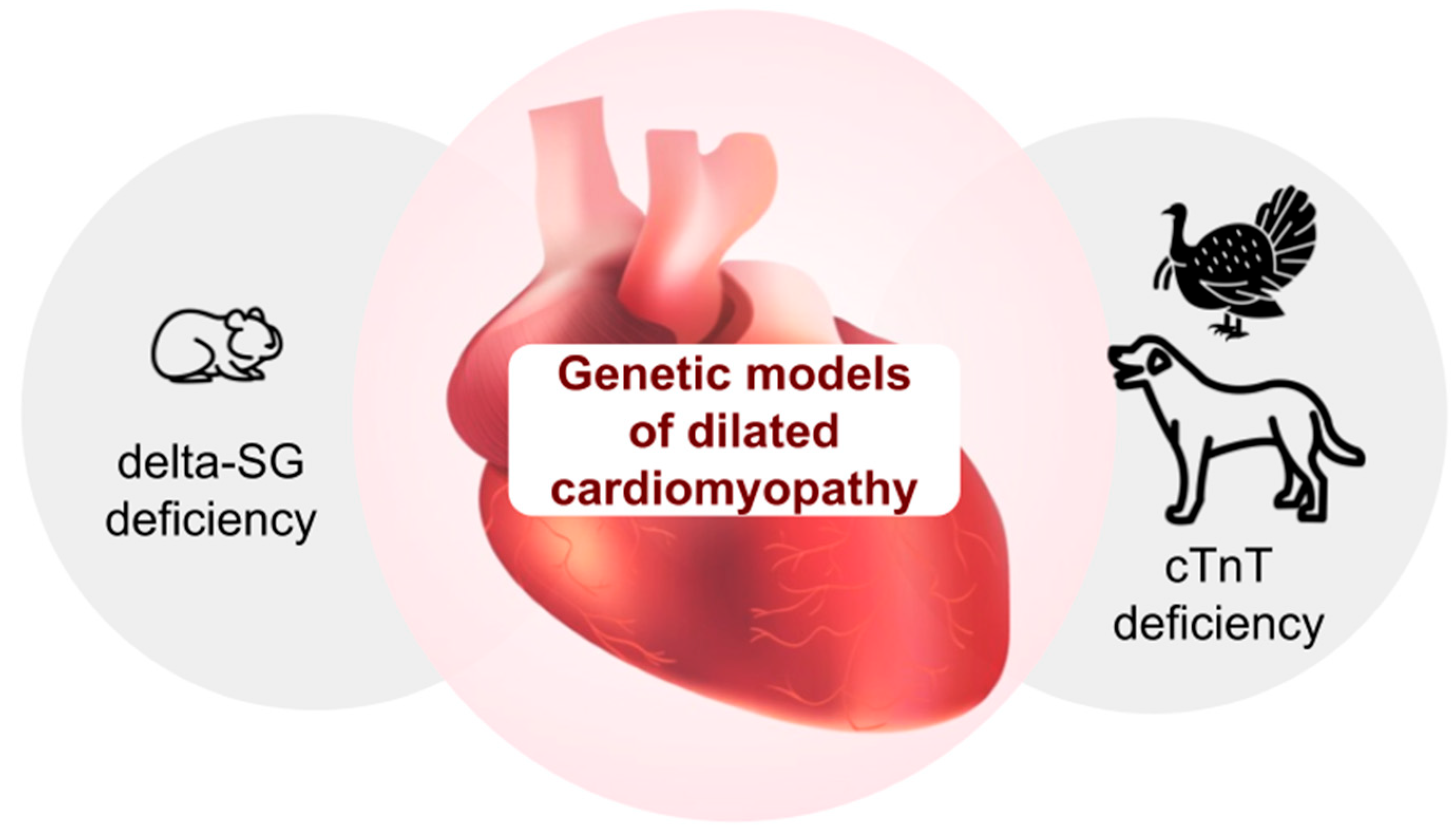
Figure 1. Genetic models of dilated cardiomyopathy (DCMP) include Syrian golden hamster model of deficiency of delta-sarcoglycan (delta-SG) and domestic turkey and dog models of deficiency of cardiac troponin T (cTnT).
Biomedicines 13 01518 g001
Figure 2. Genetic models of hypertrophic cardiomyopathy (HCMP) and restrictive cardiomyopathy (RCMP). HCMP cat models (Maine Coon cat, Persian, Ragdoll, and Sphynx) of deficiency of sarcomere proteins encoded by MYOC, MYBPC genes. RCMP cat models of deficiency of myosin, myopalladin, and cardiac troponin I (cTnI).
Biomedicines 13 01518 g002
Figure 3. Animal models of arrhythmogenic right ventricular dysplasia/cardiomyopathy (ARVC) include murine models with abnormalities in genes PKP2, DSG2, and JUP; zebrafish models with abnormalities in genes DSC2, TMEM43, and ILK; and spontaneous ARVC cat and boxer dog models.
Biomedicines 13 01518 g003
Figure 4. Specific examples of animal models of cardiovascular disease using genetically encoded tools.
Biomedicines 13 01518 g004
Figure 5. Proposed algorithm for choosing genetic animal models for studying cardiovascular diseases.
Biomedicines 13 01518 g005
Table 1. Comprehensive summary of genetic animal models for cardiovascular diseases.
ModelGenetic Basis/ModificationMain Cardiovascular PhenotypeKey ApplicationsMajor AdvantagesMain LimitationsPhysiological RelevanceGenetic ManipulabilityReproducibilityTranslational Relevance
SHR RatPolygenic (overexpression of the renin gene)Essential hypertension, stroke-proneEssential hypertension, cerebral circulatory disordersClosely mimics human essential hypertensionLimited to neurogenic hypertensionHighModerateHighModerate-High
SHR-SP RatPolygenic (derived from SHR)Severe hypertension, stroke susceptibilityStroke researchReproducible stroke phenotypeLimited lifespanHighModerateHighHigh
DSS RatPolygenic (CYP11B1)Salt-sensitive hypertensionSodium-related hypertensionGene-environment interaction modelingNo human-like nephropathyModerateLowHighModerate
FHH RatPolygenic
(Add3, Rbm20, Shoc2)
Hypertensive nephropathyRenal hypertensionRenal-cardiovascular crosstalkUnclear primary pathologyModerateLowModerateModerate
MHS RatADD1 mutation (α-adducin defect)Primary hypertension with renal dysfunctionHypertension with kidney involvementLinks hypertension to kidney pathologyLimited non-renal applicationsModerateLowHighModerate
Milan Hypertensive RatPolygenic (α-adducin mutations)Essential hypertensionMembrane transport studiesWell-characterized genetic basisLimited availabilityModerateLowHighModerate
Lyon Hypertensive RatPolygenic (Ercc6l2)Metabolic syndrome, salt-sensitive hypertensionInsulin resistance studiesHuman metabolic syndrome mimicComplex genetic architectureModerateLowModerateLow
NZGH RatPolygenic selectionSpontaneous hypertensionHypertension mechanismsStable phenotypeLow genetic tractabilityModerateLowHighModerate
MWF RatSpontaneous renal dysfunctionHypertension with proteinuriaRenal hypertensionKidney-heart axis modelingSlow progressionModerateLowModerateLow
Sabra Hypertensive Ratα2-Adrenoceptor variantsSalt-sensitive hypertensionEnvironmental hypertensionHuman salt sensitivity reproductionLimited genetic toolsModerateLowHighModerate
Buffalo RatPolygenicHypertension with insulin resistanceMetabolic hypertensionMetabolic-cardiovascular interactionsComplex phenotypeModerateLowModerateModerate
Goto-Kakizaki RatPolygenicDiabetic cardiomyopathyDiabetes-related CVDNon-obese diabetes modelMild cardiac phenotypeModerateLowHighModerate
WHHL RabbitLDLR mutationFamilial hypercholesterolemiaAtherosclerosis studiesSpontaneous human-like plaquesHigh cost, limited toolsHighModerateHighHigh
Ldlr−/− MouseLDLR knockoutHypercholesterolemiaPlaque biology researchRapid disease progressionSpecies-specific plaque differencesModerateHighHighModerate
ApoE−/− MouseApoE knockoutSevere atherosclerosisPlaque development studiesWidely used modelPlaque composition differencesModerateHighHighModerate
cMyBP-C KO MouseMYBPC3 deletionHypertrophic cardiomyopathySarcomere dysfunctionHuman mutation similarityMouse-specific physiologyModerateHighHighModerate
Feline HCM ModelMYBPC3 mutation (natural)Hypertrophic cardiomyopathySpontaneous HCM studiesNaturally occurring HCMLimited genetic manipulationHighLowModerateHigh
Canine ARVC ModelPKP2 mutation (boxers)Arrhythmogenic right ventricular cardiomyopathyARVC mechanisms, sudden cardiac deathNaturally mimics human ARVCEthical and cost challengesHighLowModerateHigh
Zebrafish (Tnnt2 KO)Tnnt2
knockout
CardiomyopathyCardiac developmentHigh-throughput screeningSimplified cardiac structureLow-ModerateHighHighLow
CPVT MouseRYR2 mutationPolymorphic VTArrhythmia mechanismsHuman CPVT recapitulationStress-dependent phenotypeModerateHighHighModerate
LQTS MouseKCNQ1/KCNH2 KOLong QT syndromeIon channel studiesDirect human mutation linkECG differencesModerateHighHighModerate
AAV9-MYH7 MouseAAV9-mediated MYH7 mutationHypertrophic cardiomyopathyGene therapy testingTissue-specific targetingTransient expressionModerateHighModerateHigh
DREADD RatChemogenetic hM3Dq receptor expressionHeart failure modulationNeural regulation of cardiac functionPrecise control of signaling pathwaysRequires ligand administrationModerateHighHighModerate
Table 2. The comparative advantages and disadvantages of popular animal species used for modeling heart diseases.
Animal ModelAdvantagesDrawbacks
Danio rerio
(zebrafish)
Short developmental period, low-cost maintenanceTwo-chamber heart, which do not reproduce all processes typical for humans
Mus musculus
(house mouse)
Short developmental period, low-cost maintenanceSmall-size heart,
high heartbeat rate
RabbitShort developmental period, high efficacy of gene modificationsModerate maintenance costs
Disclaimer/Publisher’s Note: The statements, opinions and data contained in all publications are solely those of the individual author(s) and contributor(s) and not of MDPI and/or the editor(s). MDPI and/or the editor(s) disclaim responsibility for any injury to people or property resulting from any ideas, methods, instructions or products referred to in the content.