
Reconstruction of a Chronic Quadriceps Tendon Rupture in an Elderly Polio Patient

 , ,
, ,  and
and 
Abstract
1. Introduction
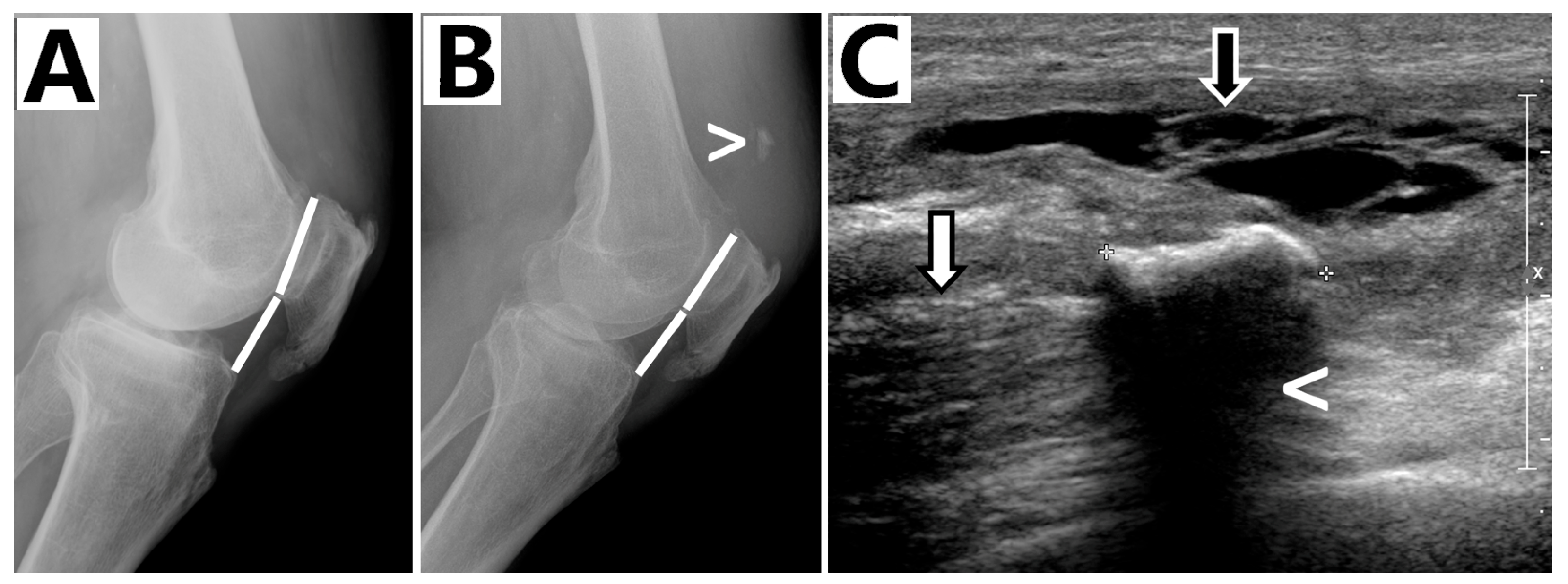
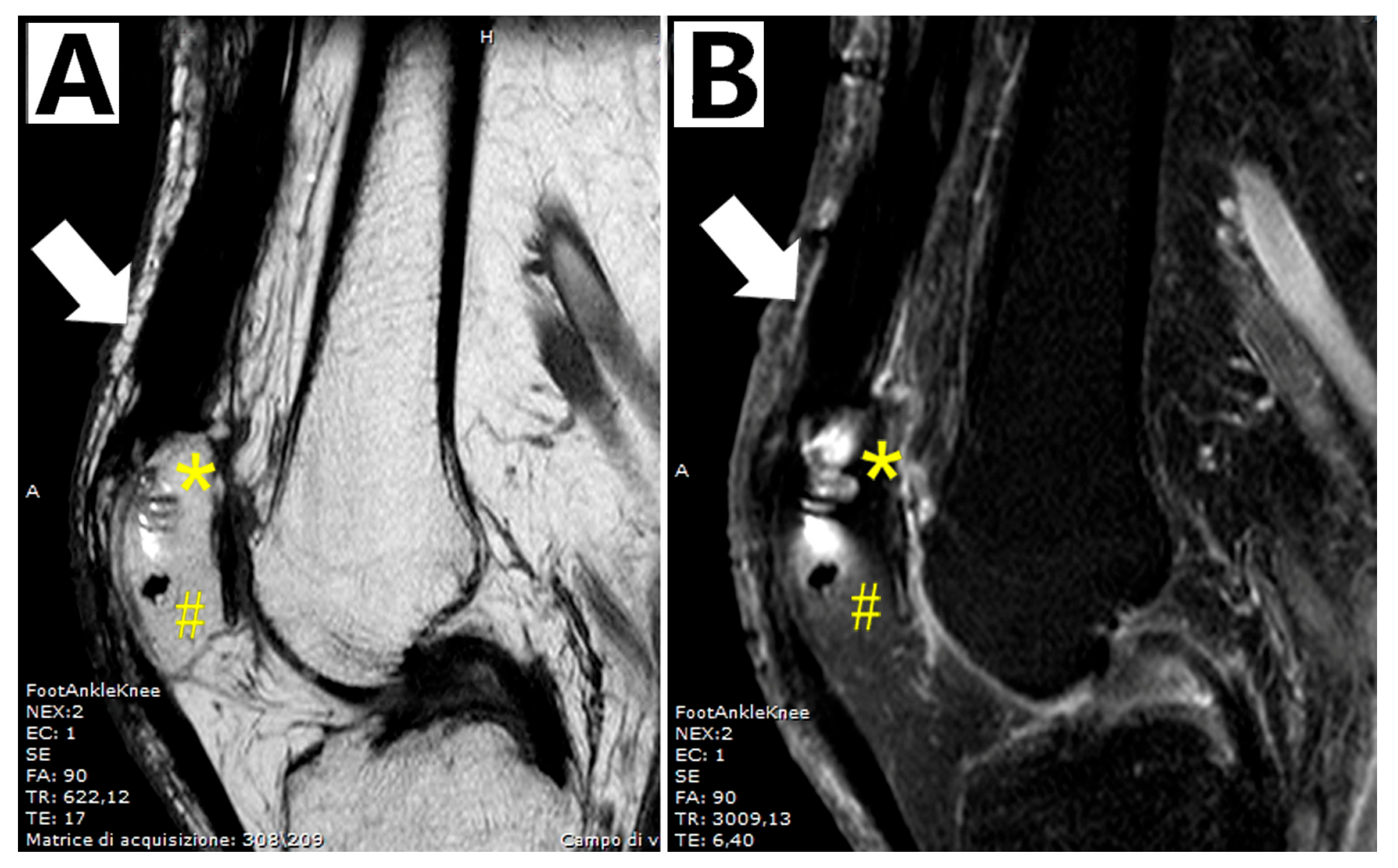
2. Case Report
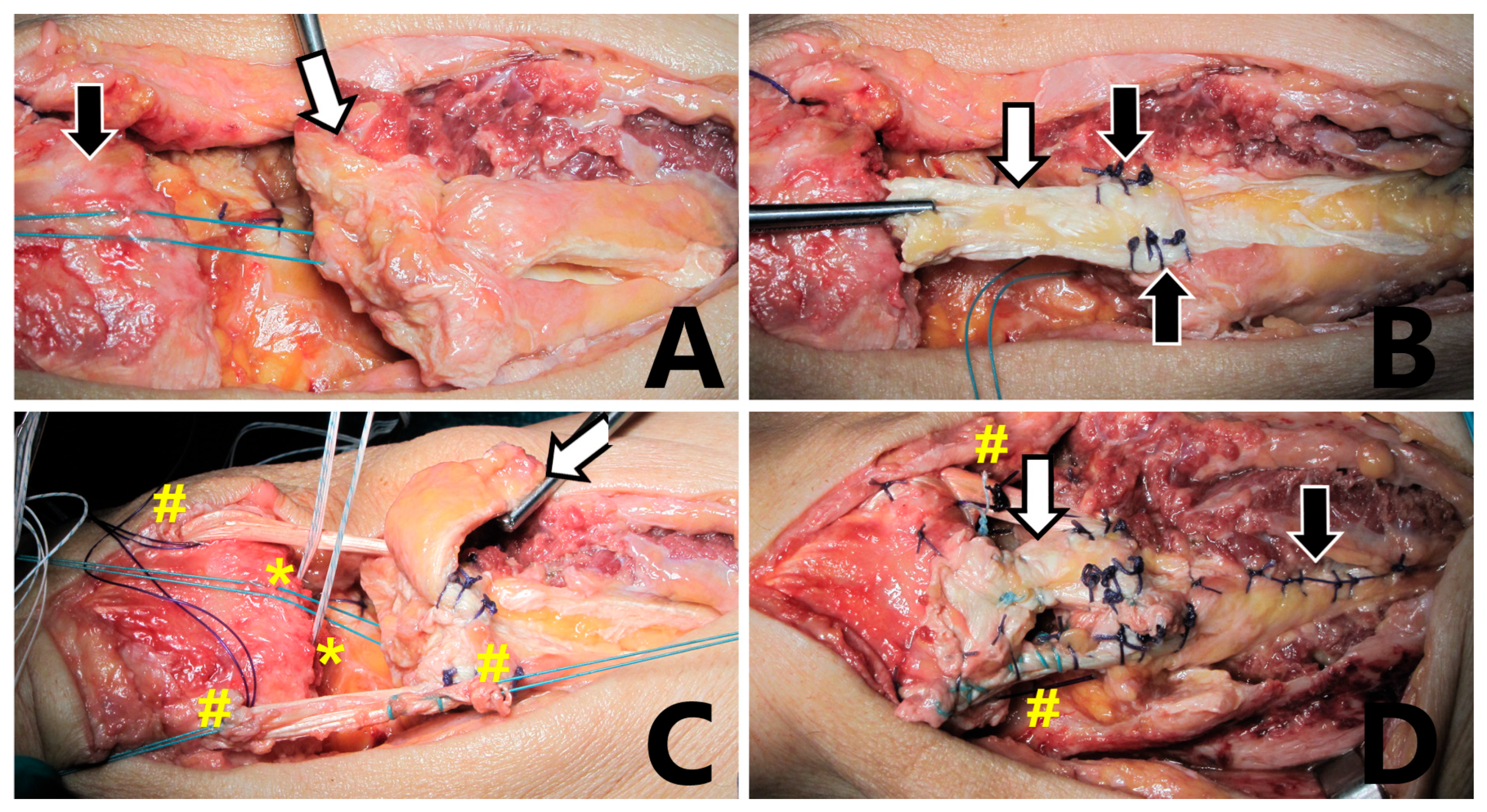
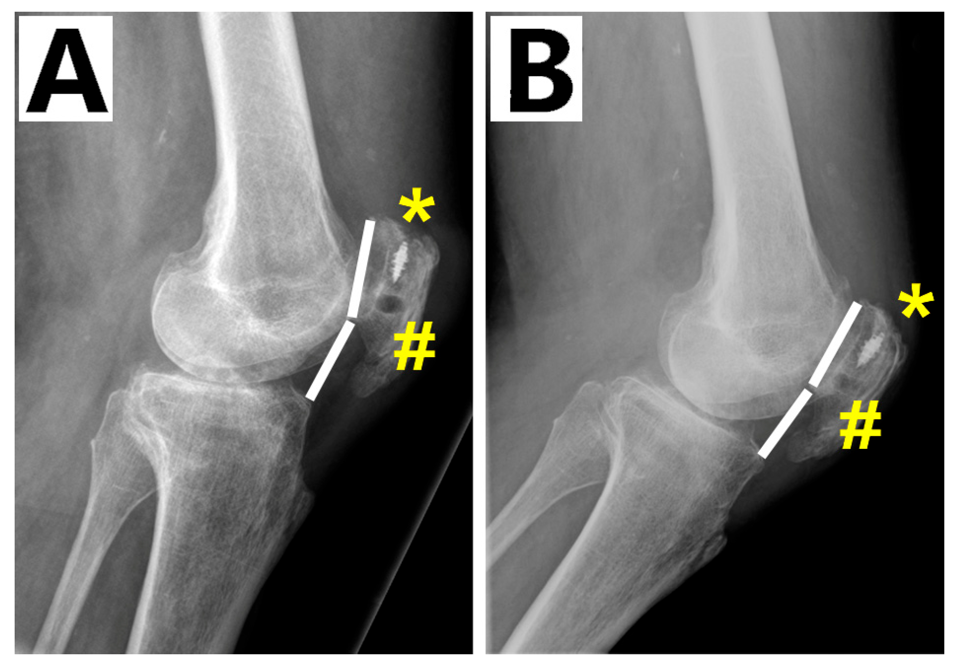
3. Surgical Technique
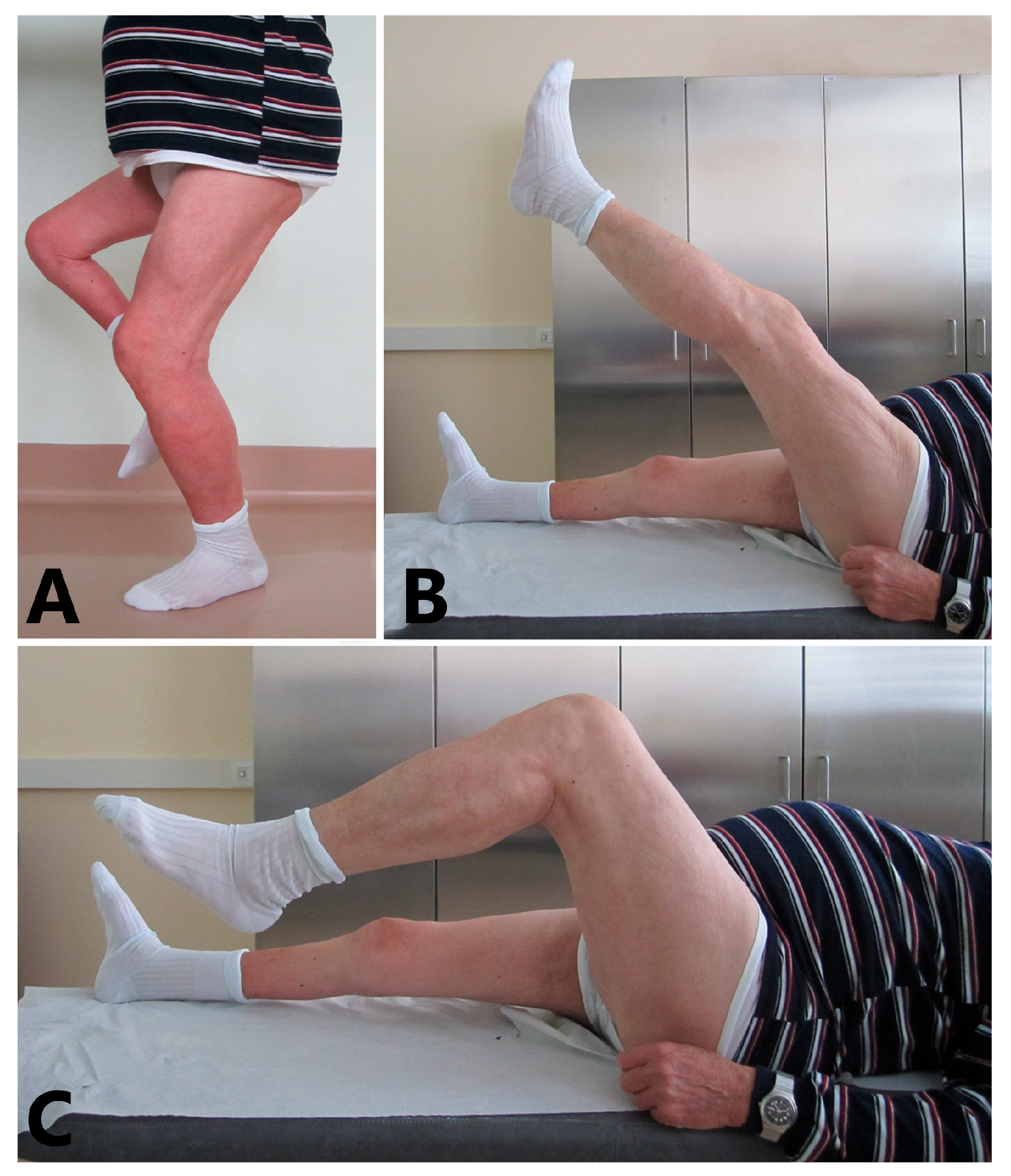
4. Discussion
Author Contributions
Funding
Institutional Review Board Statement
Informed Consent Statement
Data Availability Statement
Conflicts of Interest
References
- Elattar, O.; McBeth, Z.; Curry, E.J.; Parisien, R.L.; Galvin, J.W.; Li, X. Management of Chronic Quadriceps Tendon Rupture: A Critical Analysis Review. JBJS Rev. 2021, 9, e20. [Google Scholar] [CrossRef] [PubMed]
- Oliva, F.; Marsilio, E.; Migliorini, F.; Maffulli, N. Complex Ruptures of the Quadriceps Tendon: A Systematic Review of Surgical Procedures and Outcomes. J. Orthop. Surg. Res. 2021, 16, 547. [Google Scholar] [CrossRef] [PubMed]
- Palmeri, S.; Guelfi, M.; Corsini, A. Sports medicine group siagascot Comprehensive Management of Lower Limb Tendinopathies in Athletes: Advances and Challenges. Joints 2024, 2, e931. [Google Scholar]
- Neubauer, T.; Wagner, M.; Potschka, T.; Riedl, M. Bilateral, Simultaneous Rupture of the Quadriceps Tendon: A Diagnostic Pitfall? Rort of Three Cases and Meta-Analysis of the Literature. Knee Surg. Sports Traumatol. Arthrosc. 2007, 15, 43–53. [Google Scholar] [CrossRef] [PubMed]
- Kerin, C.; Hopgood, P.; Banks, A.J. Delayed Repair of the Quadriceps Using the Mitek Anchor System: A Case Report and Review of the Literature. Knee 2006, 13, 161–163. [Google Scholar] [CrossRef] [PubMed]
- Rehman, H.; Kovacs, P. Quadriceps Tendon Repair Using Hamstring, Prolene Mesh and Autologous Conditioned Plasma Augmentation. A Novel Technique for Repair of Chronic Quadriceps Tendon Rupture. Knee 2015, 22, 664–668. [Google Scholar] [CrossRef] [PubMed]
- Joseph, B.; Watts, H. Polio Revisited: Reviving Knowledge and Skills to Meet the Challenge of Resurgence. J. Child. Orthop. 2015, 9, 325–338. [Google Scholar] [CrossRef] [PubMed]
- McCluskey, D. Aids to the Examination of the Peripheral Nervous System. Ulst. Med. J. 1989, 201, 58. [Google Scholar]
- Scuderi, C. Ruptures of the Quadriceps Tendon. Study of Twenty Tendon Ruptures. Am. J. Surg. 1958, 95, 626–635. [Google Scholar] [CrossRef] [PubMed]
- Oliva, F.; Marsilio, E.; Migliorini, F.; Maffulli, N. Chronic Quadriceps Tendon Rupture: Quadriceps Tendon Reconstruction Using Ipsilateral Semitendinosus Tendon Graft. J. Orthop. Surg. Res. 2023, 18, 355. [Google Scholar] [CrossRef] [PubMed]
- Gulotta, L.V.; Rodeo, S.A. Biology of Autograft and Allograft Healing in Anterior Cruciate Ligament Reconstruction. Clin. Sports Med. 2007, 26, 509–524. [Google Scholar] [CrossRef] [PubMed]
- Leopardi, P.; Vico, G.D.; Rosa, D.; Cigala, F.; Maffulli, N. Reconstruction of a Chronic Quadriceps Tendon Tear in a Body Builder. Knee Surg. Sports Traumatol. Arthrosc. 2006, 14, 1007–1011. [Google Scholar] [CrossRef] [PubMed]
- Druskin, S.C.; Rodeo, S.A. Novel Treatment of a Failed Quadriceps Tendon Repair in a Diabetic Patient Using a Patella-Quadriceps Tendon Allograft. HSS J. 2013, 9, 195–199. [Google Scholar] [CrossRef] [PubMed]
- McCormick, F.; Nwachukwu, B.U.; Kim, J.; Martin, S.D. Autologous Hamstring Tendon Used for Revision of Quadiceps Tendon Tears. Orthopedics 2013, 36, e529–e532. [Google Scholar] [CrossRef] [PubMed]
- Elhessy, A.H.; Alrabai, H.M.; Eltayeby, H.H.; Gesheff, M.G.; Conway, J.D. Chronic Quadriceps Tendon Rupture Reconstruction with Sartorius Muscle Transfer: A Report of Five Cases. Plast. Reconstr. Surg.–Glob. Open 2021, 9, e3785. [Google Scholar] [CrossRef] [PubMed]
- Pintore, A.; Pintore, E.; Asparago, G.; Marsilio, E.; Torsiello, E.; Galasso, O. Sartorius Muscle Transfer for Chronic Quadriceps Tendon Rupture: A Prospective Study. Knee 2024, 53, 79–85. [Google Scholar] [CrossRef] [PubMed]
Disclaimer/Publisher’s Note: The statements, opinions and data contained in all publications are solely those of the individual author(s) and contributor(s) and not of MDPI and/or the editor(s). MDPI and/or the editor(s) disclaim responsibility for any injury to people or property resulting from any ideas, methods, instructions or products referred to in the content. |
© 2025 by the authors. Licensee MDPI, Basel, Switzerland. This article is an open access article distributed under the terms and conditions of the Creative Commons Attribution (CC BY) license (https://creativecommons.org/licenses/by/4.0/).
Share and Cite
Ronga, M.; Callegari, L.; Evola, F.R.; Crespi, C.; Rimondini, L.; Ciclamini, D. Reconstruction of a Chronic Quadriceps Tendon Rupture in an Elderly Polio Patient. Biomedicines 2025, 13, 1363. https://doi.org/10.3390/biomedicines13061363
Ronga M, Callegari L, Evola FR, Crespi C, Rimondini L, Ciclamini D. Reconstruction of a Chronic Quadriceps Tendon Rupture in an Elderly Polio Patient. Biomedicines. 2025; 13(6):1363. https://doi.org/10.3390/biomedicines13061363
Chicago/Turabian StyleRonga, Mario, Leonardo Callegari, Francesco Roberto Evola, Camilla Crespi, Lia Rimondini, and Davide Ciclamini. 2025. "Reconstruction of a Chronic Quadriceps Tendon Rupture in an Elderly Polio Patient" Biomedicines 13, no. 6: 1363. https://doi.org/10.3390/biomedicines13061363
APA StyleRonga, M., Callegari, L., Evola, F. R., Crespi, C., Rimondini, L., & Ciclamini, D. (2025). Reconstruction of a Chronic Quadriceps Tendon Rupture in an Elderly Polio Patient. Biomedicines, 13(6), 1363. https://doi.org/10.3390/biomedicines13061363