Review

Chronic Inflammation and Immune Dysregulation in Metabolic-Dysfunction-Associated Steatotic Liver Disease Progression: From Steatosis to Hepatocellular Carcinoma

1
Department of Family Medicine, Soonchunhyang University Seoul Hospital, Seoul 04401, Republic of Korea
2
Department of Family Medicine, Graduate School of Medicine, Seoul National University College of Medicine, Seoul 03080, Republic of Korea
3
Department of Neurology, Soonchunhyang University Seoul Hospital, Seoul 04401, Republic of Korea
4
Department of Translational Medicine, Graduate School of Medicine, Seoul National University College of Medicine, Seoul 03080, Republic of Korea
5
Department of Internal Medicine, Institute for Digestive Research, Digestive Disease Center, Soonchunhyang University College of Medicine, Seoul 04401, Republic of Korea
*
Author to whom correspondence should be addressed.
Biomedicines 2025, 13(5), 1260; https://doi.org/10.3390/biomedicines13051260
Submission received: 1 May 2025 / Revised: 16 May 2025 / Accepted: 19 May 2025 / Published: 21 May 2025
(This article belongs to the Special Issue Molecular Mechanism in Inflammation and Immunity)

Abstract

:
Background/Objectives: Metabolic-dysfunction-associated steatotic liver disease (MASLD) progresses from hepatic steatosis to hepatocellular carcinoma (HCC) as a result of systemic immunometabolic dysfunction. This review summarizes the key roles of the innate and adaptive immune mechanisms driving hepatic injury, fibrogenesis, and carcinogenesis in MASLD. Methods: A comprehensive literature review was performed using PubMed to identify relevant published studies. Eligible articles included original research and clinical studies addressing immunological and metabolic mechanisms in MASLD, as well as emerging therapeutic strategies. Results: We highlight the roles of cytokine networks, the gut–liver axis, and immune cell reprogramming. Emerging therapeutic strategies, including cytokine inhibitors, anti-fibrotic agents, metabolic modulators, and nutraceuticals, offer several indications for attenuating MASLD progression and reducing the prevalence of extrahepatic manifestations. Conclusions: Given the heterogeneity of MASLD, personalized combination-based approaches targeting both inflammation and metabolic stress are essential for effective disease management and the prevention of systemic complications.

1. Introduction

Metabolic dysfunction-associated steatotic liver disease (MASLD) is a recently adopted nomenclature that reflects a paradigm shift in the understanding of fatty liver diseases [1]. Previously referred to as nonalcoholic fatty liver disease (NAFLD), the term was widely used to describe a spectrum of liver disorders characterized by hepatic steatosis in individuals without significant alcohol consumption [2]. However, criticisms of this terminology have accumulated over the past decade. The label “nonalcoholic” defines the disease by what it is not, rather than by what it is, and it fails to acknowledge the key pathogenic role of metabolic dysfunction. Furthermore, the term “fatty” has been considered by many to be stigmatizing, both for patients and for public communication [3].
In response to these concerns, a comprehensive, multi-society Delphi consensus involving experts from hepatology, endocrinology, pediatrics, and public health was conducted under the guidance of major liver associations. As a result, the term MASLD was endorsed as a replacement for NAFLD, and the diagnostic criteria were redefined to require evidence of hepatic steatosis in conjunction with at least one cardiometabolic risk factor (CMRF), such as obesity, type 2 diabetes mellitus, or hypertension. Additionally, metabolic dysfunction-associated steatohepatitis (MASH) has replaced the term NASH to reflect the inflammatory and fibrotic progression of MASLD in affected individuals [1].
This reclassification acknowledges MASLD as a metabolic and systemic disease that can progress from simple steatosis to more advanced stages, including MASH, liver fibrosis, cirrhosis, and eventually hepatocellular carcinoma (HCC) [4]. Central to this progression is the persistent activation of innate and adaptive immune pathways, chronic low-grade inflammation, and cross-talk between the liver and extrahepatic organs such as the gut, adipose tissue, and the immune system [5]. Indeed, inflammation is now recognized not merely as a consequence, but as a driver of disease progression, mediated by hepatokines, cytokines, metabolic stress, and immunological dysregulation [6,7].
Recent epidemiological analyses have highlighted the growing global burden of MASLD, which is now recognized as the most common chronic liver disease, affecting over one-third of adults worldwide. MASLD is now the leading cause of chronic liver disease globally, affecting over 38% of adults and up to 14% of children, although, in regions where viral hepatitis remains prevalent, it continues to require significant public health focus [8]. According to a recent microsimulation study based on United States of America data, the prevalence of MASLD is projected to increase from 33.7% in 2020 to 41.4% by 2050, corresponding to approximately 122 million affected adults. This alarming trajectory highlights the urgent need for preventive and therapeutic strategies at the population level [9]. In addition to liver-specific outcomes such as fibrosis and carcinogenesis, MASLD imposes a significant health and economic burden through its association with cardiovascular disease, chronic kidney disease, and various extrahepatic cancers [10].
In this review, we provide an overview of the immune-mediated mechanisms that underlie the transition from steatosis to advanced liver pathology in MASLD. We revealed the roles of innate and adaptive immune cells, key cytokine networks, and chronic inflammatory signals that contribute to hepatic injury, fibrogenesis, and hepatocarcinogenesis. In addition, we discuss the emerging therapeutic approaches that target these immune pathways, aiming to prevent or reverse MASLD progression.

2. Immune Landscape in MASLD

Chronic hepatic inflammation in MASLD is orchestrated by the complex interplay between metabolic signals and the immune system. The dynamic interplay between immunometabolism and epigenetic modulation is increasingly recognized to be a key determinant in the progression of liver diseases, including MASLD, autoimmune hepatitis, and cholestatic disorders [11]. Both innate and adaptive immunity play key roles in hepatic injury, inflammation, fibrogenesis, and hepatocarcinogenesis, beginning early in steatosis and accelerating disease progression.

2.1. Innate Immunity in MASLD

Innate immune cells are the first responders to metabolic danger signals, including lipotoxicity, oxidative stress, and gut-derived pathogen-associated molecular patterns (PAMPs). These stimuli activate pattern recognition receptors (PRRs), such as toll-like receptors (TLRs) and nucleotide-binding oligomerization domain-like receptors (NLRs), on both hepatic and non-hepatic immune cells [12,13,14]. Kupffer cells, the liver-resident macrophages, play a central role in sensing free fatty acids, bacterial lipopolysaccharides (LPS), and damage-associated molecular patterns (DAMPs) [15]. Upon activation, Kupffer cells secrete pro-inflammatory cytokines such as tumor necrosis factor-alpha (TNF-α), interleukin (IL)-1β, and IL-6, contributing to hepatocellular damage and the recruitment of additional immune cells [16].
Additionally, neutrophils infiltrate the steatotic liver and promote inflammation by releasing reactive oxygen species (ROS) and neutrophil extracellular traps (NETs). NETs not only damage hepatocytes, but also activate hepatic stellate cells (HSCs), promoting fibrogenesis [17,18]. Monocyte-derived macrophages and dendritic cells (DCs) also accumulate in response to chemokines, further amplifying the local inflammatory environment. DCs, in particular, are involved in antigen presentation and priming of adaptive immune responses, bridging innate and adaptive immunity [19].
Moreover, Kupffer cells tend to polarize toward a pro-inflammatory M1-like phenotype in MASLD, characterized by elevated secretion of TNF-α and IL-1β. This polarization not only sustains hepatic inflammation, but also impairs tissue repair mechanisms associated with the anti-inflammatory M2 phenotype [20].
The contribution of liver sinusoidal endothelial cells (LSECs) is also significant. Although not classical immune cells, LSECs regulate leukocyte recruitment by expressing adhesion molecules such as vascular cell adhesion molecule-1 (VCAM-1) and intercellular adhesion molecule-1 (ICAM-1) in response to inflammation [21]. In MASLD, LSEC dysfunction enhances immune cell infiltration and supports chronic inflammation [22].
In parallel, the recognition of gut-derived LPS by TLR4 on Kupffer cells and HSCs activates the myeloid differentiation primary response 88 (MyD88)-nuclear factor kappa-light-chain-enhancer of activated B cells (NF-κB) signaling cascade, which amplifies inflammatory gene expression and drives fibrosis [12]. This highlights the crucial role of the gut–liver axis in sustaining innate immune activation.
The heterogeneity of liver macrophages, including both resident Kupffer cells and recruited monocyte-derived macrophages, plays a critical role in orchestrating inflammation in MASLD. Recent studies using single-cell technologies has revealed that these populations differ in origin and function, with infiltrating macrophages showing stronger pro-inflammatory and fibrogenic phenotypes [23]. Additionally, neutrophil NETs have emerged as active contributors to hepatic injury and fibrogenesis, linking innate immune activation to long-term disease progression [24].

2.2. Adaptive Immunity and Chronic Inflammation

As MASLD progresses, adaptive immune cells become increasingly involved. The liver is enriched with CD4+ T helper (Th) cells, CD8+ cytotoxic T cells, and B lymphocytes, all of which contribute to persistent inflammation and tissue remodeling [25,26].
Among CD4+ T cells, Th1 and Th17 subsets are particularly pathogenic. Th1 cells secrete interferon-gamma, which promotes macrophage activation and enhances cytotoxic responses [27]. Th17 cells, characterized by the production of IL-17A/F, contribute to neutrophil recruitment and liver fibrosis via the activation of HSCs [28]. Notably, IL-17 pathways are upregulated in both murine models and human MASLD, correlating with disease severity [29,30].
CD8+ T cells present cytotoxic effects on hepatocytes, exacerbating cell death and inducing chronic liver injury [31]. While CD8+ T cells initially contribute to host defense, prolonged activation in MASLD may lead to an exhausted phenotype, characterized by the upregulation of inhibitory receptors such as programmed death-1 (PD-1) and T cell immunoglobulin mucin domain-3 (TIM-3), which paradoxically sustains low-grade chronic inflammation [32].
Moreover, regulatory T (Treg) cells, which normally counterbalance inflammation, are often functionally impaired or reduced in number in MASLD, shifting the balance toward immune activation [31]. This disrupted Treg/Th17 balance is a key feature of MASLD progression and is associated with increased fibrosis [25].
B cells and their secreted antibodies may also participate in disease pathogenesis by forming immune complexes and presenting antigens, although their role remains less well defined [33]. Emerging evidence suggests the presence of ectopic lymphoid structures composed of B and T cells in advanced MASLD, indicating a shift toward localized, organized immune responses within the liver [28].

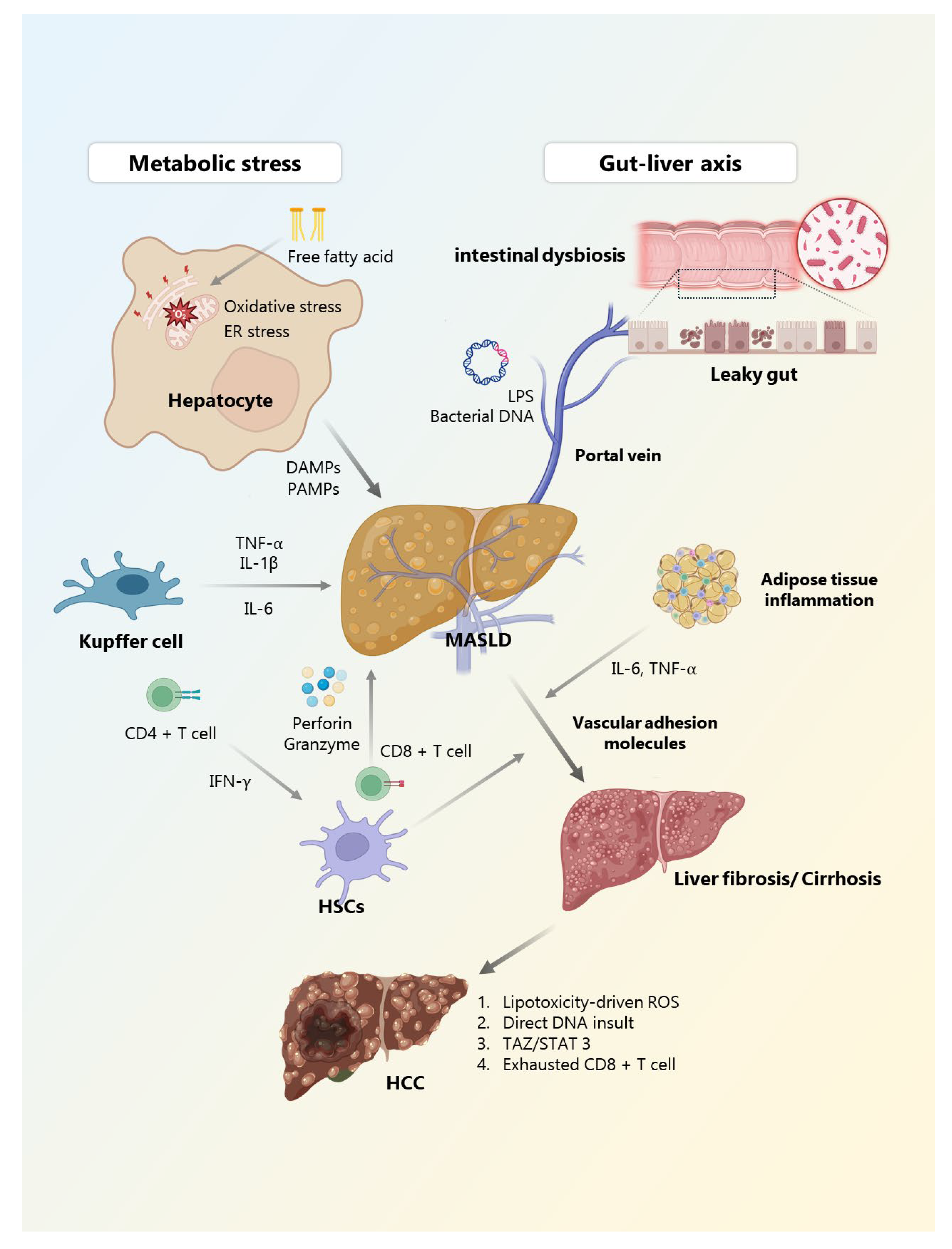
3. Chronic Inflammation in MASLD Progression

Chronic inflammation serves as a central driver in the progression of MASLD, orchestrating the transition from benign hepatic steatosis to MASH, liver fibrosis, cirrhosis, and eventually HCC. While simple steatosis is initially reversible, persistent metabolic insults and dysregulated immune responses result in a self-perpetuating cycle of hepatic injury, immune activation, and fibrogenesis. This inflammatory process has recently been conceptualized as a “domino effect”, in which the sensing of stress by innate immune cells triggers the sequential recruitment of pro-inflammatory myeloid and adaptive lymphoid cells, thereby perpetuating hepatic inflammation and fibrosis in a staged manner [33].

3.1. Metabolic Stress and Lipotoxicity as Initiating Triggers

The inflammatory cascade in MASLD is primarily initiated by lipotoxicity, resulting from the accumulation of excess free fatty acids and harmful lipid intermediates within hepatocytes [34]. These lipids induce mitochondrial dysfunction, endoplasmic reticulum (ER) stress, and oxidative stress, leading to the excessive generation of ROS. ROS not only damage hepatocyte organelles and DNA, but also amplify inflammatory signaling through redox-sensitive transcription factors such as NF-κB [35,36].
Importantly, unresolved ER stress activates the unfolded protein response, which involves signaling through inositol-requiring enzyme 1, protein kinase R-like ER kinase, and activating transcription factor 6 [37]. Chronic activation of these pathways enhances expression of pro-apoptotic and inflammatory mediators such as C/EBP homologous protein and X-box binding protein 1, thereby exacerbating hepatocellular damage and inflammation [38].
Cell death pathways, including apoptosis, necroptosis, and pyroptosis, release DAMPs such as high-mobility group box 1, adenosine triphosphate, and mitochondrial DNA, which serve as endogenous danger signals [39]. These DAMPs are recognized by PRRs like TLRs (especially TLR4 and TLR9) and NLRs (especially nucleotide-binding domain, leucine-rich-containing family, pyrin domain-containing-3 (NLRP3) inflammasome) on Kupffer cells and DCs, initiating inflammatory responses and cytokine secretion [40]. PAMPs, derived primarily from gut microbiota such as LPS, are recognized by TLR4, triggering pro-inflammatory cascades. DAMPs, such as mitochondrial DNA released from stressed hepatocytes, further activate immune responses through receptors like TLR9 and NLRP3. The NLRP3 inflammasome promotes the cleavage of pro-IL-1β via caspase-1, leading to pyroptosis and enhancing hepatic inflammation and fibrosis [40,41].

3.2. Gut–Liver Axis: Microbial Signs and Barrier Dysfunction

In addition to lipotoxic signals, the gut–liver axis plays a crucial role in accelerating hepatic inflammation. In MASLD, intestinal dysbiosis and increased permeability—often referred to as “leaky gut”—facilitate the translocation of microbial-derived PAMPs such as LPS and bacterial DNA into the portal circulation [42]. These PAMPs engage TLR4, TLR9, and other innate immune receptors on Kupffer cells, HSCs, and LSECs, triggering the release of pro-inflammatory cytokines and promoting immune cell recruitment [21,43,44].
Dysbiosis in MASLD is characterized by an altered Firmicutes-to-Bacteroidetes ratio and an enrichment of Gram-negative endotoxin-producing bacteria, such as Enterobacteriaceae [45]. Additionally, levels of beneficial microbial metabolites like butyrate are reduced [46]. This butyrate deficiency impairs Treg differentiation in the gut-associated lymphoid tissue, which could lead to systemic immune imbalance and worsened hepatic inflammation [47]. Recent finding suggested that natural compounds, such as Cornus officinalis vinegar, may ameliorate hepatic steatosis by modulating the gut microbiota and regulating lipid droplet metabolism [48].
The gut microbiota also influences the composition of short-chain fatty acids and secondary bile acids, both of which modulate immune tone and epithelial integrity in MASLD [49]. Disruptions in these microbial metabolites could further disrupt immune responses toward inflammation, exacerbating liver injury.

3.3. Immune Activation and Cytokine Networks

Activated Kupffer cells and recruited monocyte-derived macrophages produce a cascade of pro-inflammatory cytokines, including TNF-α, IL-1β, and IL-6. These mediators not only promote hepatocyte damage, but also enhance the recruitment of neutrophils and T cells, enhancing the inflammatory response [50]. Neutrophils contribute to further injury through the release of reactive oxygen species and NETs, which have been shown to activate HSCs and induce fibrogenesis [18].
The C-C motif chemokine ligand (CCL) 2-C-C chemokine receptor (CCR) 2 chemokine axis is particularly important in MASLD, as it drives the recruitment of Ly6Chi monocytes into the liver. Once recruited, these monocytes differentiate into inflammatory macrophages that promote cytokine production and support fibrogenesis [51]. Simultaneously, activation of the NLRP3 inflammasome in macrophages and hepatocytes leads to caspase-1-mediated maturation of IL-1β and IL-18, promoting an escalating inflammatory response. These cytokines stimulate additional immune cell infiltration and sustain a chronic inflammatory environment [52].
Moreover, prolonged activation of NF-κB and c-Jun N-terminal kinase pathways in hepatocytes, immune cells, and HSCs further drive the expression of inflammatory and fibrogenic genes. This signaling axis bridges metabolic stress with immune dysfunction and has been implicated in both steatohepatitis and hepatocarcinogenesis [53]. Beyond cytokines, immunometabolic reprogramming further sustains the inflammatory state. For example, lipotoxic Kupffer cells shift toward glycolysis and exhibit an M1-like pro-inflammatory polarization, which reinforces the production of TNF-α and IL-6 [54].

3.4. Adaptive Immune Contribution and Imbalance

In progressive MASLD, the innate immune response transitions into a chronic phase that involves adaptive immune components. CD4+ Th1 and Th17 cells, activated by antigen-presenting DCs and inflammatory cytokines, secrete interferon-gamma and IL-17A, which promote hepatocyte apoptosis and activate HSCs [55]. CD8+ cytotoxic T lymphocytes exacerbate tissue injury through the release of perforin and granzyme, contributing to parenchymal destruction and regeneration stress [56].
Emerging evidence suggests that prolonged antigen stimulation might drive CD8+ T cells toward an exhausted phenotype. While direct studies in MASLD are limited, chronic liver disease models show the upregulation of inhibitory receptors, such as PD-1 and TIM-3 on CD8+ T cells [57,58]. Paradoxically, these exhausted T cells may still contribute to sustained low-grade inflammation through non-cytolytic mechanisms [59].
Tregs, which are essential for maintaining immune tolerance and controlling inflammation, are often reduced or functionally impaired in MASLD, leading to unchecked immune activation. This imbalance between pro-inflammatory and anti-inflammatory T cell subsets is a hallmark of advanced stages of the disease [60]. Notably, the Th17/Treg ratio is a proposed biomarker of disease severity in MASH. Elevated Th17 cytokines (IL-17A/F and IL-22) have been linked to fibrosis progression, whereas Treg dysfunction contributes to the failure of inflammatory resolution [61].

3.5. Fibrogenesis and Systemic Inflammatory Crosstalk

Liver fibrogenesis is induced by the activation of HSCs and the deposition of extracellular matrix proteins, including type I and III collagens [62]. Transforming growth factor-beta (TGF-β), platelet-derived growth factor, and IL-13, derived from immune cells and activated hepatocytes, are major profibrogenic cytokines that maintain HSC activation and promote scar formation [63].
Furthermore, MASLD is not restricted to the liver. Chronic inflammation affects systemic circulation, promoting insulin resistance, endothelial dysfunction, and increased risk of comorbidities such as cardiovascular disease and chronic kidney disease [64,65]. Adipose tissue inflammation, marked by macrophage infiltration and altered adipokine profiles, reinforces hepatic immune dysregulation in a bidirectional manner. Additionally, systemic pro-inflammatory cytokines like IL-6 and TNF-α contribute to endothelial activation, increasing the expression of vascular adhesion molecules and promoting atherosclerosis [66]. Similar mechanisms may underlie renal inflammation and fibrosis observed in CKD comorbid with MASLD [67]. Additionally, aging-related immune dysregulation, characterized by the impaired resolution of inflammation and increased pro-inflammatory cytokines, may further exacerbate MASLD progression in elderly individuals with metabolic comorbidities [68].

3.6. Dysregulated Innate Immune Pathways in MASLD

The progression of MASLD is importantly driven by dysregulated innate immune signaling pathways. PAMPs, such as LPS derived from the gut microbiota, and DAMPs, including mitochondrial DNA released from injured hepatocytes, are key initiators of hepatic inflammation. These signals are recognized by pattern recognition receptors, particularly TLRs and NLRs, which are expressed on Kupffer cells, HSCs, and other liver-resident immune cells [69]. One of the most critical downstream mediators is the NLRP3 inflammasome. Upon activation by these danger signals, NLRP3 assembles a multiprotein complex that activates caspase-1, leading to the maturation of IL-1β and IL-18. These cytokines enhance hepatic inflammation, promote pyroptotic cell death, and recruit additional immune cells, thereby sustaining a pro-inflammatory and fibrogenic microenvironment [70].
In parallel, the Notch signaling pathway has emerged as a central regulator of hepatic immune remodeling in MASLD. Originally characterized in developmental biology, Notch signaling is now recognized for its role in the activation of hepatic progenitor cells and the modulation of macrophage polarization. Activation of Notch signaling in the liver promotes M2-like macrophage phenotypes and suppresses cytotoxic immune responses, contributing to fibrotic remodeling and the development of an immunosuppressive microenvironment [71]. Altogether, the dysregulation of PAMP/DAMP sensing, inflammasome activation, and developmental signaling pathways such as Notch constitutes a key axis in the immunopathogenesis of MASLD and its progression toward advanced liver disease.

4. From Steatosis to HCC: Immune-Modulated Progression

The progression from steatosis to HCC in the context of MASLD is not a linear transition, but rather a dynamic and multi-step process shaped by sustained inflammation, immune dysregulation, fibrotic remodeling, and oncogenic reprogramming [72]. Unlike viral or alcohol-related liver disease, MASLD-derived HCC frequently arises in non-cirrhotic livers, highlighting distinct pathophysiologic mechanisms rooted in metabolic inflammation and altered immune surveillance [73].

4.1. Chronic Inflammation and Fibrosis as Pro-Carcinogenic Drivers

Persistent low-grade inflammation in MASLD initiates a cascade of events that predispose hepatocytes to malignant transformation [74]. Kupffer cells, monocyte-derived macrophages, and neutrophils remain chronically activated, releasing ROS, nitrogen intermediates, and pro-inflammatory cytokines such as TNF-α, IL-1β, and IL-6. These mediators not only damage hepatocytes, but also induce compensatory regeneration and genomic instability [75,76].
HSCs are activated concurrently, depositing extracellular matrix proteins that result in progressive fibrosis. Fibrosis, even in the absence of cirrhosis, provides a permissive environment for tumorigenesis by altering tissue architecture, promoting hypoxia, and enabling immune evasion in the context of MASLD [77,78].

4.2. Immune Imbalance and Loss of Tumor Surveillance

The immune environment in MASLD has a paradoxical transformation as the disease progresses. While early inflammation is marked by immune activation, later stages are characterized by immune exhaustion and suppression [79]. Chronic T cell stimulation, particularly among CD8+ populations, induces an exhausted phenotype that expresses inhibitory checkpoints like PD-1 and TIM-3 [80]. Tumor-associated macrophages, lipid-associated macrophages, and Tregs further contribute to an immunosuppressive tumor microenvironment [81]. This immune landscape not only fails to eliminate premalignant cells, but may actively support tumor growth by secreting growth factors such as TGF-β and vascular endothelial growth factor.
Importantly, MASLD-related HCC is often associated with the “immune-low” subtype of liver cancer, marked by low T cell infiltration and minimal interferon signaling. This phenotype is thought to be resistant to immune checkpoint blockade therapies, highlighting the necessity for immune-based stratification in MASLD-HCC management [82].

4.3. Oxidative Stress, ER Dysfunction, and DNA Damage

Lipotoxicity-driven ROS and reactive nitrogen species induce mitochondrial damage, ER stress, and direct DNA insults in hepatocytes [83]. Accumulation of 8-hydroxy-2′-deoxyguanosine, a biomarker of oxidative DNA damage, has been observed in both human and murine models of MASLD-HCC [84]. These insults impair DNA repair mechanisms, resulting in mutations in tumor suppressors such as tumor protein p53 and activation of proto-oncogenes, including β-catenin and telomerase reverse transcriptase [85]. Unlike traditional cirrhosis-driven HCC, these mutations would emerge earlier in MASLD, prior to architectural distortion, which emphasizes the requirement for early molecular surveillance [77].
Furthermore, dysregulated autophagy and persistent ER stress amplify hepatocellular injury [86]. Proteins such as p62, a component of Mallory–Denk bodies, are prominently expressed in MASLD-HCC, promoting hepatocarcinogenesis through the activation of mammalian target of rapamycin complex 1 and cellular myelocytomatosis oncogene signaling [87].

4.4. Oncogenic Signaling Pathways

Multiple oncogenic pathways are activated in MASLD-HCC, often in response to chronic inflammation and metabolic dysfunction. The transcriptional coactivator with PDZ-binding motif (TAZ), which is upregulated by cholesterol accumulation, has been shown to drive hepatocellular transformation by enhancing oxidative stress and upregulating cytochrome B-245 beta chain/nicotinamide adenine dinucleotide phosphate hydrogen oxidase 2-mediated DNA damage [88]. Inhibition of TAZ in murine models suppresses both steatohepatitis and tumor growth, identifying it as a dual-function therapeutic target [89,90].
The wingless-type mouse mammary tumor virus integration site family (Wnt)/β-catenin pathway, often activated through mutations in catenin beta 1, defines a molecular subtype of HCC with immune exclusion and poor prognosis [91]. Notably, this pathway is frequently engaged in MASLD-HCC, contributing to resistance to immune checkpoint therapy [92]. Similarly, signal transducer and activator of transcription 3 (STAT3) signaling is sustained in obesity-related MASLD and HCC, driven by increased IL-6 and leptin. Dysregulated STAT3 activation supports tumor proliferation, survival, and immune evasion [93].
The Notch pathway has recently emerged as a MASLD-specific oncogenic driver. Even in the absence of canonical mutations, Notch signaling is upregulated in HCC arising from MASH livers [94]. The Notch pathway not only supports cholangiocarcinoma-like transcriptional programs, but also modulates immune cell infiltration and macrophage polarization, further supporting an immunosuppressive environment [95]. Evidence suggests that Notch activation contributes to hepatic fibrogenesis and carcinogenesis by influencing macrophage polarization and hepatocyte progenitor expansion, especially in the context of MASLD-associated HCC [96]. Suppression of Notch in experimental models alters the tumor phenotype, but also triggers compensatory Wnt activation, illustrating the complexity of inter-pathway crosstalk [94].

4.5. Tumor Evolution and Therapeutic Implications

Post-tumor initiation, the MASLD liver continues to promote tumor evolution through ongoing inflammation, fibrosis, and metabolic dysfunction [97]. The altered microenvironment not only supports malignant progression, but also contributes to therapeutic resistance. Studies have shown that MASLD-HCC patients respond less favorably to immune checkpoint inhibitors compared to those with viral hepatitis-related HCC, possibly due to reduced tumor immunogenicity and the higher prevalence of exhausted CD8+ T cells [79]. An overview of the immunopathogenic mechanisms and the associated immune cell populations contributing to MASLD progression is summarized below (Table 1).
These findings present the need for combinatorial treatment strategies, including agents targeting inflammation including anti-IL-6, anti-STAT3, fibrosis including anti-TGF-β, and metabolism including acetyl-CoA carboxylase (ACC) inhibitor [27,98,99,100]. In this regard, patient-derived xenograft models offer valuable translational insights into MASLD-related hepatocarcinogenesis, providing a platform for evaluating immunometabolic and epigenetic therapies [101]. Novel biomarkers such as patatin-like phospholipase domain-containing protein 3 genotypes and circulating oxidative stress markers might also stratify patients for surveillance and personalized therapy [102].

5. Therapeutic Targets and Immune Modulation

The recognition of immune dysregulation and chronic inflammation as central drivers of MASLD progression has catalyzed the development of immune-modulating therapies aimed at interfering with disease progression and improving outcomes [103]. While the traditional management of MASLD has focused on lifestyle modifications and metabolic control, the increasing understanding of the liver’s immune landscape has led to a paradigm shift. Emerging therapies now seek to target key inflammatory mediators, fibrogenic pathways, metabolic stress responses, and the immunosuppressive tumor microenvironment associated with hepatocarcinogenesis. This section outlines current and prospective therapeutic strategies, categorized by their mechanistic focus and clinical potential.

5.1. Targeting Pro-Inflammatory Cytokines and Immune Pathways

Chronic inflammation in MASLD is sustained by a network of cytokines and immune cells that amplify hepatic injury and fibrosis. Modulating these immune pathways represents a promising therapeutic option.
IL-6 and the Janus kinase/STAT3 cascade are central to metabolic inflammation. IL-6 can signal through both classical and trans-signaling pathways, the latter being predominant in chronic inflammation [104]. Trans-signaling activates the JAK1/STAT3 axis in hepatocytes, macrophages, and HSCs, promoting survival, proliferation, and fibrogenic gene expression [105]. Monoclonal antibodies targeting IL-6 or IL-6R have demonstrated efficacy in inflammatory diseases and are being explored for MASLD-related immune activation [27]. STAT3 inhibitors, including small molecules like napabucasin and TTI-101, are under investigation for their anti-inflammatory and anti-tumor potential [106,107].
IL-1β and NLRP3 inflammasome signaling play a pivotal role in pyroptosis and the amplification of innate immune responses [108]. Hepatocyte and macrophage-derived DAMPs trigger NLRP3 assembly, leading to caspase-1 activation and maturation of IL-1β and IL-18. These cytokines promote neutrophil recruitment, hepatocyte death, and HSC activation [109]. Nevertheless, neither the IL-1β inhibitor nor NLRP3 inhibitor are in the clinical trials, and urgent evaluation is required.
IL-17 and the Th17/IL-23 axis are increasingly recognized in liver fibrosis and inflammation. IL-17A promotes neutrophil chemotaxis and activates HSCs through the NF-κB and STAT3 pathways [110]. IL-23 maintains the Th17 phenotype, and a dual IL-17/IL-23 blockade is effective in psoriasis and IBD [111,112]. Trials evaluating secukinumab (anti-IL-17A) and risankizumab (anti-IL-23) would provide insights into MASLD applications, particularly in IL-17-high phenotypes.
Immune checkpoint pathways, such as PD-1/PD-L1 and CTLA-4, are essential regulators of immune exhaustion [113]. In MASLD, chronic antigen exposure and metabolic stress induce CD8+ T cell exhaustion and increased checkpoint expression [56]. While checkpoint inhibitors have limited efficacy in MASLD-related HCC due to its “immune-cold” profile, earlier intervention in the MASH stage, particularly in patients with immune infiltration, might be more effective [79]. Combination approaches involving checkpoint blockade and anti-fibrotic or metabolic agents could be the one of the effective options for MASLD-related HCC.
Recent phase II and III trials have demonstrated that agents such as resmetirom [a thyroid hormone receptor (THR)-β agonist], semaglutide (a glucagon-like peptide-1 receptor agonist), and lanifibranor [a pan-peroxisome proliferator-activated receptor (PPAR) agonist] show significant promise in improving the histological features of MASLD, including inflammation resolution and fibrosis reduction. While not yet approved, several of these candidates are in advanced development stages, emphasizing the growing momentum of targeted immunometabolic therapies for MASLD [114].

5.2. Anti-Fibrotic Therapies with Immunomodulatory Effects

Fibrosis, driven by sustained immune cell–HSC crosstalk, is the most important predictor of liver-related outcomes in MASLD [27]. As such, targeting fibrogenesis also impacts the hepatic immune environment.
TGF-β signaling is a master pathway in hepatic fibrogenesis [63]. It induces suppressor of mothers against decapentaplegic-dependent transcription of collagen and tissue inhibitor of metalloproteinase, while also presenting immunosuppressive effects [115]. Galunisertib (LY2157299), a TGF-β receptor I kinase inhibitor, has shown anti-fibrotic and anti-tumor activity in an early trial [116]. However, global TGF-β blockade carries risks, including autoimmunity and carcinogenesis, emphasizing the need for tissue-specific or temporally regulated targeting [117].
CCL2–CCR2 and CCL5–CCR5 pathways mediate the recruitment of inflammatory monocytes and macrophages [118]. Cenicriviroc, a dual CCR2/CCR5 antagonist, demonstrated anti-fibrotic effects in the CENTAUR trial, although its phase III results (AURORA) were inconclusive [119,120]. These chemokine pathways also affect immune cell composition, suggesting combinatorial strategies with immunotherapies.
Farnesoid X receptor (FXR) agonists, such as obeticholic acid (OCA), reduce hepatic steatosis, inflammation, and fibrosis. OCA regulates bile acid metabolism, inhibits NF-κB signaling, and reduces chemokine production. However, pruritus and lipid profile changes have limited its broader adoption [121]. Other FXR agonists, such as tropifexor and cilofexor, are in development [122,123].
Fibroblast growth factor (FGF) analogs like FGF19 (Aldafermin) and FGF21 (Pegbelfermin) improve insulin sensitivity, reduce lipotoxicity, and suppress inflammation via PPARγ and adenosine monophosphate-activated kinase activation. These agents modulate hepatic macrophage polarization and might synergize with anti-inflammatory therapies [124].
Lanifibranor, a pan-PPAR agonist, has shown histologic improvement in MASH and fibrosis resolution. PPARα, which activates enhances lipid oxidation, PPARγ, which modulates macrophage activation, and PPARδ, which improves mitochondrial function, would modulate immunometabolic homeostasis together [125].
Recently, THR-β agonists have emerged as promising candidates in MASLD treatment [126]. THR-β, predominantly expressed in hepatocytes, regulates lipid metabolism and mitochondrial function [127]. In MASLD, intrahepatic hypothyroidism, a state characterized by reduced conversion of T4 to T3, is frequently observed and contributes to lipid accumulation, inflammation, and fibrosis [128]. Resmetirom, a selective THR-β agonist, has demonstrated significant reductions in hepatic fat content, serum low-density lipoprotein cholesterol, and markers of fibrosis and inflammation in phase II and III trials [126,129]. It suppresses lipotoxicity, improves deiodinase activity, and downregulates pro-inflammatory pathways including NF-κB and STAT3 [130]. This evidence positions THR-β modulation as a dual metabolic and immune-targeting strategy.

5.3. Metabolic Reprogramming and Oxidative Stress Modulation

Hepatic immune cells in MASLD are undergoing metabolic reprogramming, including increased glycolysis and mitochondrial ROS production, which contributes to inflammatory phenotypes [131]. Targeting these metabolic shifts is a novel therapeutic strategy.
ACC inhibitors, such as firsocostat, reduce de novo lipogenesis and substrate overload in hepatocytes and macrophages [100]. In a phase II trial, firsocostat reduced liver fat content and demonstrated trends toward fibrosis improvement [132]. FASN inhibitors (e.g., TVB-2640) inhibit fatty acid synthesis and downstream lipid mediator production, thereby reducing lipid-induced inflammation [133]. Aminocarboxymuconate semialdehyde decarboxylase (ACMSD) inhibition by increasing nicotinamide adenine dinucleotide availability, enhances mitochondrial function, and reduces inflammatory cytokine production [134]. ACMSD inhibitors are also being evaluated for their metabolic and immune benefits in MASLD [135].
Vitamin E, as shown in the PIVENS trial, improved the histologic features of MASH in non-diabetic patients [136]. It reduces oxidative stress, improves hepatocyte survival, and modulates NF-κB activation [137]. However, its long-term safety and efficacy remain under debate [138].
Mitochondria-targeted antioxidants, such as Elamipretide, show potential in reducing hepatocellular ROS and protecting mitochondrial integrity. These agents may also suppress inflammasome activation [139].

5.4. Gut–Liver Axis: Microbiota and Immune Crosstalk

The gut microbiota shapes liver immunity through microbial metabolites and endotoxins [140]. Probiotics and synbiotics modulate microbial composition and improve intestinal barrier function [141]. Specific strains such as Lactobacillus rhamnosus and Bifidobacterium longum have been shown to reduce hepatic inflammation and fibrosis in preclinical models [142]. Clinical studies report improvements in alanine aminotransferase and hepatic steatosis scores with the modulation of gut microbiota [143]. Fecal microbiota transplantation (FMT) has shown benefits in improving gut barrier function and metabolic parameters in pilot MASLD studies [144,145]. However, long-term efficacy, donor variability, and safety concerns limit its use [146]. Postbiotics, such as short-chain fatty acid derivatives and bacterial cell wall fragments, might enhance Treg function and modulate hepatic immunity without requiring live bacteria [147]. This offers a safer and more controllable alternative to FMT.

5.5. Nutraceuticals and Adjunctive Immune Modulators

While pharmacologic therapies are evolving, nutraceuticals remain attractive adjuncts due to their safety, accessibility, and potential for systemic immune modulation.
Multivitamin use, particularly antioxidant and B-complex vitamins, has been associated with improved systemic outcomes in MASLD. In a UK Biobank analysis, multivitamin intake correlated with lower cardiovascular and renal risk, although its effects on liver-specific outcomes were modest. These findings suggest that broader antioxidants or micronutrient support may benefit the systemic inflammation and metabolic comorbidities commonly associated with MASLD [148]. In particular, vitamin D deficiency is common in MASLD and correlates with disease severity and insulin resistance [149]. As a modulator of both innate and adaptive immunity, vitamin D enhances regulatory T cell function and suppresses Th17 polarization [150]. Some trials have reported improved liver enzymes and steatosis with supplementation; however, results remain inconsistent, potentially due to differences in baseline levels, dosing, and patient characteristics [151].
Polyphenols, such as resveratrol and curcumin, and omega-3 fatty acids have demonstrated anti-inflammatory, antioxidant, and lipid-lowering properties. These agents act through NF-κB inhibition, the activation of PPARα/γ, and the modulation of gut microbiota composition. In MASLD models, they have been shown to reduce hepatic steatosis and inflammatory cytokine expression [152]. While generally well tolerated, their limited bioavailability and heterogeneous clinical trial designs require more standardized investigations to confirm the efficacy and optimal dosage in MASLD populations.
Glucosamine, traditionally used for osteoarthritis, has also shown potential anti-inflammatory and metabolic benefits relevant to MASLD. A recent large cohort-based study reported that regular glucosamine use was associated with reduced all-cause and liver-related mortality, as well as lower cardiovascular and kidney disease incidence, in patients with MASLD [153]. Although the exact mechanisms remain under investigation, the proposed effects include the inhibition of NF-κB signaling, improvements in insulin sensitivity, and the modulation of oxidative stress [154]. Given its safety profile, glucosamine may be considered to be a complementary approach, particularly in individuals with cardiometabolic comorbidities.

6. Conclusions and Future Directions

MASLD is now recognized as a systemic condition characterized by a complex interplay of metabolic stress, immune dysregulation, and chronic low-grade inflammation. As illustrated throughout this review, both innate and adaptive immune responses contribute not only to hepatic injury and fibrogenesis, but also to the progression toward HCC (Figure 1). Inflammatory mediators, immune checkpoints, and tissue remodeling signals shape a dynamic hepatic microenvironment, which can both initiate and sustain disease activity. The immunological mechanisms discussed throughout this review are increasingly supported by a growing body of research, emphasizing the convergence of immune dysregulation, metabolic stress, and fibrosis in MASLD progression. Integrating these insights with emerging translational studies is essential to characterize therapeutic strategies and improve patient outcomes.
Over the past decade, significant advances have been made in unraveling the immunopathogenic mechanisms of MASLD. As summarized in Section 5, multiple compounds, ranging from IL-6 or TGF-β inhibitors to ACC, FASN, and THR-β agonists, are in various stages of clinical development, with some already showing promise in phase II and III trials. Adjunctive interventions, such as nutraceuticals and microbiota-directed therapies, might complement pharmacological approaches by improving systemic metabolic and immune aspects.
Nevertheless, several challenges remain. First, because MASLD varies greatly between individuals, depending on factors like genetics, immune status, other health conditions, and how far the disease has progressed, it is clear that a single, uniform treatment approach would not work for everyone. This highlights the need for more personalized precision-based treatment strategies. Future studies should focus on integrating liver immune phenotyping, non-invasive biomarkers, and genomic risk factors to identify the patient subgroups most likely to benefit from specific interventions. Stratification based on immune activation or suppression states might also help predict responses to immunomodulatory therapies such as checkpoint inhibitors or IL-17 blockers. While several emerging therapies show promise in preclinical or early clinical trials, their real-world applicability remains to be validated. Comprehensive evaluations of long-term safety, adverse events, cost-effectiveness, and population-specific factors are essential before widespread adoption.
Second, while monotherapies have shown promise in selected populations, combination strategies that target multiple pathogenic pathways, such as inflammation, lipotoxicity, and fibrosis, may provide a more effective way of attenuating disease progression or reversing damage. Rationally designed trials that incorporate immune–metabolic synergy and address safety in long-term use are critically required.
Third, the development of non-invasive tools to monitor immune activity and treatment response in real time would be fundamental to guiding therapy. Circulating cytokines, immune cell-derived exosomes, and microbiome-based signatures hold potential as dynamic biomarkers.
Finally, the long-term goal of MASLD management must go beyond improving liver histology to encompass the prevention of extrahepatic complications such as cardiovascular disease, chronic kidney disease, and malignancies. An integrated approach, combining lifestyle intervention, targeted pharmacotherapy, and immune–metabolic optimization, is key to reducing the global burden of this multifaceted disease MASLD progression is highly variable, influenced by genetic susceptibility, lifestyle patterns, and comorbid conditions such as diabetes or cardiovascular disease [155]. These factors require a precision medicine approach tailored to the immunometabolic profile of individual patients.
In conclusion, MASLD represents a unique immunometabolic liver disease that demands novel, integrated, and personalized treatment paradigms. Continued advances in basic immunology, translational research, and biomarker science will enable more effective and individualized care for patients with MASLD.

Author Contributions

Conceptualization, Y.-M.J., J.-Y.L. and T.R.; methodology, Y.-M.J. and T.R.; investigation, Y.-M.J., J.-Y.L. and T.R.; writing—original draft preparation, Y.-M.J.; writing—review and editing, J.-Y.L. and T.R.; visualization, Y.-M.J. and T.R.; supervision, T.R.; project administration, T.R.; funding acquisition, T.R. All authors have read and agreed to the published version of the manuscript.

Funding

This research was funded by Basic Science Research Program through the National Research Foundation of Korea (NRF) funded by the Ministry of Education (RS-2023-00238039). The research was also supported by Soonchunhyang University Research Fund.

Institutional Review Board Statement

Not applicable.

Informed Consent Statement

Not applicable.

Data Availability Statement

No new data were created or analyzed in this study. Data sharing is not applicable to this article.

Conflicts of Interest

The authors declare no conflicts of interest.

Abbreviations

The following abbreviations are used in this manuscript:
ACCAcetyl-CoA carboxylase
ACMSDAminocarboxymuconate semialdehyde decarboxylase
CCLC-C chemokine ligand
CCRC-C chemokine receptor
DAMPsDamage-associated molecular patterns
DCDendritic cell
EREndoplasmic reticulum
FXRFarnesoid X receptor
HCCHepatocellular carcinoma
HSCHepatic stellate cell
ICAM-1Intercellular adhesion molecule-1
ILInterleukin
LPSLipopolysaccharide
LSECLiver sinusoidal endothelial cell
MASLDMetabolic dysfunction-associated steatotic liver disease
MASHMetabolic dysfunction-associated steatohepatitis
MyD88Myeloid differentiation primary response 88
NAFLDNon-alcoholic fatty liver disease
NETNeutrophil extracellular trap
NF-κBNuclear factor kappa-light-chain-enhancer of activated B cells
NLRNucleotide-binding and oligomerization domain-like receptor
NLRP3Nucleotide-binding domain, leucine-rich–containing family, pyrin domain–containing-3
OCAObeticholic acid
PAMPsPathogen-associated molecular patterns
PD-1Programmed death-1
PPARPeroxisome proliferator-activated receptor
PRRPattern recognition receptor
ROSReactive oxygen species
STAT3Signal transducer and activator of transcription 3
TAZTranscriptional coactivator with PDZ-binding motif;
TGF-βTransforming growth factor-beta;
ThT helper
TIM-3T cell immunoglobulin mucin domain-3
TLRToll-like receptor
TNF-αTumor necrosis factor-alpha
TregRegulatory T cell
VCAM-1Vascular cell adhesion molecule-1

References

  1. Rinella, M.E.; Lazarus, J.V.; Ratziu, V.; Francque, S.M.; Sanyal, A.J.; Kanwal, F.; Romero, D.; Abdelmalek, M.F.; Anstee, Q.M.; Arab, J.P.; et al. A multisociety Delphi consensus statement on new fatty liver disease nomenclature. Hepatology 2023, 78, 1966–1986. [Google Scholar] [CrossRef] [PubMed]
  2. Han, S.K.; Baik, S.K.; Kim, M.Y. Non-alcoholic fatty liver disease: Definition and subtypes. Clin. Mol. Hepatol. 2023, 29, S5–S16. [Google Scholar] [CrossRef] [PubMed]
  3. Kim, G.A.; Moon, J.H.; Kim, W. Critical appraisal of metabolic dysfunction-associated steatotic liver disease: Implication of Janus-faced modernity. Clin. Mol. Hepatol. 2023, 29, 831–843. [Google Scholar] [CrossRef]
  4. Phoolchund, A.G.S.; Khakoo, S.I. MASLD and the Development of HCC: Pathogenesis and Therapeutic Challenges. Cancers 2024, 16, 259. [Google Scholar] [CrossRef] [PubMed]
  5. Parola, M.; Pinzani, M. Liver fibrosis in NAFLD/NASH: From pathophysiology towards diagnostic and therapeutic strategies. Mol. Aspects Med. 2024, 95, 101231. [Google Scholar] [CrossRef]
  6. Milani, I.; Codini, M.; Guarisco, G.; Chinucci, M.; Gaita, C.; Leonetti, F.; Capoccia, D. Hepatokines and MASLD: The GLP1-Ras-FGF21-Fetuin-A Crosstalk as a Therapeutic Target. Int. J. Mol. Sci. 2024, 25, 10795. [Google Scholar] [CrossRef]
  7. Szabo, G.; Petrasek, J. Inflammasome activation and function in liver disease. Nat. Rev. Gastroenterol. Hepatol. 2015, 12, 387–400. [Google Scholar] [CrossRef]
  8. Younossi, Z.M.; Kalligeros, M.; Henry, L. Epidemiology of metabolic dysfunction-associated steatotic liver disease. Clin. Mol. Hepatol. 2025, 31, S32–S50. [Google Scholar] [CrossRef]
  9. Le, P.; Tatar, M.; Dasarathy, S.; Alkhouri, N.; Herman, W.H.; Taksler, G.B.; Deshpande, A.; Ye, W.; Adekunle, O.A.; McCullough, A.; et al. Estimated Burden of Metabolic Dysfunction-Associated Steatotic Liver Disease in US Adults, 2020 to 2050. JAMA Netw. Open 2025, 8, e2454707. [Google Scholar] [CrossRef]
  10. Miao, L.; Targher, G.; Byrne, C.D.; Cao, Y.Y.; Zheng, M.H. Current status and future trends of the global burden of MASLD. Trends Endocrinol. Metab. 2024, 35, 697–707. [Google Scholar] [CrossRef]
  11. Xu, Y.F.; Zhao, Z.B.; Yan, E.P.; Lian, Z.X.; Zhang, W. Complex interplay between the immune system, metabolism, and epigenetic factors in autoimmune liver diseases. Med. Adv. 2023, 1, 97–114. [Google Scholar] [CrossRef]
  12. Yu, L.; Gao, F.; Li, Y.; Su, D.; Han, L.; Li, Y.; Zhang, X.; Feng, Z. Role of pattern recognition receptors in the development of MASLD and potential therapeutic applications. Biomed. Pharmacother. 2024, 175, 116724. [Google Scholar] [CrossRef] [PubMed]
  13. Coste, S.C.; Orasan, O.H.; Cozma, A.; Negrean, V.; Sitar-Taut, A.V.; Filip, G.A.; Hangan, A.C.; Lucaciu, R.L.; Iancu, M.; Procopciuc, L.M. Metabolic Dysfunction-Associated Steatotic Liver Disease: The Associations between Inflammatory Markers, TLR4, and Cytokines IL-17A/F, and Their Connections to the Degree of Steatosis and the Risk of Fibrosis. Biomedicines 2024, 12, 2144. [Google Scholar] [CrossRef] [PubMed]
  14. Targher, G.; Byrne, C.D.; Tilg, H. MASLD: A systemic metabolic disorder with cardiovascular and malignant complications. Gut 2024, 73, 691–702. [Google Scholar] [CrossRef]
  15. Park, S.J.; Garcia Diaz, J.; Um, E.; Hahn, Y.S. Major roles of kupffer cells and macrophages in NAFLD development. Front. Endocrinol. 2023, 14, 1150118. [Google Scholar] [CrossRef]
  16. Meyer, M.; Schwarzler, J.; Jukic, A.; Tilg, H. Innate Immunity and MASLD. Biomolecules 2024, 14, 476. [Google Scholar] [CrossRef]
  17. Fa, P.; Ke, B.G.; Dupre, A.; Tsung, A.; Zhang, H. The implication of neutrophil extracellular traps in nonalcoholic fatty liver disease. Front. Immunol. 2023, 14, 1292679. [Google Scholar] [CrossRef]
  18. Babuta, M.; Morel, C.; de Carvalho Ribeiro, M.; Calenda, C.; Ortega-Ribera, M.; Thevkar Nagesh, P.; Copeland, C.; Zhuang, Y.; Wang, Y.; Cho, Y.; et al. Neutrophil extracellular traps activate hepatic stellate cells and monocytes via NLRP3 sensing in alcohol-induced acceleration of MASH fibrosis. Gut 2024, 73, 1854–1869. [Google Scholar] [CrossRef]
  19. Lukacs-Kornek, V.; Hendrikx, T.; Sutti, S. Editorial: Inflammatory responses on the road from NASH to HCC: Pathogenic mechanisms and possible therapeutic strategies. Front. Immunol. 2024, 15, 1512363. [Google Scholar] [CrossRef]
  20. Ahamed, F.; Eppler, N.; Jones, E.; Zhang, Y. Understanding Macrophage Complexity in Metabolic Dysfunction-Associated Steatotic Liver Disease: Transitioning from the M1/M2 Paradigm to Spatial Dynamics. Livers 2024, 4, 455–478. [Google Scholar] [CrossRef]
  21. Qu, J.; Wang, L.; Li, Y.; Li, X. Liver sinusoidal endothelial cell: An important yet often overlooked player in the liver fibrosis. Clin. Mol. Hepatol. 2024, 30, 303–325. [Google Scholar] [CrossRef] [PubMed]
  22. Dai, Q.; Ain, Q.; Seth, N.; Rooney, M.; Zipprich, A. Liver sinusoidal endothelial cells: Friend or foe in metabolic dysfunction- associated steatotic liver disease/metabolic dysfunction-associated steatohepatitis. Dig. Liver Dis. 2025, 57, 493–503. [Google Scholar] [CrossRef] [PubMed]
  23. Krenkel, O.; Hundertmark, J.; Abdallah, A.T.; Kohlhepp, M.; Puengel, T.; Roth, T.; Branco, D.P.P.; Mossanen, J.C.; Luedde, T.; Trautwein, C.; et al. Myeloid cells in liver and bone marrow acquire a functionally distinct inflammatory phenotype during obesity-related steatohepatitis. Gut 2020, 69, 551–563. [Google Scholar] [CrossRef] [PubMed]
  24. Nati, M.; Chung, K.J.; Chavakis, T. The Role of Innate Immune Cells in Nonalcoholic Fatty Liver Disease. J. Innate Immun. 2022, 14, 31–41. [Google Scholar] [CrossRef]
  25. Li, Z.; Wang, S.; Xu, Q.; Su, X.; Wang, Y.; Wang, L.; Zhang, Y. The double roles of T cell-mediated immune response in the progression of MASLD. Biomed. Pharmacother. 2024, 173, 116333. [Google Scholar] [CrossRef]
  26. Li, H.; Xia, N. The multifaceted roles of B lymphocytes in metabolic dysfunction-associated steatotic liver disease. Front. Immunol. 2024, 15, 1447391. [Google Scholar] [CrossRef]
  27. Sawada, K.; Chung, H.; Softic, S.; Moreno-Fernandez, M.E.; Divanovic, S. The bidirectional immune crosstalk in metabolic dysfunction-associated steatotic liver disease. Cell Metab. 2023, 35, 1852–1871. [Google Scholar] [CrossRef]
  28. Yan, M.; Man, S.; Ma, L.; Guo, L.; Huang, L.; Gao, W. Immunological mechanisms in steatotic liver diseases: An overview and clinical perspectives. Clin. Mol. Hepatol. 2024, 30, 620–648. [Google Scholar] [CrossRef]
  29. Abdelnabi, M.N.; Hassan, G.S.; Shoukry, N.H. Role of the type 3 cytokines IL-17 and IL-22 in modulating metabolic dysfunction-associated steatotic liver disease. Front. Immunol. 2024, 15, 1437046. [Google Scholar] [CrossRef]
  30. Gomes, A.L.; Teijeiro, A.; Buren, S.; Tummala, K.S.; Yilmaz, M.; Waisman, A.; Theurillat, J.P.; Perna, C.; Djouder, N. Metabolic Inflammation-Associated IL-17A Causes Non-alcoholic Steatohepatitis and Hepatocellular Carcinoma. Cancer Cell 2016, 30, 161–175. [Google Scholar] [CrossRef]
  31. Wang, H.; Tsung, A.; Mishra, L.; Huang, H. Regulatory T cell: A double-edged sword from metabolic-dysfunction-associated steatohepatitis to hepatocellular carcinoma. EBioMedicine 2024, 101, 105031. [Google Scholar] [CrossRef] [PubMed]
  32. Tsomidis, I.; Voumvouraki, A.; Kouroumalis, E. Immune Checkpoints and the Immunology of Liver Fibrosis. Livers 2025, 5, 5. [Google Scholar] [CrossRef]
  33. Mladenic, K.; Lenartic, M.; Marinovic, S.; Polic, B.; Wensveen, F.M. The “Domino effect” in MASLD: The inflammatory cascade of steatohepatitis. Eur. J. Immunol. 2024, 54, e2149641. [Google Scholar] [CrossRef]
  34. Syed-Abdul, M.M. Lipid Metabolism in Metabolic-Associated Steatotic Liver Disease (MASLD). Metabolites 2023, 14, 12. [Google Scholar] [CrossRef] [PubMed]
  35. Cao, S.S.; Kaufman, R.J. Endoplasmic reticulum stress and oxidative stress in cell fate decision and human disease. Antioxid. Redox Signal 2014, 21, 396–413. [Google Scholar] [CrossRef]
  36. Mu, C.; Wang, S.; Wang, Z.; Tan, J.; Yin, H.; Wang, Y.; Dai, Z.; Ding, D.; Yang, F. Mechanisms and therapeutic targets of mitochondria in the progression of metabolic dysfunction-associated steatotic liver disease. Ann. Hepatol. 2024, 30, 101774. [Google Scholar] [CrossRef]
  37. Watt, N.T.; McGrane, A.; Roberts, L.D. Linking the unfolded protein response to bioactive lipid metabolism and signalling in the cell non-autonomous extracellular communication of ER stress. Bioessays 2023, 45, e2300029. [Google Scholar] [CrossRef]
  38. Venkatesan, N.; Doskey, L.C.; Malhi, H. The Role of Endoplasmic Reticulum in Lipotoxicity during Metabolic Dysfunction-Associated Steatotic Liver Disease (MASLD) Pathogenesis. Am. J. Pathol. 2023, 193, 1887–1899. [Google Scholar] [CrossRef]
  39. Li, S.J.; Liu, A.B.; Yu, Y.Y.; Ma, J.H. The role and mechanism of pyroptosis and potential therapeutic targets in non-alcoholic fatty liver disease (NAFLD). Front. Cell Dev. Biol. 2024, 12, 1407738. [Google Scholar] [CrossRef]
  40. Ma, D.W.; Ha, J.; Yoon, K.S.; Kang, I.; Choi, T.G.; Kim, S.S. Innate Immune System in the Pathogenesis of Non-Alcoholic Fatty Liver Disease. Nutrients 2023, 15, 2068. [Google Scholar] [CrossRef]
  41. Wree, A.; Eguchi, A.; McGeough, M.D.; Pena, C.A.; Johnson, C.D.; Canbay, A.; Hoffman, H.M.; Feldstein, A.E. NLRP3 inflammasome activation results in hepatocyte pyroptosis, liver inflammation, and fibrosis in mice. Hepatology 2014, 59, 898–910. [Google Scholar] [CrossRef] [PubMed]
  42. Scarpellini, E.; Scarcella, M.; Tack, J.F.; Scarlata, G.G.M.; Zanetti, M.; Abenavoli, L. Gut Microbiota and Metabolic Dysfunction-Associated Steatotic Liver Disease. Antioxidants 2024, 13, 1386. [Google Scholar] [CrossRef]
  43. Simon-Codina, B.; Cacho-Pujol, J.; Moles, A.; Melgar-Lesmes, P. Reprogramming macrophages to treat liver diseases. Hepatology 2024. Epub ahead of print. [Google Scholar] [CrossRef] [PubMed]
  44. Zhang, Y.; Li, Y.; Mu, T.; Tong, N.; Cheng, P. Hepatic stellate cells specific liposomes with the Toll-like receptor 4 shRNA attenuates liver fibrosis. J. Cell Mol. Med. 2021, 25, 1299–1313. [Google Scholar] [CrossRef] [PubMed]
  45. Portincasa, P.; Khalil, M.; Graziani, A.; Fruhbeck, G.; Baffy, G.; Garruti, G.; Di Ciaula, A.; Bonfrate, L. Gut microbes in metabolic disturbances. Promising role for therapeutic manipulations? Eur. J. Intern. Med. 2024, 119, 13–30. [Google Scholar] [CrossRef]
  46. Zhang, R.; Yan, Z.; Zhong, H.; Luo, R.; Liu, W.; Xiong, S.; Liu, Q.; Liu, M. Gut microbial metabolites in MASLD: Implications of mitochondrial dysfunction in the pathogenesis and treatment. Hepatol. Commun. 2024, 8, e0484. [Google Scholar] [CrossRef]
  47. Wang, L.J.; Sun, J.G.; Chen, S.C.; Sun, Y.L.; Zheng, Y.; Feng, J.C. The role of intestinal flora in metabolic dysfunction-associated steatotic liver disease and treatment strategies. Front. Med. 2024, 11, 1490929. [Google Scholar] [CrossRef]
  48. Cao, L.; Wu, Y.; Liu, K.-Y.; Qi, N.-X.; Zhang, J.; Tie, S.-S.; Li, X.; Tian, P.-P.; Gu, S.-B. Cornus officinalis vinegar alters the gut microbiota, regulating lipid droplet changes in nonalcoholic fatty liver disease model mice. Food Med. Homol. 2024, 1, 9420002. [Google Scholar] [CrossRef]
  49. Abdelhameed, F.; Mustafa, A.; Kite, C.; Lagojda, L.; Dallaway, A.; Than, N.N.; Kassi, E.; Kyrou, I.; Randeva, H.S. Gut microbiota and metabolic dysfunction-associated steatotic liver disease (MASLD): Emerging pathogenic mechanisms and therapeutic implications. Livers 2025, 5, 11. [Google Scholar] [CrossRef]
  50. Zhao, Y.; Zhao, S.; Liu, S.; Ye, W.; Chen, W.D. Kupffer cells, the limelight in the liver regeneration. Int. Immunopharmacol. 2025, 146, 113808. [Google Scholar] [CrossRef]
  51. Guo, S.; Zhang, Q.; Guo, Y.; Yin, X.; Zhang, P.; Mao, T.; Tian, Z.; Li, X. The role and therapeutic targeting of the CCL2/CCR2 signaling axis in inflammatory and fibrotic diseases. Front. Immunol. 2024, 15, 1497026. [Google Scholar] [CrossRef] [PubMed]
  52. Yao, J.; Sterling, K.; Wang, Z.; Zhang, Y.; Song, W. The role of inflammasomes in human diseases and their potential as therapeutic targets. Signal Transduct. Target. Ther. 2024, 9, 10. [Google Scholar] [CrossRef] [PubMed]
  53. Roehlen, N.; Crouchet, E.; Baumert, T.F. Liver Fibrosis: Mechanistic Concepts and Therapeutic Perspectives. Cells 2020, 9, 875. [Google Scholar] [CrossRef] [PubMed]
  54. Yang, Y.; Wang, Y.; Guo, L.; Gao, W.; Tang, T.L.; Yan, M. Interaction between macrophages and ferroptosis. Cell Death Dis. 2022, 13, 355. [Google Scholar] [CrossRef]
  55. Fabre, T.; Kared, H.; Friedman, S.L.; Shoukry, N.H. IL-17A enhances the expression of profibrotic genes through upregulation of the TGF-beta receptor on hepatic stellate cells in a JNK-dependent manner. J. Immunol. 2014, 193, 3925–3933. [Google Scholar] [CrossRef]
  56. Pipitone, R.M.; Lupo, G.; Zito, R.; Javed, A.; Petta, S.; Pennisi, G.; Grimaudo, S. The PD-1/PD-L1 Axis in the Biology of MASLD. Int. J. Mol. Sci. 2024, 25, 3671. [Google Scholar] [CrossRef]
  57. Okwor, C.I.A.; Oh, J.S.; Crawley, A.M.; Cooper, C.L.; Lee, S.H. Expression of Inhibitory Receptors on T and NK Cells Defines Immunological Phenotypes of HCV Patients with Advanced Liver Fibrosis. iScience 2020, 23, 101513. [Google Scholar] [CrossRef]
  58. Baudi, I.; Kawashima, K.; Isogawa, M. HBV-Specific CD8+ T-Cell Tolerance in the Liver. Front. Immunol. 2021, 12, 721975. [Google Scholar] [CrossRef]
  59. Zhong, X.; Lv, M.; Ma, M.; Huang, Q.; Hu, R.; Li, J.; Yi, J.; Sun, J.; Zhou, X. State of CD8(+) T cells in progression from nonalcoholic steatohepatitis to hepatocellular carcinoma: From pathogenesis to immunotherapy. Biomed. Pharmacother. 2023, 165, 115131. [Google Scholar] [CrossRef]
  60. Savage, T.M.; Fortson, K.T.; de Los Santos-Alexis, K.; Oliveras-Alsina, A.; Rouanne, M.; Rae, S.S.; Gamarra, J.R.; Shayya, H.; Kornberg, A.; Cavero, R.; et al. Amphiregulin from regulatory T cells promotes liver fibrosis and insulin resistance in non-alcoholic steatohepatitis. Immunity 2024, 57, 303–318.e306. [Google Scholar] [CrossRef]
  61. Rau, M.; Schilling, A.K.; Meertens, J.; Hering, I.; Weiss, J.; Jurowich, C.; Kudlich, T.; Hermanns, H.M.; Bantel, H.; Beyersdorf, N.; et al. Progression from Nonalcoholic Fatty Liver to Nonalcoholic Steatohepatitis Is Marked by a Higher Frequency of Th17 Cells in the Liver and an Increased Th17/Resting Regulatory T Cell Ratio in Peripheral Blood and in the Liver. J. Immunol. 2016, 196, 97–105. [Google Scholar] [CrossRef] [PubMed]
  62. Pei, Q.; Yi, Q.; Tang, L. Liver Fibrosis Resolution: From Molecular Mechanisms to Therapeutic Opportunities. Int. J. Mol. Sci. 2023, 24, 9671. [Google Scholar] [CrossRef] [PubMed]
  63. Kim, H.Y.; Sakane, S.; Eguileor, A.; Carvalho Gontijo Weber, R.; Lee, W.; Liu, X.; Lam, K.; Ishizuka, K.; Rosenthal, S.B.; Diggle, K.; et al. The Origin and Fate of Liver Myofibroblasts. Cell Mol. Gastroenterol. Hepatol. 2024, 17, 93–106. [Google Scholar] [CrossRef] [PubMed]
  64. Zheng, H.; Sechi, L.A.; Navarese, E.P.; Casu, G.; Vidili, G. Metabolic dysfunction-associated steatotic liver disease and cardiovascular risk: A comprehensive review. Cardiovasc. Diabetol. 2024, 23, 346. [Google Scholar] [CrossRef]
  65. Bilson, J.; Mantovani, A.; Byrne, C.D.; Targher, G. Steatotic liver disease, MASLD and risk of chronic kidney disease. Diabetes Metab. 2024, 50, 101506. [Google Scholar] [CrossRef]
  66. Sandireddy, R.; Sakthivel, S.; Gupta, P.; Behari, J.; Tripathi, M.; Singh, B.K. Systemic impacts of metabolic dysfunction-associated steatotic liver disease (MASLD) and metabolic dysfunction-associated steatohepatitis (MASH) on heart, muscle, and kidney related diseases. Front. Cell Dev. Biol. 2024, 12, 1433857. [Google Scholar] [CrossRef]
  67. Frak, W.; Kucmierz, J.; Szlagor, M.; Mlynarska, E.; Rysz, J.; Franczyk, B. New Insights into Molecular Mechanisms of Chronic Kidney Disease. Biomedicines 2022, 10, 2846. [Google Scholar] [CrossRef]
  68. Ramesh, P.R.; Krishnan, P.; Prabu, S.; Srinivasan, V.; Niranjan, V. Diagnosis and management of metabolic dysfunction- associated steatotic liver disease in South Asians- A clinical review. Obes. Pillars 2024, 12, 100142. [Google Scholar] [CrossRef]
  69. Shang, D.F.; Xu, W.Q.; Zhao, Q.; Zhao, C.L.; Wang, S.Y.; Han, Y.L.; Li, H.G.; Liu, M.H.; Zhao, W.X. Molecular mechanisms of pyroptosis in non-alcoholic steatohepatitis and feasible diagnosis and treatment strategies. Pharmacol. Res. 2025, 216, 107754. [Google Scholar] [CrossRef]
  70. Huang, Y.; Xu, W.; Zhou, R. NLRP3 inflammasome activation and cell death. Cell Mol. Immunol. 2021, 18, 2114–2127. [Google Scholar] [CrossRef]
  71. Chen, W.; Liu, Y.; Chen, J.; Ma, Y.; Song, Y.; Cen, Y.; You, M.; Yang, G. The Notch signaling pathway regulates macrophage polarization in liver diseases. Int. Immunopharmacol. 2021, 99, 107938. [Google Scholar] [CrossRef]
  72. Nakamura, T.; Masuda, A.; Nakano, D.; Amano, K.; Sano, T.; Nakano, M.; Kawaguchi, T. Pathogenic Mechanisms of Metabolic Dysfunction-Associated Steatotic Liver Disease (MASLD)-Associated Hepatocellular Carcinoma. Cells 2025, 14, 428. [Google Scholar] [CrossRef]
  73. Wang, X.; Zhang, L.; Dong, B. Molecular mechanisms in MASLD/MASH-related HCC. Hepatology 2024. [Google Scholar] [CrossRef] [PubMed]
  74. Provera, A.; Vecchio, C.; Sheferaw, A.N.; Stoppa, I.; Pantham, D.; Dianzani, U.; Sutti, S. From MASLD to HCC: What’s in the middle? Heliyon 2024, 10, e35338. [Google Scholar] [CrossRef] [PubMed]
  75. Matsuda, M.; Seki, E. Hepatic Stellate Cell-Macrophage Crosstalk in Liver Fibrosis and Carcinogenesis. Semin. Liver Dis. 2020, 40, 307–320. [Google Scholar] [CrossRef]
  76. Xu, G.X.; Wei, S.; Yu, C.; Zhao, S.Q.; Yang, W.J.; Feng, Y.H.; Pan, C.; Yang, K.X.; Ma, Y. Activation of Kupffer cells in NAFLD and NASH: Mechanisms and therapeutic interventions. Front. Cell Dev. Biol. 2023, 11, 1199519. [Google Scholar] [CrossRef] [PubMed]
  77. Anstee, Q.M.; Reeves, H.L.; Kotsiliti, E.; Govaere, O.; Heikenwalder, M. From NASH to HCC: Current concepts and future challenges. Nat. Rev. Gastroenterol. Hepatol. 2019, 16, 411–428. [Google Scholar] [CrossRef]
  78. Chen, L.; Ye, X.; Yang, L.; Zhao, J.; You, J.; Feng, Y. Linking fatty liver diseases to hepatocellular carcinoma by hepatic stellate cells. J. Natl. Cancer Cent. 2024, 4, 25–35. [Google Scholar] [CrossRef]
  79. Cheu, J.W.; Wong, C.C. The immune microenvironment of steatotic hepatocellular carcinoma: Current findings and future prospects. Hepatol. Commun. 2024, 8. [Google Scholar] [CrossRef]
  80. Ma, J.; Zheng, B.; Goswami, S.; Meng, L.; Zhang, D.; Cao, C.; Li, T.; Zhu, F.; Ma, L.; Zhang, Z.; et al. PD1(Hi) CD8(+) T cells correlate with exhausted signature and poor clinical outcome in hepatocellular carcinoma. J. Immunother. Cancer 2019, 7, 331. [Google Scholar] [CrossRef]
  81. Kim, S.J.; Hyun, J. Altered lipid metabolism as a predisposing factor for liver metastasis in MASLD. Mol. Cells 2024, 47, 100010. [Google Scholar] [CrossRef] [PubMed]
  82. Xu, W.; Nie, C.; Lv, H.; Chen, B.; Wang, J.; Wang, S.; Zhao, J.; He, Y.; Chen, X. Molecular subtypes based on Wnt-signaling gene expression predict prognosis and tumor microenvironment in hepatocellular carcinoma. Front. Immunol. 2022, 13, 1010554. [Google Scholar] [CrossRef]
  83. Allameh, A.; Niayesh-Mehr, R.; Aliarab, A.; Sebastiani, G.; Pantopoulos, K. Oxidative Stress in Liver Pathophysiology and Disease. Antioxidants 2023, 12, 1653. [Google Scholar] [CrossRef]
  84. Kakehashi, A.; Suzuki, S.; Ishii, N.; Okuno, T.; Kuwae, Y.; Fujioka, M.; Gi, M.; Stefanov, V.; Wanibuchi, H. Accumulation of 8-hydroxydeoxyguanosine, L-arginine and Glucose Metabolites by Liver Tumor Cells Are the Important Characteristic Features of Metabolic Syndrome and Non-Alcoholic Steatohepatitis-Associated Hepatocarcinogenesis. Int. J. Mol. Sci. 2020, 21, 7746. [Google Scholar] [CrossRef] [PubMed]
  85. Gungor, M.Z.; Uysal, M.; Senturk, S. The Bright and the Dark Side of TGF-beta Signaling in Hepatocellular Carcinoma: Mechanisms, Dysregulation, and Therapeutic Implications. Cancers 2022, 14, 940. [Google Scholar] [CrossRef]
  86. Kim, Y.S.; Kim, S.G. Endoplasmic reticulum stress and autophagy dysregulation in alcoholic and non-alcoholic liver diseases. Clin. Mol. Hepatol. 2020, 26, 715–727. [Google Scholar] [CrossRef] [PubMed]
  87. Kakehashi, A.; Suzuki, S.; Wanibuchi, H. Recent Insights into the Biomarkers, Molecular Targets and Mechanisms of Non-Alcoholic Steatohepatitis-Driven Hepatocarcinogenesis. Cancers 2023, 15, 4566. [Google Scholar] [CrossRef]
  88. Rives, C.; Fougerat, A.; Ellero-Simatos, S.; Loiseau, N.; Guillou, H.; Gamet-Payrastre, L.; Wahli, W. Oxidative Stress in NAFLD: Role of Nutrients and Food Contaminants. Biomolecules 2020, 10, 1702. [Google Scholar] [CrossRef]
  89. Wang, X.; Zheng, Z.; Caviglia, J.M.; Corey, K.E.; Herfel, T.M.; Cai, B.; Masia, R.; Chung, R.T.; Lefkowitch, J.H.; Schwabe, R.F.; et al. Hepatocyte TAZ/WWTR1 Promotes Inflammation and Fibrosis in Nonalcoholic Steatohepatitis. Cell Metab. 2016, 24, 848–862. [Google Scholar] [CrossRef]
  90. He, Y.; Hwang, S.; Cai, Y.; Kim, S.J.; Xu, M.; Yang, D.; Guillot, A.; Feng, D.; Seo, W.; Hou, X.; et al. MicroRNA-223 Ameliorates Nonalcoholic Steatohepatitis and Cancer by Targeting Multiple Inflammatory and Oncogenic Genes in Hepatocytes. Hepatology 2019, 70, 1150–1167. [Google Scholar] [CrossRef]
  91. Gajos-Michniewicz, A.; Czyz, M. WNT/beta-catenin signaling in hepatocellular carcinoma: The aberrant activation, pathogenic roles, and therapeutic opportunities. Genes. Dis. 2024, 11, 727–746. [Google Scholar] [CrossRef] [PubMed]
  92. Werner, W.; Kuzminskaya, M.; Lurje, I.; Tacke, F.; Hammerich, L. Overcoming Resistance to Immune Checkpoint Blockade in Liver Cancer with Combination Therapy: Stronger Together? Semin. Liver Dis. 2024, 44, 159–179. [Google Scholar] [CrossRef] [PubMed]
  93. Malakmahmoudi, N.; Pisu, R.; Laconi, E.; Marongiu, F. Dietary Rhythms and MASLD-Related Hepatocellular Carcinoma. Cancers 2024, 16, 3481. [Google Scholar] [CrossRef]
  94. Jeong, B.K.; Choi, W.I.; Choi, W.; Moon, J.; Lee, W.H.; Choi, C.; Choi, I.Y.; Lee, S.H.; Kim, J.K.; Ju, Y.S.; et al. A male mouse model for metabolic dysfunction-associated steatotic liver disease and hepatocellular carcinoma. Nat. Commun. 2024, 15, 6506. [Google Scholar] [CrossRef]
  95. Zhu, C.; Ho, Y.J.; Salomao, M.A.; Dapito, D.H.; Bartolome, A.; Schwabe, R.F.; Lee, J.S.; Lowe, S.W.; Pajvani, U.B. Notch activity characterizes a common hepatocellular carcinoma subtype with unique molecular and clinicopathologic features. J. Hepatol. 2021, 74, 613–626. [Google Scholar] [CrossRef]
  96. Geisler, F.; Strazzabosco, M. Emerging roles of Notch signaling in liver disease. Hepatology 2015, 61, 382–392. [Google Scholar] [CrossRef] [PubMed]
  97. Wang, S.; Friedman, S.L. Found in translation-Fibrosis in metabolic dysfunction-associated steatohepatitis (MASH). Sci. Transl. Med. 2023, 15, eadi0759. [Google Scholar] [CrossRef]
  98. Noh, H.R.; Sui, G.; Lee, J.W.; Wang, F.; Park, J.S.; Ma, Y.; Ma, H.; Jeong, J.W.; Shin, D.S.; Wu, X.; et al. Jolkinolide B Ameliorates Liver Inflammation and Lipogenesis by Regulating JAK/STAT3 Pathway. Biomol. Ther. 2024, 32, 793–800. [Google Scholar] [CrossRef]
  99. Crouchet, E.; Dachraoui, M.; Juhling, F.; Roehlen, N.; Oudot, M.A.; Durand, S.C.; Ponsolles, C.; Gadenne, C.; Meiss-Heydmann, L.; Moehlin, J.; et al. Targeting the liver clock improves fibrosis by restoring TGF-beta signaling. J. Hepatol. 2025, 82, 120–133. [Google Scholar] [CrossRef]
  100. Mateo-Marin, M.A.; Alves-Bezerra, M. Targeting acetyl-CoA carboxylases for the treatment of MASLD. J. Lipid Res. 2024, 65, 100676. [Google Scholar] [CrossRef]
  101. Gu, A.; Li, J.; Li, M.Y.; Liu, Y. Patient-derived xenograft model in cancer: Establishment and applications. MedComm 2020 2025, 6, e70059. [Google Scholar] [CrossRef]
  102. Ahmadizar, F.; Younossi, Z.M. Exploring Biomarkers in Nonalcoholic Fatty Liver Disease Among Individuals With Type 2 Diabetes Mellitus. J. Clin. Gastroenterol. 2025, 59, 36–46. [Google Scholar] [CrossRef]
  103. Dong, T.; Li, J.; Liu, Y.; Zhou, S.; Wei, X.; Hua, H.; Tang, K.; Zhang, X.; Wang, Y.; Wu, Z.; et al. Roles of immune dysregulation in MASLD. Biomed. Pharmacother. 2024, 170, 116069. [Google Scholar] [CrossRef] [PubMed]
  104. Montgomery, A.; Tam, F.; Gursche, C.; Cheneval, C.; Besler, K.; Enns, W.; Manku, S.; Rey, K.; Hanson, P.J.; Rose-John, S.; et al. Overlapping and distinct biological effects of IL-6 classic and trans-signaling in vascular endothelial cells. Am. J. Physiol. Cell Physiol. 2021, 320, C554–C565. [Google Scholar] [CrossRef]
  105. Li, Y.; Zhao, J.; Yin, Y.; Li, K.; Zhang, C.; Zheng, Y. The Role of IL-6 in Fibrotic Diseases: Molecular and Cellular Mechanisms. Int. J. Biol. Sci. 2022, 18, 5405–5414. [Google Scholar] [CrossRef] [PubMed]
  106. Tsimberidou, A.M.; Vining, D.J.; Arora, S.P.; de Achaval, S.; Larson, J.; Kauh, J.; Cartwright, C.; Avritscher, R.; Alibhai, I.; Tweardy, D.J.; et al. Phase I Trial of TTI-101, a First-in-Class Oral Inhibitor of STAT3, in Patients with Advanced Solid Tumors. Clin. Cancer Res. 2025, 31, 965–974. [Google Scholar] [CrossRef] [PubMed]
  107. Lee, C.; Cheung, S.T. STAT3: An Emerging Therapeutic Target for Hepatocellular Carcinoma. Cancers 2019, 11, 1646. [Google Scholar] [CrossRef]
  108. Gaidt, M.M.; Hornung, V. Alternative inflammasome activation enables IL-1beta release from living cells. Curr. Opin. Immunol. 2017, 44, 7–13. [Google Scholar] [CrossRef]
  109. Gan, C.; Cai, Q.; Tang, C.; Gao, J. Inflammasomes and Pyroptosis of Liver Cells in Liver Fibrosis. Front. Immunol. 2022, 13, 896473. [Google Scholar] [CrossRef]
  110. Sun, D.; Li, W.; Ding, D.; Tan, K.; Ding, W.; Wang, Z.; Fu, S.; Hou, G.; Zhou, W.P.; Gu, F. IL-17a promotes hepatocellular carcinoma by increasing FAP expression in hepatic stellate cells via activation of the STAT3 signaling pathway. Cell Death Discov. 2024, 10, 230. [Google Scholar] [CrossRef]
  111. Liu, T.; Li, S.; Ying, S.; Tang, S.; Ding, Y.; Li, Y.; Qiao, J.; Fang, H. The IL-23/IL-17 Pathway in Inflammatory Skin Diseases: From Bench to Bedside. Front. Immunol. 2020, 11, 594735. [Google Scholar] [CrossRef] [PubMed]
  112. Noviello, D.; Mager, R.; Roda, G.; Borroni, R.G.; Fiorino, G.; Vetrano, S. The IL23-IL17 Immune Axis in the Treatment of Ulcerative Colitis: Successes, Defeats, and Ongoing Challenges. Front. Immunol. 2021, 12, 611256. [Google Scholar] [CrossRef] [PubMed]
  113. Zabeti Touchaei, A.; Vahidi, S. MicroRNAs as regulators of immune checkpoints in cancer immunotherapy: Targeting PD-1/PD-L1 and CTLA-4 pathways. Cancer Cell Int. 2024, 24, 102. [Google Scholar] [CrossRef]
  114. Chang, Y.; Jeong, S.W.; Jang, J.Y. Recent updates on pharmacologic therapy in non-alcoholic fatty liver disease. Clin. Mol. Hepatol. 2024, 30, 129–133. [Google Scholar] [CrossRef]
  115. Deng, Z.; Fan, T.; Xiao, C.; Tian, H.; Zheng, Y.; Li, C.; He, J. TGF-beta signaling in health, disease, and therapeutics. Signal Transduct. Target. Ther. 2024, 9, 61. [Google Scholar] [CrossRef]
  116. Pahk, K.; Lee, S.G.; Joung, C.; Kim, E.O.; Kwon, H.W.; Kim, D.H.; Hwang, J.I.; Kim, S.; Kim, W.K. SP-1154, a novel synthetic TGF-beta inhibitor, alleviates obesity and hepatic steatosis in high-fat diet-induced mice. Biomed. Pharmacother. 2022, 145, 112441. [Google Scholar] [CrossRef]
  117. Byrne, W.L.; Mills, K.H.; Lederer, J.A.; O’Sullivan, G.C. Targeting regulatory T cells in cancer. Cancer Res. 2011, 71, 6915–6920. [Google Scholar] [CrossRef] [PubMed]
  118. Tokuyama, H.; Ueha, S.; Kurachi, M.; Matsushima, K.; Moriyasu, F.; Blumberg, R.S.; Kakimi, K. The simultaneous blockade of chemokine receptors CCR2, CCR5 and CXCR3 by a non-peptide chemokine receptor antagonist protects mice from dextran sodium sulfate-mediated colitis. Int. Immunol. 2005, 17, 1023–1034. [Google Scholar] [CrossRef]
  119. Lefebvre, E.; Moyle, G.; Reshef, R.; Richman, L.P.; Thompson, M.; Hong, F.; Chou, H.L.; Hashiguchi, T.; Plato, C.; Poulin, D.; et al. Antifibrotic Effects of the Dual CCR2/CCR5 Antagonist Cenicriviroc in Animal Models of Liver and Kidney Fibrosis. PLoS ONE 2016, 11, e0158156. [Google Scholar] [CrossRef]
  120. Anstee, Q.M.; Neuschwander-Tetri, B.A.; Wai-Sun Wong, V.; Abdelmalek, M.F.; Rodriguez-Araujo, G.; Landgren, H.; Park, G.S.; Bedossa, P.; Alkhouri, N.; Tacke, F.; et al. Cenicriviroc Lacked Efficacy to Treat Liver Fibrosis in Nonalcoholic Steatohepatitis: AURORA Phase III Randomized Study. Clin. Gastroenterol. Hepatol. 2024, 22, 124–134.e121. [Google Scholar] [CrossRef]
  121. Almeqdadi, M.; Gordon, F.D. Farnesoid X Receptor Agonists: A Promising Therapeutic Strategy for Gastrointestinal Diseases. Gastro Hep Adv. 2024, 3, 344–352. [Google Scholar] [CrossRef] [PubMed]
  122. Sanyal, A.J.; Lopez, P.; Lawitz, E.J.; Lucas, K.J.; Loeffler, J.; Kim, W.; Goh, G.B.B.; Huang, J.F.; Serra, C.; Andreone, P.; et al. Tropifexor for nonalcoholic steatohepatitis: An adaptive, randomized, placebo-controlled phase 2a/b trial. Nat. Med. 2023, 29, 392–400. [Google Scholar] [CrossRef]
  123. Patel, K.; Harrison, S.A.; Elkhashab, M.; Trotter, J.F.; Herring, R.; Rojter, S.E.; Kayali, Z.; Wong, V.W.; Greenbloom, S.; Jayakumar, S.; et al. Cilofexor, a Nonsteroidal FXR Agonist, in Patients With Noncirrhotic NASH: A Phase 2 Randomized Controlled Trial. Hepatology 2020, 72, 58–71. [Google Scholar] [CrossRef]
  124. Henriksson, E.; Andersen, B. FGF19 and FGF21 for the Treatment of NASH-Two Sides of the Same Coin? Differential and Overlapping Effects of FGF19 and FGF21 From Mice to Human. Front. Endocrinol. 2020, 11, 601349. [Google Scholar] [CrossRef]
  125. Barb, D.; Kalavalapalli, S.; Leiva, E.G.; Bril, F.; Huot-Marchand, P.; Dzen, L.; Rosenberg, J.T.; Junien, J.L.; Broqua, P.; Rocha, A.O.; et al. Pan-PPAR agonist lanifibranor improves insulin resistance and hepatic steatosis in patients with T2D and MASLD. J. Hepatol. 2025, 82, 979–991. [Google Scholar] [CrossRef]
  126. Harrison, S.A.; Bedossa, P.; Guy, C.D.; Schattenberg, J.M.; Loomba, R.; Taub, R.; Labriola, D.; Moussa, S.E.; Neff, G.W.; Rinella, M.E.; et al. A Phase 3, Randomized, Controlled Trial of Resmetirom in NASH with Liver Fibrosis. N. Engl. J. Med. 2024, 390, 497–509. [Google Scholar] [CrossRef] [PubMed]
  127. Soares De Oliveira, L.; Kaserman, J.E.; Van Der Spek, A.H.; Lee, N.J.; Undeutsch, H.J.; Werder, R.B.; Wilson, A.A.; Hollenberg, A.N. Thyroid hormone receptor beta (THRbeta1) is the major regulator of T3 action in human iPSC-derived hepatocytes. Mol. Metab. 2024, 90, 102057. [Google Scholar] [CrossRef]
  128. Ramanathan, R.; Patwa, S.A.; Ali, A.H.; Ibdah, J.A. Thyroid Hormone and Mitochondrial Dysfunction: Therapeutic Implications for Metabolic Dysfunction-Associated Steatotic Liver Disease (MASLD). Cells 2023, 12, 2806. [Google Scholar] [CrossRef] [PubMed]
  129. Karim, G.; Bansal, M.B. Resmetirom: An Orally Administered, Smallmolecule, Liver-directed, beta-selective THR Agonist for the Treatment of Non-alcoholic Fatty Liver Disease and Non-alcoholic Steatohepatitis. touchREV Endocrinol. 2023, 19, 60–70. [Google Scholar] [CrossRef]
  130. Ratziu, V.; Scanlan, T.S.; Bruinstroop, E. Thyroid hormone receptor-beta analogues for the treatment of metabolic dysfunction-associated steatohepatitis (MASH). J. Hepatol. 2025, 82, 375–387. [Google Scholar] [CrossRef]
  131. Horn, P.; Tacke, F. Metabolic reprogramming in liver fibrosis. Cell Metab. 2024, 36, 1439–1455. [Google Scholar] [CrossRef] [PubMed]
  132. Alkhouri, N.; Herring, R.; Kabler, H.; Kayali, Z.; Hassanein, T.; Kohli, A.; Huss, R.S.; Zhu, Y.; Billin, A.N.; Damgaard, L.H.; et al. Safety and efficacy of combination therapy with semaglutide, cilofexor and firsocostat in patients with non-alcoholic steatohepatitis: A randomised, open-label phase II trial. J. Hepatol. 2022, 77, 607–618. [Google Scholar] [CrossRef] [PubMed]
  133. Loomba, R.; Mohseni, R.; Lucas, K.J.; Gutierrez, J.A.; Perry, R.G.; Trotter, J.F.; Rahimi, R.S.; Harrison, S.A.; Ajmera, V.; Wayne, J.D.; et al. TVB-2640 (FASN Inhibitor) for the Treatment of Nonalcoholic Steatohepatitis: FASCINATE-1, a Randomized, Placebo-Controlled Phase 2a Trial. Gastroenterology 2021, 161, 1475–1486. [Google Scholar] [CrossRef]
  134. Castro-Portuguez, R.; Sutphin, G.L. Kynurenine pathway, NAD(+) synthesis, and mitochondrial function: Targeting tryptophan metabolism to promote longevity and healthspan. Exp. Gerontol. 2020, 132, 110841. [Google Scholar] [CrossRef]
  135. Liu, Y.J.; Kimura, M.; Li, X.; Sulc, J.; Wang, Q.; Rodriguez-Lopez, S.; Scantlebery, A.M.L.; Strotjohann, K.; Gallart-Ayala, H.; Vijayakumar, A.; et al. ACMSD inhibition corrects fibrosis, inflammation, and DNA damage in MASLD/MASH. J. Hepatol. 2025, 82, 174–188. [Google Scholar] [CrossRef]
  136. Sanyal, A.J.; Chalasani, N.; Kowdley, K.V.; McCullough, A.; Diehl, A.M.; Bass, N.M.; Neuschwander-Tetri, B.A.; Lavine, J.E.; Tonascia, J.; Unalp, A.; et al. Pioglitazone, vitamin E, or placebo for nonalcoholic steatohepatitis. N. Engl. J. Med. 2010, 362, 1675–1685. [Google Scholar] [CrossRef]
  137. Lee, J.; Kim, J.; Lee, R.; Lee, E.; Choi, T.G.; Lee, A.S.; Yoon, Y.I.; Park, G.C.; Namgoong, J.M.; Lee, S.G.; et al. Therapeutic strategies for liver diseases based on redox control systems. Biomed. Pharmacother. 2022, 156, 113764. [Google Scholar] [CrossRef]
  138. Kaye, A.D.; Thomassen, A.S.; Mashaw, S.A.; MacDonald, E.M.; Waguespack, A.; Hickey, L.; Singh, A.; Gungor, D.; Kallurkar, A.; Kaye, A.M.; et al. Vitamin E (alpha-Tocopherol): Emerging Clinical Role and Adverse Risks of Supplementation in Adults. Cureus 2025, 17, e78679. [Google Scholar] [CrossRef]
  139. Fock, E.M.; Parnova, R.G. Protective Effect of Mitochondria-Targeted Antioxidants against Inflammatory Response to Lipopolysaccharide Challenge: A Review. Pharmaceutics 2021, 13, 144. [Google Scholar] [CrossRef]
  140. Silveira, M.A.D.; Bilodeau, S.; Greten, T.F.; Wang, X.W.; Trinchieri, G. The gut-liver axis: Host microbiota interactions shape hepatocarcinogenesis. Trends Cancer 2022, 8, 583–597. [Google Scholar] [CrossRef]
  141. Zhang, C.; Fang, T.; Shi, L.; Wang, Y.; Deng, X.; Wang, J.; Zhou, Y. The synbiotic combination of probiotics and inulin improves NAFLD though modulating gut microbiota. J. Nutr. Biochem. 2024, 125, 109546. [Google Scholar] [CrossRef] [PubMed]
  142. Cheng, L.; Shi, J.; Peng, H.; Tong, R.; Hu, Y.; Yu, D. Probiotics and liver fibrosis: An evidence-based review of the latest research. J. Funct. Foods 2023, 109, 105773. [Google Scholar] [CrossRef]
  143. Zhou, X.; Wang, J.; Zhou, S.; Liao, J.; Ye, Z.; Mao, L. Efficacy of probiotics on nonalcoholic fatty liver disease: A meta-analysis. Medicine 2023, 102, e32734. [Google Scholar] [CrossRef]
  144. Augustijn, Q.J.J.; Grefhorst, A.; de Groen, P.; Wortelboer, K.; Seegers, J.F.M.; Gul, I.S.; Suenaert, P.; Verheij, J.; de Vos, W.M.; Herrema, H.; et al. Randomised double-blind placebo-controlled trial protocol to evaluate the therapeutic efficacy of lyophilised faecal microbiota capsules amended with next-generation beneficial bacteria in individuals with metabolic dysfunction-associated steatohepatitis. BMJ Open 2025, 15, e088290. [Google Scholar] [CrossRef] [PubMed]
  145. Hou, P.F.; Yao, Y.; Wu, Y.; Yu, H.T.; Qin, Y.; Yi, L.; Mi, M.T. Fecal microbiota transplantation improves hepatic steatosis induced by HFD in a mouse model associated with liver ILC1 regulation and indole-3-carbinol level. Front. Nutr. 2025, 12, 1500293. [Google Scholar] [CrossRef]
  146. Karimi, M.; Shirsalimi, N.; Hashempour, Z.; Salehi Omran, H.; Sedighi, E.; Beigi, F.; Mortezazadeh, M. Safety and efficacy of fecal microbiota transplantation (FMT) as a modern adjuvant therapy in various diseases and disorders: A comprehensive literature review. Front. Immunol. 2024, 15, 1439176. [Google Scholar] [CrossRef]
  147. Mazziotta, C.; Tognon, M.; Martini, F.; Torreggiani, E.; Rotondo, J.C. Probiotics Mechanism of Action on Immune Cells and Beneficial Effects on Human Health. Cells 2023, 12, 184. [Google Scholar] [CrossRef] [PubMed]
  148. Ryu, T.; Chae, S.Y.; Lee, J.; Han, J.W.; Yang, H.; Chung, B.S.; Yang, K. Multivitamin supplementation and its impact in metabolic dysfunction-associated steatotic liver disease. Sci. Rep. 2025, 15, 8675. [Google Scholar] [CrossRef]
  149. Aggeletopoulou, I.; Tsounis, E.P.; Triantos, C. Vitamin D and Metabolic Dysfunction-Associated Steatotic Liver Disease (MASLD): Novel Mechanistic Insights. Int. J. Mol. Sci. 2024, 25, 4901. [Google Scholar] [CrossRef]
  150. Dzopalic, T.; Bozic-Nedeljkovic, B.; Jurisic, V. The role of vitamin A and vitamin D in modulation of the immune response with a focus on innate lymphoid cells. Cent. Eur. J. Immunol. 2021, 46, 264–269. [Google Scholar] [CrossRef]
  151. Li, D.; Fu, N.; Wu, H. Effect of vitamin D supplementation on various parameters in patients with non-alcoholic fatty liver disease: An updated meta-analysis. Clin. Exp. Hepatol. 2023, 9, 396–404. [Google Scholar] [CrossRef] [PubMed]
  152. Cano, R.; Bermudez, V.; Galban, N.; Garrido, B.; Santeliz, R.; Gotera, M.P.; Duran, P.; Boscan, A.; Carbonell-Zabaleta, A.K.; Duran-Aguero, S.; et al. Dietary Polyphenols and Gut Microbiota Cross-Talk: Molecular and Therapeutic Perspectives for Cardiometabolic Disease: A Narrative Review. Int. J. Mol. Sci. 2024, 25, 9118. [Google Scholar] [CrossRef] [PubMed]
  153. Ryu, T.; Chang, Y.; Yoo, J.J.; Lee, S.H.; Jeong, S.W.; Kim, S.G.; Kim, Y.S.; Kim, H.S.; Yang, K.; Jang, J.Y. Glucosamine supplementation attenuates progression of metabolic dysfunction-associated steatotic liver disease and related comorbidities. Clin. Nutr. 2025, 47, 119–128. [Google Scholar] [CrossRef] [PubMed]
  154. Veskovic, M.; Sutulovic, N.; Hrncic, D.; Stanojlovic, O.; Macut, D.; Mladenovic, D. The Interconnection between Hepatic Insulin Resistance and Metabolic Dysfunction-Associated Steatotic Liver Disease-The Transition from an Adipocentric to Liver-Centric Approach. Curr. Issues Mol. Biol. 2023, 45, 9084–9102. [Google Scholar] [CrossRef]
  155. Zhang, W.; Lu, W.; Jiao, Y.; Li, T.; Wang, H.; Wan, C. Identifying disease progression biomarkers in metabolic associated steatotic liver disease (MASLD) through weighted gene co-expression network analysis and machine learning. J. Transl. Med. 2025, 23, 472. [Google Scholar] [CrossRef]
Figure 1. Immune cell interactions and chronic inflammation in MASLD progression to HCC. This figure summarizes the pathophysiological progression of MASLD from simple steatosis to HCC. The figure presents key cellular players, including Kupffer cells, infiltrating macrophages, and T cells, in addition to molecular mediators such as cytokines, DAMPs, and PAMPs, which orchestrate the immunometabolic crosstalk central to MASLD progression. Chronic inflammation and sustained immune dysregulation, driven by metabolic stress, lipotoxicity, and gut–liver axis alterations, contribute to hepatocyte injury, activation of hepatic stellate cells, and progression of liver fibrosis. Over time, these mechanisms interact to promote cirrhosis and malignant transformation. MASLD, metabolic-dysfunction-associated steatotic liver disease; ER, endoplasmic reticulum; LPS, lipopolysaccharide; DAMPs, damage-associated molecular patterns; PAMPs, pathogen-associated molecular patterns; TNF-α, tumor necrosis factor-alpha; IL, interleukin; IFN-γ, interferon-gamma; HSC, hepatic stellate cell; ROS, reactive oxygen species; TAZ, transcriptional coactivator with PDZ-binding motif; STAT3, signal transducer and activator of transcription 3; HCC, hepatocellular carcinoma.
Biomedicines 13 01260 g001
Table 1. Immunopathogenic mechanisms and immune cells in MASLD progression.
Stage/TriggerImmune CellsKey Mediators/PathwaysHepatic Outcome
LipotoxicityKupffer cells [15]
DCs [19]
NF-κB [12]
ROS [17]
DAMPs [15]
Inflammation [20]
Immune cell recruitment [22]
Gut-derived signalsKupffer cells [12]
LSECs [21]
HSCs [44]
LPS [42]
TLR4/TLR9 [43]
NLRP3 [14]
Inflammation [47]
Fibrosis [44]
Chronic inflammationNeutrophils [18]
Macrophages [51]
CD4+/CD8+ T cells [27,32]
IL-6 [66]
IL-1β [76]
TNF-α [66]
NETs [18]
Hepatocyte injury [18]
Fibrosis [33]
Adaptive responseTh17 [61]
Tregs [60]
CD8+ T cells [58]
IL-17A/F [61]
IFN-γ [26]
PD-1 [58]
Immune imbalance
Sustained inflammation [60]
Fibrogenesis, CarcinogenesisHSCs [77]
TAMs [81]
exhausted T cells [57]
TGF-β [63]
STAT3 [93]
Wnt/β-catenin [91]
Fibrosis
Immune escape
HCC development [79,97]
DC, dendritic cells; NF-κB, nuclear factor kappa-light-chain-enhancer of activated B cells; ROS, reactive oxygen species; DAMP, damage-associated molecular patterns; LSEC, liver sinusoidal endothelia cell; HSC, hepatic stellate cell; LPS, lipopolysaccharide; TLR, toll-like receptor; NLRP3, nucleotide-binding domain, leucine-rich-containing family, pyrin domain-containing-3; IL, interleukin; TNF-α, tumor necrosis factor-alpha; NET, neutrophil extracellular trap; Th, T helper; Treg, regulatory T cell; IFN-γ, interferon-gamma; PD-1, programmed death-1; TAM, tumor-associated macrophage; STAT3, signal transducer and activator of transcription 3; Wnt, wingless-type mouse mammary tumor virus integration site family; HCC, hepatocellular carcinoma.
Disclaimer/Publisher’s Note: The statements, opinions and data contained in all publications are solely those of the individual author(s) and contributor(s) and not of MDPI and/or the editor(s). MDPI and/or the editor(s) disclaim responsibility for any injury to people or property resulting from any ideas, methods, instructions or products referred to in the content.

