Review

The Role of mTOR in Amyotrophic Lateral Sclerosis

by
José Augusto Nogueira-Machado
*,
Fabiana Rocha-Silva
and
Nathalia Augusta Gomes
Stricto Sensu Postgraduate Program in Medicine/Biomedicine, Santa Casa de Belo Horizonte College, Belo Horizonte 30110-005, Brazil
*
Author to whom correspondence should be addressed.
Biomedicines 2025, 13(4), 952; https://doi.org/10.3390/biomedicines13040952
Submission received: 22 January 2025 / Revised: 7 April 2025 / Accepted: 9 April 2025 / Published: 13 April 2025
(This article belongs to the Special Issue mTOR Signaling in Disease and Therapy)

Abstract

:
Background: Amyotrophic lateral sclerosis (ALS) is a rare, progressive, and incurable disease characterized by muscle weakness and paralysis. Recent studies have explored a possible link between ALS pathophysiology and mTOR signaling. Recent reports have linked the accumulation of protein aggregates, dysfunctional mitochondria, and homeostasis to the development of ALS. mTOR plays a pivotal role in controlling autophagy and affecting energy metabolism, in addition to supporting neuronal growth, plasticity, and the balance between apoptosis and autophagy, all of which are important for homeostasis. Aim: This mini-review approaches the regulatory roles of mTOR signaling pathways, their interaction with other metabolic pathways, and their potential to modulate ALS progression. Significance: It discusses how these metabolic signaling pathways affect the neuromuscular junction, producing symptoms of muscle weakness and atrophy similar to those seen in patients with ALS. The discussion includes the concepts of neurocentric and peripheral and the possible connection between mTOR and neuromuscular dysfunction in ALS. Conclusions: It highlights the therapeutic potential of mTOR signaling and interconnections with other metabolic routes, making it a promising biomarker and therapeutic target for ALS.

1. Introduction

Amyotrophic lateral sclerosis (ALS) is an incurable disease characterized by progressive muscle weakness, resulting in paralysis [1,2]. There are two types of ALS: an idiopathic form known as sporadic ALS (sALS) and a genetically inherited type called familial ALS (fALS), which has over 30 identifiable subtypes of mutated genes. sALS develops epigenetic mutations that regulate the synthesis of mutated proteins, such as SOD1 and TARDBP [3,4]. The mechanisms associated with the onset and progression of this complex neurodegenerative disorder remain unknown. Nevertheless, the symptoms are similar among the several types and subtypes, and muscular weakness is the main one. Muscle weakness in both arms and legs, followed by respiratory muscle paralysis, is associated with neurodegenerative disorders. We must look into their genetics, proteomics, and environmental inducers to fully understand how they affect signaling pathways, especially those that control muscle function. Amyotrophic lateral sclerosis, one of the most significant motor neuron diseases, causes muscle degeneration and leads to paralysis [5]. The pathophysiological mechanisms of ALS are complex, involving intricate interactions among multiple factors and metabolic signaling. It is important to emphasize that frontotemporal dementia (FTD) shares significant pathological, genetic, and clinical similarities with ALS [6]. The pathophysiological processes underlying ALS are currently believed to be multifactorial. Four significant ALS-associated genes—C9orf72, SOD1, TARDBP (encoding the TDP-43 protein), and FUS—have multiple mutations and at least 40 other genes associated with fALS [7]. Studies have been performed on the role of innate immunity in ALS. Its pathophysiology involves pathogen-associated molecular patterns (PAMPs) such as High-mobility group box 1 protein (HMGB1), Receptor for advanced glycation end-products (RAGE), and Toll-like receptor 4 (TLR4) signaling axes, suggesting the perspective of the involvement of neuroinflammation in the disease [8]. On the other hand, it is also suggested that the downregulation of the IGF-1 (insulin-like growth factor 1) and GLP-1 (glucagon-like peptide-1) signaling pathways significantly affects the progression of ALS pathogenesis [5].
Figure 1 shows hypothetical metabolic interactions involving mTOR. Several growth factors, including IGF-1 and GLP-1, activate mTOR through the phosphoinositide signaling cascade involving Akt (PKB) signaling. The PI3K/Akt pathway can also activate mTOR. The complexity of the process becomes clear when PI3K/Akt/mTOR signaling enhances autophagy, and the activation of mTOR by growth factors can inhibit it, and apoptosis is activated. This imbalance, mTOR-induced, increases misfolded protein aggregation and deposition in the cytoplasm and endoplasmic reticulum.
The process could involve mutated SOD1, changes in antioxidative mechanisms leading to oxidative stress, and endoplasmic reticulum stress with impaired mitochondrial function. These processes are observed in ALS and other neurodegenerative disorders. The main symptom of ALS is progressive neuromuscular atrophy, and there is a proposed link between the dysregulation of IGF-1/GLP-1 signaling and ALS. By inhibiting the PI3K/Akt/mTOR and MAPK/ERK pathways, IGF-1 and GLP-1 prevent neuronal death resulting from amyloidogenesis, cerebral glucose deprivation, and neuroinflammation. Neurodegenerative disease progression accelerates in patients with IGF-1 resistance and GLP-1 deficiency [9,10,11,12,13]. The complexity of the process becomes evident when PI3K/Akt/mTOR signaling stimulates autophagy (Figure 1). At the same time, growth factors also activate mTOR, which subsequently inhibits it, and this inhibition leads to the activation of apoptosis. Despite the extensive literature on this topic, the relationship between signaling pathways related to mTOR activation and homeostasis remains unclear. Understanding these metabolic interactions in ALS is particularly complex. All interaction studies are open for discussion and study, including those on healthy individuals and those with specific pathologies.
It has been proposed that the IGF-1/Akt/mTOR signaling pathway improves muscle growth and controls the mTORC1-related protein synthesis pathways. These pathways are involved in both the hypertrophy and atrophy of the skeletal muscles, and they are less active in cases of muscular atrophy [14]. Therefore, the IGF-1/Akt/mTOR signaling pathway is believed to have a role in ALS. This review examines mTOR’s metabolic signaling interactions, which result in an imbalance between autophagy and apoptosis. This imbalance impacts cellular homeostasis. After extensive studies, mTOR could be a potential therapeutic target or biomarker for ALS and other neurodegenerative diseases that involve aggregation and misfolded proteins, among other conditions.

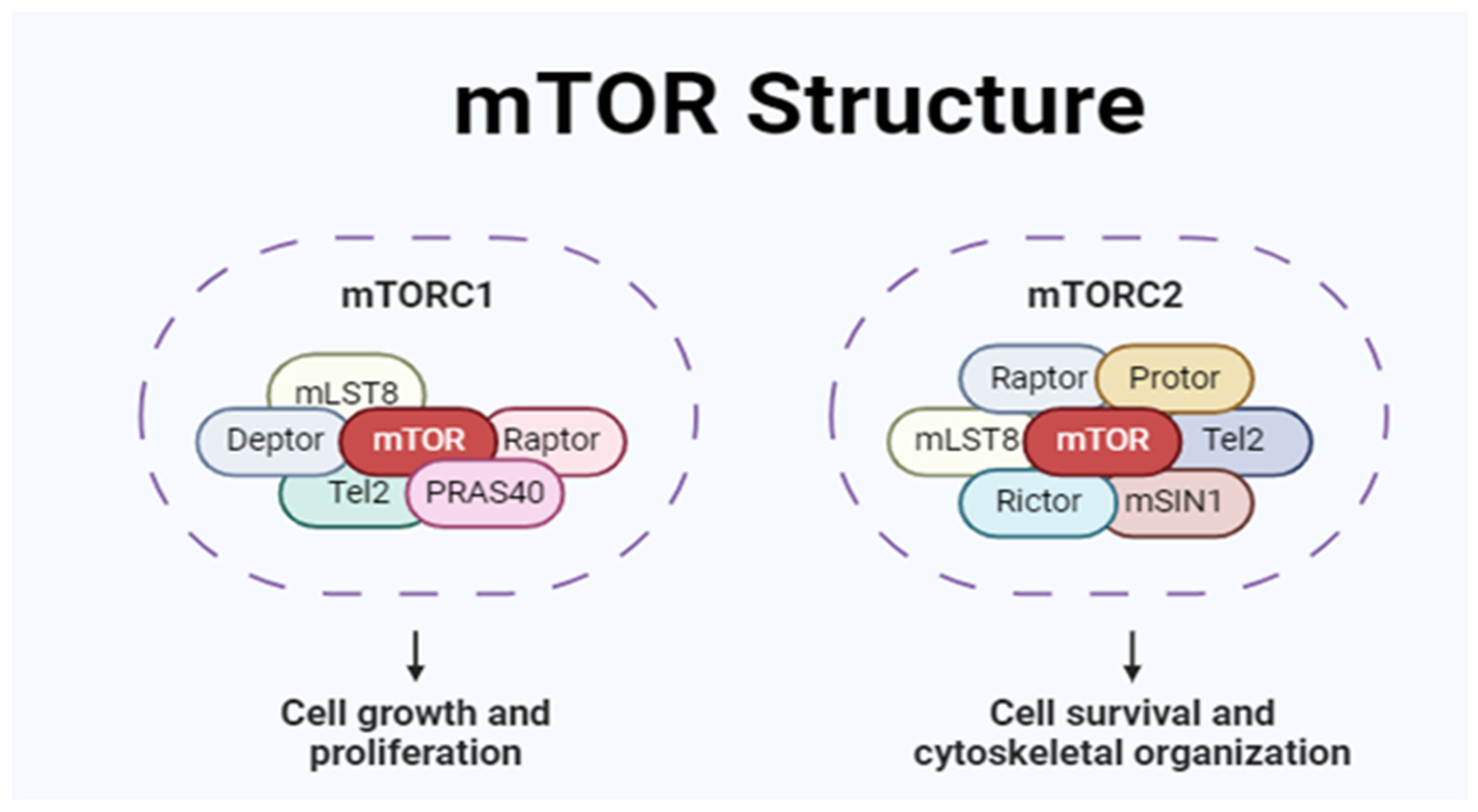
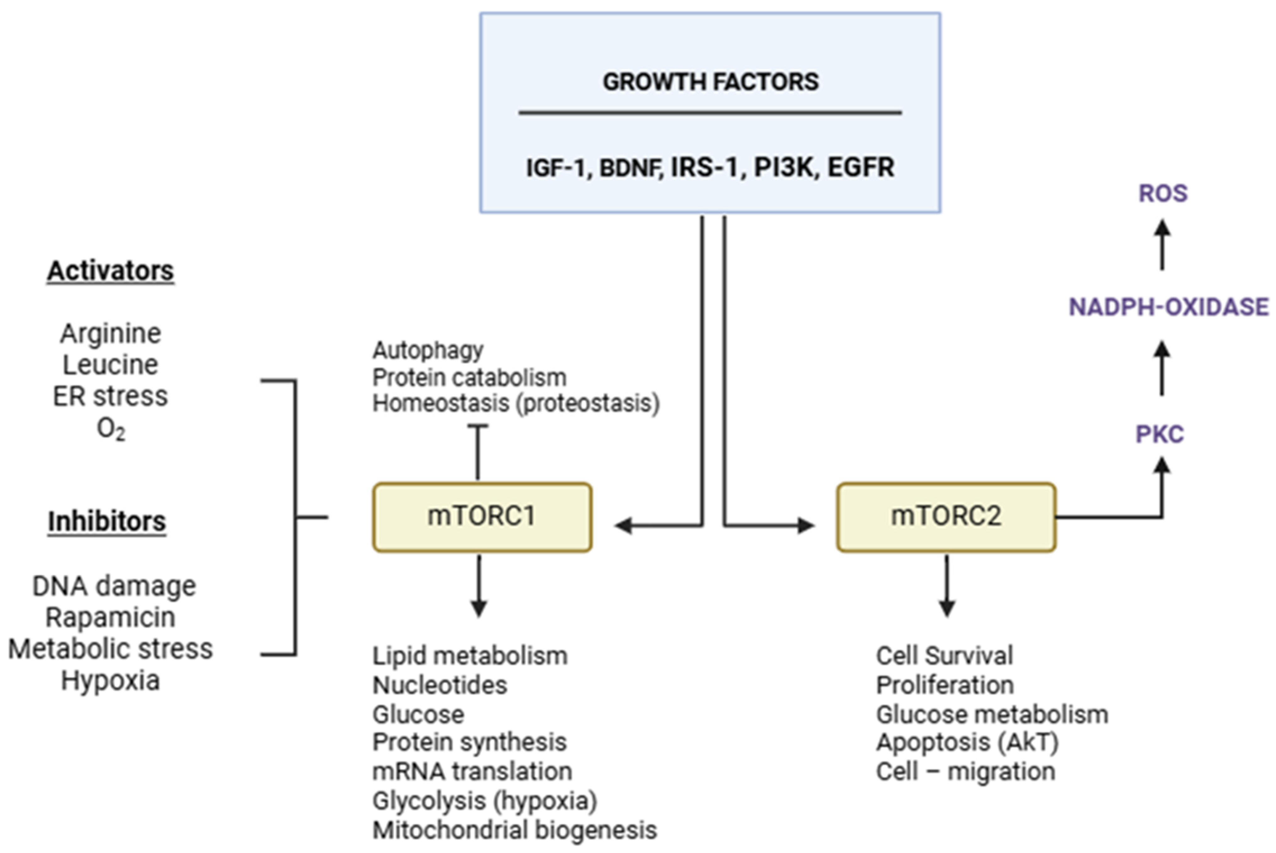
2. mTOR Structure and Function

mTOR belongs to the PI3K-related kinase (PIKK) family of protein kinases. There are two separate mTOR structures with catalytic units that control different cellular functions at the same time. The mTORC functionality is associated with neurodegenerative disorders. Three proteins comprise one of them, mTORC1: Deptor, PRAS40, GβL/mLST8, and the regulatory protein raptor (rapamycin-sensitive). The other structure, mTORC2, consists of the proteins GβL/mLST8, mSIN1, PRR5/Protor, Deptor, and Rictor (Figure 2). Studies have proposed that the mTORC2 protein Rictor is partially insensitive to rapamycin inhibition (Figure 2).
Growth factors, nutrition, and energy metabolism activate mTOR function. It regulates autophagy, contributes to lipids, proteins, and glucose synthesis, inhibits autophagy, controls ribosome function and mRNA translation, and enhances mitochondrial respiration. Therefore, the worsening of several diseases, such as cancer, diabetes, aging, and neurological disorders like ALS, has been associated with dysfunctional mTOR signaling [6,7,8,9,10,11]. Neuronal growth factors and other growth factors activate mTORC1. Decreased mTORC1 functionality can reduce memory retention and worsen muscle degeneration [6,7,8]. However, it can also stimulate lysosomal biogenesis, impacting homeostasis and influencing apoptosis, cellular development, division, proliferation, survival, and aging [11]. Understanding mTOR’s function is complex due to its multifaceted role. The same activators can trigger both pathways, yet each may have distinct characteristics (Figure 3).
mTOR inhibition triggers autophagy and proteolysis, which releases free amino acids for protein synthesis [12,13,14,15,16,17,18]. Both mTORC1 and mTORC2 have opposing effects on Akt. mTORC1 inhibits insulin/Akt signaling and also blocks mTORC2 activation. Conversely, the inhibition of mTORC1 using specific compounds, such as rapamycin, activates both Akt and mTORC2 [19,20,21,22]. Extracellular growth factors activate both mTORC1 and mTORC2 (Figure 2). However, the dysfunction or activation of mTORC1 leads to disturbances in proteostasis (impaired homeostasis) [23]. Another vital system for homeostasis is the action of AMPK (AMP-activated protein kinase), which may affect mTOR function. Both AMPK and mTORC function as energy sensors. Abundant nutrients activate mTOR, while calorie restriction activates AMPK. In this case, AMPK activates autophagy by the ubiquitin–proteasome system (UPS), a protein complex that induces proteolysis in misfolded proteins after being ubiquitinated and maintains homeostasis, detoxifies cells, and removes waste [24,25,26]. Despite the complexity of the mechanism, mTOR and AMPK are metabolic pathways that can be used to identify novel biomarkers or therapeutic targets. Studies have demonstrated that exercise, leucine, and IGF-1 activate mTORC1 signaling, promoting muscle hypertrophy in mice [21,27,28].
Mice given carbohydrate-restricted diets exhibited reduced mTOR activity, which had detrimental effects on memory and aging and increased lifespan, as shown by Robert et al. [29]. Mice lacking raptor, a component of mTOR, suffer premature death due to significant muscular atrophy and decreased body weight [29]. Rictor-knockout mice did not exhibit these results. It indicates that mTORC1, rather than mTORC2, is responsible for many of the essential functions of mTOR in skeletal muscle [21,29,30,31]. Muscular atrophy and weakness are common symptoms in all forms of ALS, suggesting a potential role for mTOR in the neurodegenerative pathophysiology. Interestingly, while the short-term, “in vivo” activation of mTORC1 signaling promotes muscle growth, long-term (chronic) activation can lead to significant muscle atrophy, body mass loss, and early death [29]. Persistent inhibition of autophagy and dysfunction in homeostasis within muscle tissue may be contributing factors. Grasping the functioning of mTORC1 across various signaling pathways related to insulin, proteins, lipids, amino acids, physical activity, and mechanical muscle force is essential. This perspective opens up a new way to look into treatments for various age-related diseases.

3. mTOR and ALS

ALS is a highly intricate neurological disease. In addition to the sporadic form, there are more than 30 subtypes of the genetic type, each with identifiable mutant genes. However, almost all studies concentrate on sporadic amyotrophic lateral sclerosis, with few reports on particular kinds of fALS. All types and subtypes of ALS share the same symptoms of atrophy and muscle weakness. Since mTOR is associated with muscular growth, a dysregulated mTOR function may play a role in the pathophysiology of ALS and possibly other neurodegenerative diseases [30].
Patients with familial ALS type 8 (ALS8) show elevated intracellular mTOR levels when compared to healthy controls. This increase in intracellular mTOR is suggested to be due to a possible dysfunction in the exocytosis process [31]. This finding has only been observed in cells from ALS8 patients and cannot be generalized to all types of familial ALS. The high intracellular mTOR content can inhibit autophagy and increase the presence of misfolded and aggregated proteins in the cytoplasm, as well as dysregulate the endoplasmic reticulum and alter enzymatic function, all of which could significantly affect both sporadic ALS and familial ALS. All these factors are directly or indirectly affected by mTOR.
Strong mTOR activation by nutrients and growth hormones, such as IGF-1 and insulin, promotes autophagy inhibition, possibly dysregulating homeostasis in ALS. It suggests a connection between the pathogenesis of ALS and the dysregulation of mTOR signaling and autophagy. Apart from oxidative stress, both sporadic amyotrophic lateral sclerosis and familial amyotrophic lateral sclerosis exhibit common features such as misfolded proteins, malfunctioning lysosomes, dysregulated mitochondria, changes in DNA and RNA activities, alterations in protein translation, and accumulation within the nucleus and cytoplasm. Mutant proteins are deleterious and affect cellular homeostasis. However, cells utilize various strategies to maintain cellular homeostasis, including well-balanced and activated autophagy. Gene mutations or epigenetic changes can cause ALS. These changes affect genomic and proteomic responses in different types of cells, which in turn affects the function of the neural plaque in ALS and other neurodegenerative diseases. This process involves mTOR-inhibiting autophagy. Dysfunction in mTOR signaling and astrocyte reactivity has been associated with neurodegenerative disorders and central nervous system damage [32,33,34,35].
Granatiero et al. [36] reported that in astrocytes with SOD1 mutations, the activation of mTOR is associated with the increased expression of the insulin-like growth factor 1 receptor (IGF1R). Elevated levels of the IGF1R activate the mTOR pathway and subsequently lead to the inhibition of autophagy. Interestingly, inhibiting the IGF1R-mTOR pathway has been shown to mitigate the harmful effects on motor neurons in the astrocytes of SOD1 mutant mice, indicating a potential connection between mTOR activity and motor neuron damage in ALS, highlighting mTOR’s complex and nuanced role [36].
However, the occasional conflicting reports about mTOR’s potential role in sALS highlight the need for further research. For instance, mTOR inhibition has been shown to both accelerate ALS progression and induce motor neuron degeneration in models with mutant SOD1. However, in other studies, similar mTOR inhibition in transgenic mice with elevated TDP-43 levels has led to protection rather than degeneration [33,34,35]. These findings underscore the complexity of mTOR’s function and emphasize the need for continued research. The quest for a definitive explanation is challenging.
mTOR is a natural autophagy inhibitor that alters the balance of autophagy and apoptosis. It could negatively influence homeostasis (Figure 2). Its inhibition could be a strategic therapeutic resource for controlling protein aggregation and accumulation inside cells from ALS patients and other neurodegenerative disorders. However, the possibility of negatively affecting cellular reactivity needs to be considered. Studies using rapamycin to inhibit mTOR have demonstrated its ability to reverse the autophagy inhibition it induces, leading to reduced internal cellular toxicity and improved homeostasis. Several factors, including hormones, intense physical activity, mechanical stress, and dietary intake, contribute to mTOR activation and muscle hypertrophy. Each factor impacts the mTOR signaling pathway, which is involved in muscle growth and interconnected with several other metabolic routes [37,38,39,40]. Mutations in valosin-containing protein (VCP) are associated with muscle weakness in mice. Dysfunction in mTOR and VCP may aggravate muscle weakness in amyotrophic lateral sclerosis (ALS), according to studies by James et al. [35]. It suggests that activated mTOR may play a role in the progression and exacerbation of ALS. Both the apoptotic and autophagic pathways in ALS are affected by mTOR signaling. An imbalance in these pathways can dysregulate homeostasis and cause disease [8,9,10] (Figure 3). Reports indicate that findings on mTOR inhibition are inconsistent, suggesting that results may change based on the type of ALS, specific gene mutations, and the metabolic signaling involved. In SOD1 mutants, mTOR inhibition worsens ALS, while similar mTOR inhibition in elevated TDP-43 levels protects against neuronal degeneration [41,42,43,44,45,46] despite the pathogenesis of ALS being associated with TDP-43 [47,48,49]. Amyotrophic lateral sclerosis and frontotemporal dementia have in common with each other their genetic and pathological characteristics. As the disorder progresses, there is an increase in neuronal degeneration, the formation of protein inclusions, chronic inflammation, and neurodegeneration.
Other signs observed in both ALS and FTD include the formation of misfolded TDP-43 and altered SOD1 or FUS. Furthermore, mitochondrial dysfunction, oxidative stress, protein imbalance, altered autophagy, ER stress-mediated cell death, stress granule formation, and altered RNA processing accelerate the disease’s progression [41,42,43]. Changes in homeostasis, cytotoxicity, and nucleocytoplasmic protein distribution correlate with TDP-43 aggregation and RNA dysregulation [39]. Multiple mutations in the TARDBP gene have been associated with ALS [50,51,52,53]. As a result, abnormal functions of TDP-43, including protein aggregation, relocation from the nucleus to the cytoplasm, and the accumulation of ubiquitinated and hyperphosphorylated TDP-43 in inclusion bodies, play a pivotal role in the disease’s pathophysiology.
These adverse effects are intensified in both sporadic and familial ALS [40]. Cytoplasmic inclusions of the transactive response (TAR) DNA-binding protein (TDP-43) are seen in about 90% of ALS patients. This intriguing phenomenon seems connected to reduced autophagy, potentially within an mTOR-activated system, which results in an increased accumulation of TDP-43′s C-terminal fragment. However, inhibiting mTOR pathways triggers autophagy, thereby regulating the cytoplasmic deposition of proteins. Rapamycin-induced mTOR inhibition stimulates autophagy, reducing TDP-43 (25 kDa fragment), regulating its accumulation, and returning it to its usual intracellular location [44,45,46]. These findings demonstrate the critical role of autophagy and the mTOR pathway in the pathophysiology of ALS.
Autophagy is a cellular process that clears protein aggregates, reduces caspase activation, and prevents apoptosis. This protective mechanism shields cells from the harmful effects of protein aggregates on their functions. Neurodegenerative diseases such as ALS have been associated with the accumulation of these aggregates. Apoptosis and autophagy are interconnected processes that can regulate each other in a balanced manner. The mechanism known as mTOR influences this balance. When mTOR is active, autophagy is upregulated. However, mTOR’s substrates regulate autophagy and multiple other biological processes. Inhibiting mTOR can affect crucial cellular functions like protein translation and ribosome biogenesis. Both autophagy and apoptosis are essential for maintaining cellular homeostasis. Autophagy removes misfolded proteins, damaged organelles, and long-lived cytosolic proteins, while apoptosis involves steps that prepare the targeted materials for transport to the lysosome. Various processes associated with apoptotic cell death lead to the formation of apoptotic bodies, which are then engulfed by macrophages or other cells [47,48,49,50]. The function of homeostasis depends on the balance of both phenomena. An imbalance between them can lead to neurodegenerative diseases like ALS, which are characterized by altered levels and activation in mTOR. The inhibition of autophagy decreases the cells’ ability to eliminate cellular waste. The abnormal and toxic protein aggregates worsen cellular stress in neurological disorders, resulting in cellular malfunction and disease.
Consequently, activating autophagy could help in restoring this pathologic state. Once the PI3K/Akt/mTOR complex is formed, the PI3K/AKT signaling (survival pathways) alters the activity of mTOR and Akt, activating autophagy-protecting neurons [48,51,52,53,54,55,56,57,58]. The deregulation of autophagy or apoptosis plays an essential role in the etiology and progression of numerous diseases. This imbalance has been associated with several neurodegenerative diseases, including ALS.
ALS and spinal muscular atrophy (SMA) share some of the exact pathophysiological mechanisms regarding synaptic plasticity and synaptogenesis, suggesting that mTOR may be involved [59,60,61,62,63]. The wild-type VAPB of the ALS8 protein functions at the endoplasmic reticulum (ER) membrane, regulating vesicular transport, maintaining homeostasis, and connecting the ER to the Golgi apparatus. In ALS8, on the other hand, overexpressing the mutant VAPBP56S causes wild-type VAPB to clump together and become active, which changes how the mitochondria-endoplasmic reticulum complex (MERC) works. The mTOR pathway activates these processes, including autophagy, apoptosis, and increased protein synthesis through C1q/TNF-related protein 3 (CTRP3) [62,63,64,65,66]. One possible explanation for motor neuron death is that when VAPB is mutated, mTOR cannot restore cellular homeostasis. The mechanism of mTOR is complex. Phosphorylated mTOR inhibits catabolism and enhances cell growth, whereas the non-phosphorylated form stimulates autophagy [35]. Autophagy activation through mTOR inhibition seems to be a promising therapeutic target for ALS and other neurodegenerative disorders. However, due to the toxicity of mTOR inhibitors, several studies have investigated potential substitutes for rapamycin. Additionally, mTOR impacts ALS and other neurodegenerative diseases. The complexity of mTOR’s metabolic signaling interactions currently prevents its use as a therapeutic target. Studies aimed at reducing the detrimental effects of mTOR inhibition in ALS and other neurodegenerative diseases require a thorough investigation of its mechanisms and the metabolic pathways involved in its signaling. Muscle atrophy, weakness, and paralysis often result from ubiquitin–proteasome, autophagy, and mitochondrial dysfunction, which are the main characteristics of ALS. These issues involve mTOR/AMPK signaling. ALS is a multifaceted neuromuscular disease characterized by muscle atrophy, motor neuron loss, and neuromuscular junction dysfunction. The majority of studies on ALS have focused on the central nervous system. However, recent approaches indicate how important peripheral tissues—especially skeletal muscle—are to disease onset and control, which may impact the neuromuscular junction (NMJ) and motor neurons, similarly to ALS-like muscle atrophy. An innovative strategy for ALS research could be utilized to investigate the interaction between muscles and neurons at the motor plate. Studies in other models have indicated that muscle protein release may influence neuron function [39,66]. A recently discovered protein, C1q/TNF-related protein 3 (CTRP3), assists muscles in regulating axonal local translation. This regulatory mechanism depends on the involvement of mTOR. Moreover, ALS has been linked to reduced axonal local protein synthesis [67]. A proposed pathophysiological connection exists between amyotrophic lateral sclerosis (ALS) and spinal muscular atrophy (SMA), indicating that these conditions may share fundamental underlying mechanisms [67]. Moreover, mTOR may significantly influence memory, synaptic plasticity, and the protein-mediated processes involved in synaptogenesis [67,68]. Reconceptualizing muscles as secretory organs presents an innovative and potential direction for ALS research [63,66]. Researchers have cautiously approached muscle as a secretory organ. According to these studies, the muscle protein CTRP-3 may impact the PI3K/AKT/mTOR signaling pathway and regulate the production of neuronal proteins. Decreased muscle CTRP-3 production may affect plaque neuron metabolism, leading to neuromuscular dysfunction. A protein like CTRP-3 can modify the neural plate’s function by inhibiting adjacent neurons’ neurotransmitter release, affecting muscle performance. Therefore, a thorough evaluation of the CRTP3-PI3K/Akt/mTOR axis in the context of muscular dysfunction in ALS is necessary.

4. Conclusions

The involvement of mTOR in ALS pathophysiology is highly suggestive, but studies are still in the early stages. The role of mTOR signaling in degenerative diseases such as ALS and other related conditions lies in its ability to inhibit autophagy, which prevents the elimination of misfolded proteins (cellular waste), contributing negatively to the disease. mTOR is associated with conditions that impact muscle function, including hypertrophy and atrophy. From this perspective, its role in ALS can be envisioned because muscle weakness and atrophy are the most common symptoms across the several types and subtypes of ALS and suggest a connection between mTOR and ALS. Previous studies in animal models of ALS indicate that dysfunction in skeletal muscle and the neuromuscular junction occurs before the degeneration of motor neurons and the onset of clinical symptoms. Evaluating the muscle as a secretory organ and how the protein CTRP-3 affects the PI3K/AKT/mTOR signaling pathway, which can control protein synthesis in neurons, could support the idea of neuromuscular dysfunction. Therefore, mTOR dysfunction in muscle secretion may influence the mTOR pathway and other signaling processes that regulate neurons and muscles, ultimately affecting the peripheral neuromotor function system. Nevertheless, this approach remains a hypothesis for potential discussion. Patients with ALS exhibit distinct features of muscle degeneration, while dementia is usually absent, which sets them apart from other neurodegenerative diseases. Therefore, emphasizing a peripheral focus rather than a purely neurocentric perspective may highlight the significance of mTOR signaling in ALS. Despite this, studies on ALS with a muscular focus are limited [66,67,69,70]. Crayle et al. [68] presented an intriguing proposition associating the lack of IGBP7 (insulin-like growth factor binding protein 7) expression with the activation of IGF-1, an mTOR activator. The authors associated IGBP7 absence with the reversal of clinical symptoms of ALS. Theoretically, it seems controversial because mTOR activation inhibits autophagy, which enhances the aggregation of protein and its deposition in the cytoplasm, which could exacerbate the disease. In truth, despite several studies and partial conclusions, we still know little or nothing definitive about ALS in all aspects of the disease, including onset and progression. mTOR and its metabolic connections may be a novel therapeutic resource, but this requires extensive study. The only certainty we have is that we know very little about ALS. With our current knowledge, we only promote an insignificant delay in the progression of the disease without any cure or control.

Author Contributions

J.A.N.-M. wrote, reviewed, and edited the manuscript. N.A.G. and F.R.-S. drew the figures. All authors have read and agreed to the published version of the manuscript.

Funding

The authors declare that FAPEMIG (Fundação de Amparo à Pesquisa do Estado de Minas Gerais) provided financial support for the research, with process numbers APQ 03767-23 and 000097-21.

Conflicts of Interest

The authors declare no conflicts of interest, financial or otherwise.

References

  1. Rowland, L.P.; Shneider, N.A. Amyotrophic Lateral Sclerosis. New Engl. J. Med. 2001, 344, 1688–1700. [Google Scholar] [CrossRef] [PubMed]
  2. Rothstein, J.D. Current Hypotheses for the Underlying Biology of Amyotrophic Lateral Sclerosis. Ann. Neurol. 2009, 65 (Suppl. S1), S3–S9. [Google Scholar] [CrossRef] [PubMed]
  3. De Souza, P.V.S.; de Pinto, W.B.V.R.; Chieia, M.A.T.; Oliveira, A.S.B. Clinical and Genetic Basis of Familial Amyotrophic Lateral Sclerosis. Arq. De Neuro-Psiquiatr. 2015, 73, 1026–1037. [Google Scholar] [CrossRef] [PubMed]
  4. Kalia, L.V.; Lang, A.E. Parkinson’s Disease. Lancet 2015, 386, 896–912. [Google Scholar] [CrossRef]
  5. Shandilya, A.; Mehan, S. Dysregulation of IGF-1/GLP-1 Signaling in the Progression of ALS: Potential Target Activators and Influences on Neurological Dysfunctions. Neurol. Sci. Off. J. Ital. Neurol. Soc. Ital. Soc. Clin. Neurophysiol. 2021, 42, 3145–3166. [Google Scholar] [CrossRef]
  6. Tang, S.J.; Reis, G.; Kang, H.; Gingras, A.-C.; Sonenberg, N.; Schuman, E.M. A Rapamycin-Sensitive Signaling Pathway Contributes to Long-Term Synaptic Plasticity in the Hippocampus. Proc. Natl. Acad. Sci. USA 2002, 99, 467–472. [Google Scholar] [CrossRef]
  7. Hoeffer, C.A.; Klann, E. MTOR Signaling: At the Crossroads of Plasticity, Memory and Disease. Trends Neurosci. 2010, 33, 67–75. [Google Scholar] [CrossRef]
  8. Stoica, L.; Zhu, P.J.; Huang, W.; Zhou, H.; Kozma, S.C.; Costa-Mattioli, M. Selective Pharmacogenetic Inhibition of Mammalian Target of Rapamycin Complex I (MTORC1) Blocks Long-Term Synaptic Plasticity and Memory Storage. Proc. Natl. Acad. Sci. USA 2011, 108, 3791–3796. [Google Scholar] [CrossRef]
  9. Schieke, S.M.; Phillips, D.; McCoy, J.P.J.; Aponte, A.M.; Shen, R.-F.; Balaban, R.S.; Finkel, T. The Mammalian Target of Rapamycin (mTOR) Pathway Regulates Mitochondrial Oxygen Consumption and Oxidative Capacity. J. Biol. Chem. 2006, 281, 27643–27652. [Google Scholar] [CrossRef]
  10. Ramanathan, A.; Schreiber, S.L. Direct Control of Mitochondrial Function by MTOR. Proc. Natl. Acad. Sci. USA 2009, 106, 22229–22232. [Google Scholar] [CrossRef]
  11. Saxton, R.A.; Sabatini, D.M. MTOR Signaling in Growth, Metabolism, and Disease. Cell 2017, 168, 960–976. [Google Scholar] [CrossRef] [PubMed]
  12. Porstmann, T.; Santos, C.R.; Griffiths, B.; Cully, M.; Wu, M.; Leevers, S.; Griffiths, J.R.; Chung, Y.-L.; Schulze, A. SREBP Activity Is Regulated by MTORC1 and Contributes to Akt-Dependent Cell Growth. Cell Metab. 2008, 8, 224–236. [Google Scholar] [CrossRef] [PubMed]
  13. Ben-Sahra, I.; Howell, J.J.; Asara, J.M.; Manning, B.D. Stimulation of de Novo Pyrimidine Synthesis by Growth Signaling through MTOR and S6K1. Science 2013, 339, 1323–1328. [Google Scholar] [CrossRef]
  14. Ben-Sahra, I.; Hoxhaj, G.; Ricoult, S.J.H.; Asara, J.M.; Manning, B.D. MTORC1 Induces Purine Synthesis through Control of the Mitochondrial Tetrahydrofolate Cycle. Science 2016, 351, 728–733. [Google Scholar] [CrossRef]
  15. Brunn, G.J.; Hudson, C.C.; Sekulić, A.; Williams, J.M.; Hosoi, H.; Houghton, P.J.; Lawrence, J.C.J.; Abraham, R.T. Phosphorylation of the Translational Repressor PHAS-I by the Mammalian Target of Rapamycin. Science 1997, 277, 99–101. [Google Scholar] [CrossRef]
  16. Martina, J.A.; Chen, Y.; Gucek, M.; Puertollano, R. MTORC1 Functions as a Transcriptional Regulator of Autophagy by Preventing Nuclear Transport of TFEB. Autophagy 2012, 8, 903–914. [Google Scholar] [CrossRef]
  17. Roczniak-Ferguson, A.; Petit, C.S.; Froehlich, F.; Qian, S.; Ky, J.; Angarola, B.; Walther, T.C.; Ferguson, S.M. The Transcription Factor TFEB Links MTORC1 Signaling to Transcriptional Control of Lysosome Homeostasis. Sci. Signal. 2012, 5, ra42. [Google Scholar] [CrossRef]
  18. Settembre, C.; Zoncu, R.; Medina, D.L.; Vetrini, F.; Erdin, S.; Erdin, S.; Huynh, T.; Ferron, M.; Karsenty, G.; Vellard, M.C.; et al. A Lysosome-to-Nucleus Signalling Mechanism Senses and Regulates the Lysosome via MTOR and TFEB. EMBO J. 2012, 31, 1095–1108. [Google Scholar] [CrossRef]
  19. Zhang, Y.; Manning, B.D. MTORC1 Signaling Activates NRF1 to Increase Cellular Proteasome Levels. Cell Cycle 2015, 14, 2011–2017. [Google Scholar] [CrossRef]
  20. Dibble, C.C.; Manning, B.D. Signal Integration by MTORC1 Coordinates Nutrient Input with Biosynthetic Output. Nat. Cell Biol. 2013, 15, 555–564. [Google Scholar] [CrossRef]
  21. Bodine, S.C.; Stitt, T.N.; Gonzalez, M.; Kline, W.O.; Stover, G.L.; Bauerlein, R.; Zlotchenko, E.; Scrimgeour, A.; Lawrence, J.C.; Glass, D.J.; et al. Akt/MTOR Pathway Is a Crucial Regulator of Skeletal Muscle Hypertrophy and Can Prevent Muscle Atrophy in Vivo. Nat. Cell Biol. 2001, 3, 1014–1019. [Google Scholar] [CrossRef] [PubMed]
  22. Zhang, Y.; Nicholatos, J.; Dreier, J.R.; Ricoult, S.J.H.; Widenmaier, S.B.; Hotamisligil, G.S.; Kwiatkowski, D.J.; Manning, B.D. Coordinated Regulation of Protein Synthesis and Degradation by MTORC1. Nature 2014, 513, 440–443. [Google Scholar] [CrossRef] [PubMed]
  23. Laplante, M.; Sabatini, D.M. MTOR Signaling in Growth Control and Disease. Cell 2012, 149, 274–293. [Google Scholar] [CrossRef]
  24. Hosokawa, N.; Hara, T.; Kaizuka, T.; Kishi, C.; Takamura, A.; Miura, Y.; Iemura, S.; Natsume, T.; Takehana, K.; Yamada, N.; et al. Nutrient-Dependent MTORC1 Association with the ULK1-Atg13-FIP200 Complex Required for Autophagy. Mol. Biol. Cell 2009, 20, 1981–1991. [Google Scholar] [CrossRef]
  25. Jung, C.H.; Jun, C.B.; Ro, S.-H.; Kim, Y.-M.; Otto, N.M.; Cao, J.; Kundu, M.; Kim, D.-H. ULK-Atg13-FIP200 Complexes Mediate MTOR Signaling to the Autophagy Machinery. Mol. Biol. Cell 2009, 20, 1992–2003. [Google Scholar] [CrossRef]
  26. Kim, J.; Kundu, M.; Viollet, B.; Guan, K.-L. AMPK and MTOR Regulate Autophagy through Direct Phosphorylation of Ulk1. Nat. Cell Biol. 2011, 13, 132–141. [Google Scholar] [CrossRef]
  27. Anthony, J.C.; Yoshizawa, F.; Anthony, T.G.; Vary, T.C.; Jefferson, L.S.; Kimball, S.R. Leucine Stimulates Translation Initiation in Skeletal Muscle of Postabsorptive Rats via a Rapamycin-Sensitive Pathway. J. Nutr. 2000, 130, 2413–2419. [Google Scholar] [CrossRef]
  28. Rommel, C.; Bodine, S.C.; Clarke, B.A.; Rossman, R.; Nunez, L.; Stitt, T.N.; Yancopoulos, G.D.; Glass, D.J. Mediation of IGF-1-Induced Skeletal Myotube Hypertrophy by PI(3)K/Akt/MTOR and PI(3)K/Akt/GSK3 Pathways. Nat. Cell Biol. 2001, 3, 1009–1013. [Google Scholar] [CrossRef]
  29. Roberts, M.N.; Wallace, M.A.; Tomilov, A.A.; Zhou, Z.; Marcotte, G.R.; Tran, D.; Perez, G.; Gutierrez-Casado, E.; Koike, S.; Knotts, T.A.; et al. A Ketogenic Diet Extends Longevity and Healthspan in Adult Mice. Cell Metab. 2017, 26, 539–546.e5. [Google Scholar] [CrossRef]
  30. Bentzinger, C.F.; Romanino, K.; Cloëtta, D.; Lin, S.; Mascarenhas, J.B.; Oliveri, F.; Xia, J.; Casanova, E.; Costa, C.F.; Brink, M.; et al. Skeletal Muscle-Specific Ablation of Raptor, but Not of Rictor, Causes Metabolic Changes and Results in Muscle Dystrophy. Cell Metab. 2008, 8, 411–424. [Google Scholar] [CrossRef]
  31. Gomes, N.A.; das Chagas Lima, E.; Silva, F.; de Oliveira Volpe, C.M.; Villar-Delfino, P.H.; de Sousa, C.F.; Rocha-Silva, F.; Nogueira-Machado, J.A. Overexpression of MTOR in Leukocytes from ALS8 Patients. Curr. Neuropharmacol. 2023, 21, 482–490. [Google Scholar] [CrossRef] [PubMed]
  32. Zhang, X.; Li, L.; Chen, S.; Yang, D.; Wang, Y.; Zhang, X.; Wang, Z.; Le, W. Rapamycin Treatment Augments Motor Neuron Degeneration in SOD1(G93A) Mouse Model of Amyotrophic Lateral Sclerosis. Autophagy 2011, 7, 412–425. [Google Scholar] [CrossRef] [PubMed]
  33. Saxena, S.; Roselli, F.; Singh, K.; Leptien, K.; Julien, J.-P.; Gros-Louis, F.; Caroni, P. Neuroprotection through Excitability and MTOR Required in ALS Motoneurons to Delay Disease and Extend Survival. Neuron 2013, 80, 80–96. [Google Scholar] [CrossRef] [PubMed]
  34. Wang, I.-F.; Tsai, K.-J.; Shen, C.-K.J. Autophagy Activation Ameliorates Neuronal Pathogenesis of FTLD-U Mice: A New Light for Treatment of TARDBP/TDP-43 Proteinopathies. Autophagy 2013, 9, 239–240. [Google Scholar] [CrossRef]
  35. Ching, J.K.; Weihl, C.C. Rapamycin-Induced Autophagy Aggravates Pathology and Weakness in a Mouse Model of VCP-Associated Myopathy. Autophagy 2013, 9, 799–800. [Google Scholar] [CrossRef]
  36. Granatiero, V.; Sayles, N.M.; Savino, A.M.; Konrad, C.; Kharas, M.G.; Kawamata, H.; Manfredi, G. Modulation of the IGF1R-MTOR Pathway Attenuates Motor Neuron Toxicity of Human ALS SOD1G93A Astrocytes. Autophagy 2021, 17, 4029–4042. [Google Scholar] [CrossRef]
  37. Glass, D.J. Molecular Mechanisms Modulating Muscle Mass. Trends Mol. Med. 2003, 9, 344–350. [Google Scholar] [CrossRef]
  38. Yoon, M.-S. MTOR as a Key Regulator in Maintaining Skeletal Muscle Mass. Front. Physiol. 2017, 8, 788. [Google Scholar] [CrossRef]
  39. Prasad, A.; Bharathi, V.; Sivalingam, V.; Girdhar, A.; Patel, B.K. Molecular Mechanisms of TDP-43 Misfolding and Pathology in Amyotrophic Lateral Sclerosis. Front. Mol. Neurosci. 2019, 12, 25. [Google Scholar] [CrossRef]
  40. Ayala, Y.M.; De Conti, L.; Avendaño-Vázquez, S.E.; Dhir, A.; Romano, M.; D’Ambrogio, A.; Tollervey, J.; Ule, J.; Baralle, M.; Buratti, E.; et al. TDP-43 Regulates Its MRNA Levels through a Negative Feedback Loop. EMBO J. 2011, 30, 277–288. [Google Scholar] [CrossRef]
  41. Ransohoff, R.M. How Neuroinflammation Contributes to Neurodegeneration. Science 2016, 353, 777–783. [Google Scholar] [CrossRef] [PubMed]
  42. Jimenez-Pacheco, A.; Franco, J.M.; Lopez, S.; Gomez-Zumaquero, J.M.; Magdalena Leal-Lasarte, M.; Caballero-Hernandez, D.E.; Cejudo-Guillén, M.; Pozo, D. Epigenetic Mechanisms of Gene Regulation in Amyotrophic Lateral Sclerosis. Adv. Exp. Med. Biol. 2017, 978, 255–275. [Google Scholar] [CrossRef] [PubMed]
  43. Sreedharan, J.; Neukomm, L.J.; Brown, R.H.J.; Freeman, M.R. Age-Dependent TDP-43-Mediated Motor Neuron Degeneration Requires GSK3, Hat-Trick, and Xmas-2. Curr. Biol. 2015, 25, 2130–2136. [Google Scholar] [CrossRef]
  44. Mitchell, J.C.; Constable, R.; So, E.; Vance, C.; Scotter, E.; Glover, L.; Hortobagyi, T.; Arnold, E.S.; Ling, S.-C.; McAlonis, M.; et al. Wild Type Human TDP-43 Potentiates ALS-Linked Mutant TDP-43 Driven Progressive Motor and Cortical Neuron Degeneration with Pathological Features of ALS. Acta Neuropathol. Commun. 2015, 3, 36. [Google Scholar] [CrossRef] [PubMed]
  45. Caccamo, A.; Majumder, S.; Deng, J.J.; Bai, Y.; Thornton, F.B.; Oddo, S. Rapamycin Rescues TDP-43 Mislocalization and the Associated Low Molecular Mass Neurofilament Instability. J. Biol. Chem. 2009, 284, 27416–27424. [Google Scholar] [CrossRef]
  46. Ravikumar, B.; Duden, R.; Rubinsztein, D.C. Aggregate-Prone Proteins with Polyglutamine and Polyalanine Expansions Are Degraded by Autophagy. Hum. Mol. Genet. 2002, 11, 1107–1117. [Google Scholar] [CrossRef]
  47. Ravikumar, B.; Berger, Z.; Vacher, C.; O’Kane, C.J.; Rubinsztein, D.C. Rapamycin Pre-Treatment Protects against Apoptosis. Hum. Mol. Genet. 2006, 15, 1209–1216. [Google Scholar] [CrossRef]
  48. Berger, Z.; Ravikumar, B.; Menzies, F.M.; Oroz, L.G.; Underwood, B.R.; Pangalos, M.N.; Schmitt, I.; Wullner, U.; Evert, B.O.; O’Kane, C.J.; et al. Rapamycin Alleviates Toxicity of Different Aggregate-Prone Proteins. Hum. Mol. Genet. 2006, 15, 433–442. [Google Scholar] [CrossRef]
  49. Kim, D.-H.; Sarbassov, D.D.; Ali, S.M.; King, J.E.; Latek, R.R.; Erdjument-Bromage, H.; Tempst, P.; Sabatini, D.M. MTOR Interacts with Raptor to Form a Nutrient-Sensitive Complex That Signals to the Cell Growth Machinery. Cell 2002, 110, 163–175. [Google Scholar] [CrossRef]
  50. Sarbassov, D.D.; Ali, S.M.; Sengupta, S.; Sheen, J.-H.; Hsu, P.P.; Bagley, A.F.; Markhard, A.L.; Sabatini, D.M. Prolonged Rapamycin Treatment Inhibits MTORC2 Assembly and Akt/PKB. Mol. Cell 2006, 22, 159–168. [Google Scholar] [CrossRef]
  51. Yang, Y.-P.; Liang, Z.-Q.; Gu, Z.-L.; Qin, Z.-H. Molecular Mechanism and Regulation of Autophagy. Acta Pharmacol. Sin. 2005, 26, 1421–1434. [Google Scholar] [CrossRef] [PubMed]
  52. Fleming, A.; Noda, T.; Yoshimori, T.; Rubinsztein, D.C. Chemical Modulators of Autophagy as Biological Probes and Potential Therapeutics. Nat. Chem. Biol. 2011, 7, 9–17. [Google Scholar] [CrossRef] [PubMed]
  53. Heras-Sandoval, D.; Pérez-Rojas, J.M.; Hernández-Damián, J.; Pedraza-Chaverri, J. The Role of PI3K/AKT/MTOR Pathway in the Modulation of Autophagy and the Clearance of Protein Aggregates in Neurodegeneration. Cell. Signal. 2014, 26, 2694–2701. [Google Scholar] [CrossRef] [PubMed]
  54. Carrì, M.T.; Cozzolino, M. SOD1 and Mitochondria in ALS: A Dangerous Liaison. J. Bioenerg. Biomembr. 2011, 43, 593–599. [Google Scholar] [CrossRef]
  55. Heath-Engel, H.M.; Chang, N.C.; Shore, G.C. The Endoplasmic Reticulum in Apoptosis and Autophagy: Role of the BCL-2 Protein Family. Oncogene 2008, 27, 6419–6433. [Google Scholar] [CrossRef]
  56. Hellwig, C.T.; Passante, E.; Rehm, M. The Molecular Machinery Regulating Apoptosis Signal Transduction and Its Implication in Human Physiology and Pathophysiologies. Curr. Mol. Med. 2011, 11, 31–47. [Google Scholar] [CrossRef]
  57. Hetz, C.; Thielen, P.; Fisher, J.; Pasinelli, P.; Brown, R.H.; Korsmeyer, S.; Glimcher, L. The Proapoptotic BCL-2 Family Member BIM Mediates Motoneuron Loss in a Model of Amyotrophic Lateral Sclerosis. Cell Death Differ. 2007, 14, 1386–1389. [Google Scholar] [CrossRef]
  58. Hetz, C.; Thielen, P.; Matus, S.; Nassif, M.; Court, F.; Kiffin, R.; Martinez, G.; Cuervo, A.M.; Brown, R.H.; Glimcher, L.H. XBP-1 Deficiency in the Nervous System Protects against Amyotrophic Lateral Sclerosis by Increasing Autophagy. Genes Dev. 2009, 23, 2294–2306. [Google Scholar] [CrossRef]
  59. Hetz, C.A. ER Stress Signaling and the BCL-2 Family of Proteins: From Adaptation to Irreversible Cellular Damage. Antioxid. Redox Signal. 2007, 9, 2345–2355. [Google Scholar] [CrossRef]
  60. Kye, M.J.; Niederst, E.D.; Wertz, M.H.; Gonçalves, I.D.C.G.; Akten, B.; Dover, K.Z.; Peters, M.; Riessland, M.; Neveu, P.; Wirth, B.; et al. SMN Regulates Axonal Local Translation via MiR-183/MTOR Pathway. Hum. Mol. Genet. 2014, 23, 6318–6331. [Google Scholar] [CrossRef]
  61. Biondi, O.; Branchu, J.; Ben Salah, A.; Houdebine, L.; Bertin, L.; Chali, F.; Desseille, C.; Weill, L.; Sanchez, G.; Lancelin, C.; et al. IGF-1R Reduction Triggers Neuroprotective Signaling Pathways in Spinal Muscular Atrophy Mice. J. Neurosci. Off. J. Soc. For Neurosci. 2015, 35, 12063–12079. [Google Scholar] [CrossRef] [PubMed]
  62. Shi, Y.; Shen, H.-M.; Gopalakrishnan, V.; Gordon, N. Epigenetic Regulation of Autophagy Beyond the Cytoplasm: A Review. Front. Cell Dev. Biol. 2021, 9, 675599. [Google Scholar] [CrossRef] [PubMed]
  63. Yin, S.; Liu, L.; Gan, W. The Roles of Post-Translational Modifications on MTOR Signaling. Int. J. Mol. Sci. 2021, 22, 1784. [Google Scholar] [CrossRef]
  64. Chen, J.; Bassot, A.; Giuliani, F.; Simmen, T. Amyotrophic Lateral Sclerosis (ALS): Stressed by Dysfunctional Mitochondria-Endoplasmic Reticulum Contacts (MERCs). Cells 2021, 10, 71789. [Google Scholar] [CrossRef]
  65. Ramesh, N.; Pandey, U.B. Autophagy Dysregulation in ALS: When Protein Aggregates Get Out of Hand. Front. Mol. Neurosci. 2017, 10, 263. [Google Scholar] [CrossRef]
  66. Rehorst, W.A.; Thelen, M.P.; Nolte, H.; Türk, C.; Cirak, S.; Peterson, J.M.; Wong, G.W.; Wirth, B.; Krüger, M.; Winter, D.; et al. Muscle Regulates MTOR Dependent Axonal Local Translation in Motor Neurons via CTRP3 Secretion: Implications for a Neuromuscular Disorder, Spinal Muscular Atrophy. Acta Neuropathol. Commun. 2019, 7, 154. [Google Scholar] [CrossRef]
  67. Costa-Mattioli, M.; Sossin, W.S.; Klann, E.; Sonenberg, N. Translational control of long-lasting synaptic plasticity and memory. Neuron 2009, 61, 10–26. [Google Scholar] [CrossRef]
  68. Crayle, J.I.; Rampersaud, E.; Myers, J.R.; Wuu, J.; Taylor, J.P.; Wu, G.; Benatar, M.; Bedlack, R.S. Genetic Associations with an Amyotrophic Lateral Sclerosis Reversal Phenotype. Neurology 2024, 103, e209696. [Google Scholar] [CrossRef]
  69. Gao, J.; Sterling, E.; Hankin, R.; Sikal, A.; Yao, Y. Therapeutics Targeting Skeletal Muscle in Amyotrophic Lateral Sclerosis. Biomolecules 2024, 14, 878. [Google Scholar] [CrossRef]
  70. Kubinski, S.; Claus, P. Protein network analysis reveals a functional connectivity of dysregulated processes in ALS and SMA. Neurosci. Insights 2022, 17. [Google Scholar] [CrossRef]
Figure 1. The model demonstrates metabolic signaling, illustrating how mTOR affects autophagy and apoptosis. The arrows mean activation; decreasing or increasing function for autophagy and apoptosis, respectively, while mTOR inhibits autophagy ( Biomedicines 13 00952 i001 ) IRS = insulin receptor substrate; PI3K = phosphatidylinositol (PI) 3-kinase; Akt = serine/th reonine kinase Akt, also known as protein kinase B (PKB); PIP2 = phosphatidylinositol biphosphate; PIP3 = phosphatidylinositol triphosphate.
Biomedicines 13 00952 g001
Figure 2. The mTOR pathway plays a crucial role in cellular regulation and consists of two main complexes: mTORC1 and mTORC2. mTORC1 is composed of proteins such as Deptor, PRAS40, GβL/mLST8, and the rapamycin-sensitive regulatory protein known as raptor. In contrast, mTORC2 includes GβL/mLST8, mSIN1, PRR5/Protor, Deptor, and Rictor. Understanding these complexes is essential for investigating their functions and potential therapeutic applications in cellular processes.
Biomedicines 13 00952 g002
Figure 3. The functions of mTORC1 and mTORC2 are complex and involve various interactions with other signaling pathways, activation mechanisms, and self-regulatory processes. IRS-1 = insulin receptor substrate 1; IGF-1 = insulin growth factor 1; BDNF = brain-derived neurotrophic factor; PI3K = phosphoinositide 3-kinase; EGFR = epidermal growth factor receptor.
Biomedicines 13 00952 g003
Disclaimer/Publisher’s Note: The statements, opinions and data contained in all publications are solely those of the individual author(s) and contributor(s) and not of MDPI and/or the editor(s). MDPI and/or the editor(s) disclaim responsibility for any injury to people or property resulting from any ideas, methods, instructions or products referred to in the content.

