
Therapeutic Plasma Exchange in Corticosteroid-Refractory Multiple Sclerosis Relapses: Mechanisms, Efficacy, and Integration into Clinical Practice


Abstract
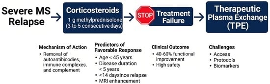
1. Introduction
2. Mechanistic Rationale for TPE in MS
3. Methodological Approach
4. Clinical Efficacy of Therapeutic Plasma Exchange

| Study | Design | N | Population | Timing to TPE, Median in Days * | Response Rate (%) | Outcome Measure |
|---|---|---|---|---|---|---|
| Weiner et al., 1989 [39] | RCT/sham controlled as adjunctive therapy | 76 | RRMS | ≤2 | 64 | DSS |
| Weinshenker et al., 1999 [30] | RCT/sham- controlled crossover | 22 | Mixed demyelinating diseases | ≤14 | 42 | TND |
| Keegan et al., 2002 [32] | Retrospective | 59 | Mixed demyelinating diseases | 17 | 44 | TND |
| Llufriu et al., 2009 [34] | Retrospective | 18 | RRMS | 27 | 28 ** | EDSS |
| Ehler et al., 2015 [35] | Retrospective | 37 | RMS | 44 | 81.1 | EDSS |
| Correia et al., 2018 [36] | Retrospective | 46 | RMS | 33 *** | 80.4 | EDSS |
| Marrodan et al., 2021 [37] | Retrospective | 23 | RRMS | 15 *** | 78 | EDSS |
| Blechinger et al., 2021 [9] | retrospective | 118 | RMS | 39 | 78.8 | EDSS |
| Bunganic et al., 2022 [8] | Retrospective | 155 | RRMS | 49 | 50 | EDSS |
| Iacono et al., 2024 [7] | Retrospective | 59 | Mixed neuroimmunological diseases | 26 | 80 **** | EDSS and MRS |
| Mesaros et al., 2024 [38] | Retrospective | 107 | RMS | 32 | 80.9 | EDSS |
5. Predictors of Therapeutic Response
5.1. Age and Disease Duration
5.2. Relapse Phenotype and Severity
5.3. MRI Features of Active Inflammation
5.4. Timing of Intervention
5.5. Baseline Neurological Status and Chronic Deficits
- -
- Poor baseline neurological function (e.g., EDSS ≥ 7.5) at relapse onset, is strongly predictive of poor outcomes and likely reflects extensive, irreversible tissue [34].
- -
- Absence of MRI inflammatory activity, such as lack of gadolinium enhancement or diffusion restriction, has been linked to minimal clinical benefit [35].
- -
- Presence of fixed neurological deficits from prior relapses can further complicate assessments of new inflammatory activity and limit recovery potential.
- -
- Slowly progressive symptoms evolution rather than abrupt relapses onset, often indicates a non-inflammatory or degenerative process (e.g., progressive MS) that is inherently less responsive to immunomodulatory interventions such as TPE [45].
5.6. CSF and Serological Biomarkers
6. Safety Profile of TPE
6.1. Common and Anticipated Adverse Effects
- -
- Hemodynamic instability: Hypotension is among the most frequently reported side effects, often resulting from rapid intravascular volume shifts, autonomic dysfunction in neurologically impaired patients, or an inadequate compensatory response to volume replacement. Risk can be mitigated by employing slower exchange rates, ensuring adequate pre-procedural hydration, and judicious use of vasopressors when necessary [7].
- -
- Electrolyte imbalances: Hypocalcemia related to citrate anticoagulation is common, presenting with perioral paresthesias, muscle cramps, or in severe cases, arrhythmias. Prophylactic calcium supplementation (e.g., calcium gluconate infusion) during the procedure and close monitoring of ionized calcium levels are effective preventing [48,49].
- -
- Gastrointestinal and constitutional symptoms: Nausea, vomiting, dizziness, chills, and fatigue may occur particularly during initial sessions. These events are generally self-limiting and respond well to symptomatic management [49].
6.2. Vascular Access and Catheter-Related Risks
- -
- Catheter-associated thrombosis and infection: non-tunneled CVCs carry an increased e risk of catheter-associated bloodstream infections, particularly in immunosuppressed individuals. Preventive strategies include strict aseptic techniques, use of antimicrobial-impregnated catheters, and minimizing catheter dwell time [50,51].
- -
- -
- Hemorrhagic events: Although uncommon, bleeding complications may occur secondary to heparin anticoagulation or depletion of coagulation factors [49]. Regular monitoring of coagulation parameters and avoidance of concurrent anticoagulation, unless clearly indicated, are recommended.
6.3. Immunologic and Allergic Reactions
- -
- -
- Allergic manifestations: Mild reactions including urticaria, pruritus, or flushing are relatively common and usually manageable with antihistamines. Severe reactions such as anaphylaxis are exceedingly rare but require immediate discontinuation of the procedure and emergency management [49].
6.4. Serious and Rare Adverse Events
- -
- Sepsis and bloodstream infections: Particularly in elderly or immunocompromised individuals, bacteremia may result from catheter colonization or manipulation. Early recognition and empiric antimicrobial therapy are critical.
- -
- Coagulopathies and bleeding: Repeated exchanges may deplete clotting factors, underscoring the need for periodic fibrinogen monitoring and, in select cases, the administration of FFP or cryoprecipitate.
- -
- Metabolic derangements: Electrolyte abnormalities such as hypokalemia, hypomagnesemia, or hypernatremia may occur, especially in patients with underlying renal dysfunction or when large fluid volumes are administered. Careful monitoring and targeted replacement are required.
6.5. Long-Term Safety and Immunologic Tolerance
7. Practical Considerations and Protocol Variability
Strategies to Optimize Implementation
- -
- -
- Defined Criteria for TPE Initiation: Incorporation of explicit definitions of corticosteroid non-response and clear timing thresholds for TPE initiation into relapse management algorithms ensuring timely escalation during the critical therapeutic window [8].
- -
- Streamlined Referral Pathways: Implementation of fast-track care models that enable rapid transitions from outpatient neurology services to apheresis centers, minimizing treatment delays and optimizing recovery potential [61].
- -
- Innovative Access Models: Deployment of telemedicine-based consults and mobile apheresis units to extend access in resource-limited regions, thereby reducing disparities in access to advanced immunomodulatory care [62].
8. Gaps in Current Knowledge
8.1. Lack of Contemporary Multicenter Randomized Controlled Trials (RCTs)
8.2. Heterogeneous Definitions of Corticosteroid-Refractory Relapse
8.3. Inconsistent Outcome Measures and Limited Use of Composite Endpoints
8.4. Understudied Role in Progressive MS with Superimposed Relapses
9. Integration into Clinical Practice
10. Future Directions
10.1. Comparative Efficacy Trials
10.2. Biomarker Discovery and Validation
10.3. Economic and Health Systems Research
10.4. Combination and Sequential Therapeutic Strategies
11. Conclusions
Author Contributions
Funding
Institutional Review Board Statement
Informed Consent Statement
Data Availability Statement
Conflicts of Interest
References
- Hirst, C.; Ingram, G.; Pearson, O.; Pickersgill, T.; Scolding, N.; Robertson, N. Contribution of relapses to disability in multiple sclerosis. J. Neurol. 2008, 255, 280–287. [Google Scholar] [CrossRef] [PubMed]
- Lublin, F.D.; Häring, D.A.; Ganjgahi, H.; Ocampo, A.; Hatami, F.; Čuklina, J.; Aarden, P.; Dahlke, F.; Arnold, D.L.; Wiendl, H.; et al. How patients with multiple sclerosis acquire disability. Brain 2022, 145, 3147–3161. [Google Scholar] [CrossRef]
- Repovic, P. Management of Multiple Sclerosis Relapses. Contin. Lifelong Learn. Neurol. 2019, 25, 655–669. [Google Scholar] [CrossRef]
- Ross, A.P.; Ben-Zacharia, A.; Harris, C.; Smrtka, J. Multiple sclerosis, relapses, and the mechanism of action of adrenocorticotropic hormone. Front. Neurol. 2013, 4, 21. [Google Scholar] [CrossRef]
- Leone, M.A.; Bonissoni, S.; Collimedaglia, L.; Tesser, F.; Calzoni, S.; Stecco, A.; Naldi, P.; Monaco, F. Factors predicting incomplete recovery from relapses in multiple sclerosis: A prospective study. Mult. Scler. 2008, 14, 485–493. [Google Scholar] [CrossRef] [PubMed]
- Stoppe, M.; Busch, M.; Krizek, L.; Then Bergh, F. Outcome of MS relapses in the era of disease-modifying therapy. BMC Neurol. 2017, 17, 151. [Google Scholar] [CrossRef]
- Iacono, S.; Schirò, G.; Salemi, G.; Scirè, E.; Aridon, P.; Melfa, M.; Andolina, M.; Sorbello, G.; Calì, A.; Brighina, F.; et al. Efficacy and Safety of Rescue Treatment with Plasma Exchange in Patients with Acute Inflammatory Neurological Disorders: A Single Center Experience. Neurol. Int. 2024, 16, 761–775. [Google Scholar] [CrossRef]
- Bunganic, R.; Blahutova, S.; Revendova, K.; Zapletalova, O.; Hradilek, P.; Hrdlickova, R.; Ganesh, A.; Cermakova, Z.; Bar, M.; Volny, O. Therapeutic plasma exchange in multiple sclerosis patients with an aggressive relapse: An observational analysis in a high-volume center. Sci. Rep. 2022, 12, 18374. [Google Scholar] [CrossRef]
- Blechinger, S.; Ehler, J.; Bsteh, G.; Winkelmann, A.; Leutmezer, F.; Meister, S.; Santer, A.; Hecker, M.; Berger, T.; Rommer, P.; et al. Therapeutic plasma exchange in steroid-refractory multiple sclerosis relapses. A retrospective two-center study. Ther. Adv. Neurol. Disord. 2021, 14, 1756286420975642. [Google Scholar] [CrossRef] [PubMed]
- Fraussen, J.; Claes, N.; de Bock, L.; Somers, V. Targets of the humoral autoimmune response in multiple sclerosis. Autoimmun. Rev. 2014, 13, 1126–1137. [Google Scholar] [CrossRef]
- Stork, L.; Ellenberger, D.; Beißbarth, T.; Friede, T.; Lucchinetti, C.F.; Brück, W.; Metz, I. Differences in the Reponses to Apheresis Therapy of Patients With 3 Histopathologically Classified Immunopathological Patterns of Multiple Sclerosis. JAMA Neurol. 2018, 75, 428–435. [Google Scholar] [CrossRef]
- Rodríguez Murúa, S.; Farez, M.F.; Quintana, F.J. The Immune Response in Multiple Sclerosis. Annu. Rev. Pathol. 2022, 17, 121–139. [Google Scholar] [CrossRef] [PubMed]
- Zhang, X.; Chen, F.; Sun, M.; Wu, N.; Liu, B.; Yi, X.; Ge, R.; Fan, X. Microglia in the context of multiple sclerosis. Front. Neurol. 2023, 14, 1157287. [Google Scholar] [CrossRef] [PubMed]
- Hammond, B.P.; Panda, S.P.; Kaushik, D.K.; Plemel, J.R. Microglia and Multiple Sclerosis. Adv. Neurobiol. 2024, 37, 445–456. [Google Scholar] [CrossRef]
- Comi, G.; Bar-Or, A.; Lassmann, H.; Uccelli, A.; Hartung, H.P.; Montalban, X.; Sørensen, P.S.; Hohlfeld, R.; Hauser, S.L. Role of B Cells in Multiple Sclerosis and Related Disorders. Ann. Neurol. 2021, 89, 13–23. [Google Scholar] [CrossRef]
- Lucchinetti, C.F.; Bruck, W.; Parisi, J.; Scheithauer, B.; Rodriguez, M.; Lassmann, H. Heterogeneity of multiple sclerosis lesions: Implications for the pathogenesis of demyelination. Ann. Neurol. 2000, 47, 707–717. [Google Scholar] [CrossRef]
- Touil, H.; Li, R.; Zuroff, L.; Moore, C.S.; Healy, L.; Cignarella, F.; Piccio, L.; Ludwin, S.; Prat, A.; Gommerman, J.; et al. Cross-talk between B cells, microglia and macrophages, and implications to central nervous system compartmentalized inflammation and progressive multiple sclerosis. EBioMedicine 2023, 96, 104789. [Google Scholar] [CrossRef]
- Schafflick, D.; Xu, C.A.; Hartlehnert, M.; Cole, M.; Schulte-Mecklenbeck, A.; Lautwein, T.; Wolbert, J.; Heming, M.; Meuth, S.G.; Kuhlmann, T.; et al. Integrated single cell analysis of blood and cerebrospinal fluid leukocytes in multiple sclerosis. Nat. Commun. 2020, 11, 247. [Google Scholar] [CrossRef] [PubMed]
- Cencioni, M.T.; Mattoscio, M.; Magliozzi, R.; Bar-Or, A.; Muraro, P.A. B cells in multiple sclerosis—From targeted depletion to immune reconstitution therapies. Nat. Rev. Neurol. 2021, 17, 399–414. [Google Scholar] [CrossRef]
- Bajrami, A.; Magliozzi, R.; Pisani, A.I.; Pizzini, F.B.; Crescenzo, F.; Marastoni, D.; Calabrese, M. Volume changes of thalamus, hippocampus and cerebellum are associated with specific CSF profile in MS. Mult. Scler. 2022, 28, 550–560. [Google Scholar] [CrossRef]
- Reali, C.; Magliozzi, R.; Roncaroli, F.; Nicholas, R.; Howell, O.W.; Reynolds, R. B cell rich meningeal inflammation associates with increased spinal cord pathology in multiple sclerosis. Brain Pathol. 2020, 30, 779–793. [Google Scholar] [CrossRef] [PubMed]
- Foettinger, F.; Pilz, G.; Wipfler, P.; Harrer, A.; Kern, J.M.; Trinka, E.; Moser, T. Immunomodulatory Aspects of Therapeutic Plasma Exchange in Neurological Disorders-A Pilot Study. Int. J. Mol. Sci. 2023, 24, 6552. [Google Scholar] [CrossRef]
- Jacob, S.; Mazibrada, G.; Irani, S.R.; Jacob, A.; Yudina, A. The Role of Plasma Exchange in the Treatment of Refractory Autoimmune Neurological Diseases: A Narrative Review. J. Neuroimmune Pharmacol. 2021, 16, 806–817. [Google Scholar] [CrossRef] [PubMed]
- Kimura, K.; Lin, Y.; Yamaguchi, H.; Sato, W.; Takewaki, D.; Minote, M.; Doi, Y.; Okamoto, T.; Takahashi, R.; Kondo, T. Th1—CD11c+ B Cell Axis Associated with Response to Plasmapheresis in Multiple Sclerosis. Ann. Neurol. 2021, 90, 595–611. [Google Scholar] [CrossRef] [PubMed]
- Jamshidian, A.; Abd-Nikfarjam, B.; Khademi, Z.; Shaygannejad, V.; Salehi, M. Therapeutic plasma exchange may adjust IL-6 and TGF-β signals in relapsed MS patients peripheral blood. J. Clin. Apher. 2020, 35, 72–78. [Google Scholar] [CrossRef]
- Tonev, D.; Momchilova, A. Oxidative Stress and the Nuclear Factor Erythroid 2-Related Factor 2 (Nrf2) Pathway in Multiple Sclerosis: Focus on Certain Exogenous and Endogenous Nrf2 Activators and Therapeutic Plasma Exchange Modulation. Int. J. Mol. Sci. 2023, 24, 17223. [Google Scholar] [CrossRef]
- Jamshidian, A.; Gharagozloo, M. Can plasma exchange therapy induce regulatory T lymphocytes in multiple sclerosis patients? Clin. Exp. Immunol. 2012, 168, 75–77. [Google Scholar] [CrossRef]
- Cortese, I.; Chaudhry, V.; So, Y.T.; Cantor, F.; Cornblath, D.R.; Rae-Grant, A. Evidence-based guideline update: Plasmapheresis in neurologic disorders [RETIRED]: Report of the Therapeutics and Technology Assessment Subcommittee of the American Academy of Neurology. Neurology 2011, 76, 294–300. [Google Scholar] [CrossRef]
- Szczepiorkowski, Z.M.; Winters, J.L.; Bandarenko, N.; Kim, H.C.; Linenberger, M.L.; Marques, M.B.; Sarode, R.; Schwartz, J.; Weinstein, R.; Shaz, B.H. Guidelines on the use of therapeutic apheresis in clinical practice—Evidence-based approach from the Apheresis Applications Committee of the American Society for Apheresis. J. Clin. Apher. 2010, 25, 83–177. [Google Scholar] [CrossRef]
- Weinshenker, B.G. Therapeutic plasma exchange for acute inflammatory demyelinating syndromes of the central nervous system. J. Clin. Apher. 1999, 14, 144–148. [Google Scholar] [CrossRef]
- Brochet, B.; Deloire, M.; Germain, C.; Ouallet, J.C.; Wittkop, L.; Dulau, C.; Perez, P.; Thevenot, F.; De Sèze, J.; Zéphir, H.; et al. Double-blind, randomized controlled trial of therapeutic plasma exchanges vs sham exchanges in moderate-to-severe relapses of multiple sclerosis. J. Clin. Apher. 2020, 35, 281–289. [Google Scholar] [CrossRef]
- Keegan, M.; Pineda, A.A.; McClelland, R.L.; Darby, C.H.; Rodriguez, M.; Weinshenker, B.G. Plasma exchange for severe attacks of CNS demyelination: Predictors of response. Neurology 2002, 58, 143–146. [Google Scholar] [CrossRef]
- Kleiter, I.; Gahlen, A.; Borisow, N.; Fischer, K.; Wernecke, K.D.; Hellwig, K.; Pache, F.; Ruprecht, K.; Havla, J.; Kümpfel, T.; et al. Apheresis therapies for NMOSD attacks: A retrospective study of 207 therapeutic interventions. Neurol. Neuroimmunol. Neuroinflamm. 2018, 5, e504. [Google Scholar] [CrossRef] [PubMed]
- Llufriu, S.; Castillo, J.; Blanco, Y.; Ramió-Torrentà, L.; Río, J.; Vallès, M.; Lozano, M.; Castellà, M.D.; Calabia, J.; Horga, A.; et al. Plasma exchange for acute attacks of CNS demyelination: Predictors of improvement at 6 months. Neurology 2009, 73, 949–953. [Google Scholar] [CrossRef]
- Ehler, J.; Koball, S.; Sauer, M.; Mitzner, S.; Hickstein, H.; Benecke, R.; Zettl, U.K. Response to Therapeutic Plasma Exchange as a Rescue Treatment in Clinically Isolated Syndromes and Acute Worsening of Multiple Sclerosis: A Retrospective Analysis of 90 Patients. PLoS ONE 2015, 10, e0134583. [Google Scholar] [CrossRef]
- Correia, I.; Ribeiro, J.J.; Isidoro, L.; Batista, S.; Nunes, C.; Macário, C.; Borges, C.; Tomaz, J.; Sousa, L. Plasma exchange in severe acute relapses of multiple sclerosis—Results from a Portuguese cohort. Mult. Scler. Relat. Disord. 2018, 19, 148–152. [Google Scholar] [CrossRef]
- Marrodan, M.; Crema, S.; Rubstein, A.; Alessandro, L.; Fernandez, J.; Correale, J.; Ysrraelit, M.C. Therapeutic plasma exchange in MS refractory relapses: Long-term outcome. Mult. Scler. Relat. Disord. 2021, 55, 103168. [Google Scholar] [CrossRef]
- Mesaros, S.; Pekmezovic, T.; Martinovic, V.; Ivanovic, J.; Tamas, O.; Dinic, M.; Drulovic, J. Beneficial therapeutic plasma exchange response in the treatment of severe relapses in patients with multiple sclerosis. Acta Neurol. Belg. 2024, 124, 1885–1890. [Google Scholar] [CrossRef] [PubMed]
- Weiner, H.L.; Dau, P.C.; Khatri, B.O.; Petajan, J.H.; Birnbaum, G.; McQuillen, M.P.; Fosburg, M.T.; Feldstein, M.; Orav, E.J. Double-blind study of true vs. sham plasma exchange in patients treated with immunosuppression for acute attacks of multiple sclerosis. Neurology 1989, 39, 1143–1149. [Google Scholar] [CrossRef] [PubMed]
- Keegan, M.; König, F.; McClelland, R.; Brück, W.; Morales, Y.; Bitsch, A.; Panitch, H.; Lassmann, H.; Weinshenker, B.; Rodriguez, M.; et al. Relation between humoral pathological changes in multiple sclerosis and response to therapeutic plasma exchange. Lancet 2005, 366, 579–582. [Google Scholar] [CrossRef]
- Kleiter, I.; Gold, R. Present and Future Therapies in Neuromyelitis Optica Spectrum Disorders. Neurotherapeutics 2016, 13, 70–83. [Google Scholar] [CrossRef]
- Bergeron, E.; Bouffard, M.A. Evidence-based management of optic neuritis. Curr. Opin. Ophthalmol. 2024, 35, 73–82. [Google Scholar] [CrossRef]
- Chen, J.J.; Flanagan, E.P.; Pittock, S.J.; Stern, N.C.; Tisavipat, N.; Bhatti, M.T.; Chodnicki, K.D.; Tajfirouz, D.A.; Jamali, S.; Kunchok, A.; et al. Visual Outcomes Following Plasma Exchange for Optic Neuritis: An International Multicenter Retrospective Analysis of 395 Optic Neuritis Attacks. Am. J. Ophthalmol. 2023, 252, 213–224. [Google Scholar] [CrossRef]
- Aungsumart, S.; Apiwattanakul, M. Clinical outcomes and predictive factors related to good outcomes in plasma exchange in severe attack of NMOSD and long extensive transverse myelitis: Case series and review of the literature. Mult. Scler. Relat. Disord. 2017, 13, 93–97. [Google Scholar] [CrossRef]
- Rajabi, M.; Shafaeibajestan, S.; Asadpour, S.; Alyari, G.; Taei, N.; Kohkalani, M.; Raoufinia, R.; Afarande, H.; Saburi, E. Primary Progressive Multiple Sclerosis: New Therapeutic Approaches. Neuropsychopharmacol Rep. 2025, 45, e70039. [Google Scholar] [CrossRef]
- Di Sabatino, E.; Ferraro, D.; Gaetani, L.; Emiliano, E.; Parnetti, L.; Di Filippo, M. CSF biomarkers of B-cell activation in multiple sclerosis: A clinical perspective. J. Neurol. 2025, 272, 211. [Google Scholar] [CrossRef] [PubMed]
- Schwartz, J.; Padmanabhan, A.; Aqui, N.; Balogun, R.A.; Connelly-Smith, L.; Delaney, M.; Dunbar, N.M.; Witt, V.; Wu, Y.; Shaz, B.H. Guidelines on the Use of Therapeutic Apheresis in Clinical Practice-Evidence-Based Approach from the Writing Committee of the American Society for Apheresis: The Seventh Special Issue. J. Clin. Apher. 2016, 31, 149–162. [Google Scholar] [CrossRef] [PubMed]
- Jin, S.S.; Dugar, A.; Hoofnagle, A.N.; Sanchez, A.P.; Ward, D.M.; Ix, J.H.; Ginsberg, C. Therapeutic Plasma Exchange and Changes in Calcium, Phosphate, Parathyroid Hormone, and Fibroblast Growth Factor-23. J. Clin. Endocrinol. Metab. 2025, dgaf400. [Google Scholar] [CrossRef]
- Klingele, M.; Allmendinger, C.; Thieme, S.; Baerens, L.; Fliser, D.; Jan, B. Therapeutic apheresis within immune-mediated neurological disorders: Dosing and its effectiveness. Sci. Rep. 2020, 10, 7925. [Google Scholar] [CrossRef] [PubMed]
- Malchesky, P.S.; Koo, A.P.; Roberson, G.A.; Hadsell, A.T.; Rybicki, L.A. Apheresis technologies and clinical applications: The 2002 international apheresis registry. Ther. Apher. Dial. 2004, 8, 124–143. [Google Scholar] [CrossRef]
- Allon, M. Current management of vascular access. Clin. J. Am. Soc. Nephrol. 2007, 2, 786–800. [Google Scholar] [CrossRef]
- Kaplan, A. Complications of apheresis. Semin. Dial. 2012, 25, 152–158. [Google Scholar] [CrossRef]
- Salazar, E.; Gowani, F.; Segura, F.; Passe, H.; Seamster, L.; Chapman, B.; Joubert, F.; Hopson, S.; Easley, T.; Garcia, S.; et al. Ultrasound-based criteria for adequate peripheral venous access in therapeutic apheresis procedures. J. Clin. Apher. 2021, 36, 797–801. [Google Scholar] [CrossRef]
- Cervantes, C.E.; Bloch, E.M.; Sperati, C.J. Therapeutic Plasma Exchange: Core Curriculum 2023. Am. J. Kidney Dis. 2023, 81, 475–492. [Google Scholar] [CrossRef]
- McLeod, B.C. Therapeutic apheresis: Use of human serum albumin, fresh frozen plasma and cryosupernatant plasma in therapeutic plasma exchange. Best. Pract. Res. Clin. Haematol. 2006, 19, 157–167. [Google Scholar] [CrossRef]
- Kaname, S.; Ong, M.L.; Mathias, J.; Gatta, F.; Law, L.; Wang, Y. Outcomes in patients with thrombotic microangiopathy associated with a trigger following plasma exchange: A systematic literature review. Transfus. Apher. Sci. 2025, 64, 104048. [Google Scholar] [CrossRef]
- Fodil, S.; Urbina, T.; Bredin, S.; Mayaux, J.; Lafarge, A.; Missri, L.; Maury, E.; Demoule, A.; Pene, F.; Mariotte, E.; et al. Bloodstream infections among patients receiving therapeutic plasma exchanges in the intensive care unit: A 10 year multicentric study. Ann. Intensive Care. 2024, 14, 117. [Google Scholar] [CrossRef] [PubMed]
- Rashidi, M.; Naghavi, S.; Ramezani, N.; Ashtari, F.; Shaygannejad, V.; Hosseini, S.M.; Adibi, I. Early clinical response and complications of therapeutic plasma exchange in central nervous system demyelinating diseases. J. Cent. Nerv. Syst. Dis. 2024, 16, 11795735241262738. [Google Scholar] [CrossRef] [PubMed]
- Trebst, C.; Reising, A.; Kielstein, J.T.; Hafer, C.; Stangel, M. Plasma exchange therapy in steroid-unresponsive relapses in patients with multiple sclerosis. Blood Purif. 2009, 28, 108–115. [Google Scholar] [CrossRef] [PubMed]
- Abedi, F.; Zarei, B.; Elyasi, S. Albumin: A comprehensive review and practical guideline for clinical use. Eur. J. Clin. Pharmacol. 2024, 80, 1151–1169. [Google Scholar] [CrossRef]
- Saheb, S.; Gallo, A. Urgent therapeutic plasma exchange. Transfus. Apher. Sci. 2020, 59, 102991. [Google Scholar] [CrossRef]
- Roddam, H.; Rog, D.; Janssen, J.; Wilson, N.; Cross, L.; Olajide, O.; Dey, P. Inequalities in access to health and social care among adults with multiple sclerosis: A scoping review of the literature. Mult. Scler. Relat. Disord. 2019, 28, 290–304. [Google Scholar] [CrossRef]
- Kurtzke, J.F. Rating neurologic impairment in multiple sclerosis: An expanded disability status scale (EDSS). Neurology 1983, 33, 1444–1452. [Google Scholar] [CrossRef]
- Fischer, J.S.; Rudick, R.A.; Cutter, G.R.; Reingold, S.C. The Multiple Sclerosis Functional Composite Measure (MSFC): An integrated approach to MS clinical outcome assessment. National MS Society Clinical Outcomes Assessment Task Force. Mult. Scler. J. 1999, 5, 244–250. [Google Scholar] [CrossRef]
- McGuigan, C.; Hutchinson, M. The multiple sclerosis impact scale (MSIS-29) is a reliable and sensitive measure. J. Neurol. Neurosurg. Psychiatry 2004, 75, 266–269. [Google Scholar]
- Miller, D.M.; Bethoux, F.; Victorson, D.; Nowinski, C.J.; Buono, S.; Lai, J.S.; Wortman, K.; Burns, J.L.; Moy, C.; Cella, D. Validating Neuro-QoL short forms and targeted scales with people who have multiple sclerosis. Mult. Scler. 2016, 22, 830–841. [Google Scholar] [CrossRef]
- Nowinski, C.J.; Miller, D.M.; Cella, D. Evolution of Patient-Reported Outcomes and Their Role in Multiple Sclerosis Clinical Trials. Neurotherapeutics 2017, 14, 934–944. [Google Scholar] [CrossRef] [PubMed]
- Schmierer, K.; Giovannoni, G. MS can be considered a primary progressive disease in all cases, but some patients have superimposed relapses—Commentary. Mult. Scler. 2021, 27, 1006–1007. [Google Scholar] [CrossRef] [PubMed]
- Boedecker, S.C.; Luessi, F.; Engel, S.; Kraus, D.; Klimpke, P.; Holtz, S.; Meinek, M.; Marczynski, P.; Weinmann, A.; Wein-mann-Menke, J. Immunoadsorption and plasma exchange-Efficient treatment options for neurological autoimmune diseases. J. Clin. Apher. 2022, 37, 70–81. [Google Scholar] [CrossRef]
- Vardakas, I.; Dorst, J.; Huss, A.; Mayer, B.; Fangerau, T.; Taranu, D.; Tumani, H.; Senel, M. Plasma Exchange vs. Immunoadsorption: Effects on Immunological Markers and Predictive Value in Steroid-Refractory MS Attacks. Mult. Scler. J. Exp. Transl. Clin. 2025, 11, 20552173251321797. [Google Scholar] [CrossRef]
- Bayry, J.; Hartung, H.P.; Kaveri, S.V. IVIg for relapsing-remitting multiple sclerosis: Promises and uncertainties. Trends Pharmacol. Sci. 2015, 36, 419–421. [Google Scholar] [CrossRef]
- Borsky, P.; Holmannova, D.; Parova, H.; Horvath, S.; Sramek, P.; Brooke, R.T.; Milciute, M.; Gordevicius, J.; Fiala, Z.; Andrys, C.; et al. Human clinical trial of plasmapheresis effects on biomarkers of aging (efficacy and safety trial). Sci. Rep. 2025, 15, 21059. [Google Scholar] [CrossRef] [PubMed]
- Li, X.; Zhang, J.; Sun, C.; Zhang, Y.; Cai, R.; Fu, R.; Zheng, J.; Huang, D. Application of biological age assessment of Chinese population in potential anti-ageing technology. Immun. Ageing 2018, 15, 33. [Google Scholar] [CrossRef] [PubMed]
- Vardakas, I.; Dorst, J.; Huss, A.; Mayer, B.; Fangerau, T.; Taranu, D.; Tumani, H.; Senel, M. Serum neurofilament light chain and glial fibrillary acidic protein for predicting response to apheresis in steroid-refractory multiple sclerosis relapses. Eur. J. Neurol. 2024, 31, e16323. [Google Scholar] [CrossRef] [PubMed]
- Klemencic Kozul, T.; Yudina, A.; Donovan, C.; Pinto, A.; Osman, C. Cost-minimisation analysis of plasma exchange versus IVIg in the treatment of autoimmune neurological conditions. BMC Health Serv. Res. 2022, 22, 904. [Google Scholar] [CrossRef]
- Darabi, K.; Berg, A.H. Rituximab can be combined with daily plasma exchange to achieve effective B-cell depletion and clinical improvement in acute autoimmune TTP. Am. J. Clin. Pathol. 2006, 125, 592–597. [Google Scholar] [CrossRef][Green Version]
| Category | Predictor | Effect of Response |
|---|---|---|
| Demographics | Age < 45 years | Positive |
| Disease duration | <5 years | Positive |
| Clinical phenotype | Optic neuritis, myelitis | Positive |
| MRI findings | Gadolinium enhancement | Positive |
| Time initiation | <14 days | Positive |
| Baseline EDSS | >7.5 | Poorer outcome |
| No MRI activity | Absent of Gadolinium enhancement | Limited benefit |
| Adverse Event | Incidence | Mechanism | Mitigation |
|---|---|---|---|
| Hypotension | 9–23% | Volume shift | Slow infusion, fluids |
| Hypocalcemia/metabolic alkalosis | 0.3–7.8% | Citrate anticoagulation | Calcium supplementation |
| Nausea/Dizziness | 11–18% | Volume/electrolyte shifts | Symptomatic treatment |
| Catheter infections | <10% | Noncompliance with aseptic technique during the insertion and maintenance of intravascular catheters | Aseptic technique, early removal |
| Allergic reactions | 3–12% using FFP 0.2–0.3% using albumin | Recipient’s immune response to foreign proteins in the FFP | Premedication |
| Reference | Criterion | Definition/Threshold |
|---|---|---|
| Bunganic et al., 2022 [8] | Timeframe | No clinical improvement within 10–14 days after the final dose of IVMP |
| Trebst et al., 2009 [59] | Neurological status | No meaningful improvement in EDSS or functional domains (motor, sensory, vision) |
| Kleiter et al., 2018 [33] | Optic Neuritis | Persistent visual acuity ≤ 0.3 (Snellen 20/70 or worse) after 2 weeks post-IVMP |
| Weinshenker et al., 1999 [30] | Motor/sensory systems | Failure to recover strength or gait function in clinically affected limbs |
| Ehler et al., 2015 [35] | Physician assessment | Patient remains with moderate/severe disability, defined as no return to baseline function or <1-point EDSS gain |
Disclaimer/Publisher’s Note: The statements, opinions and data contained in all publications are solely those of the individual author(s) and contributor(s) and not of MDPI and/or the editor(s). MDPI and/or the editor(s) disclaim responsibility for any injury to people or property resulting from any ideas, methods, instructions or products referred to in the content. |
© 2025 by the authors. Licensee MDPI, Basel, Switzerland. This article is an open access article distributed under the terms and conditions of the Creative Commons Attribution (CC BY) license (https://creativecommons.org/licenses/by/4.0/).
Share and Cite
Marrodan, M.; Ysrraelit, M.C.; Correale, J. Therapeutic Plasma Exchange in Corticosteroid-Refractory Multiple Sclerosis Relapses: Mechanisms, Efficacy, and Integration into Clinical Practice. Biomedicines 2025, 13, 2399. https://doi.org/10.3390/biomedicines13102399
Marrodan M, Ysrraelit MC, Correale J. Therapeutic Plasma Exchange in Corticosteroid-Refractory Multiple Sclerosis Relapses: Mechanisms, Efficacy, and Integration into Clinical Practice. Biomedicines. 2025; 13(10):2399. https://doi.org/10.3390/biomedicines13102399
Chicago/Turabian StyleMarrodan, Mariano, Maria C. Ysrraelit, and Jorge Correale. 2025. "Therapeutic Plasma Exchange in Corticosteroid-Refractory Multiple Sclerosis Relapses: Mechanisms, Efficacy, and Integration into Clinical Practice" Biomedicines 13, no. 10: 2399. https://doi.org/10.3390/biomedicines13102399
APA StyleMarrodan, M., Ysrraelit, M. C., & Correale, J. (2025). Therapeutic Plasma Exchange in Corticosteroid-Refractory Multiple Sclerosis Relapses: Mechanisms, Efficacy, and Integration into Clinical Practice. Biomedicines, 13(10), 2399. https://doi.org/10.3390/biomedicines13102399