Review

Inflammation in the Peripheral Nervous System after Injury

1
School of Life Sciences, Key Laboratory of Neuroregeneration of Jiangsu and Ministry of Education, Co-Innovation Center of Neuroregeneration, Nantong University, Nantong 226019, China
2
Medical School, Nantong University, Nantong 226001, China
*
Authors to whom correspondence should be addressed.
Biomedicines 2024, 12(6), 1256; https://doi.org/10.3390/biomedicines12061256

Submission received: 28 April 2024 / Revised: 1 June 2024 / Accepted: 3 June 2024 / Published: 5 June 2024
(This article belongs to the Section Neurobiology and Clinical Neuroscience)

Abstract

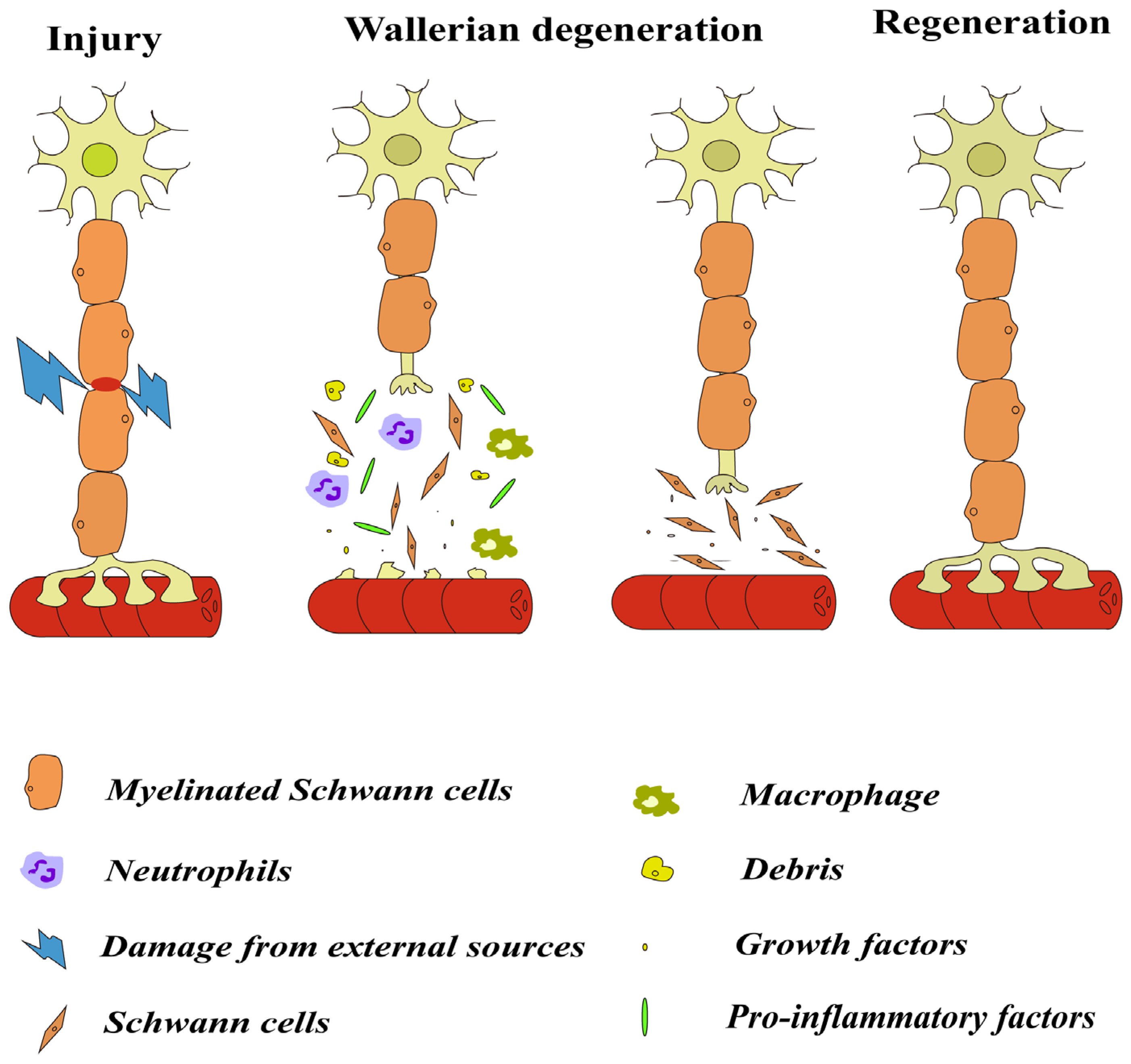
Nerve injury is a common condition that occurs as a result of trauma, iatrogenic injury, or long-lasting stimulation. Unlike the central nervous system (CNS), the peripheral nervous system (PNS) has a strong capacity for self-repair and regeneration. Peripheral nerve injury results in the degeneration of distal axons and myelin sheaths. Macrophages and Schwann cells (SCs) can phagocytose damaged cells. Wallerian degeneration (WD) makes the whole axon structure degenerate, creating a favorable regenerative environment for new axons. After nerve injury, macrophages, neutrophils and other cells are mobilized and recruited to the injury site to phagocytose necrotic cells and myelin debris. Pro-inflammatory and anti-inflammatory factors involved in the inflammatory response provide a favorable microenvironment for peripheral nerve regeneration and regulate the effects of inflammation on the body through relevant signaling pathways. Previously, inflammation was thought to be detrimental to the body, but further research has shown that appropriate inflammation promotes nerve regeneration, axon regeneration, and myelin formation. On the contrary, excessive inflammation can cause nerve tissue damage and pathological changes, and even lead to neurological diseases. Therefore, after nerve injury, various cells in the body interact with cytokines and chemokines to promote peripheral nerve repair and regeneration by inhibiting the negative effects of inflammation and harnessing the positive effects of inflammation in specific ways and at specific times. Understanding the interaction between neuroinflammation and nerve regeneration provides several therapeutic ideas to improve the inflammatory microenvironment and promote nerve regeneration.
Keywords: inflammation; nerve regeneration; Wallerian degeneration; immune cells; cytokine; chemokine inflammation; nerve regeneration; Wallerian degeneration; immune cells; cytokine; chemokine