Article

Activity of Epsilon-poly-L-lysine against Multidrug-Resistant Pseudomonas aeruginosa and Klebsiella pneumoniae Isolates of Urinary Tract Infections

1
MicroART-Antibiotic Resistance Team, Department of Veterinary Sciences, University of Trás-os-Montes and Alto Douro, 5000-801 Vila Real, Portugal
2
Department of Genetics and Biotechnology, University of Trás-os-Montes and Alto Douro, 5000-801 Vila Real, Portugal
3
Functional Genomics and Proteomics Unit, University of Trás-os-Montes and Alto Douro, 5000-801 Vila Real, Portugal
4
Associated Laboratory for Green Chemistry, University NOVA of Lisbon, 1099-085 Caparica, Portugal
5
CQ-VR, Chemistry Research Centre, Food and Wine Chemistry Laboratory, University of Trás-os-Montes and Alto Douro, 5000-801 Vila Real, Portugal
6
Area of Biochemistry and Molecular Biology, University of La Rioja, 26006 Logroño, Spain
7
Institut National de Recherche Pour l’Agriculture, l’Alimentation et l’Environnement (INRAE), Université Clermont Auvergne (UCA), UMR Microbiologie Environnement Digestif Santé (MEDiS), 63122 Saint-Genès-Champanelle, France
8
Clinical Pathology Department, Hospital Centre of Trás-os-Montes and Alto Douro, 5000-508 Vila Real, Portugal
9
CECAV—Veterinary and Animal Research Centre, University of Trás-os-Montes and Alto Douro, 5000-801 Vila Real, Portugal
10
Veterinary and Animal Research Centre, Associate Laboratory for Animal and Veterinary Science (AL4AnimalS), 5000-801 Vila Real, Portugal
*
Author to whom correspondence should be addressed.
These authors contributed equally to this work.
Biomedicines 2024, 12(3), 638; https://doi.org/10.3390/biomedicines12030638
Submission received: 19 January 2024 / Revised: 21 February 2024 / Accepted: 11 March 2024 / Published: 13 March 2024
(This article belongs to the Topic Advances in Vaccines and Antimicrobial Therapy)

Abstract

:
Pseudomonas aeruginosa and Klebsiella pneumoniae are notorious for their resistance to antibiotics and propensity for biofilm formation, posing significant threats to human health. Epsilon-poly-L-lysine (ε-PL) emerges as a naturally occurring antimicrobial poly(amino acid), which positions it as a prospective agent for addressing challenges linked to multidrug resistance. ε-PL symbolizes a promising avenue in the pursuit of efficacious therapeutic strategies and warrants earnest consideration within the realm of clinical treatment. Thus, our objective was to determine the antibiotic susceptibility profiles of 38 selected P. aeruginosa and ESBL-producing K. pneumoniae clinical isolates and determine the ability of ε-PL to inhibit biofilm formation. After PCR analysis, detection of genes related to β-lactamases was observed among the selected isolates of P. aeruginosa [blaSPM (35.7%), blaKPC (35.7%), blaSHV (14.3%), blaCTX-M (14.3%), blaOXA (14.3%), blaTEM (7.1%), blaPER (7.1%), blaVIM (7.1%), and blaVIM-2 (7.1%)] and K. pneumoniae [blaCTX-M (91.7%), blaTEM (83.3%), blaKPC (16.7%), blaNDM (12.5%), and blaOXA (4.2%)]. The results of testing the activity of ε-PL against the clinical isolates showed relatively high minimum inhibitory concentrations (MICs) for the P. aeruginosa (range: 8–64 µg/mL) and K. pneumoniae isolates (range: 16–32 µg/mL). These results suggest the need for prior optimization of ε-PL concerning its viability as an alternative to antibiotics for treating infections caused by P. aeruginosa and K. pneumoniae of clinical origin. It is noteworthy that, in the context of a low antibiotic discovery rate, ε-PL could play a significant role in this quest, considering its low toxicity and the unlikely development of resistance. Upon exposure to ε-PL, P. aeruginosa and K. pneumoniae isolates exhibited a reduction in biofilm production, with ε-PL concentration showing an inverse relationship, particularly in isolates initially characterized as strong or moderate producers, indicating its potential as a natural antimicrobial agent with further research needed to elucidate optimal concentrations and application methods across different bacterial species. Further research is needed to optimize its use and explore its potential in various applications.

1. Introduction

Pseudomonas aeruginosa and Klebsiella pneumoniae are opportunistic gram-negative pathogens that can colonize and cause human infections [1]. These pathogens are one of the most common causes of nosocomial infections and are frequently responsible for a variety of healthcare-related infections, including pneumonia, bloodstream, meningitis, urine, and wound or surgical site infections, among others [2,3,4,5,6]. P. aeruginosa and K. pneumoniae can potentially develop resistance to many classes of antibiotics, such as carbapenems, and treating infections with these bacteria can be very challenging [7,8,9]. Multidrug-resistant (MDR) and extensively drug-resistant (XDR) Gram-negative bacteria are major public health problems worldwide. The main difference between these two bacteria is that P. aeruginosa is a glucose-non-fermenting and oxidase-positive bacteria, and K. pneumoniae is a glucose-fermenting and oxidase-negative bacteria. Also, another difference is their type of respiration, namely that K. pneumoniae is facultatively anaerobic as opposed to P. aeruginosa, which is considered an aerobic bacteria. Consequently, biofilm production and formation have different metabolic processes [10,11]. Biofilm formation is a highly conserved mechanism of bacterial adaptation and is associated with developing resistance to many antibiotics [12]. Thus, to combat multidrug-resistant P. aeruginosa and K. pneumoniae, alternative treatment strategies for reducing MDR bacteria have become a prominent research topic.
ε-PL is a hydrophilic cationic linear homo-poly-amino acid, generally composed of 25 to 35 identical L-lysine residues with a unique structure characterized by the peptidic bonds between the ε-amino and α-carboxy groups [13]. Within the scope of research on ε-PL, new modification strategies stand out for obtaining structures beyond linear configurations, encompassing dendritic, hyperbranched, and functionalized forms of ε-PL. These variations aim to enhance specific characteristics, broaden applications, and meet diverse needs [13]. ε-PL can inhibit various microorganisms, such as most bacteria [14], and it is “Generally Recognized As Safe” (GRAS) as a food preservative. The antimicrobial activity of ε-PL is closely related to the number of repetitive L-lysine residues. It has been shown that more than 10 residues are required for ε-PL to exert adequate antibacterial activity. It has already been demonstrated that α-PL is a much less potent antimicrobial compound than ε-PL [15]. Studies testing the antimicrobial capacity of this peptide have shown that it can be electrostatically adsorbed to the surface of the bacteria, followed by the removal of the outer membrane. Consequently, this results in the abnormal distribution of the cytoplasm, causing damage to the bacteria [16].
This study sought to investigate a group of specifically chosen P. aeruginosa and ESBL-producing K. pneumoniae isolates sourced from urine samples. Our focus was to explore the capability of these strains in forming biofilms and to assess the antimicrobial activity of ε-PL against these pathogens.

2. Materials and Methods

2.1. Samples and Bacterial Strains

Thirty-eight selected P. aeruginosa (n = 14) and ESBL-producing K. pneumoniae (n = 24) isolates recovered from urine samples at the Medical Centre of Trás-os-Montes and Alto Douro between September 2021 and June 2022 were included in this study. These strains were identified using VITEK 2® COMPACT (bioMérieux, Auvergne-Rhône-Alpes, France), and their identification was confirmed in the medical microbiology laboratory by seeding P. aeruginosa and K. pneumoniae isolates on Pseudomonas Agar Base supplemented with CN (Liofilchem, Rosetodegli, Abruzzi, Italy) medium and HiCrome Klebsiella Selective Agar Base medium (HiMedia Laboratories, Maharashtra, India), respectively. Both pathogens were incubated at 37 °C for 24–48 h. The isolates were cryopreserved at −20 °C in skim milk.

2.2. Antimicrobial Susceptibility Testing

The phenotypic resistance characterization of the isolates was performed by the Kirby–Bauer disk diffusion method following EUCAST standards (2022) [17]. Twelve antibiotics were tested for P. aeruginosa (charge on disks): piperacillin (30 μg), ticarcillin-clavulanic acid (75–10 μg), ceftazidime (10 μg), cefepime (30 μg), aztreonam (30 μg), imipenem (10 μg), doripenem (10 μg), meropenem (10 μg), amikacin (30 μg), tobramycin (10 μg), gentamicin (10 μg), and ciprofloxacin (5 μg). The minimum inhibitory concentration (MIC) by microdilution method was performed for piperacillin-tazobactam antibiotics (resistant when MIC > 16 mg/L) since no breakpoints for disk diffusion method are available in the EUCAST guidelines. Thirteen antibiotics were tested for K. pneumoniae isolates following EUCAST standards (2022): ticarcillin-clavulanic acid (75–10 μg), cefoxitin (30 μg), ceftazidime (10 μg), cefepime (30 μg), aztreonam (30 μg), imipenem (10 μg), meropenem (10 μg), ertapenem (10 μg), amikacin (30 μg), gentamicin (10 μg), ciprofloxacin (5 μg), trimethoprim–sulfamethoxazole (1.25/23.75 μg), and chloramphenicol (30 μg). Three antibiotics were tested for K. pneumoniae isolates following CLSI standards (2021) [18]: cefotaxime (30 μg), nalidixic acid (30 μg), and tetracycline (30 μg).

2.3. Determination of Minimum Inhibitory Concentration

The microdilution method was used to determine the minimum inhibitory concentration (MIC) of linear ε-PL (~4700 g/mol; Handary, Brussels, Belgium). Briefly, each isolate was seeded on Brain Heart Infusion (BHI) agar (Frilabo, Maia, Portugal) and incubated at 37 °C for 24 h. Then, the bacterial cells were subcultured in tubes containing Mueller-Hinton (MH) broth (Frilabo, Maia, Portugal) and incubated overnight at 37 °C with 150 rpm in the ES-20/60 Orbital Shaker-Incubator (Biosan, Riga, Latvia). The overnight culture was diluted in fresh MH broth to a turbidity standard of 0.5 McFarland using a spectrophotometer. Then, Epsilon-poly-L-lysine was diluted in sterilized distilled water at different concentrations (0 µg/mL to 8182 µg/mL), and 75 µL of each concentration was placed in a polystyrene flat bottom 96-well plate. Also, 75 µL of bacterial suspension was placed in the same 96-well microtiter plate and incubated for 24 h at 37 °C with 150 rpm. Bacterial growth was determined at 630 nm using a microplate reader, the BioTek ELx808U (BioTek, Winooski, VT, USA). MIC was defined as the lowest concentration that prevents bacterial growth.

2.4. Biofilm Formation and Biomass Quantification

The bacterial adhesion of all isolates was assessed using a microtitre plate-based assay as previously described with some modifications [19]. Briefly, to perform the assay, one colony from each bacterial culture that had grown overnight on brain heart infusion (BHI) agar was suspended in Luria-Bertani (LB) broth (Oxoid®, Basingstoke, UK) and incubated for 24 h at 37 °C. Then, the bacterial suspension was diluted to 0.5 on the McFarland scale using Tryptic Soy Broth (TSB) (Oxoid®, Basingstoke, UK). Then, 100 µL of each dilution was added to each well of a 96-well flat-bottom microplate. Pseudomonas aeruginosa ATCC® 27853 was included in all microplates as a positive control, and TSB without bacterial inoculum was used as a negative control. The microplates were incubated for 24 h at 37 °C. For quantitative assays, eight replicate wells were used for each treatment. After incubation, bacterial cells in suspension were removed by turning the microplates over, and they were washed twice with distilled water. This step helps remove stray cells and media components that may be stained in the next step, significantly reducing background staining. Subsequently, 125 µL of a 0.1% (v/v) solution of crystal violet (CV) (Frilabo, Maia, Portugal) was introduced into each well of the microplate, followed by an incubation period at room temperature for 10–15 min. Post-incubation, the CV solution was decanted, and the microplates underwent 3–4 washes with distilled water. Following this, meticulous drying of the plates was conducted using paper towels to eliminate any residual cells and stains, after which they were left to air-dry overnight.
To determine the biomass of the biofilm, 125 µL of 30% (v/v) acetic acid was added to each well of the microplate to dissolve the crystal violet. After an additional incubation period at room temperature for 10–15 min, optical density readings were taken at 630 nm (OD630nm) using a microplate reader (BioTek ELx808U, BioTek, Winooski, VT, USA). An uninoculated well containing 30% acetic acid served as a reference blank.
Following the MIC results, the same protocol was utilized for the production of biofilms, namely with the use of crystal violet, with one exception. Instead of 125 µL of 0.1% (v/v) CV solution, we added 175 µL since each well once contained 150 µL of liquid (75 µL of bacterial suspension and 75 µL of ε-PL solution), and we wanted the CV solution to cover all the biofilm that had eventually formed. The subsequent steps, including incubation, washing, drying, and biomass quantification, adhered to the protocol. Optical density was measured using a microplate reader (BioTek ELx808U, Lonza’s Vision Zero), with an uninoculated well containing 30% acetic acid serving as the blank control.

2.5. Antimicrobial Resistance and Virulence Genes

All isolates were studied for the presence of antimicrobial resistance genes according to the phenotypic results. DNA extraction was performed by following the boil method protocol [20]. Briefly, three colonies of overnight-growth bacteria were placed in a test tube containing 500 µL of distilled water and boiled for 8 min in a water bath. After, the tubes were vortexed vigorously and centrifuged for 2 min at 12,000 rpm. Then, 490 µL of the supernatant was recovered. Afterward, DNA concentration was determined using a NanoDrop system, and polymerase chain reaction (PCR) was performed. For P. aeruginosa isolates, the following resistance genes were tested: blaTEM, blaSHV, blaCTX, blaPER, blaSME, blaKPC, blaGim, blaSmp, blaVim, blaVim-2, blaNDM, blaOXA, acc(3)-I, aac(3)-II, aac(3)-III, aac(3)-IV, and ant(2′)-Ia. For K. pneumoniae isolates, the following resistance genes were tested: blaTEM, blaSHV, blaCTX, blaKPC, blaNDM, blaOXA, acc(3)-II, aac(3)-IV, tetA, tetB, cmlA, catA, sul1, sul2, sul3, and dfrA [21,22,23,24,25,26,27,28,29,30,31,32,33,34,35,36,37,38,39].

2.6. Multilocus Sequence Typing

Multilocus sequence typing (MLST) for selected P. aeruginosa and K. pneumoniae isolates was performed by PCR and sequencing of seven housekeeping genes (acsA, aroE, guaA, mutL, nuoD, ppsA, and trpE for P. aeruginosa; gapA, infB, mdh, pgi, phoE, rpoB, and tonB for K. pneumoniae). Allelic profiles and sequence types (STs) were compared with the PubMLST and BIGSdb-Pasteur databases (http://pubmlst.org/paeruginosa/; https://bigsdb.pasteur.fr/klebsiella/, accessed on 28 April 2023).

3. Results and Discussion

3.1. Antimicrobial Susceptibility Testing

All phenotypic profiles of each P. aeruginosa and K. pneumoniae isolate are indicated in Table 1. P. aeruginosa isolates included in this study showed, in most cases, resistance to imipenem (85.7%). All P. aeruginosa isolates were susceptible to amikacin and colistin. On the other hand, ESBL-producing K. pneumoniae isolates showed resistance to cefotaxime (100%), cefepime (100%), and aztreonam (100%). Moreover, one of the K. pneumoniae isolates showed phenotypic resistance to imipenem (4.2%), and five additional isolates showed resistance to other carbapenems, such as ertapenem and/or meropenem (20.8%).

3.2. Antimicrobial Resistance Genes

After PCR analysis, some β-lactamase-related genes were detected in P. aeruginosa isolates, such as blaSPM (35.7% of isolates), blaKPC (35.7%), blaSHV (14.3%), blaCTX-M (14.3%), blaOXA (14.3%), blaTEM (7.1%), blaPER (7.1%), blaVIM (7.1%), and blaVIM-2 (7.1%) (Table 1). Multiple studies also report the presence of these genes in clinical P. aeruginosa isolates. Recently, in 2020, Ahmed et al. reported that 51% of P. aeruginosa from cancer patients were positive for the carbapenemase genes encoding VIM, GIM, and IMP enzymes, 38% for SPM and AIM, 30% for BIC, 20% for NDM and TEM, 17% for KPC, and 15% for OXA [40]. The first report of blaVIM-2 and blaPER-1 in Latin America was published in 2018 in a P. aeruginosa strain recovered from a cerebrospinal fluid sample [41]. The emergence of these β-lactamase genes in P. aeruginosa is a major concern in treating infections caused by this bacterium since they are associated with various resistance mechanisms to β-lactam antibiotics. The resistance to aminoglycosides expressed by two isolates was associated with the aac(3)-IV gene. AAC enzymes are commonly detected in aminoglycoside-resistant strains, as well as APH enzymes. Like the current study, Holbrook and Garneau-Tsodikova have also detected the aac(3)-IV gene in 10.7% of the isolates, together with aph(3′)-Ia (70.5%), aac(6′)-Ib (41.0%), and ant(2″)-Ia (5.7%) [42].
Since all K. pneumoniae isolates were ESBL-producers, most of the isolates carried, as expected, ESBL-related genes (Table 1). The most prevalent β-lactamase genes detected in our study were blaCTX-M (91.7%) and blaSHV (91.7%), followed by blaTEM (83.3%). All the blaCTX-M genes confer an ESBL phenotype, although only some variants of blaTEM or blaSHV are related to the ESBL phenotype. All K. pneumoniae have an intrinsic SHV resistance gene. However, in some cases, it could be mutated to an ESBL variant, or the isolate could acquire a new ESBL-SHV variant that can only be known by sequencing the obtained amplicons. The presence of these β-lactamases in K. pneumoniae continues to be reported worldwide. Multiple studies keep detecting these genes and their variants [43,44,45]. Moreover, the blaKPC (16.7%) and blaNDM (12.5%) carbapenemase genes were also found among our isolates. The emergence and spread of carbapenemase genes among clinical isolates is a significant concern since carbapenem antibiotics are often used as a last resort for treating infections caused by multidrug-resistant bacteria. The most common carbapenemase genes detected in K. pneumoniae are from the KPC [46,47], NDM [48,49], and OXA-48 families [50,51]. Different β-lactamase gene combinations were observed among our isolates, but the most predominant was blaCTX-M + blaTEM + blaSHV (n = 12). Also, one K. pneumoniae strain (HS79) showed a combination of five different β-lactamase genes (blaCTX-M + blaTEM + blaKPC + blaOXA + blaNDM). In only one strain (HS50), no carbapenemase genes were detected, besides this strain being resistant to ertapenem. However, other carbapenemase genes have also been reported in K. pneumoniae, such as VIM [52,53], IMP [54,55], and GES [56,57], that may be conferring resistance to ertapenem in this isolate. In relation to resistance to sulfonamides and trimethoprim, we detected sul2 (87.5%), sul1 (41.7%), sul3 (12.5%), and dfrA (8.3%) genes among the studied isolates. Like our study, Mbelle et al. also reported high rates of prevalence of sul2 (86%) and sul1 (78%) [58]. The sul3 and dfrA genes are also commonly found in K. pneumoniae. Carvalho et al. [59] reported, in 2021, the presence of sul2 in 10 K. pneumoniae clinical isolates and sul3 in 5 isolates, but no dfrA was detected. However, the presence of dfrA1 in four isolates and dfrA17 in one isolate was reported by Yu et al. [60] after whole-genome sequencing of seven K. pneumoniae strains. Relatively to aminoglycoside resistance, we verified the presence of aac(3)-II (75%) and aadA1 (62.5%). The same was reported by Lomonaco et al. [43], who detected the presence of these two genes in ten MDR clinical isolates of K. pneumoniae from the United States of America. Moreover, tetA (12.5%) was the only gene detected that confers resistance to tetracyclines. The absence of tetA or tetB genes was observed in certain tetracycline-resistant isolates. This could be attributed to several factors, such as the presence of alternative tetracycline resistance genes like tetC, tetD, or tetG [61] or the emergence of tetracycline resistance through mutations in bacterial ribosomes or other cellular components independent of specific resistance genes [62]. The resistance to chloramphenicol was proven by the presence of the cmlA gene (37.5%) (Table 1). The cmlA gene is a common mechanism of resistance to chloramphenicol. It is often located on mobile genetic elements such as plasmids, which can facilitate its spread between bacterial species and contribute to the emergence of multidrug-resistant strains. cmlA-mediated resistance to chloramphenicol has been reported in various bacterial species, including K. pneumoniae [53].

3.3. Multilocus Sequence Typing

After the biofilm production results, we selected seven K. pneumoniae and seven P. aeruginosa isolates for sequence typing. The results are presented in Table 2.
These sequence types (STs) are commonly found in K. pneumoniae strains: ST15, ST307, and ST348 are known to be widespread and have been associated with hospital-acquired infections [63,64,65], while ST584 is a relatively new ST that has been identified in K. pneumoniae strains from clinical and environmental sources [66,67]. The STs found in P. aeruginosa have been associated with hospital-acquired infections: ST285, ST274, ST1404, and ST228 have been identified in hospitals in several European countries. The other STs have been reported in hospitals in Brazil, China, the United States, and Saudi Arabia [68]. Interestingly, we found two new allelic combinations for HU5 and HU6 isolates. HU5 seems to be a single locus variant (SLV) of ST1228 with a different trpE allele. HU6 seems to be an SLV of ST170/ST997/ST1315/ST2454 with a different mutL allele. The identification of new allelic combinations in P. aeruginosa is not uncommon, as this bacterium has a high level of genetic diversity [69]. SLVs are typically defined as strains that differ from each other by a single nucleotide polymorphism (SNP) in one of the seven MLST loci, and they are often considered to be related to each other in an evolutionary sense.

3.4. Determination of Minimum Inhibitory Concentration (MIC) for Epsilon-poly-L-lysine

In vitro activity of ε-PL against the P. aeruginosa and K. pneumoniae isolates showed relatively high MICs. MIC values ranged between 8 and 64 µg/mL in P. aeruginosa isolates, with 64.3% having a MIC value of 32 µg/mL and between 16 and 32 µg/mL in K. pneumoniae isolates, with 58.3% also having a MIC value of 32 µg/mL (Figure 1). The high MIC values observed for ε-PL against the tested P. aeruginosa and K. pneumoniae isolates suggest that these bacteria have relatively low susceptibility to the antimicrobial effects of ε-PL. The results from the electron microscopy analysis of E. coli O157:H7 cells, reported by Zhang et al. [70], suggest that ε-PL can cause damage to the bacterial cell wall or membrane even at concentrations lower (16 µg/mL) than those observed in our study. This supports the idea that the relatively high MICs observed in the current study may be due to factors such as the limited permeability of the bacterial cell wall or membrane to ε-PL rather than the inability of ε-PL to exert its antimicrobial effects once inside the bacterial cell. However, further studies would be needed to confirm this hypothesis. Moreover, the activity should be tested in the future under different methodological conditions (media and bacteria density, among others) to check if these variables could affect the activity of the agent. In another recent study, it was observed that ε-PL caused alterations in the morphology of P. aeruginosa cells, resulting in protrusions on the cell wall, as evidenced by scanning electron microscopy [71]. Additionally, ε-PL demonstrated the ability to depolarize and permeabilize the bacterial membrane over time, which may enhance the effectiveness of antibiotics. The MIC in the referred study turned out to be higher, around 50 µg/mL, using the reference strain PAO1. Experiments also evaluated the efficacy of ε-PL in combination with imipenem against both sensitive and antibiotic-resistant strains of P. aeruginosa. A synergy between ε-PL and imipenem was observed, resulting in a significant reduction in bacterial growth, especially against resistant strains. These findings suggest that ε-PL could be a promising option for treating P. aeruginosa infections, particularly those resistant to conventional antibiotics. The ability of ε-PL to increase bacterial membrane permeability may enhance the effectiveness of antibiotics, making it a potential therapeutic adjuvant against resistant bacterial infections. Regarding K. pneumoniae, this field of research remains an enigma, thereby underscoring the significance of this study.

3.5. Biofilm Formation and Biomass Quantification

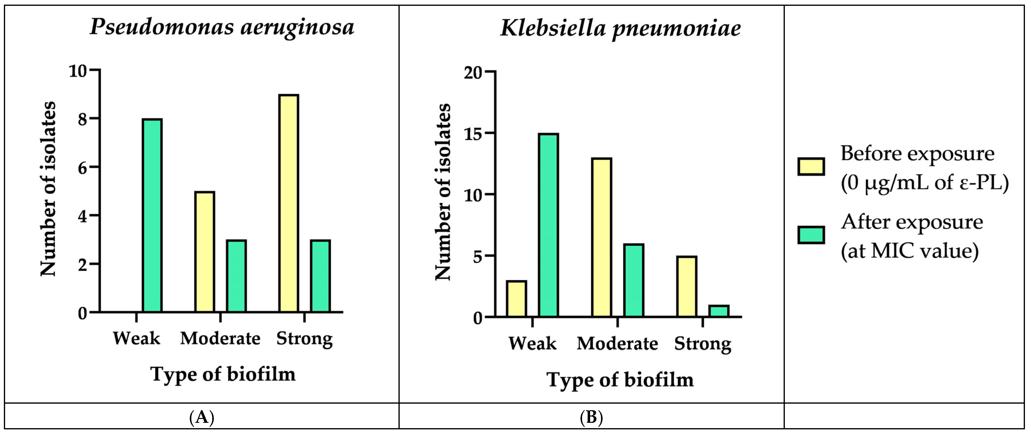
We verified that all P. aeruginosa isolates (n = 14) were biofilm producers (100%), of which five were moderate producers (35.7%) and nine were strong producers (64.3%) (Figure 2A). Among K. pneumoniae isolates, we verified that 21 of them were biofilm producers (87.5%), of which 5 were strong producers (20.8%), 13 were moderate producers (54.2%), and 3 were weak producers (12.5%) (Figure 2B). After the exposure to ε-PL, we verified a decrease in biofilm production in 11 P. aeruginosa and 16 K. pneumoniae strains, i.e., in these strains, we verified that the concentration of ε-PL was inversely proportional to the production of biofilm, which means that the strains that were initially strong/moderate biofilm producers at MIC values became weak/null biofilm producers (Figure 2). Several studies have investigated the effects of ε-PL on biofilm production in different bacterial species. Some studies have shown similar results to the current study. For example, a study conducted on Salmonella typhimurium showed that ε-PL inhibits biofilm formation in a dose-dependent manner. Additionally, ε-PL at low concentrations did not affect the viability of bacterial cells. This suggests that ε-PL may have a selective effect on biofilms, inhibiting their formation while leaving bacterial cells intact [72]. Another study investigated the effect of ε-PL on the cell structure and biofilm formation in Cronobacter sakazakii. The results showed that ε-PL had a MIC of 256 μg/mL against C. sakazakii. ε-PL was found to reduce the surface hydrophobicity and motility of C. sakazakii, which affects the formation of biofilm. ε-PL also demonstrated a significant effect on the inhibition and eradication of biofilm, and the removal efficiency of biofilm was significantly improved when combined with physical oscillation. This means that ε-PL destroyed the cell structure of C. sakazakii, and the bacteriostatic effect was achieved while inhibiting biofilm formation and removing mature biofilm [73]. Overall, these studies indicate that ε-PL may serve as a natural antimicrobial agent that can hinder the formation of biofilms in diverse bacterial species. However, further research is needed to determine the optimal concentration and application methods for different bacterial species and to investigate the potential mechanisms of action of ε-PL on biofilm production. Surprisingly, one of the K. pneumoniae isolates (HS55) showed an opposite relationship between ε-PL concentration and biofilm production, moving from non-producer to weak biofilm producer at 4 µg/mL of ε-PL. It is not uncommon for some bacteria to display different responses to antibacterial agents due to variations in their genetic background or environmental conditions. For instance, a study reported that low concentrations of triclosan, an antimicrobial agent, can promote the binding of S. aureus to inanimate surfaces such as plastic and glass [74].
Another type of consideration that must be made is related to the potential of ε-PL in a clinical setting. ε-PL, with its antimicrobial properties against Gram-negative bacteria like P. aeruginosa and K. pneumoniae, holds significant promise as a potential therapeutic agent. In a clinical setting, ε-PL might be considered for topical application, where ε-PL could be formulated into creams, ointments, or gels for the treatment of localized skin infections or wounds [75,76]. Additionally, ε-PL could be utilized in medical devices such as catheters or wound dressings to prevent bacterial colonization and biofilm formation [72].
Furthermore, ε-PL could be explored in the future for the treatment of systemic infections caused by MDR bacteria. However, further research would be needed to determine the potential side effects associated with systemic ε-PL administration, as well as the optimal dosage and administration route. Overall, the successful translation of ε-PL from preclinical studies to clinical applications would require rigorous evaluation of its safety, efficacy, and pharmacokinetic properties in human trials.

4. Conclusions

In this study it was performed the genetic characterization of β-lactamase genes in the collection of P. aeruginosa and K. pneumoniae clinical isolates used as target for testing the activity of ε-PL agent; it has been shown the carriage of a wide range of relevant β-lactamases, including those related to ESBL and carbapenemases, according to the phenotypes of the selected isolates.
On the other hand, this study found high MICs in isolates of both bacterial species for ε-PL, with the majority of P. aeruginosa and K. pneumoniae isolates having a MIC value of 32 µg/mL. These MIC values suggest that these bacteria require relatively high concentration of ε-PL compound to be inhibited under the studied conditions, emphasizing the need for preliminary screening and/or optimization of the polymer, considering the specificity of the intended application. Despite these values, it is important to note that this natural polymer is highly customizable, demonstrating broad efficacy, low toxicity, and a low probability of leading to the development of resistant strains. In the context of this work and considering these properties, new multidisciplinary research efforts are prompted to enhance its efficacy against P. aeruginosa and K. pneumoniae and to leverage its clinical application.

Author Contributions

T.d.S., C.S. and M.P.-H. carried out laboratory work and data analysis. T.d.S. and C.S. drafted the manuscript. The final version was written through the contributions of all authors. P.P., G.I., M.R. and C.T. were responsible for the provision of study materials. T.d.S., C.S., M.R., O.A., S.S., E.C., M.P.-H., C.T., P.P., M.H. and G.I. implemented data analyses and helped to draft the manuscript. All authors have read and agreed to the published version of the manuscript.

Funding

This work was supported by the projects UIDP/00772/2020 and LA/P/0059/2020, funded by the Portuguese Foundation for Science and Technology (FCT). This research was also funded by FCT related to PhD grants through the references DFA/BD/5332/2020 and 2020.06967.BD.

Institutional Review Board Statement

Not applicable.

Informed Consent Statement

Not applicable.

Data Availability Statement

Data are contained within the article.

Conflicts of Interest

The authors declare no conflicts of interest.

References

  1. Riquelme, S.A.; Ahn, D.; Prince, A. Pseudomonas aeruginosa and Klebsiella pneumoniae Adaptation to Innate Immune Clearance Mechanisms in the Lung. J. Innate Immun. 2018, 10, 442–454. [Google Scholar] [CrossRef] [PubMed]
  2. Yu, V.L.; Hansen, D.S.; Wen, C.K.; Sagnimeni, A.; Klugman, K.P.; Von Gottberg, A.; Goossens, H.; Wagener, M.M.; Benedi, V.J.; Casellas, J.M.; et al. Virulence Characteristics of Klebsiella and Clinical Manifestations of K. pneumoniae Bloodstream Infections. Emerg. Infect. Dis. 2007, 13, 986. [Google Scholar] [CrossRef] [PubMed]
  3. van Delden, C. Pseudomonas aeruginosa Bloodstream Infections: How Should We Treat Them? Int. J. Antimicrob. Agents 2007, 30, 71–75. [Google Scholar] [CrossRef] [PubMed]
  4. Curran, C.S.; Bolig, T.; Torabi-Parizi, P. Mechanisms and Targeted Therapies for Pseudomonas aeruginosa Lung Infection. Am. J. Respir. Crit. Care Med. 2018, 197, 708–727. [Google Scholar] [CrossRef]
  5. Ferreiro, J.L.L.; Otero, J.Á.; González, L.G.; Lamazares, L.N.; Blanco, A.A.; Sanjurjo, J.R.B.; Conde, I.R.; Soneira, M.F.; De La Fuente Aguado, J. Pseudomonas aeruginosa Urinary Tract Infections in Hospitalized Patients: Mortality and Prognostic Factors. PLoS ONE 2017, 12, e0178178. [Google Scholar] [CrossRef]
  6. Mariana Cristea, O.; Silvia Avrămescu, C.; Bălășoiu, M.; Popescu, F.; Popescu, F.; Amzoiu, M. Original Paper Urinary Tract Infection with Klebsiella pneumoniae in Patients with Chronic Kidney Disease. Curr. Health Sci. J. 2017, 43, 137–148. [Google Scholar] [CrossRef]
  7. Kode, M.; Waso-Reyneke, M.; Reyneke, B.; Denissen, J.; Clements-Decker, T.; Havenga, B.; Khan, S.; Khan, W. Integration of Bdellovibrio spp. with SODIS and Moringa Oleifera Flocculation to Target Multi-Drug Resistant Klebsiella pneumoniae and Pseudomonas aeruginosa. J. Environ. Chem. Eng. 2022, 10, 107962. [Google Scholar] [CrossRef]
  8. Maraolo, A.E.; Cascella, M.; Corcione, S.; Cuomo, A.; Nappa, S.; Borgia, G.; De Rosa, F.G.; Gentile, I. Management of Multidrug-Resistant Pseudomonas aeruginosa in the Intensive Care Unit: State of the Art. Expert Rev. Anti-Infect. Ther. 2017, 15, 861–871. [Google Scholar] [CrossRef]
  9. Bassetti, M.; Righi, E.; Carnelutti, A.; Graziano, E.; Russo, A. Multidrug-Resistant Klebsiella pneumoniae: Challenges for Treatment, Prevention and Infection Control. Expert Rev. Anti-Infect. Ther. 2018, 16, 749–761. [Google Scholar] [CrossRef]
  10. Thi, M.T.T.; Wibowo, D.; Rehm, B.H.A. Pseudomonas aeruginosa Biofilms. Int. J. Mol. Sci. 2020, 21, 8671. [Google Scholar] [CrossRef]
  11. Wang, G.; Zhao, G.; Chao, X.; Xie, L.; Wang, H. The Characteristic of Virulence, Biofilm and Antibiotic Resistance of Klebsiella pneumoniae. Int. J. Environ. Res. Public Health 2020, 17, 6278. [Google Scholar] [CrossRef] [PubMed]
  12. de Sousa, T.; Hébraud, M.; Enes Dapkevicius, M.L.N.; Maltez, L.; Pereira, J.E.; Capita, R.; Alonso-Calleja, C.; Igrejas, G.; Poeta, P. Genomic and Metabolic Characteristics of the Pathogenicity in Pseudomonas aeruginosa. Int. J. Mol. Sci. 2021, 22, 12892. [Google Scholar] [CrossRef]
  13. Warszynski, P.; Li, Y.; Zhou, C.; Chen, S.; Huang, S. Recent Advances in Epsilon-Poly-L-Lysine and L-Lysine-Based Dendrimer Synthesis, Modification, and Biomedical Applications. Front. Chem. 2021, 1, 659304. [Google Scholar] [CrossRef]
  14. Shukla, S.C.; Singh, A.; Pandey, A.K.; Mishra, A. Review on Production and Medical Applications of ε-Polylysine. Biochem. Eng. J. 2012, 65, 70–81. [Google Scholar] [CrossRef]
  15. Takehara, M.; Hibino, A.; Saimura, M.; Hirohara, H. High-Yield Production of Short Chain Length Poly(ε-L-Lysine) Consisting of 5–20 Residues by Streptomyces aureofaciens, and Its Antimicrobial Activity. Biotechnol. Lett. 2010, 32, 1299–1303. [Google Scholar] [CrossRef] [PubMed]
  16. Wei, L.; Wu, R.; Wang, C.; Wu, Z. Effects of ε-Polylysine on Pseudomonas aeruginosa and Aspergillus fumigatus Biofilm in Vitro. Med. Sci. Monit. 2017, 23, 4225–4229. [Google Scholar] [CrossRef] [PubMed]
  17. The European Committee on Antimicrobial Susceptibility Testing. Breakpoint Tables for Interpretation of MICs and Zone Diameters, version 12.0; The European Committee on Antimicrobial Susceptibility Testing: Växjö, Sweden, 2022. [Google Scholar]
  18. CLSI. Performance Standards for Antimicrobial Susceptibility Testing, 31st ed.; Clinical and Laboratory Standards Institute: Malvern, PA, USA, 2021; ISBN 9781684401048. [Google Scholar]
  19. O’Toole, G.A. Microtiter Dish Biofilm Formation Assay. J. Vis. Exp. 2011, 47, e2437. [Google Scholar] [CrossRef]
  20. Dashti, A.A.; Jadaon, M.M.; Abdulsamad, A.M.; Dashti, H.M. Heat Treatment of Bacteria: A Simple Method of DNA Extraction for Molecular Techniques. Kuwait Med. J. 2009, 41, 117–122. [Google Scholar]
  21. Perreten, V.; Boerlin, P. A New Sulfonamide Resistance Gene (Sul3) in Escherichia coli Is Widespread in the Pig Population of Switzerland. Antimicrob. Agents Chemother. 2003, 47, 1169–1172. [Google Scholar] [CrossRef]
  22. Schnellmann, C.; Gerber, V.; Rossano, A.; Jaquier, V.; Panchaud, Y.; Doherr, M.G.; Thomann, A.; Straub, R.; Perreten, V. Presence of New mecA and Mph(C) Variants Conferring Antibiotic Resistance in Staphylococcus spp. Isolated from the Skin of Horses before and after Clinic Admission. J. Clin. Microbiol. 2006, 44, 4444–4454. [Google Scholar] [CrossRef]
  23. Maynard, C.; Fairbrother, J.M.; Bekal, S.; Sanschagrin, F.; Levesque, R.C.; Brousseau, R.; Masson, L.; Larivière, S.; Harel, J. Antimicrobial Resistance Genes in Enterotoxigenic Escherichia coli O149:K91 Isolates Obtained over a 23-Year Period from Pigs. Antimicrob. Agents Chemother. 2003, 47, 3214–3221. [Google Scholar] [CrossRef] [PubMed]
  24. Mazel, D.; Dychinco, B.; Webb, V.A.; Davies, J. Antibiotic Resistance in the ECOR Collection: Integrons and Identification of a Novel Aad Gene. Antimicrob. Agents Chemother. 2000, 44, 1568–1574. [Google Scholar] [CrossRef] [PubMed]
  25. Sáenz, Y.; Briñas, L.; Domínguez, E.; Ruiz, J.; Zarazaga, M.; Vila, J.; Torres, C. Mechanisms of Resistance in Multiple-Antibiotic-Resistant Escherichia coli Strains of Human, Animal, and Food Origins. Antimicrob. Agents Chemother. 2004, 48, 3959–3967. [Google Scholar] [CrossRef] [PubMed]
  26. Guardabassi, L.; Dijkshoorn, L.; Collard, J.M.; Olsen, J.E.; Dalsgaard, A. Distribution and In-Vitro Transfer of Tetracycline Resistance Determinants in Clinical and Aquatic Acinetobacter Strains. J. Med. Microbiol. 2000, 49, 929–936. [Google Scholar] [CrossRef] [PubMed]
  27. Zhao, X.; Yang, J.; Ju, Z.; Chang, W.; Sun, S. Molecular Characterization of Antimicrobial Resistance in Escherichia coli from Rabbit Farms in Tai’an, China. BioMed Res. Int. 2018, 2018, 8607647. [Google Scholar] [CrossRef] [PubMed]
  28. Madsen, L.; Aarestrup, F.M.; Olsen, J.E. Characterisation of Streptomycin Resistance Determinants in Danish Isolates of Salmonella typhimurium. Vet. Microbiol. 2000, 75, 73–82. [Google Scholar] [CrossRef] [PubMed]
  29. Zhang, L.; Hinz, A.J.; Nadeau, J.P.; Mah, T.F. Pseudomonas aeruginosa tssC1 Links Type VI Secretion and Biofilm-Specific Antibiotic Resistance. J. Bacteriol. 2011, 193, 5510–5513. [Google Scholar] [CrossRef]
  30. Van de Klundert, J.A.M.; Vliegenthart, J.S.; Rather, P.N.; Shaw, K.J.; Hare, R.S.; Miller, G.H. Nomenclature of Aminoglycoside Resistance Genes: A Comment. Antimicrob. Agents Chemother. 1993, 37, 927–928. [Google Scholar] [CrossRef]
  31. Vila, J.; Ruiz, J.; Marco, F.; Barcelo, A.; Goni, P.; Giralt, E.; De Anta, T.J. Association between Double Mutation in gyrA Gene of Ciprofloxacin- Resistant Clinical Isolates of Escherichia coli and MICs. Antimicrob. Agents Chemother. 1994, 38, 2477–2479. [Google Scholar] [CrossRef]
  32. Schaber, J.A.; Carty, N.L.; McDonald, N.A.; Graham, E.D.; Cheluvappa, R.; Griswold, J.A.; Hamood, A.N. Analysis of Quorum Sensing-Deficient Clinical Isolates of Pseudomonas aeruginosa. J. Med. Microbiol. 2004, 53, 841–853. [Google Scholar] [CrossRef]
  33. Neyestanaki, D.K.; Mirsalehian, A.; Rezagholizadeh, F.; Jabalameli, F.; Taherikalani, M.; Emaneini, M. Determination of Extended Spectrum Beta-Lactamases, Metallo-Beta-Lactamases and AmpC-Beta-Lactamases among Carbapenem Resistant Pseudomonas aeruginosa Isolated from Burn Patients. Burns 2014, 40, 1556–1561. [Google Scholar] [CrossRef] [PubMed]
  34. Mona, S.; Nour, M.A.K.; ElSheshtawy, N.M. Genetic Identification of Pseudomonas aeruginosa Virulence Genes among Different Isolates. J. Microb. Biochem. Technol. 2015, 7, 274–277. [Google Scholar] [CrossRef]
  35. Hong, S.S.; Kim, K.; Huh, J.Y.; Jung, B.; Kang, M.S.; Hong, S.G. Multiplex PCR for Rapid Detection of Genes Encoding Class A Carbapenemases. Ann. Lab. Med. 2012, 32, 359–361. [Google Scholar] [CrossRef] [PubMed]
  36. Galdino, A.C.M.; Viganor, L.; Ziccardi, M.; Nunes, A.P.F.; dos Santos, K.R.N.; Branquinha, M.H.; Santos, A.L.S. Heterogeneous Production of Proteases from Brazilian Clinical Isolates of Pseudomonas aeruginosa. Enferm. Infecc. Microbiol. Clin. 2017, 35, 630–637. [Google Scholar] [CrossRef] [PubMed]
  37. Ellington, M.J.; Kistler, J.; Livermore, D.M.; Woodford, N. Multiplex PCR for Rapid Detection of Genes Encoding Acquired Metallo-β-Lactamases. J. Antimicrob. Chemother. 2007, 59, 321–322. [Google Scholar] [CrossRef] [PubMed]
  38. Shanthi Amudhan, M.; Sekar, U.; Kamalanathan, A.; Balaraman, S. blaIMP and blaVIM Mediated Carbapenem Resistance in Pseudomonas and Acinetobacter Species in India. J. Infect. Dev. Ctries. 2012, 6, 757–762. [Google Scholar] [CrossRef] [PubMed]
  39. Alm, R.A.; Mattick, J.S. Identification of a Gene, pilV, Required for Type 4 Fimbrial Biogenesis in Pseudomonas aeruginosa, Whose Product Possesses a Pre-pilin-like Leader Sequence. Mol. Microbiol. 1995, 16, 485–496. [Google Scholar] [CrossRef] [PubMed]
  40. Ahmed, N.; Ali, Z.; Riaz, M.; Zeshan, B.; Wattoo, J.I.; Aslam, M.N. Evaluation of Antibiotic Resistance and Virulence Genes among Clinical Isolates of Pseudomonas aeruginosa from Cancer Patients. Asian Pac. J. Cancer Prev. 2020, 21, 1333–1338. [Google Scholar] [CrossRef]
  41. Papa-Ezdra, R.; Bado, I.; Caiata, L.; Vignoli, R.; Seija, V. First Report of Pseudomonas aeruginosa Co-Harbouring blaVIM-2 and blaPER-1 in Latin America. J. Glob. Antimicrob. Resist. 2018, 15, 121–122. [Google Scholar] [CrossRef]
  42. Holbrook, S.Y.L.; Garneau-Tsodikova, S. Evaluation of Aminoglycoside and Carbapenem Resistance in a Collection of Drug-Resistant Pseudomonas aeruginosa Clinical Isolates. Microb. Drug Resist. 2018, 24, 1020–1030. [Google Scholar] [CrossRef]
  43. Lomonaco, S.; Crawford, M.A.; Lascols, C.; Timme, R.E.; Anderson, K.; Hodge, D.R.; Fisher, D.J.; Pillai, S.P.; Morse, S.A.; Khan, E.; et al. Resistome of Carbapenem- and Colistin-Resistant Klebsiella pneumoniae Clinical Isolates. PLoS ONE 2018, 13, e0198526. [Google Scholar] [CrossRef] [PubMed]
  44. Founou, R.C.; Founou, L.L.; Allam, M.; Ismail, A.; Essack, S.Y. Whole Genome Sequencing of Extended Spectrum β-Lactamase (ESBL)-Producing Klebsiella pneumoniae Isolated from Hospitalized Patients in KwaZulu-Natal, South Africa. Sci. Rep. 2019, 9, 6266. [Google Scholar] [CrossRef] [PubMed]
  45. Hernández-García, M.; Castillo-Polo, J.A.; Cordero, D.G.; Pérez-Viso, B.; García-Castillo, M.; de la Fuente, J.S.; Morosini, M.I.; Cantón, R.; Ruiz-Garbajosa, P. Impact of Ceftazidime-Avibactam Treatment in the Emergence of Novel KPC Variants in the ST307-Klebsiella pneumoniae High-Risk Clone and Consequences for Their Routine Detection. J. Clin. Microbiol. 2022, 60, e02245-21. [Google Scholar] [CrossRef] [PubMed]
  46. Tekeli, A.; Dolapci, I.; Evren, E.; Oguzman, E.; Karahan, Z.C. Characterization of Klebsiella pneumoniae Coproducing KPC and NDM-1 Carbapenemases from Turkey. Microb. Drug Resist. 2020, 26, 118–125. [Google Scholar] [CrossRef] [PubMed]
  47. Mendes, G.; Ramalho, J.F.; Bruschy-Fonseca, A.; Lito, L.; Duarte, A.; Melo-Cristino, J.; Caneiras, C. First Description of Ceftazidime/Avibactam Resistance in a ST13 KPC-70-Producing Klebsiella pneumoniae Strain from Portugal. Antibiotics 2022, 11, 167. [Google Scholar] [CrossRef]
  48. Biedrzycka, M.; Urbanowicz, P.; Guzek, A.; Brisse, S.; Gniadkowski, M.; Izdebski, R. Dissemination of Klebsiella pneumoniae ST147 NDM-1 in Poland, 2015–2019. J. Antimicrob. Chemother. 2021, 76, 2538–2545. [Google Scholar] [CrossRef] [PubMed]
  49. Novais, Â.; Ferraz, R.V.; Viana, M.; da Costa, P.M.; Peixe, L. NDM-1 Introduction in Portugal through a ST11 KL105 Klebsiella pneumoniae Widespread in Europe. Antibiotics 2022, 11, 92. [Google Scholar] [CrossRef] [PubMed]
  50. Messaoudi, A.; Haenni, M.; Bouallègue, O.; Saras, E.; Chatre, P.; Chaouch, C.; Boujâafar, N.; Mansour, W.; Madec, J.Y. Dynamics and Molecular Features of OXA-48-like-Producing Klebsiella pneumoniae Lineages in a Tunisian Hospital. J. Glob. Antimicrob. Resist. 2020, 20, 87–93. [Google Scholar] [CrossRef]
  51. Gurung, S.; Kafle, S.; Dhungel, B.; Adhikari, N.; Shrestha, U.T.; Adhikari, B.; Banjara, M.R.; Rijal, K.R.; Ghimire, P. Detection of Oxa-48 Gene in Carbapenem-Resistant Escherichia coli and Klebsiella pneumoniae from Urine Samples. Infect. Drug Resist. 2020, 13, 2311–2321. [Google Scholar] [CrossRef]
  52. Protonotariou, E.; Poulou, A.; Politi, L.; Sgouropoulos, I.; Metallidis, S.; Kachrimanidou, M.; Pournaras, S.; Tsakris, A.; Skoura, L. Hospital Outbreak Due to a Klebsiella pneumoniae ST147 Clonal Strain Co-Producing KPC-2 and VIM-1 Carbapenemases in a Tertiary Teaching Hospital in Northern Greece. Int. J. Antimicrob. Agents 2018, 52, 331–337. [Google Scholar] [CrossRef]
  53. Dziri, R.; Ayari, I.; Barguellil, F.; Ouzari, H.I.; El Asli, M.S.; Klibi, N. First Report of NDM and VIM Coproducing Klebsiella pneumoniae in Tunisia and Emergence of Novel Clones. Microb. Drug Resist. 2019, 25, 1282–1286. [Google Scholar] [CrossRef] [PubMed]
  54. Khodadadian, R.; Rahdar, H.A.; Javadi, A.; Safari, M.; Khorshidi, A. Detection of VIM-1 and IMP-1 Genes in Klebsiella pneumoniae and Relationship with Biofilm Formation. Microb. Pathog. 2018, 115, 25–30. [Google Scholar] [CrossRef] [PubMed]
  55. Zhang, Y.; Wang, X.; Wang, Q.; Chen, H.; Li, H.; Wang, S.; Wang, R.; Wang, H. Emergence of Tigecycline Nonsusceptible and IMP-4 Carbapenemase-Producing K2-ST65 Hypervirulent Klebsiella pneumoniae in China. Microbiol. Spectr. 2021, 9, e01305-21. [Google Scholar] [CrossRef] [PubMed]
  56. Literacka, E.; Izdebski, R.; Urbanowicz, P.; Zabicka, D.; Klepacka, J.; Sowa-Sierant, I.; Zak, I.; Garus-Jakubowska, A.; Hryniewicz, W.; Gniadkowski, M. Spread of Klebsiella pneumoniae ST45 Producing GES-5 Carbapenemase or GES-1 Extended-Spectrum β-Lactamase in Newborns and Infants. Antimicrob. Agents Chemother. 2020, 64, e00595-20. [Google Scholar] [CrossRef] [PubMed]
  57. Lee, A.J.; Suh, H.S. Klebsiella pneumoniae ST11 Producing GES-5 Carbapenemase Isolated from Tertiary-Care Hospital. Clin. Lab. 2021, 67, 2189–2194. [Google Scholar] [CrossRef] [PubMed]
  58. Mbelle, N.M.; Feldman, C.; Sekyere, J.O.; Maningi, N.E.; Modipane, L.; Essack, S.Y. Pathogenomics and Evolutionary Epidemiology of Multi-Drug Resistant Clinical Klebsiella pneumoniae Isolated from Pretoria, South Africa. Sci. Rep. 2020, 10, 1232. [Google Scholar] [CrossRef] [PubMed]
  59. Carvalho, I.; Chenouf, N.S.; Carvalho, J.A.; Castro, A.P.; Silva, V.; Capita, R.; Alonso-Calleja, C.; de Lurdes Nunes Enes Dapkevicius, M.; Igrejas, G.; Torres, C.; et al. Multidrug-Resistant Klebsiella pneumoniae Harboring Extended Spectrum β-Lactamase Encoding Genes Isolated from Human Septicemias. PLoS ONE 2021, 16, e0250525. [Google Scholar] [CrossRef]
  60. Yu, X.; Zhang, W.; Zhao, Z.; Ye, C.; Zhou, S.; Wu, S.; Han, L.; Han, Z.; Ye, H. Molecular Characterization of Carbapenem-Resistant Klebsiella pneumoniae Isolates with Focus on Antimicrobial Resistance. BMC Genom. 2019, 20, 822. [Google Scholar] [CrossRef]
  61. Skočková, A.; Cupáková, Š.; Karpíšková, R.; Janštová, B.; Skočková, A.; Cupáková, Š.; Karpíšková, R.; Janštová, B. Detection of Tetracycline Resistance Genes in Escherichia coli from Raw Cow’s Milk. J. Microbiol. Biotechnol. Food Sci. 2012, 1, 777–784. [Google Scholar]
  62. Grossman, T.H. Tetracycline Antibiotics and Resistance. Cold Spring Harb. Perspect. Med. 2016, 6, a025387. [Google Scholar] [CrossRef]
  63. Vubil, D.; Figueiredo, R.; Reis, T.; Canha, C.; Boaventura, L.; Da Silva, G.J. Outbreak of KPC-3-Producing ST15 and ST348 Klebsiella pneumoniae in a Portuguese Hospital. Epidemiol. Infect. 2017, 145, 595–599. [Google Scholar] [CrossRef] [PubMed]
  64. Aires-De-Sousa, M.; De La Rosa, J.M.O.; Gonçalves, M.L.; Pereira, A.L.; Nordmann, P.; Poirel, L. Epidemiology of Carbapenemase-Producing Klebsiella pneumoniae in a Hospital, Portugal. Emerg. Infect. Dis. 2019, 25, 1632. [Google Scholar] [CrossRef] [PubMed]
  65. Perdigão, J.; Modesto, A.; Pereira, A.L.; Neto, O.; Matos, V.; Godinho, A.; Phelan, J.; Charleston, J.; Spadar, A.; de Sessions, P.F.; et al. Whole-Genome Sequencing Resolves a Polyclonal Outbreak by Extended-Spectrum Beta-Lactam and Carbapenem-Resistant Klebsiella pneumoniae in a Portuguese Tertiary-Care Hospital. Microb. Genom. 2020, 7, 349. [Google Scholar] [CrossRef] [PubMed]
  66. Bachiri, T.; Bakour, S.; Ladjouzi, R.; Thongpan, L.; Rolain, J.M.; Touati, A. High Rates of CTX-M-15-Producing Escherichia coli and Klebsiella pneumoniae in Wild Boars and Barbary Macaques in Algeria. J. Glob. Antimicrob. Resist. 2017, 8, 35–40. [Google Scholar] [CrossRef] [PubMed]
  67. Koreň, J.; Andrezál, M.; Drahovská, H.; Hubenáková, Z.; Liptáková, A.; Maliar, T. Next-Generation Sequencing of Carbapenem-Resistant Klebsiella pneumoniae Strains Isolated from Patients Hospitalized in the University Hospital Facilities. Antibiotics 2022, 11, 1538. [Google Scholar] [CrossRef] [PubMed]
  68. Zhao, Y.; Chen, D.; Ji, B.; Zhang, X.; Anbo, M.; Jelsbak, L. Whole-Genome Sequencing Reveals High-Risk Clones of Pseudomonas aeruginosa in Guangdong, China. Front. Microbiol. 2023, 14, 1134. [Google Scholar] [CrossRef] [PubMed]
  69. Gomila, M.; Del Carmen Gallegos, M.; Fernández-Baca, V.; Pareja, A.; Pascual, M.; Díaz-Antolín, P.; García-Valdés, E.; Lalucat, J. Genetic Diversity of Clinical Pseudomonas aeruginosa Isolates in a Public Hospital in Spain. BMC Microbiol. 2013, 13, 138. [Google Scholar] [CrossRef] [PubMed]
  70. Zhang, X.; Shi, C.; Liu, Z.; Pan, F.; Meng, R.; Bu, X.; Xing, H.; Deng, Y.; Guo, N.; Yu, L. Antibacterial Activity and Mode of Action of ε-Polylysine against Escherichia coli O157:H7. J. Med. Microbiol. 2018, 67, 838–845. [Google Scholar] [CrossRef]
  71. Cezard, A.; Fouquenet, D.; Vasseur, V.; Jeannot, K.; Launay, F.; Si-Tahar, M.; Hervé, V. Poly-L-Lysine to Fight Antibiotic Resistances of Pseudomonas aeruginosa. Int. J. Mol. Sci. 2023, 24, 2851. [Google Scholar] [CrossRef]
  72. Shen, C.; Islam, M.T.; Masuda, Y.; Honjoh, K.I.; Miyamoto, T. Transcriptional Changes Involved in Inhibition of Biofilm Formation by ε-Polylysine in Salmonella typhimurium. Appl. Microbiol. Biotechnol. 2020, 104, 5427–5436. [Google Scholar] [CrossRef]
  73. Shi, C.L.; Wang, X.W.; Li, A.Q.; Qian, S.H.; Wang, Z.; Zhao, S.G.; Liu, Y.; Xue, Z.L. Effect of ε-Polylysine on the Cell Structure and Biofilm Formation of Cronobacter sakazakii. Biotechnol. Bull. 2022, 38, 147. [Google Scholar] [CrossRef]
  74. Syed, A.K.; Ghosh, S.; Love, N.G.; Boles, B.R. Triclosan Promotes Staphylococcus aureus Nasal Colonization. mBio 2014, 5, e01015-13. [Google Scholar] [CrossRef]
  75. Wang, L.; Zhang, C.; Zhang, J.; Rao, Z.; Xu, X.; Mao, Z.; Chen, X. Epsilon-Poly-L-Lysine: Recent Advances in Biomanufacturing and Applications. Front. Bioeng. Biotechnol. 2021, 9, 748976. [Google Scholar] [CrossRef]
  76. Li, S.; Mao, Y.; Zhang, L.; Wang, M.; Meng, J.; Liu, X.; Bai, Y.; Guo, Y. Recent Advances in Microbial ε-Poly-L-Lysine Fermentation and Its Diverse Applications. Biotechnol. Biofuels Bioprod. 2022, 15, 65. [Google Scholar] [CrossRef]
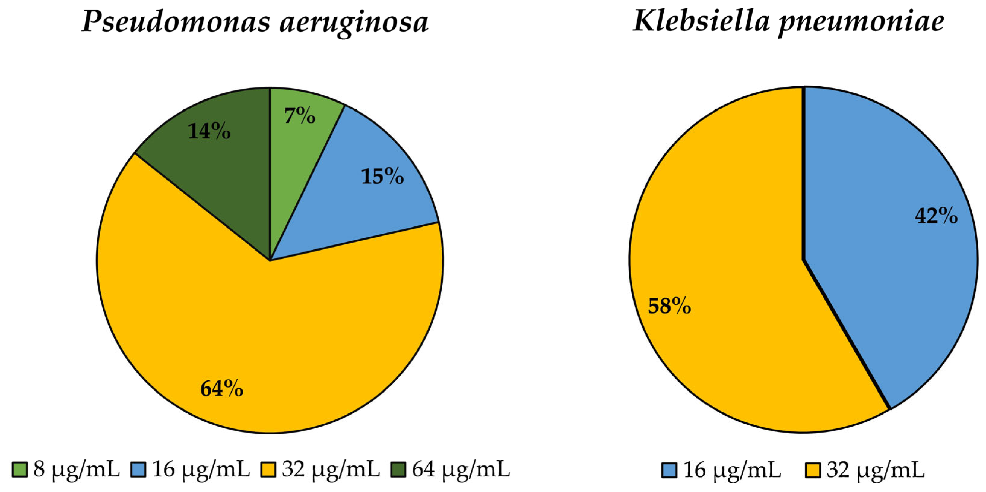
Figure 1. Determination of minimum inhibitory concentration for Epsilon-poly-L-lysine against P. aeruginosa (Pa) and K. pneumoniae (Kp) isolates.
Biomedicines 12 00638 g001
Figure 2. Effect of the ε-PL on the biofilm formation against 14 P. aeruginosa and 24 K. pneumoniae isolates. (A) Before exposure to ε-PL, the 14 P. aeruginosa isolates were strong and moderate biofilm producers. After exposure to ε-PL at the MIC value, a reduction in biofilm production was verified in 11 isolates. The number of strong and moderate biofilm producers’ isolates has reduced, and the number of weak biofilm producers has increased. (B) Before exposure to ε-PL, 21 K. pneumoniae isolates were strong, moderate, and weak biofilm producers. After exposure to ε-PL at the MIC value, a reduction in biofilm production was verified in 16 isolates. The number of strong and moderate biofilm producers’ isolates has reduced, and the number of weak biofilm producers has increased.
Biomedicines 12 00638 g002
Table 1. Phenotypic and genotypic characterization of resistance to different antibiotics in the selected P. aeruginosa (Pa) and K. pneumoniae (Kp) isolates.
SpeciesIsolateMDRPhenotype of ResistanceResistance Genes
PaHU1IMI, MEM, LEVblaKPC
HU2IMIblaKPC
HU4+PTZ, CAZ, ATM, IMI, MEM, DORblaKPC
HU5IMI, MEM, LEVblaSPM
HU6IMIblaKPC, blaSPM
HU7IMIblaSPM
HU8+PTZ, TTC, CAZ, ATM, CIP, blaCTX-M, blaVIM2, blaSPM
HU9IMIblaSHV
HU10IMIblaSHV, blaVIM
HU11IMIblaTEM, blaSPM
HU12IMIblaCTX-M, blaOXA
HU13TOB, CNaac(3)-IV
HU14IMI, CNblaPER, blaOXA, aac(3)-IV
HU15IMIblaKPC
KpHS2+TTC, CAZ, CTX, FEP, ATM, CN, TET, CIP, SXTblaCTX-M, aac(3)-II, aadA1, sul2
HS8+TTC, FOX, CAZ, CTX, FEP, ATM, CN, TET, CIP, NA, SXTblaCTX-M, blaTEM, blaSHV, aac(3)-II, sul2
HS14+TTC, CAZ, CTX, FEP, ATM, AK, CIP, NA, SXTblaCTX-M, blaTEM, blaSHV, aac(3)-II, sul2
HS17+TTC, FOX, CAZ, CTX, FEP, ATM, CN, CIP, NA, SXT, CHLblaCTX-M, blaTEM, blaSHV, aac(3)-II, aadA1, cmlA, sul2
HS29+TTC, CAZ, CTX, FEP, ATM, TET, CIP, SXTblaCTX-M, blaTEM, blaSHV, tetA, sul2
HS30+TTC, FOX, CAZ, CTX, FEP, ATM, MEM, ERT, IMI, AK, CN, CIP, SXTblaTEM, blaSHV, blaKPC, aac(3)-II, aadA1, sul2
HS34+TTC, CAZ, CTX, FEP, ATM, CN, CIP, NA, SXT, CHLblaCTX-M, blaSHV, aac(3)-II, aadA1, cmlA, sul2
HS36+TTC, FOX, CAZ, CTX, FEP, ATM, MEM, ERT, AK, CN, CIP, NA, SXTblaCTX-M, blaTEM, blaSHV, blaKPC, aac(3)-II, aadA1, sul2
HS42+TTC, CAZ, CTX, FEP, ATM, CN, CIP, NA, SXTblaCTX-M, blaTEM, blaSHV, aac(3)-II, aadA1, sul2, sul1
HS50+TTC, CAZ, CTX, FEP, ATM, ERT, AK, CN, SXTblaCTX-M, blaTEM, blaSHV, aac(3)-II, aadA1, sul1, sul2
HS54+TTC, CAZ, CTX, FEP, ATM, ERT, CIP, SXTblaTEM, blaSHV, blaNDM, sul2, sul1
HS55+TTC, CAZ, CTX, FEP, ATM, CN, CIP, NA, SXT, CHLblaCTX-M, blaTEM, blaSHV, aac(3)-II, aadA1, cmlA, sul2
HS56+TTC, FOX, CAZ, CTX, FEP, ATM, MEM, ERT, AK, CN, CIP, NA, CHLblaCTX-M, blaSHV, blaKPC, blaNDM, aac(3)-II, aadA1, cmlA
HS57+TTC, CAZ, CTX, FEP, ATM, TET, CIP, SXTblaCTX-M, blaTEM, blaSHV, tetA, sul2
HS58+TTC, CAZ, CTX, FEP, ATM, CN, TET, CIP, NA, SXT, CHLblaCTX-M, blaSHV, aac(3)-II, cmlA, sul1
HS67+CAZ, CTX, FEP, ATM, CN, CIP, SXTblaCTX-M, blaTEM, blaSHV, aac(3)-II, aadA1, sul2
HS77+TTC, CAZ, CTX, FEP, ATM, CN, CIP, NA, SXT, CHLblaCTX-M, blaTEM, blaSHV, aac(3)-II, aadA1, sul1, sul2, sul3
HS79+TTC, FOX, CAZ, CTX, FEP, ATM, ERT, CIPblaCTX-M, blaTEM, blaKPC, blaOXA, blaNDM
HS81+TTC, CAZ, CTX, FEP, ATM, CN, CIP, NA, SXT, CHLblaCTX-M, blaTEM, blaSHV, aac(3)-II, aadA1, cmlA, sul1, sul2, sul3
HS84+TTC, CAZ, CTX, FEP, ATM, CN, CIP, NA, SXT, CHLblaCTX-M, blaTEM, blaSHV, aac(3)-II, aadA1, cmlA, sul1, sul2, dfrA
HS87+CTX, FEP, ATM, CIP, NA, SXTblaCTX-M, blaTEM, blaSHV, sul1, sul2, dfrA
HS91+TTC, CAZ, CTX, FEP, ATM, CIP, NA, SXTblaCTX-M, blaTEM, blaSHV, sul1, sul2
HS92+TTC, CAZ, CTX, FEP, ATM, AK, CIP, NA, SXTblaCTX-M, blaTEM, blaSHV, aac(3)-II, aadA1, sul1, sul2
HS101+CAZ, CTX, FEP, ATM, CN, TET, CIP, SXT, CHLblaCTX-M, blaTEM, blaSHV, aac(3)-II, aadA1, tetA, cmlA, sul1, sul2
TTC—ticarcillin-clavulanate; FOX—cefoxitin; CAZ—ceftazidime; CTX—cefotaxime; FEP—cefepime; ATM—aztreonam; MEM—meropenem; ERT—ertapenem; IMI—imipenem; AK—amikacin; CN—gentamicin; TET—tetracycline; CIP—ciprofloxacin; NA—nalidixic acid; SXT—trimethoprim-sulfamethoxazole; CHL—chloramphenicol; DOR—doripenem; PTZ—piperacillin; TOB—tobramycin.
Table 2. MLST types of seven clinical P. aeruginosa isolates and seven clinical K. pneumoniae isolates.
P. aeruginosaK. pneumoniae
IsolateSTIsolateST
HU2699HS8307
HU5X *HS29348
HU6X *HS3415
HU71338HS36307
HU10285HS50584
HU14274HS5515
HU151404HS58307
* New allelic combination.
Disclaimer/Publisher’s Note: The statements, opinions and data contained in all publications are solely those of the individual author(s) and contributor(s) and not of MDPI and/or the editor(s). MDPI and/or the editor(s) disclaim responsibility for any injury to people or property resulting from any ideas, methods, instructions or products referred to in the content.