Review

Metabolic Dysfunction in Parkinson’s Disease: Unraveling the Glucose–Lipid Connection

by
Jeswinder Sian-Hulsmann
1,
Peter Riederer
2 and
Tanja Maria Michel
2,*
1
Department of Human Anatomy and Medical Physiology, University of Nairobi, P.O. Box 30197, Nairobi 00100, Kenya
2
Research Unit of Psychiatry, Department of Psychiatry, Odense, Region of Southern Denmark, University Hospital of Southern Denmark, 5000 Odense, Denmark
*
Author to whom correspondence should be addressed.
Biomedicines 2024, 12(12), 2841; https://doi.org/10.3390/biomedicines12122841
Submission received: 24 September 2024 / Revised: 18 November 2024 / Accepted: 22 November 2024 / Published: 13 December 2024

Abstract

:
Despite many years of research into the complex neurobiology of Parkinson’s disease, the precise aetiology cannot be pinpointed down to one causative agent but rather a multitude of mechanisms. Current treatment options can alleviate symptomsbut only slightly slow down the progression and not cure the disease and its underlying causes. Factors that play a role in causing the debilitating neurodegenerative psycho-motoric symptoms include genetic alterations, oxidative stress, neuroinflammation, general inflammation, neurotoxins, iron toxicity, environmental influences, and mitochondrial dysfunction. Recent findings suggest that the characteristic abnormal protein aggregation of alpha-synuclein and destruction of substantia nigra neurons might be due to mitochondrial dysfunction related to disturbances in lipid and glucose metabolism along with insulin resistance. The latter mechanism of action might be mediated by insulin receptor substrate docking to proteins that are involved in neuronal survival and signaling related to cell destruction. The increased risk of developing Type 2 Diabetes Mellitus endorses a connection between metabolic dysfunction and neurodegeneration. Here, we explore and highlight the potential role of glycolipid cellular insults in the pathophysiology of the disorder, opening up new promising avenues for the treatment of PD. Thus, antidiabetic drugs may be employed as neuromodulators to hinder the progression of the disorder.

1. Introduction

General Aspects of Parkinson’s Disease

Parkinson’s disease (PD) [1] is a progressive neurodegenerative disorder that exhibits abnormal movement, including resting tremor, bradykinesia, akinesia, hypokinesia and rigidity [2]. These are often coupled with non-motor symptoms such as depression and sleep disturbances and cognitive alterations, like Parkinson’s dementia disorder or Lewy body disorder, which may worsen as the disease progresses [3]. The loss of dopaminergic neurons in the substantia nigra (SN) pars compacta contributes mainly to the loss of voluntary movement observed in the disorder. Another characteristic pathological hallmark is the presence of Lewy bodies (LBs), which comprise misfolded alpha-synuclein aggregates. Many processes have been implicated in the destruction of the nigra cells and the generation of LBs [4].
Despite many years of research and some success at treating the symptoms of PD, the causative factors remain to be elucidated. Moreover, the precise mode(s) in which they interact in the complex labyrinth of events is still not completely understood; hence, treatment strategies to prevent neurodegeneration and stop the cascades implied are limited. However, the molecular pathways leading to the clinical symptoms include oxidative stress, neuroinflammation, dysfunction of protein clearance mechanisms/defective endolysosomal pathway, genetic components, and defective mitochondrial function [5]. However, the complex interactions remain to be elucidated more closely. Moreover, increasing evidence points to disruption of mitochondrial functioning, lipid and especially glucose metabolism in neurodegenerative diseases such as Alzheimer’s dementia and, lately, PD. In the following sections, we will give an overview of the implications and key role of metabolism in the complex molecular underpinnings of PD, which is a stepping stone to opening up new avenues for treatment options and targets.

2. Specific Aspects of PD and Metabolism: Mitochondria, Glucose, and Lipid Metabolism

2.1. Mitochondrial Dysfunction in PD

Langston and colleagues [6] first implicated mitochondrial dysfunction in PD. They described Parkinsonian like symptoms in illicit users of meperidine analogue 1-methyl-4-phenyl-1,2,5,6-tetrahydropyridine (MPTP). The active agent in MPTP is 1-Methyl-4-phenylpyridinium (MPP+), which is a mitochondrial complex 1 inhibitor in dopaminergic neurons.
Subsequently, a marked reduction in the activity of mitochondrial NADH-ubiquinone reductase (complex 1) was found in the SN from post-mortem brain tissue of PD patients [7,8,9]. This observation indicated mitochondrial dysfunction, which represents a crucial pathological factor since it has been reported repeatedly in sporadic and familial forms of illness [10,11].
The underlying cause(s) of PD might be many, but a strong link is seen for mutations in mitochondrial DNA that may alter the physiological dynamics, the presence of some mutated protein associated with mitochondria, or changes in the morphology of PD that may alter its function [12,13]. Mitochondrial dysfunctions can be linked to mutations that may initiate intense oxidative and inflammatory cellular damage and are associated with conditions such as Diabetes, myopathies, and neuropathies [14,15]. Indeed, in familial PD, several genes have been implicated in underlying mitochondrial dysfunction, including PARK 2, 6, 7, and 8 [16,17,18,19].
Furthermore, the mitochondria are a hub for producing toxic superoxide and other free radicals within the cell, released from oxidative phosphorylation processes [20]. This makes the mitochondria vulnerable to free radical-driven cytotoxic processes such as oxidative stress [21,22]. Additionally, since oxidative stress occurs early in the disorder’s progression, it may contribute significantly to mitochondrial dysfunction. Indeed, the depletion of the antioxidant reduced glutathione (GSH) in the SN in asymptomatic PD or incidental Lewy body disease endorses the occurrence of oxidative stress [23].
Mitochondrial dysfunction can contribute to a number of processes that are catastrophic for the cells, such as producing reactive free radicals leading to oxidative stress, alpha-synuclein aggregation, neuroinflammation [24], and disturbances in glucose and lipid metabolism. Of note is that dysfunction of both glucose and lipid metabolism causes a lack of bioenergetic supply of mitochondria [25,26]. Environmental neurotoxins, such as many pesticides, are so-called “uncouplers” of mitochondrial oxidative phosphorylation, resulting in a decrease in adenosine triphosphate (ATP) production [27]. This destroys not only pests and insects but can also cause neurological damage in humans, particularly over long-term exposure.
Thiamine-dependent processes are primarily of importance in glucose metabolism. Thiamine acts as a coenzyme for transketolase and for the pyruvate dehydrogenase and alpha-ketoglutarate dehydrogenase complexes, which play a fundamental role in intracellular glucose metabolism. Thiamine is involved in the development of the mitochondrial membrane and synaptic membrane function [28]. Alpha-ketoglutarate dehydroxygenase is a thiamine-dependent enzyme of the glycolysis pathway, and its activity is decreased in PD [29]. Increased dopamine release has been detected in experimental studies in rat striatum. In addition to that, thiamine, as well as dopamine itself, has been suggested to be a critical factor in increasing oxidative stress and peroxisomal mechanisms, as well as in gene expression and protein processing [30]. Mitochondrial pyruvate dehydrogenase and 2-oxoglutarate dehydrogenase are of major importance in delivering ATP, a primary source of energy supply for all neuronal tissues.
Additionally, nigral mitochondrial malfunction may exacerbate the production of lactate, a by-product of glycolysis. Subsequently, lactate can evoke neuronal destruction, perhaps via prompting or augmenting microgliosis and inflammatory-mediated cytotoxic processes that encourage misfolding of alpha-synuclein. Lactate may also affect other metabolic pathways, such as lipid metabolism, producing toxic lipid droplets that tend to cause neuronal destruction via lipid peroxidation and reactive oxygen-mediated oxidative stress. Thus, an imbalance in glucose homeostasis can have dire consequences on cellular bioenergetics and may affect the metabolism of lipids. The dysfunction of glucose metabolism may produce cellular energy failure, which may hinder general cellular functions of the dopaminergic neurons, such as neurotransmitter release/metabolism, vesicle transport, protein quality control, and neuron/glial survival. Mitochondrial malfunction may represent an important pathological feature that can disturb glucose metabolism and produce reactive oxygen species that prompt neurotoxic reactions, including oxidative stress and neuroinflammation. Additionally, it can contribute to the accumulation of misfolded protein (alpha-synuclein) aggregates.
This article reviews evidence of metabolic dysfunction, mainly glucose and lipid metabolism, as a potential candidate in the pathophysiology of PD. The evidence strongly supports the involvement of impaired cellular bioenergetics in neurodegeneration. This offers new pharmacological avenues for managing the illness, targeting the impaired glucose/lipid metabolism processes. Therefore, new lines of treatment might be hindering metabolic dysfunction-associated cell death. However, such novel treatments would be an add-on to gold-standard levodopa treatment. Diabetes is an ideal model to provide a better understanding of the consequences of disturbed glucose homeostasis. Although the mechanisms correlating neurodegeneration in PD and disturbed glucose homeostasis in the brain are still not well understood.

2.2. Implications of Mitochondrial Dysfunction and Disturbances in Cellular Bioenergetics of Glucose in PD

Oxidative phosphorylation occurs during cellular respiration [31]. It is involved in ATP or energy production and comprises two processes: electron transport chain (ETC) and chemiosmosis. Complex 1 is one of the four membrane-embedded protein complexes involved in electron chain transport (ECT), which consists of oxidation/reduction reactions to transfer electrons from one complex to another. The ETC builds a proton gradient in the inner mitochondrial membrane, which is then used to produce stored energy in the form of ATP. However, oxidative phosphorylation has the propensity to generate reactive oxygen and nitrogen species as a by-product, although this is relatively low under physiological conditions [32]. Decreased neuronal ATP levels have been observed in PD [15], which may need to be carefully interpreted since ATP measurements are a critical manoeuvre.
Mitochondrial dysfunction and deficiency in complex 1 can disturb energy-generating reactions such as oxidative phosphorylation, reducing cellular ATP levels and impeding cellular physiological functioning. In addition to that, it can also promote the production of free radicals that are toxic to cells [33]. Eventually, if these persist, cellular deleterious effects can be the result (Figure 1). In the presence of the inability of the cellular antioxidant defence system (such as glutathione peroxidase, glutathione, catalase, and superoxide dismutase) to overcome free radical production, resulting in oxidative stress, loss of cell membrane integrity, cell death, and cell senescence are the result. Therefore, the reduction in complex 1 activity [9] in the SN in PD represents a significant pathological factor. Hence, it may be associated with the dopaminergic neuronal defect characteristic of the disorder. The SN may be particularly vulnerable to decreased or malfunctioning mitochondria in light of its high mitochondrial density in order to provide a higher oxidative phosphorylation to meet the great ATP demands of the dopaminergic neurons [34].

2.3. Mitochondrial Genes and PD

There may also be an underlying genetic involvement in the neuronal energy deficit seen in the disorder. Mutation of the PD gene, PGK1, which encodes for phosphoglycerate kinase 1, a crucial enzyme required for glycolysis, increases the susceptibility to PD [36]. Conversely, augmentation of phosphoglycerate kinase 1 (PGK-1) activity alleviates PD symptoms [37] probably by providing ATP to release extracellular dopamine in the striatum [38]. PGK1 is a rate-limiting enzyme for the production of ATP in glycolysis in nerve terminals. More importantly, it has neuroprotective abilities since it permits the synapses to operate under conditions of hypoglycemia/hypometabolism [39]. This notion is supported by the modest repression of cell death of a PGK 1 activator, Terazosin, in many animal models [40]. This drug enhances PGK 1 activity for energy generation. It is regulated by a chaperone protein DJ-1 (PARK 7) or deglycase, and the PARK 7 gene mutations are associated with familial PD. Therefore, the neuroprotective role DJ-1′s might be twofold: it enhances PGK1 activity, thereby reducing hypometabolic defective synaptic function and hindering the accumulation of 3-phosphoglycerol-lysine resides in protein [41].
Although, significant pathophysiological heterogeneity has been demonstrated [42] in patients with PD, subtyping the disorder has been challenging [43]. Nevertheless, a recent study [44] suggests that idiopathic PD can be subdivided into two subtypes based on the severity and regional distribution of mitochondrial complex 1 deficiency in neurons. Furthermore, these subtypes also illustrate distinguishable cellular and clinical features.
More importantly, recent studies have highlighted the impact of complex 1 deficiency in the SN on the progression of the illness [45]. This reduction in mitochondrial complex 1 activity in PD patients versus controls concords with previous findings in asymptomatic PD/incidental Lewy body disease [23].

2.4. Is PD a “New Diabetes Type”—The Missing Link Between Insulin and Dopamine?

The seminal histochemical studies of Falck and Hellman [46], Lakomy and Chodkowska [47], and Cegrell [48] demonstrated the presence of catecholamines in pancreatic cells. Malaisse et al. [49] showed that insulin secretion can be stimulated by adrenergic receptors and acetylcholine and inhibited by alpha-adrenergic receptors. As summarised in detail by De Iuliis et al. [50] the presence of dopamine in secretory granules of pancreatic ß-cells leads to the assumption that hyperglycaemic effects might be driven by dopamine through insulin storage or the releasing mechanisms [50,51]. Indeed, tyrosine hydroxylase and aromatic amino acid decarboxylase have been identified in ß-cells, and monoamine oxidase is co-localized with insulin in secretory granules of ß-cells and VMAT1 (vesicular monoamine transporter 1) and DAT (dopamine transporter) were found in pancreatic cells [52,53] As well, D1- and D2-like dopamine receptors are expressed in ß-cells [54,55]. Thus, it seems that the role of dopamine in pancreatic insulin secretion is inhibitory and modulated by dopamine receptors [50].
Interestingly, there are no reports on the influence of levodopa treatment on pancreatic cells. It is unknown whether levodopa treatment of PD poses a risk for inducing/stimulating Diabetes mellitus or whether it is protective. Clinical and epidemiological studies are urgently needed to answer such and other related questions.
In the brain, insulin receptors have been identified in neurons and astrocytes of most brain regions, including the hippocampus, cerebral cortex, ventral tegmental area (VTA), and substantia nigra [50,56,57]. Insulin regulates glucose transporter (Glut) 4 and GLUT 8 [58]. Insulin triggers two pathways via insulin receptor substrate (IRS) docking proteins: the phosphoinositide-3-kinase cascade (PIK 3-involved in neuronal survival) and the Ras-Raf-mitogen-activated protein kinase (MAPK/ERK) signalling (involved in cell death) [50,59]. Furthermore, insulin promotes neuronal survival, plasticity and neuronal antioxidant defence as well as protection against neuronal apoptosis [60] via the SHC/ERK1/2 pathway [50].
These pathways have been shown to play an important role in both Alzheimer’s disease (AD) as well as in attention-deficit-hyperactivity-disorder (ADHD) [61]. Indeed, there is ample evidence pointing at the influence of glucose and insulin in mammalian Target of Rapamycin Complex 1 (mTOR C1) and Complex 2 (mTOR C2) pathways [61]. This might explain some of the processes happening in PD, too. Increased GABAergic inhibitory neurons in the striatum with upregulation of D1-receptor activity and downregulation of D2-receptors in medium spiny neurons is seen in transgenic mice, whose mTORC1 signalling is hyperactivated in inhibitory neurons in the striatum, while cortical neurons are left unaffected. These findings suggest critical involvement of the mTOR pathway in locomotor abnormalities in PD [62], (provisionally accepted work). <murine models of dopamine neuron-specific deletions of Raptor or Rictor, emphasize on the importance of both mTORC1 and mTORC2 for dopamine. Studies show that the inhibition of mTORC1 impacts dopamine neuron structure and function. This causes somatodendritic and axonal hypotrophy, increased excitability, and decreased dopamine production and release, which contrasts the consequences of inhibition of mTORC2 with some subtle effects on the output of VTA dopamine neurons [63]. The disruption of these pathways leads to pronounced deficits in dopamine release. Thus, this demonstrates their importance for dopaminergic functions [63]. In addition to that, rapamycin, an mTOR inhibitor and inducer of autophagy, potentiates oxidative stress-induced cell death, while the autophagy inhibitor 3-methyladenine protects the dopamine cells [64]. Dopamine receptors D2 and D3 are positive regulators of autophagy, while dopamine receptors D1 and D5 are negative regulators [65] (Figure 1 and Figure 2).
Of note in this regard is that a number of known drugs influence the mTOR pathways. For example, morphine that reduces the insulin receptor substrate/Protein kinase B (IRS/Akt) activity decreases K+ channel expression in the VTA, morphine increases mTORC1 and decreases mTORC2 signalling in the VTA [67,68]. In addition to that, striatal mTORC1 blockade may prevent D2-receptor-dependent extrapyramidal motor side effects of haloperidol in psychiatric illness [69,70].
In summary, both insulin and dopamine actions meet at the mTOR protein interaction, thus providing evidence for a robust linking of energy-providing pathways and neuronal dopaminergic activity in PD.

2.5. The Role of Hyperglycaemia in PD

The abnormal function of the mitochondria reported in PD is most likely directly related to the disturbed glucose metabolism and cellular bioenergetics. This notion is supported by the increased gluconeogenesis and abnormal changes in glucose breakdown in vivo seen in idiopathic PD patients [12,71,72]. Similarly, in Diabetes Mellitus, the pathological elevation of hepatic glucose formation accounts for the characteristic hyperglycaemia manifested in the condition [73]. In vivo and in vitro studies demonstrate the cytotoxic effects of uncontrolled hyperglycaemic levels. Indeed, long-term brain hyperglycaemia has been shown to inflict neurodegeneration, particularly to the nigrostriatal motor tract and mesocortical–limbic pathways in rats [74]. Culture studies using dopaminergic (pheochromocytoma) PC12 cells showed that the hyperglycaemic-related mode of action of dopamine neural destruction is exerted by oxidative stress by glycated modification [75,76] and apoptosis [77]. Furthermore, the elevated glucose levels in the diseased state account for the increased production of its toxic metabolite by-product, methylglyoxal. This is a 2-6-fold increase compared to physiological levels [35,78,79]. Alternatively, triacylglycerol hydrolysis can be generated from the lipid metabolic pathway [80].
Nevertheless, methylglyoxal intervenes in endothelial functioning and is instrumental in insulin resistance (IR) [81]. Under normoglycemic conditions, methylglyoxal is detoxified to lactate [82] by the actions of the two enzymes glyoxalases (1 and 2) in the presence of GSH (Figure 1). Therefore, the depleted GSH levels in the SN in PD [83,84] may exacerbate the toxic effects of methylglyoxal. It can exert cellular deleterious effects and inhibit cell growth via glycated modification by modifying proteins and lipids [85], disturbing calcium homeostasis and leading to cell destruction [86]. More importantly, methylglyoxal can augment the striatal dopamine depletion by reacting with dopamine to produce a toxic tetrahydro isoquinolin that mediates cellular destruction via apoptosis [79] and alpha-synuclein pathology-related glutaminergic hyperactivity [87].
Evidence from a meta-analysis [88,89] and epidemiological studies [90,91,92,93,94,95] showed that Type 2 diabetes increases the risk and development of idiopathic PD (by 1.21-fold). Indeed, PD patients with pre-existing Type 2 Diabetes suffer from a more rapid disease progression along with reduced survival rates. Interestingly, this seems unrelated to alpha-synuclein pathology [96]. Therefore, alpha-synuclein-containing LBs may be considered as a consequence of neuronal death rather than a cause.
Lixisenatide, a glucagon-like peptide agonist used in treating Diabetes Mellitus Type 2 and obesity, has been shown to have neuroprotective attributes in the PD mouse model [97]. Administration of lixisenatide to patients in the early stages of the course of PD (less than three years since diagnosis, responsive to treatment, and no motor complications) over 12 months modulated the progression of motor dysfunction [97,98]. The implications of these findings are fascinating and highlight the possible involvement of metabolic deficits or dysfunction in the pathogenesis and/or the progression of the illness. Although further studies are warranted with larger patient groups, different drug doses of lixisenatide are needed to access optimal efficacy coupled with minimum toxicity and longer duration.

2.6. The Effect of Cerebral Insulin Resistance in PD

Since Type 2 Diabetes is a consequence of peripheral insulin resistance (IR), IR in the brain might be observed in PD, pointing to IR as the common denominator for the two conditions [99,100,101]. Disruption in insulin metabolism or signalling in the brain may have enormous cellular consequences, given its vital role in brain function and neuronal survival [102]. These neuroprotective actions of insulin (Figure 1) are probably related to its ability to hinder the actions of cytotoxic processes such as oxidative stress, beta-amyloid toxicity, and apoptotic cellular destruction [103,104].
Perhaps peripheral IR and brain IR [105] might prompt brain pathologies, including neuroinflammation, oxidative stress [106] and a “leaky” blood–brain barrier (Figure 1) [107]. However, these cytotoxic processes are intertwined. Thus, pro-inflammatory cytokines released from a state of chronic neuroinflammation may also contribute to the central IR. Additionally, neuroinflammation may prompt or, at least partly, contribute to the alpha-synuclein pathology observed in the SN in PD. Interestingly, brain IR may contribute to alpha-synuclein pathology by blocking the insulin-degrading enzyme inhibition of alpha-synuclein fibril production, which subsequently supports its aggregation [108,109] and amyloid polypeptide [78]. Therefore, disturbed brain insulin signalling and IR can potentially induce neuronal destruction. The progressive loss of SN neurons in PD may, in part, subscribe to the disturbed IR/reduction in insulin production/glucose metabolism since the SN expresses insulin receptors [110,111] and the mid-brain contains detectable levels of insulin [112]. Indeed, reduced insulin receptors in the SN and greater IR have been found in PD [113]. The depleted brain insulin levels may hinder the release of dopamine from the striatum since insulin is involved in the action potential-dependent release of dopamine in this area [114].
Although central (alpha-synuclein aggregation and dopamine cell death) and peripheral (islet amyloid polypeptide aggregation and apoptosis) IR is associated with different pathological features, a so-called “cross-talk” or interaction between the two aggregates has been reported [115,116]. Amyloid polypeptide aggregation might support the abnormal accumulation of alpha-synuclein.
IR has been found to play a notable role in symptoms associated with metabolic- syndrome, such as elevation of lipid peroxidation and abnormalities in lipid profile [117]. Lipids play an essential role in cellular physiological and pathological processes.

2.7. Lipid Metabolism in PD

Lipid peroxidation occurs due to reactive free radical species-mediated oxidative stress. It oxidizes the lipids in cell membranes, breaking up the double bindings of the fatty acids that form the bi-lipid cell membrane, leading to the loss of cell membrane function and fluidity, resulting in cell death [118]. It is a marker for systemic IR and many neurodegenerative disorders such as PD [119] and Alzheimer’s disease [120,121]. Elevated lipid peroxidation indicates disturbed lipid metabolism, which may play an essential role in the pathogenesis and development of many disorders.
The SN in PD showed a reduction of polyunsaturated fatty acids, a substrate, and an index for lipid peroxidation [119]. Indeed, the elevation of lipid peroxidation was supported by the increase of an intermediate of this process, malondialdehyde. Thus, the decrease in polyunsaturated fatty acids is probably related to their utilization. The dysfunctional nigral mitochondria may be responsible for disturbing the lipid metabolism pathways in PD [122,123], since they modulate lipid metabolism and storage. On the other hand, lipid storage disorders also have the potential to disrupt mitochondrial function [14].
Lipid transport and regulation play a vital role in the physiological functioning of brain cells such as neurons and glia [124]. Interestingly, histological studies using a fluorescent probe (BODYIPY) displayed atypical lipid accumulation in dopaminergic neurons (Figure 1) and microglia (but not astrocytes) in the SN in PD [125]. The cellular preference for fat build-up is congruous with regions associated with neuroinflammation (Figure 1). This finding raises intriguing questions about the potential association of lipid accumulation and mitochondrial complex 1 reduction. Recently, statistically significant elevations in lipids such as cholesterol, sphingomyelin, and phosphatidylcholine have been described in the cerebrospinal fluid of PD patients [126]. The accumulation of lipids may pose a risk for disease pathogenesis or contribute to neurodegeneration via mechanisms such as free radical-mediated oxidative stress.
Free radicals might prompt the formation of lipids in neuronal cells. The lipids are transported by the glial–neuron lactate shuttle (via monocarboxylate transporters and fatty acid transport proteins) into the glia, where it is converted to lipid droplets [127]. The monocarboxylate transporter permits lactate secretion from the glia. Afterward, lactate is taken up by the neurons and converted to pyruvate and acetyl-coenzyme A. Subsequently, the breakdown product is used for the synthesis of fatty acids. Consecutively, they are transported via fatty acid protein to the glia. Moreover, lactate is not only a breakdown product of glycolysis but plays a crucial role in neurodegeneration [66] and induces metabolic reprogramming [128]. The latter is demonstrated by the elevated lactate concentration in the cerebrospinal fluid of PD patients compared to control subjects, which corresponds to the progression of the disease [129]. The neurodegenerative effects of lactate (Figure 2) might be mediated by the activation of glia, which in turn releases pro-inflammatory cytokines (such as Interleukin-1beta and 6, tumour necrosis factor-alpha), initiating the cytotoxic neuroinflammatory cascade [24]. Its production might be increased by the upregulation of hexokinase-2 in dopaminergic neurons in PD. This might promote degenerative processes such as apoptosis and alpha-synuclein aggregation [130]. This notion is supported by inhibiting hexokinase-2 expression, which decreases lactate production and related apoptotic destruction of dopamine neurons [130].
Lactate can prompt lipids to produce and mobilize lipid droplets (Figure 2) [128,131]. Lipid droplets execute essential physiological and pathological roles in energy homeostasis by mitigating harmful cellular effects [132]. Furthermore, oxidative stress in the brain promotes the accumulation of lipid droplets in glial cells [133,134,135]. Its ability to trigger inflammation might confer its pathological nature. Since defective lipid homeostasis, signalling, and lipid droplet accumulation have been associated with an increased risk of PD, metabolic disturbances might play a vital role in the development of the disease.

2.8. Bioenergetic Drug Treatment in Parkinson’s Disease
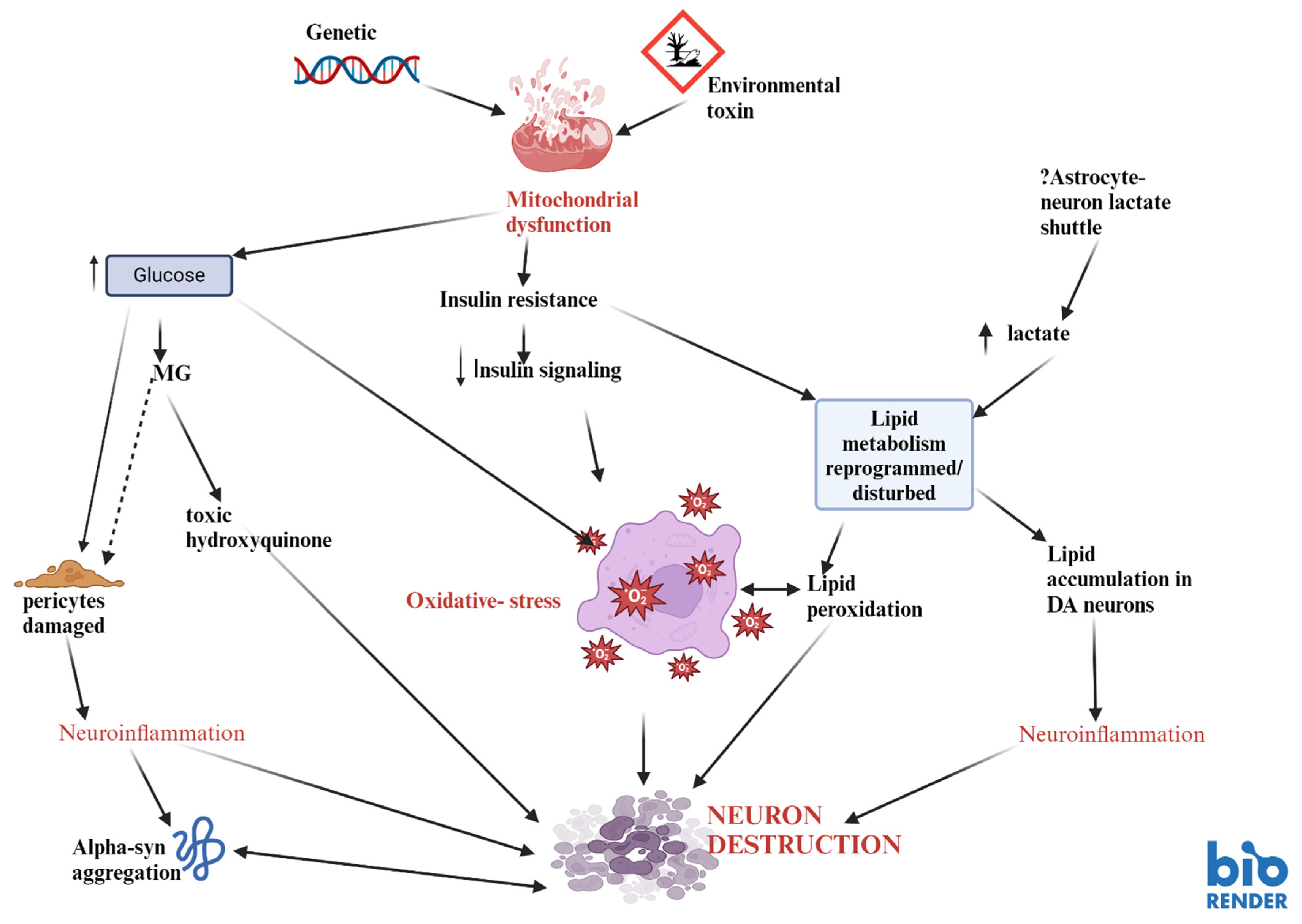
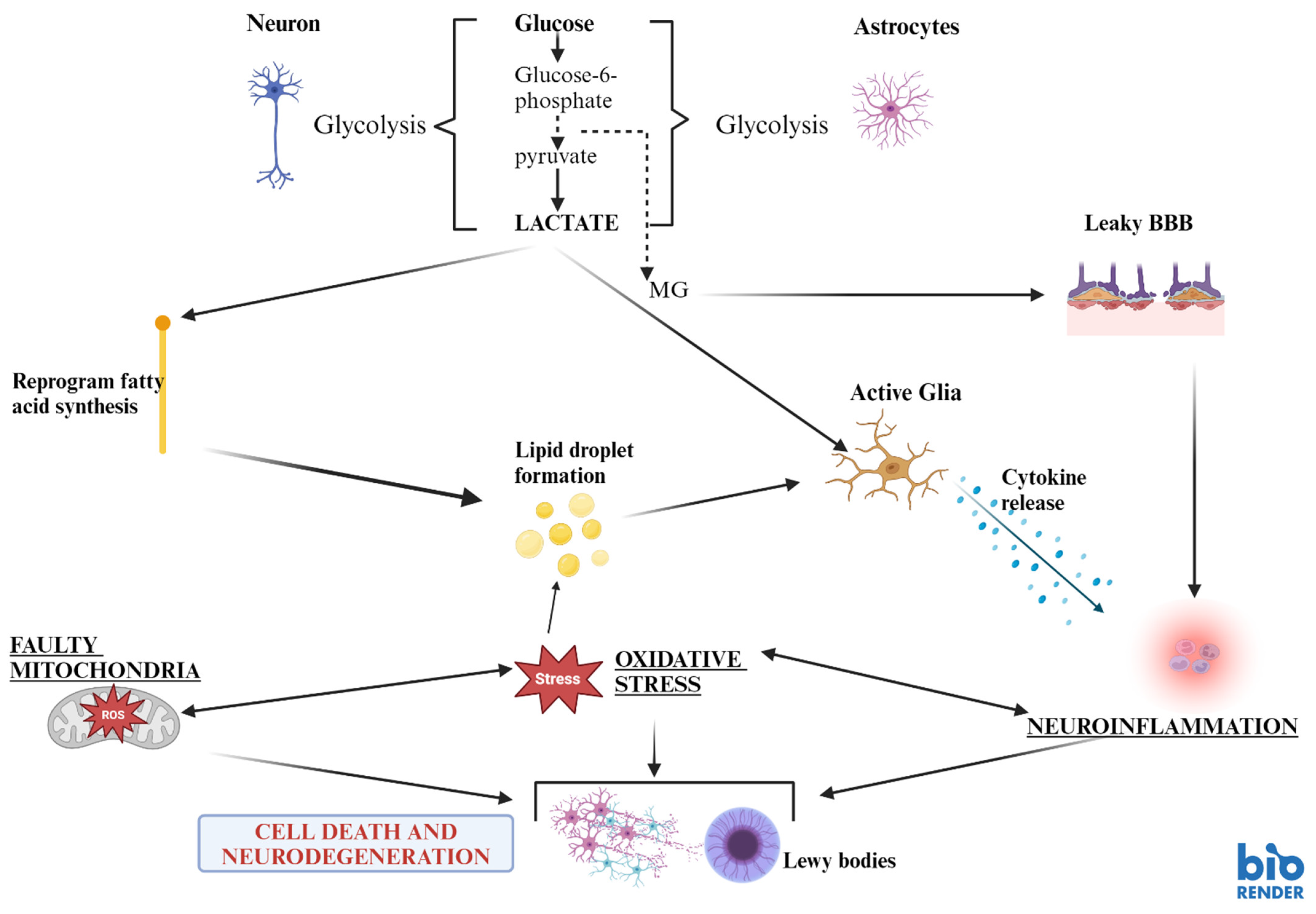
Therapies used for the treatment of Type 2 Diabetes may be promising candidates to supplement dopaminergic drug treatment strategies in PD.
As reported by De Iuliis et al. [50] the compound “Exenatide”, a Glucagon-like Peptide 1 (GLP-1) analogue, improved motor behaviour [41,111,136,137,138]. In addition to that, Athauda et al. [139,140] found higher IRS1 phosphorylation and Akt and mTOR expression in the individuals who received Exenatide, which was associated with better motor scores.
DPP-4 (Dipeptidyl Peptidase-4) is an enzyme that plays a role in glucose metabolism. DDP4 inhibitors block the enzyme dipeptidyl peptidase-4. Therefore, they could be used to treat Type 2 Diabetes since they increase insulin production and reduce the amount of glucose produced by the liver. This helps lower blood sugar levels in people with Type 2 Diabetes. Common DPP-4 inhibitors include medications like Sitagliptin, Saxagliptin, and Linagliptin. DPP-4 inhibitors lower the risk for the development of PD [141,142].
Thiazolidinediones (TZDs) improve insulin sensitivity in peripheral tissues such as muscle and fat. They activate PPAR-γ (peroxisome proliferator-activated receptor gamma), which regulates glucose and lipid metabolism. Thiazolidinediones significantly decrease the incidence of PD [143].
Pioglitazone is a type of TZD. There has been some interest in pioglitazone for its possible neuroprotective effects, potentially decreasing the risk or progression of PD. However, the evidence is not definitive, and more research is needed to confirm this potential. Pioglitazone showed no significant decline in the incidence of PD [141], but a reduction in the incidence of PD was reported when administered together with statins [144].
Ping et al. [145] describe that the commonly prescribed medication for Type 2 Diabetes, metformin, may be associated with an increased incidence of PD. Metformin influences mitochondrial function and glucose metabolism, which may play a role in neurodegenerative processes happening in PD. However, more research is required to fully understand the mechanisms and the risks.
The water-soluble vitamin thiamine (Vitamin B1) is essential for energy metabolism and the functioning of the nervous system. It is critically involved in glucose metabolism in the Krebs cycle and is required for the synthesis of neurotransmitters. Thiamine deficiency can lead to neurological conditions such as Wernicke–Korsakoff syndrome and Beriberi, and it has been suggested that thiamine could have a neuroprotective role in other conditions as well. In the context of PD, there is ongoing research into the potential protective role of thiamine, as it supports mitochondrial function and neuronal health. While thiamine deficiency has been linked to cognitive impairments, its supplementation has been investigated for its therapeutic effects in neurodegenerative diseases like PD, with promising results [30,146].
Thiamin is a cofactor of several enzymes active in the glycolysis pathway. Therefore, thiamine has been connected to the PD pathology and candidate pathways which involve transcription factors Sp1, p53, Bcl2, caspase-3, tyrosine hydroxylase, glycogen synthase kinase-3ß, vascular endothelial growth factor, advanced glycation end products, nuclear factor kappa B, mitogen-activated protein kinase, and the reduced form of nicotinamide adenine nucleotide phosphate [147,148]. Indeed, high-dose thiamine can effectively reverse PD motor and non-motor symptoms [30]. This is in line with experimental studies in the rat model of MPTP-induced Parkinsonian in rats [149] and experimental thiamine deficiency [124,150].

3. Discussion

Parkinson’s disease (PD) is a devastating neurological disorder with a number of subtypes (e.g., early-onset and late-onset PD, tremor-dominant, akinetic-rigid PD, PD with tremor, rigidity and bradyphrenia, PD with dementia, Lewy body dementia, vascular PD) [151,152]. The triggers underlying PD are unknown, although several have been described in recent years, e.g., carbon monoxide, insecticides, pesticides, chemicals, viruses, iron, manganese, plumbum, and genetic mutations [153,154]. Oxidative stress and inflammation, as well as mitochondriopathy, have been discussed as being of major pathological importance [20,62,155]. Therefore, we have focused on comorbid disorders that might be triggering the molecular cascade leading to PD. We have focused on metabolic disease and Type 2 Diabetes to investigate them as possible risk factors prompting, e.g., early-onset PD.
The ultimate goal of this review is to unravel molecular aspects of PD and to open up new avenues for treatment in order to minimize these risk factors. Type 2 Diabetes (DM) comprises several aspects of interest as risk factors for triggering PD. (1) This is of interest because disturbances in the glucose and lipid metabolism seen in DM lead to the loss of metabolites important for the brain’s energy supply. Indeed, a loss of ATP production in mitochondria has been suggested to be responsible for mitochondriopathy in PD, leading to the degeneration of nigrostriatal dopaminergic neurons. (2) The role of Type 2 Diabetes has been extensively studied in Alzheimer’s Disease (AD) ([60,122,156] for review) and conclusions from such research can be of importance for PD, too. (3) Thiamine (Vitamin B1) is a key player in glucose metabolism and is vitally involved in mitochondrial membrane development and synaptic membrane function [28]. Thiamine might show therapeutic benefits in mitochondrial respiratory chain deficit [146]. Thus, it is of significance to further elucidate the link between glucose, insulin, lipids, and dopamine with respect to cellular bioenergetic (hyperglycaemia, insulin resistance) dysfunction in mitochondria. Bioenergetic drug treatment strategies are of interest to enlarge the armamentarium to fight PD. Here, we report in detail on all these mechanisms Therefore, we extend previous work on this topic in PD. In addition to that, the review uncovers evidence from the molecular investigation, which may lay the foundation for clinical studies designed to improve the brain’s energy metabolism in patients with PD and in patients with DM and/or disturbances of lipid metabolism. Potential promising clinical applications [157] should be investigated further with a focus on glucose metabolism to open up new and exciting possibilities for innovative therapeutic interventions in PD. Hence, this report should stimulate further efforts to elucidate biochemical aspects of PD other than, e.g., alpha-synuclein-related pathology. Given the increasing evidence reported, the latter might even be a result of cellular energy loss, oxidative stress, inflammation, and mitochondriopathy.
In summary, this overview emphasized on the important role of energy loss due to deficits in the glucose/lipid metabolism in the pathogenesis of PD. This is critical for understanding the molecular mechanisms discussed in PD, like oxidative stress, inflammation, mitochondriopathy, and the pathological processes and interactions induced by alpha-synuclein. The review summarizes molecular evidence and wants to open up new clinical strategies to fight the molecular underpinnings that lead to the devastating disorder PD.

4. Conclusions

The mitochondrial dysfunction described in PD may be closely linked to irregular glucose metabolism and brain IR. Injecting recombinant complex 1 or NADH-quinone oxidoreductase adeno-associated virus into the SN pars compacta of MPTP-treated mice has shown promising results. This treatment significantly improved dopamine neuronal survival and motor disabilities potentially by compensating for the compromised complex 1 activity [22]. The implications of this are significant, suggesting that such a treatment could be effective in the prodromal phase of idiopathic PD, offering hope for salvaging the remaining neurons from the pathological processes.
Brain hyperglycaemia in PD might be closely associated with insulin depletion, malfunction in glucose metabolism, or perhaps the inability of cerebral insulin to cope with the pathological elevation of glucose. Consequently, vulnerable regions in the brain, such as the SN, are susceptible to the onslaught of cellular destruction processes. Elevated brain glucose may elevate the production of its cytotoxic metabolite methylglyoxal, which is related to dopamine depletion, IR, and apoptotic cellular destruction in susceptible mid-brain areas.
It seems that the alterations in the peripheral/central IR pathways have the propensity to facilitate neuronal destruction. This might be primarily related to the loss of the neuroprotective role of insulin in PD. This reflects the operation of complicated pathways that prompt many questions worth exploring further: Is PD a metabolic disorder? Does the peripheral IR lead to central IR or vice versa? How does IR lead to neuronal death? In PD, is the brain IR a consequence of faulty nigral mitochondrial complex 1? Is there a shared pathogenesis between PD and DM? What factors support or confer the development of PD in Type 2 Diabetic patients? Are there other neurodegenerative disorders that exhibit similar processes as in Type 2 Diabetes?
It is becoming increasingly clear that PD and Type 2 Diabetes share common pathophysiological processes. Metabolic disturbances are central to these mechanisms. Ultimately, other factors such as environmental/neurotoxins/genetic factors may finally determine the development of PD in some patients with Diabetes. This underscores the importance of understanding and addressing metabolic disturbances in these diseases.
The nigral mitochondrial dysfunction in PD may consequently disturb the regulation of lipid homeostasis metabolism and promote cellular degenerative processes. Altered lipid metabolism/storage might impact cellular bioenergetics and insulin signalling and disturb glucose/lipid pathways. Other factors may also be involved in disruption in the lipid homeostasis. Modified glucolipid pathways may contribute to PD’s elevated lactate levels in the cerebrospinal fluid. The increased lactate from neurons and glia may be considered an important therapeutic target to reduce catastrophic cellular destruction since it activates the neuroinflammatory pathways.
The impairment in glucose metabolism may disturb the functioning of the blood–brain barrier. Hyperglycaemia can disturb pericyte function and damage the tight junction features in the barrier via oxidative stress and neuroinflammation/inflammation [158,159]. Additionally, IR depletes the pericyte population [160]. Pericytes are an essential constituent of this physical barrier. They exert an integral role in the physiological functioning of the microglia, which provide integrity and vascular supply to the blood–brain barrier. The raised lipids in the cerebrospinal fluid from PD patients may also support a disrupted blood–brain barrier (Figure 2).
It appears that perhaps the focus needs to be shifted from mechanisms underlying abnormal alpha-synuclein aggregation in the brain toward the disruption of the glucolipid metabolic pathways to provide a better understanding of the complex pathogenesis of PD. The disease may have a multisystemic pathogenesis coupled with multifactorial factorial (genetic risk factors, environmental, neurotoxin exposure) interactions such asenergy deficits, particularly in the synapses. This might be a (predisposing) vulnerability factor for the degenerating dopamine neurons. PGK 1 may represent a critical pharmacological point of action for exercising bioenergetic control for neuroprotection and modulation. Additionally, antidiabetic drugs may prove to be a potential game changer in delaying the progression of PD.
Brain insulin resistance may disrupt lipid metabolism, leading to cytotoxic processes such as lipid peroxidation and oxidative stress. Additionally, the elevated brain lactate levels (perhaps due to a faulty astrocyte–neuron lactate shuttle) may also disturb or reprogram lipid breakdown. This may be responsible for the fat accumulation in the dopamine neurons, which may enhance their susceptibility to neuroinflammation.
Methylglyoxal is a toxic by-product of glycolysis; it can destroy the integrity of the blood–brain barrier, rendering it “leaky” and leaving the brain open to access to neurotoxins and inflammatory mediators that can induce neuroinflammation.

Author Contributions

Conceptualization, J.S.-H. and T.M.M.; methodology, J.S.-H. and T.M.M.; validation, J.S.-H., P.R. and T.M.M.; investigation, J.S.-H. and P.R.; resources, J.S.-H., T.M.M. and P.R.; data curation, J.S.-H.; writing—original draft preparation, J.S.-H.; writing—review and editing, J.S.-H., T.M.M. and P.R.; visualization, J.S.-H.; supervision, T.M.M.; project administration, T.M.M.; funding acquisition, P.R. All authors have read and agreed to the published version of the manuscript.

Funding

This research received no external funding.

Data Availability Statement

The original contributions presented in the study are included in the article, further inquiries can be directed to the corresponding authors.

Acknowledgments

We thank Elleni Cotti for her help in editing the different versions and communication between authors.

Conflicts of Interest

The authors declare no conflict of interest.

References

  1. Parkinson, J. An Essay on the Shaking Palsy. J. Neuropsychiatry Clin. Neurosci. 2002, 14, 223–236. [Google Scholar] [CrossRef] [PubMed]
  2. Birkmayer, W.; Riederer, P. The Clinical Pathology of Parkinson’s Disease. Biochemistry, Clinical Pathology, and Treatment; Springer: Berlin/Heidelberg, Germany, 1983; pp. 46–88. [Google Scholar]
  3. Rodriguez, M.; Morales, I.; Rodriguez-Sabate, C.; Sanchez, A.; Castro, R.; Brito, J.M.; Sabate, M. The degeneration and replacement of dopamine cells in Parkinson’s disease: The role of aging. Front. Neuroanat. 2014, 8, 80. [Google Scholar] [CrossRef] [PubMed]
  4. Obeso, J.A.; Rodriguez-Oroz, M.C.; Goetz, C.G.; Marin, C.; Kordower, J.H.; Rodriguez, M.; Hirsch, E.C.; Farrer, M.; Schapira, A.H.V.; Halliday, G. Missing pieces in the Parkinson’s disease puzzle. Nat. Med. 2010, 16, 653–661. [Google Scholar] [CrossRef] [PubMed]
  5. Vos, M.; Klein, C. The Importance of Drosophila melanogaster Research to UnCover Cellular Pathways Underlying Parkinson’s Disease. Cells 2021, 10, 579. [Google Scholar] [CrossRef] [PubMed]
  6. Langston, J.; Ballard, P.; Tetrud, J.; Irwin, I. Chronic Parkinsonism in humans due to a product of meperidine-analog synthesis. Science 1983, 219, 979–980. [Google Scholar] [CrossRef]
  7. Mizuno, Y.; Ohta, S.; Tanaka, M.; Takamiya, S.; Suzuki, K.; Sato, T.; Oya, H.; Ozawa, T.; Kagawa, Y. Deficiencies in complex I subunits of the respiratory chain in Parkinson’s disease. Biochem. Biophys. Res. Commun. 1989, 163, 1450–1455. [Google Scholar] [CrossRef]
  8. Reichmann, H.; Riederer, P. Biochemische Analyse der Atmungskettenkomplexe verschiedener Hirnregionen von Patienten mit Morbus Parkinson. In Morbus Parkinson und Andere Basalganglienerkrankungen; BMFT Symposium Bad Kissingen: Bonn, Germany, 1989. [Google Scholar]
  9. Schapira, A.H.; Cooper, J.M.; Dexter, D.; Jenner, P.; Clark, J.B.; Marsden, C.D. Mitochondrial complex I deficiency in Parkinson’s disease. Lancet 1989, 1, 1269. [Google Scholar] [CrossRef]
  10. Gao, X.Y.; Yang, T.; Gu, Y.; Sun, X.-H. Mitochondrial Dysfunction in Parkinson’s Disease: From Mechanistic Insights to Therapy. Front. Aging Neurosci. 2022, 14, 885500. [Google Scholar] [CrossRef]
  11. Ryan, B.J.; Hoek, S.; Fon, E.A.; Wade-Martins, R. Mitochondrial dysfunction and mitophagy in Parkinson’s: From familial to sporadic disease. Trends Biochem. Sci. 2015, 40, 200–210. [Google Scholar] [CrossRef]
  12. Borsche, M.; Märtens, A.; Hörmann, P.; Brückmann, T.; Lohmann, K.; Tunc, S.; Klein, C.; Hiller, K.; Balck, A. In Vivo Investigation of Glucose Metabolism in Idiopathic and PRKN -Related Parkinson’s disease. Mov. Disord. 2023, 38, 697. [Google Scholar] [CrossRef]
  13. Borsche, M.; Pereira, S.L.; Klein, C.; Grünewald, A. Mitochondria and Parkinson’s Disease: Clinical, Molecular, and Translational Aspects. J. Park. Dis. 2020, 11, 45–60. [Google Scholar] [CrossRef] [PubMed]
  14. Debashree, B.; Kumar, M.; Keshava-Prasad, T.S.; Natarajan, A.; Christopher, R.; Nalini, A.; Bindu, P.S.; Gayathri, N.; Srinivas-Bharath, M.M. Mitochondrial dysfunction in human skeletal muscle biopsies of lipid storage disorder. J. Neurochem. 2018, 145, 323–341. [Google Scholar] [CrossRef] [PubMed]
  15. Saxena, U. Bioenergetics failure in neurodegenerative diseases: Back to the future. Expert Opin. Ther. Targets 2012, 16, 351–354. [Google Scholar] [CrossRef] [PubMed]
  16. Bonifati, V.; Rizzu, P.; Van Baren, M.J.; Schaap, O.; Breedveld, G.; Krieger, E.; Dekker, M.J.; Squitieri, F.; Ibanez, P.; Heutink, P. Mutations in the DJ-1 Gene Associated with Autosomal Recessive Early-Onset Parkinsonism. Science 2022, 299, 256–259. [Google Scholar] [CrossRef]
  17. Kitada, T.; Asakawa, S.; Hattori, N.; Matsumine, H.; Yamamura, Y.; Minoshima, S.; Yokochi, M.; Mizuno, Y.; Shimizu, N. Mutations in the parkin gene cause autosomal recessive juvenile parkinsonism. Nature 1998, 392, 605–608. [Google Scholar] [CrossRef]
  18. Polymeropoulos, M.H.; Lavedan, C.; Leroy, E.; Ide, S.E.; Dehejia, A.; Dutra, A.; Pike, B.; Root, H.; Rubenstein, J.; Boyer, R.; et al. Mutation in the alpha-synuclein gene identified in families with Parkinson’s disease. Science 1997, 276, 2045–2047. [Google Scholar] [CrossRef]
  19. Zimprich, A.; Biskup, S.; Leitner, P.; Lichtner, P.; Farrer, M.; Lincoln, S.; Kachergus, J.; Hulihan, M.; Uitti, R.J.; Calne, D.B.; et al. Mutations in LRRK2 cause autosomal-dominant parkinsonism with pleomorphic pathology. Neuron 2004, 44, 601–607. [Google Scholar] [CrossRef]
  20. Riederer, P.; Monoranu, C.; Strobel, S.; Iordache, T.; Sian-Huelsmann, J. Iron as the concert master in the pathogenic orchestra playing in sporadic Parkinson’s disease. J. Neural Transm. 2021, 128, 1577–1598. [Google Scholar] [CrossRef]
  21. Cooper, O.; Hallett, P.; Isacson, O. Upstream lipid and metabolic systems are potential causes of Alzheimer’s disease, Parkinson’s disease and dementias. FEBS J. 2024, 291, 632–645. [Google Scholar] [CrossRef] [PubMed] [PubMed Central]
  22. Li, H.; Zhang, J.; Shen, Y.; Ye, Y.; Jiang, Q.; Chen, L.; Sun, B.; Chen, Z.; Shen, L.; Fang, H.; et al. Targeting Mitochondrial Complex I Deficiency in MPP+/MPTP-induced Parkinson’s Disease Cell Culture and Mouse Models by Transducing Yeast NDI1 Gene. Biol. Proced. Online 2024, 26, 9. [Google Scholar] [CrossRef]
  23. Dexter, D.T.; Sian, J.; Rose, S.; Hindmarsh, J.G.; Mann, V.M.; Cooper, J.M.; Wells, F.R.; Daniel, S.E.; Lees, A.J.; Schapira, A.H.V.; et al. Indices of oxidative stress and mitochondrial function in individuals with incidental Lewy body disease. Ann. Neurol. 1994, 35, 38–44. [Google Scholar] [CrossRef] [PubMed]
  24. Sian-Hulsmann, J.; Riederer, P. Virus-induced brain pathology and the neuroinflammation-inflammation continuum: The neurochemists view. J. Neural Transm. 2024, 131, 1429–1453. [Google Scholar] [CrossRef] [PubMed]
  25. Li, J.; Chen, L.; Qin, Q.; Wang, D.; Zhao, J.; Gao, H.; Yuan, X.; Zhang, J.; Zou, Y.; Mao, Z.; et al. Upregulated hexokinase 2 expression induces the apoptosis of dopaminergic neurons by promoting lactate production in Parkinson’s disease. Neurobiol. Dis. 2022, 163, 105605. [Google Scholar] [CrossRef] [PubMed]
  26. Qi, A.; Liu, L.; Zhang, J.; Chen, S.; Xu, S.; Chen, Y.; Zhang, L.; Cai, C. Plasma Metabolic Analysis Reveals the Dysregulation of Short-Chain Fatty Acid Metabolism in Parkinson’s Disease. Mol. Neurobiol. 2023, 60, 2619–2631. [Google Scholar] [CrossRef] [PubMed]
  27. Hollingsworth, R.L.; Sharif, H.; Griswold, A.; Fontana, P.; Mintseris, J.; Dagbay, K.B.; Paulo, J.A.; Gygi, S.P.; Bachovchin, D.A.; Wu, H. DPP9 sequesters the C terminus of NLRP1 to repress inflammasome activation. Nature 2021, 592, 778–783. [Google Scholar] [CrossRef]
  28. Kloss, O.; Eskin, N.A.M.; Suh, M. Thiamin deficiency on fetal brain development with and without prenatal alcohol exposure. Biochem. Cell Biol. 2018, 96, 169–177. [Google Scholar] [CrossRef]
  29. Mizuno, Y.; Matuda, S.; Yoshino, H.; Mori, H.; Hattori, N.; Ikebe, S. An immunohistochemical study on alpha-ketoglutarate dehydrogenase complex in Parkinson’s disease. Ann. Neurol. 1994, 35, 204–210. [Google Scholar] [CrossRef]
  30. Constantini, A.; Immacolata-Pala, M.; Comagnoni, L.; Colangeli, M. High-dose thiamine as initial treatment of Parkinson’s disease. BMJ Case Rep. 2013. [Google Scholar] [CrossRef]
  31. Bartolák-Suki, E.; Imsirovic, J.; Nishibori, Y.; Krishnan, R.; Suki, B. Regulation of Mitochondrial Structure and Dynamics by the Cytoskeleton and Mechanical Factors. Int. J. Mol. Sci. 2017, 18, 1812. [Google Scholar] [CrossRef]
  32. Lass, A.; Agarwal, S.; Sohal, R.S. Mitochondrial Ubiquinone Homologues, Superoxide Radical Generation, and Longevity in Different Mammalian Species. J. Biol. Chem. 1997, 272, 19199–19204. [Google Scholar] [CrossRef]
  33. Keane, J.; Gray, B.; McNaughton, L. The Effect of rhGH and IGF-1 on Mitochondrial Superoxide Generation in Human Lymphocytes in Vitro. Med. Sci. Sports Exerc. 2011, 43, 434. [Google Scholar] [CrossRef]
  34. Pacelli, C.; Giguère, N.; Bourque, M.J.; Lévesque, M.; Slack, R.S.; Trudeau, L.-É. Elevated Mitochondrial Bioenergetics and Axonal Arborization Size Are Key Contributors to the Vulnerability of Dopamine Neurons. Curr. Biol. 2015, 25, 2349–2360. [Google Scholar] [CrossRef] [PubMed]
  35. Dai, C.; Tan, C.; Zhao, L.; Liang, Y.; Liu, G.; Liu, H.; Zhong, Y.; Liu, Z.; Mo, L.; Liu, X.; et al. Glucose metabolism impairment in Parkinson’s disease. Brain Res. Bull. 2023, 199, 110672. [Google Scholar] [CrossRef] [PubMed]
  36. Sakaue, S.; Kasai, T.; Mizuta, I.; Suematsu, M.; Osone, S.; Azuma, Y.; Imamura, T.; Tokuda, T.; Kanno, H.; El-Agnaf, O.M.A.; et al. Early-onset parkinsonism in a pedigree with phosphoglycerate kinase deficiency and a heterozygous carrier: Do PGK-1 mutations contribute to vulnerability to parkinsonism? npj Park. Dis. 2017, 3, 13. [Google Scholar] [CrossRef]
  37. Tang, B.L. Glucose, glycolysis, and neurodegenerative diseases. J. Cell. Physiol. 2020, 235, 8044–8805. [Google Scholar] [CrossRef]
  38. Zhang, Y.X.; Yamashita, H.; Ohshita, T.; Sawamoto, N.; Nakamura, S. ATP increases extracellular dopamine level through stimulation of P2Y purinoceptors in the rat striatum. Brain Res. 1995, 691, 205–212. [Google Scholar] [CrossRef]
  39. Kokotos, A.C.; Antoniazzi, A.M.; Unda, S.R.; Ko, M.S.; Park, D.; Eliezer, D.; Kaplitt, M.G.; Camilli, P.D.; Ryan, T.A. Phosphoglycerate kinase is a central leverage point in Parkinson’s Disease driven neuronal metabolic deficits. bioRxiv 2023. [Google Scholar] [CrossRef]
  40. Cai, R.; Zhang, Y.; Simmering, J.E.; Schultz, J.L.; Li, Y.; Fernandez-Carasa, I.; Consiglio, A.; Raya, A.; Polgreen, P.M.; Narayanan, N.S.; et al. Enhancing glycolysis attenuates Parkinson’s disease progression in models and clinical databases. J. Clin. Investig. 2019, 129, 4539–4549. [Google Scholar] [CrossRef]
  41. Akhmadi, A.; Yeskendir, A.; Dey, N.; Mussakhmetov, A.; Shatkenova, Z.; Kulyyassov, A.; Andreeva, A.; Utepbergenov, D. DJ-1 protects proteins from acylation by catalyzing the hydrolysis of highly reactive cyclic 3-phosphoglyceric anhydride. Nat. Commun. 2024, 15, 2004. [Google Scholar] [CrossRef]
  42. Wüllner, U.; Borghammer, P.; Choe, C.U.; Csoti, I.; Falkenburger, B.; Gasser, T.; Lingor, P.; Riederer, P. The heterogeneity of Parkinson’s disease. J. Neural Transm. 2023, 130, 827–838. [Google Scholar] [CrossRef]
  43. Shakya, S.; Prevett, J.; Hu, X.; Xiao, R. Characterization of Parkinson’s Disease Subtypes and Related Attributes. Front. Neurol. 2022, 13, 81003813. [Google Scholar] [CrossRef] [PubMed]
  44. Flønes, I.H.; Toker, L.; Sandnes, D.A.; Castelli, M.; Mostafavi, S.; Lura, N.; Shadad, O.; Fernandez-Vizarra, E.; Painous, C.; Pérez-Soriano, A.; et al. Mitochondrial complex I deficiency stratifies idiopathic Parkinson’s disease. Nat. Commun. 2024, 15, 3631. [Google Scholar] [CrossRef] [PubMed]
  45. González-Rodríguez, P.; Zampese, E.; Stout, K.A.; Guzman, J.N.; Ilijic, E.; Yang, B.; Tkatch, T.; Stavarache, M.A.; Wokosin, D.L.; Gao, L.; et al. Disruption of mitochondrial complex I induces progressive parkinsonism. Nature 2021, 599, 650–656. [Google Scholar] [CrossRef] [PubMed]
  46. Falck, B.; Hellman, B. Evidence for the presence of biogenic amines in pancreatic islets. Experientia 1963, 19, 139–140. [Google Scholar] [CrossRef]
  47. Lakomy, M.; Chodkowska, D. Cholinergic innervation of pig pancreas. Acta Histochem. 1984, 75, 63–68. [Google Scholar] [CrossRef]
  48. Cegrell, L. The occurrence of biogenic monoamines in the mammalian endocrine pancreas. Acta Physiol. Scand. Suppl. 1968, 314, 1–60. [Google Scholar]
  49. Malaisse, W.; Malaisse-Lagae, F.; Wright, P.H.; Ashmore, J. Effects of adrenergic and cholinergic agents upon insulin secretion in vitro. Endocrinology 1967, 80, 975–978. [Google Scholar] [CrossRef]
  50. De Iuliis, A.; Montinaro, E.; Fatati, G.; Plebani, M.; Colosimo, C. Diabetes mellitus and Parkinson’s disease: Dangerous liaisons between insulin and dopamine. Neural Regen. Res. 2022, 17, 523–533. [Google Scholar]
  51. Lu, Y.Q.; Yuan, L.; Sun, Y.; Dou, H.W.; Su, J.H.; Hou, Z.P.; Li, J.Y.; Li, W. Long-term hyperglycemia aggravates α-synuclein aggregation and dopaminergic neuronal loss in a Parkinson’s disease mouse model. Transl. Neurodegener. 2022, 11, 14. [Google Scholar]
  52. Anlauf, M.; Eissele, R.; Schäfer, M.K.-H.; Eiden, L.E.; Arnold, R.; Pauser, U.; Klöppel, G.; Weihe, E. Expression of the Two Isoforms of the Vesicular Monoamine Transporter (VMAT1 and VMAT2) in the Endocrine Pancreas and Pancreatic Endocrine Tumors. J. Histochem. Cytochem. 2003, 51, 1027–1040. [Google Scholar] [CrossRef]
  53. Mezey, E.; Eisenhofer, G.; Harta, G.; Hansson, S.; Gould, L.; Hunyady, B.; Hoffman, B.J. A Novel Nonneuronal Catecholaminergic System: Exocrine Pancreas Synthesizes and Releases Dopamine. Proc. Natl. Acad. Sci. USA 1996, 93, 10377–10382. [Google Scholar] [CrossRef] [PubMed]
  54. Bini, J.; Sanchez-Rangel, E.; Gallezot, J.D.; Naganawa, M.; Nabulsi, N.; Lim, K.; Najafzadeh, S.; Shirali, A.; Ropchan, J.; Matuskey, D.; et al. PET Imaging of Pancreatic Dopamine D2 and D3 Receptor Density with 11C-(+)-PHNO in Type 1 Diabetes. J. Nucl. Med. 2020, 61, 570–576. [Google Scholar] [CrossRef] [PubMed]
  55. Rubí, B.; Ljubicic, S.; Pournourmohammadi, S.; Carobbio, S.; Armanet, M.; Bartley, C.; Maechler, P. Dopamine D2-like receptors are expressed in pancreatic beta cells and mediate inhibition of insulin secretion. J. Biol. Chem. 2005, 280, 36824–36832. [Google Scholar] [CrossRef] [PubMed]
  56. Figlewicz, D.P.; Evans, S.B.; Murphy, J.; Hoen, M.; Baskin, D.G. Expression of receptors for insulin and leptin in the ventral tegmental area/substantia nigra (VTA/SN) of the rat. Brain Res. 2003, 964, 107–115. [Google Scholar] [CrossRef] [PubMed]
  57. Havrankova, J.; Roth, J.; Brownstein, M. Insulin receptors are widely distributed in the central nervous system of the rat. Nature 1978, 272, 827–829. [Google Scholar] [CrossRef]
  58. Jurcovicova, J. Glucose transport in brain—Effect of inflammation. Endocr. Regul. 2014, 48, 35–48. [Google Scholar] [CrossRef]
  59. White, M.F.; Kahn, C.R. The insulin signaling system. J. Biol. Chem. 1994, 269, 1–4. [Google Scholar] [CrossRef]
  60. Duarte, A.I.; Santos, P.; Oliveira, C.R.; Santos, M.S.; Rego, A.C. Insulin neuroprotection against oxidative stress is mediated by Akt and GSK-3beta signaling pathways and changes in protein expression. Biochim. Biophys. Acta 2008, 1783, 994–1002. [Google Scholar] [CrossRef]
  61. Grünblatt, E.; Homolak, J.; Babic-Perhoc, A.; Davor, V.; Knezovic, A.; Osmanovic-Barilar, J.; Riederer, P.; Walitza, S.; Tackenberg, C.; Salkovic-Petrisic, M. From attention-deficit hyperactivity disorder to sporadic Alzheimer’s disease-Wnt/mTOR pathways hypothesis. Front. Neurosci. 2023, 17, 1104985. [Google Scholar] [CrossRef]
  62. Chen, L.; Saito, R.; Noda Narita, S.; Kassai, H.; Aiba, A. Hyperactive mTORC1 in striatum dysregulates dopamine receptor expression and odor preference behavior. Front. Neurosci. 2024, 30, 18. [Google Scholar] [CrossRef]
  63. Kosillo, P.; Ahmed, K.M.; Aisenberg, E.E.; Karalis, V.; Roberts, B.M.; Cragg, S.J.; Bateup, H.S. Dopamine neuron morphology and output are differentially controlled by mTORC1 and mTORC2. eLife 2022, 11, e75398. [Google Scholar] [CrossRef]
  64. Choi, K.C.; Kim, S.H.; Ha, J.Y.; Kim, S.T.; Son, J.H. A novel mTOR activating protein protects dopamine neurons against oxidative stress by repressing autophagy related cell death. J. Neurochem. 2010, 112, 366–376. [Google Scholar] [CrossRef] [PubMed]
  65. Wang, D.; Ji, X.; Liu, J.; Li, Z.; Zhang, X. Dopamine Receptor Subtypes Differentially Regulate Autophagy. Int. J. Mol. Sci. 2018, 19, 1540. [Google Scholar] [CrossRef] [PubMed]
  66. Yang, C.; Pan, R.-Y.; Guan, F.; Yuan, Z. Lactate metabolism in neurodegenerative diseases. Neural Regen. Res. 2024, 19, 69–74. [Google Scholar] [CrossRef] [PubMed]
  67. Liu, X.; Li, Y.; Yu, L.; Vickstrom, C.R.; Liu, Q.S. VTA mTOR Signaling Regulates Dopamine Dynamics, Cocaine-Induced Synaptic Alterations, and Reward. Neuropsychopharmacology 2018, 43, 1066–1077. [Google Scholar] [CrossRef] [PubMed]
  68. Mazei-Robison, M.S.; Koo, J.W.; Friedman, A.K.; Lansink, C.S.; Robison, A.J.; Vinish, M.; Krishnan, V.; Kim, S.; Siuta, M.A.; Galli, A.; et al. Role for mTOR signaling and neuronal activity in morphine-induced adaptations in ventral tegmental area dopamine neurons. Neuron 2011, 72, 977–990. [Google Scholar] [CrossRef]
  69. Barroso-Chinea, P.; Luis-Ravelo, D.; Fumagallo-Reading, F.; Castro-Hernandez, J.; Salas-Hernandez, J.; Rodriguez-Nuñez, J.; Febles-Casquero, A.; Cruz-Muros, I.; Afonso-Oramas, D.; Abreu-Gonzalez, P.; et al. DRD3 (dopamine receptor D3) but not DRD2 activates autophagy through MTORC1 inhibition preserving protein synthesis. Autophagy 2020, 16, 1279–1295. [Google Scholar] [CrossRef]
  70. Ramírez-Jarquín, U.N.; Shahani, N.; Pryor, W.; Usiello, A.; Subramaniam, S. The mammalian target of rapamycin (mTOR) kinase mediates haloperidol-induced cataleptic behavior. Transl. Psychiatry 2020, 10, 336. [Google Scholar] [CrossRef]
  71. Labandeira, C.M.; Fraga-Bau, A.; Arias-Ron, D.; Alvarez-Rodriguez, E.; Vicente-Alba, P.; Lago-Garma, J.; Rodriguez-Perez, A.I. Parkinson’s disease and diabetes mellitus: Common mechanisms and treatment repurposing. Neural Regen. Res. 2022, 17, 1652–1658. [Google Scholar] [CrossRef]
  72. Liu, M.; Jiao, Q.; Du, X.; Bi, M.; Chen, X.; Jiang, H. Potential Crosstalk Between Parkinson’s Disease and Energy Metabolism. Aging Dis. 2021, 12, 2003. [Google Scholar] [CrossRef]
  73. Zhang, X.; Yang, S.; Chen, J.; Su, Z. Unraveling the Regulation of Hepatic Gluconeogenesis. Front. Endocrinol. 2019, 9, 802. [Google Scholar] [CrossRef] [PubMed]
  74. Renaud, J.; Bassareo, V.; Beaulieu, J.; Pinna, A.; Schlich, M.; Lavoie, C.; Murtas, D.; Simola, N.; Martinoli, M.G. Dopaminergic neurodegeneration in a rat model of long-term hyperglycemia: Preferential degeneration of the nigrostriatal motor pathway. Neurobiol. Dis. 2018, 69, 117–128. [Google Scholar] [CrossRef] [PubMed]
  75. Münch, G.; Lüth, H.J.; Wong, A.; Arendt, T.; Hirsch, E.; Ravid, R.A.; Riederer, P. Crosslinking of α-synuclein by advanced glycation endproducts—An early pathophysiological step in Lewy body formation? J. Chem. Neuroanat. 2000, 20, 253–257. [Google Scholar] [CrossRef]
  76. Su, C.J.; Shen, Z.; Cui, R.X.; Huang, Y.; Xu, D.L.; Zhao, F.L.; Pan, J.; Shi, A.-M.; Liu, T.; Yu, Y.-L. Thioredoxin-Interacting Protein (TXNIP) Regulates Parkin/PINK1-mediated Mitophagy in Dopaminergic Neurons Under High-glucose Conditions: Implications for Molecular Links Between Parkinson’s Disease and Diabetes. Neurosci. Bull. 2020, 36, 346–358. [Google Scholar] [CrossRef] [PubMed]
  77. Renaud, J.; Bournival, J.; Zottig, X.; Martinoli, M.G. Resveratrol Protects DAergic PC12 Cells from High Glucose-Induced Oxidative Stress and Apoptosis: Effect on p53 and GRP75 Localization. Neurotox. Res. 2013, 25, 110–123. [Google Scholar] [CrossRef] [PubMed]
  78. Lu, J.; Randell, E.; Han, Y.; Adeli, K.; Krahn, J.; Meng, Q.H. Increased plasma methylglyoxal level, inflammation, and vascular endothelial dysfunction in diabetic nephropathy. Clin. Biochem. 2011, 44, 307–311. [Google Scholar] [CrossRef]
  79. Xie, B.; Lin, F.; Ullah, K.; Peng, L.; Ding, W.; Dai, R.; Qing, H.; Deng, Y. A newly discovered neurotoxin ADTIQ associated with hyperglycemia and Parkinson’s disease. Biochem. Biophys. Res. Commun. 2015, 459, 361–366. [Google Scholar] [CrossRef]
  80. Lai, S.W.T.; Lopez Gonzalez, E.J.; Zoukari, T.; Ki, P.; Shuck, S.C. Methylglyoxal and its Adducts: Induction, Repair, and Association with Disease. Chem. Res. Toxicol. 2022, 35, 1720–1746. [Google Scholar] [CrossRef] [PubMed] [PubMed Central]
  81. Memon, M.A.; Khan, R.N.; Riaz, S.; Ain, Q.U.; Ahmed, M.; Kumar, N. Methylglyoxal and insulin resistance in berberine-treated type 2 diabetic patients. J. Res. Med. Sci. Off. J. Isfahan Univ. Med. Sci. 2018, 23, 110. [Google Scholar] [CrossRef]
  82. Allaman, I.; Bélanger, M.; Magistretti, P.J. Methylglyoxal, the dark side of glycolysis. Front. Neurosci. 2015, 9, 23. [Google Scholar] [CrossRef]
  83. Sian, J.; Dexter, D.T.; Lees, A.J.; Daniel, S.; Agid, Y.; Javoy-Agid, F.; Jenner, P.; Marsden, C.D. Alterations in glutathione levels in Parkinson’s disease and other neurodegenerative disorders affecting basal ganglia. Ann. Neurol. 1994, 36, 348–355. [Google Scholar] [CrossRef] [PubMed]
  84. Sofic, E.; Lange, K.W.; Jellinger, K.; Riederer, P. Reduced and oxidized glutathione in the substantia nigra of patients with Parkinson’s disease. Neurosci. Lett. 1992, 142, 128–130. [Google Scholar] [CrossRef] [PubMed]
  85. Rowan, S.; Bejarano, E.; Taylor, A. Mechanistic targeting of advanced glycation end products in age-related diseases. Biochim. Biophys. Acta (BBA)—Mol. Basis Dis. 2018, 1864, 3631–3643. [Google Scholar] [CrossRef]
  86. Yang, Z.; Zhang, W.; Lu, H.; Cai, S. Methylglyoxal in the Brain: From Glycolytic Metabolite to Signalling Molecule. Molecules 2022, 27, 7905. [Google Scholar] [CrossRef]
  87. Chegão, A.; Guarda, M.; Alexandre, B.M.; Shvachiy, L.; Temido-Ferreira, M.; Marques-Morgado, I.; Fernandes Gomes, B.; Matthiesen, R.; Lopes, L.V.; Florindo, P.R.; et al. Glycation modulates glutamatergic signaling and exacerbates Parkinson’s disease-like phenotypes. npj Park. Dis. 2022, 8, 51. [Google Scholar] [CrossRef]
  88. Bantounou, M.; Shoaib, K.; Mazzoleni, A.; Modalavalasa, H.; Kumar, N.; Philip, S. The association between type 2 diabetes mellitus and Parkinson’s disease; a systematic review and meta-analysis. Brain Disord. 2014, 15, 100158. [Google Scholar] [CrossRef]
  89. Chohan, H.; Senkevich, K.; Patel, R.K.; Bestwick, J.P.; Jacobs, B.M.; Bandres Ciga, S.; Gan-Or, Z.; Noyce, A.J. Type 2 Diabetes as a Determinant of Parkinson’s Disease Risk and Progression. Mov. Disord. 2021, 36, 1420–1429. [Google Scholar] [CrossRef]
  90. Cullinane, P.W.; de Pablo Fernandez, E.; König, A.; Outeiro, T.F.; Jaunmuktane, Z.; Warner, T.T. Type 2 Diabetes and Parkinson’s Disease: A Focused Review of Current Concepts. Mov. Disord. Off. J. Mov. Disord. Soc. 2023, 38, 162–177. [Google Scholar] [CrossRef]
  91. Han, K.; Kim, B.; Lee, S.H.; Kim, M.K. A nationwide cohort study on diabetes severity and risk of Parkinson disease. npj Park. Dis. 2023, 9, 11. [Google Scholar] [CrossRef]
  92. Lipman, I.J.; Boykin, M.E.; Flora, R.E. Glucose intolerance in Parkinson’s disease. J. Chronic Dis. 1974, 27, 573–579. [Google Scholar] [CrossRef]
  93. Sabari, S.S.; Balasubramani, K.; Iyer, M.; Sureshbabu, H.W.; Venkatesan, D.; Gopalakrishnan, A.V.; Narayanaswamy, A.; Senthil Kumar, N.; Vellingiri, B. Type 2 Diabetes (T2DM) and Parkinson’s Disease (PD): A Mechanistic Approach. Mol. Neurobiol. 2023, 60, 4547–4573. [Google Scholar] [CrossRef] [PubMed]
  94. Sandyk, R. The relationship between diabetes mellitus and Parkinson’s disease. Int. J. Neurosci. 1993, 69, 125–130. [Google Scholar] [CrossRef] [PubMed]
  95. Sergi, D.; Renaud, J.; Simola, N.; Martinoli, M.G. Diabetes, a Contemporary Risk for Parkinson’s Disease: Epidemiological and Cellular Evidences. Front. Aging Neurosci. 2019, 11, 302. [Google Scholar] [CrossRef] [PubMed]
  96. De Pablo-Fernández, E.; Courtney, R.; Rockliffe, A.; Gentleman, S.; Holton, J.L.; Warner, T.T. Faster disease progression in Parkinson’s disease with type 2 diabetes is not associated with increased α-synuclein, tau, amyloid-β or vascular pathology. Neuropathol. Appl. Neurobiol. 2021, 47, 1080–1091. [Google Scholar] [CrossRef]
  97. Meissner, W.G.; Remy, P.; Giordana, C.; Maltête, D.; Pascal-Derkinderen, J.L.; Houéto, M.A.; Benatru, I.; Boraud, T.; Brefel-Courbon, C.; Carrière, N.; et al. Trial of Lixisenatide in Early Parkinson’s Disease. N. Engl. J. Med. 2024, 390, 1176–1185. [Google Scholar] [CrossRef]
  98. Adam, D. Diabetes drug slows the development of Parkinson’s disease. Nature, 2024; epub ahead of print. [Google Scholar] [CrossRef]
  99. Hong, C.T.; Chen, K.Y.; Wang, W.; Chiu, J.Y.; Wu, D.; Chao, T.Y.; Hu, C.J.; Chau, K.D.; Bamodu, O.A. Insulin Resistance Promotes Parkinson’s Disease through Aberrant Expression of α-Synuclein, Mitochondrial Dysfunction, and Deregulation of the Polo-Like Kinase 2 Signaling. Cells 2020, 9, 740. [Google Scholar] [CrossRef]
  100. Ruiz-Pozo, V.A.; Tamayo-Trujillo, R.; Cadena-Ullauri, S.; Frias-Toral, E.; Guevara-Ramírez, P.; Paz-Cruz, E.; Chapela, S.; Montalván, M.; Morales-López, T.; Simancas-Racines, D.; et al. The Molecular Mechanisms of the Relationship between Insulin Resistance and Parkinson’s Disease Pathogenesis. Nutrients 2023, 5, 3585. [Google Scholar] [CrossRef]
  101. Wang, R.; Jin, Z.; Zhen, Q.; Qi, L.; Liu, C.; Wang, P.; Liu, Y.; Fang, J.; Liu, Y.; Su, Y.; et al. Hyperglycemia affects axial signs in patients with Parkinson’s disease through mechanisms of insulin resistance or non-insulin resistance. Neurol. Sci. 2024, 45, 2011–2019. [Google Scholar] [CrossRef]
  102. Soto, M.; Cai, W.; Konishi, M.; Kahn, C.R. Insulin signaling in the hippocampus and amygdala regulates metabolism and neurobehavior. Proc. Natl. Acad. Sci. USA 2019, 116, 6379–6384. [Google Scholar] [CrossRef]
  103. Reno, C.M.; Tanoli, T.; Bree, A.; Daphna-Iken, D.; Cui, C.; Maloney, S.E.; Wozniak, D.F.; Fisher, S.J. Antecedent glycemic control reduces severe hypoglycemia-induced neuronal damage in diabetic rats. Am. J. Physiol.-Endocrinol. Metab. 2013, 304, E1331–E1337. [Google Scholar] [CrossRef]
  104. Rensink, A.A.M.; Otte-Höller, I.; de Boer, R.; Bosch, R.R.; ten Donkelaar, H.J.; de Waal, R.M.W.; Verbeek, M.M.; Kremer, B. Insulin inhibits amyloid β-induced cell death in cultured human brain pericytes. Neurobiol. Aging 2024, 25, 93–103. [Google Scholar] [CrossRef] [PubMed]
  105. Bassil, F.; Delamarre, A.; Canron, M.H.; Dutheil, N.; Vital, A.; Négrier-Leibreich, M.-L.; Bezard, E.; Fernagut, P.O.; Meissner, W.G. Impaired brain insulin signaling in Parkinson’s disease. Neuropathol. Appl. Neurobiol. 2022, 48, e12760. [Google Scholar] [CrossRef]
  106. Kawahito, S.; Kitahata, H.; Oshita, S. Problems associated with glucose toxicity: Role of hyperglycemia-induced oxidative stress. World J. Gastroenterol. 2009, 15, 4137. [Google Scholar] [CrossRef] [PubMed]
  107. Al-Bachari, S.; Naish, J.H.; Parker, G.J.M.; Emsley, H.C.; Parkes, L.M. Blood–Brain Barrier Leakage Is Increased in Parkinson’s Disease. Front. Physiol. 2020, 11, 593026. [Google Scholar] [CrossRef] [PubMed]
  108. Sharma, S.K.; Chorell, E.; Steneberg, P.; Vernersson-Lindahl, E.; Edlund, H.; Wittung-Stafshede, P. Insulin-degrading enzyme prevents α-synuclein fibril formation in a nonproteolytical manner. Sci. Rep. 2015, 5, 12531. [Google Scholar] [CrossRef]
  109. Zhao, L. Insulin-Degrading Enzyme as a Downstream Target of Insulin Receptor Signaling Cascade: Implications for Alzheimer’s Disease Intervention. J. Neurosci. 2004, 24, 11120–11126. [Google Scholar] [CrossRef]
  110. Anandhan, A.; Jacome, M.S.; Lei, S.; Hernandez-Franco, P.; Pappa, A.; Panayiotidis, M.I.; Powers, R.; Franco, R. Metabolic Dysfunction in Parkinson’s Disease: Bioenergetics, Redox Homeostasis and Central Carbon Metabolism. Brain Res. Bull. 2017, 133, 12–30. [Google Scholar] [CrossRef] [PubMed] [PubMed Central]
  111. Athauda, D.; Foltynie, T. Insulin resistance and Parkinson’s disease: A new target for disease modification? Prog. Neurobiol. 2016, 145–146, 98–120. [Google Scholar] [CrossRef]
  112. Baskin, D.G.; Porte, D.; Guest, K.; Dorsa, D.M. Regional Concentrations of Insulin in the Rat Brain. Endocrinology 1983, 112, 898–903. [Google Scholar] [CrossRef]
  113. Morris, J.K.; Vidoni, E.D.; Perea, R.D.; Rada, R.; Johnson, D.K.; Lyons, K.; Pahwa, R.; Burns, J.M.; Honea, R.A. Insulin resistance and gray matter volume in neurodegenerative disease. Neuroscience 2014, 13, 139–147. [Google Scholar] [CrossRef]
  114. Stouffer, M.A.; Woods, C.A.; Patel, J.C.; Lee, C.R.; Witkovsky, P.; Bao, L.; Machold, R.P.; Jones, K.T.; de Vaca, S.C.; Reith, M.E.A.; et al. Insulin enhances striatal dopamine release by activating cholinergic interneurons and thereby signals reward. Nat. Commun. 2015, 6, 8543. [Google Scholar] [CrossRef] [PubMed]
  115. Li, S.; Liu, Y.; Lu, S.; Xu, J.; Liu, X.; Yang, D.; Yang, Y.; Hou, L.; Li, N. A crazy trio in Parkinson’s disease: Metabolism alteration, α-synuclein aggregation, and oxidative stress. Mol. Cell. Biochem. 2024; epub ahead of print. [Google Scholar] [CrossRef] [PubMed]
  116. Yu, H.; Sun, T.; He, X.; Wang, Z.; Zhao, K.; An, J.; Wen, L.; Li, J.; Li, W.; Feng, J. Association between Parkinson’s Disease and Diabetes Mellitus: From Epidemiology, Pathophysiology and Prevention to Treatment. Aging Dis. 2022, 13, 1591–1605. [Google Scholar] [CrossRef] [PubMed]
  117. Holloway, G.P.; Thrush, A.B.; Heigenhauser, G.J.F.; Tandon, N.N.; Dyck, D.J.; Bonen, A.; Spriet, L.L. Skeletal muscle mitochondrial FAT/CD36 content and palmitate oxidation are not decreased in obese women. Am. J. Physiol.-Endocrinol. Metab. 2007, 292, E1782–E1789. [Google Scholar] [CrossRef] [PubMed]
  118. Barrera, G. Oxidative Stress and Lipid Peroxidation Products in Cancer Progression and Therapy. ISRN Oncol. 2012, 2012, 137289. [Google Scholar] [CrossRef]
  119. Dexter, D.T.; Carter, C.J.; Wells, F.R.; Javoy-Agid, F.; Agid, Y.; Lees, A.; Jenner, P.; Marsden, C.D. Basal lipid peroxidation in substantia nigra is increased in Parkinson’s disease. J. Neurochem. 1989, 52, 381–389. [Google Scholar] [CrossRef]
  120. Salkovic-Petrisic, M.; Osmanovic, J.; Grünblatt, E.; Riederer, P.; Hoyer, S. Modeling sporadic Alzheimer’s disease: The insulin resistant brain state generates multiple long-term morphobiological abnormalities including hyperphosphorylated tau protein and amyloid-β. J. Alzheimer’s Dis. 2009, 18, 729–750. [Google Scholar] [CrossRef]
  121. Salkovic-Petrisic, M.; Tribl, F.; Schmidt, M.; Hoyer, S.; Riederer, P. Alzheimer-like changes in protein kinase B and glycogen synthase kinase-3 in rat frontal cortex and hippocampus after damage to the insulin signalling pathway. J. Neurochem. 2006, 96, 1005–1015. [Google Scholar] [CrossRef]
  122. Aguer, C.; Harper, M.E. Skeletal muscle mitochondrial energetics in obesity and type 2 diabetes mellitus: Endocrine aspects. Best Pract. Res. Clin. Endocrinol. Metab. 2012, 26, 805–819. [Google Scholar] [CrossRef]
  123. Sukhorukov, V.N.; Orekhov, A.N. Molecular Aspects of Inflammation and Lipid Metabolism in Health and Disease: The Role of the Mitochondria. Int. J. Mol. Sci. 2024, 25, 6299. [Google Scholar] [CrossRef]
  124. Liu, D.; Ke, Z.; Luo, J. Thiamine Deficiency and Neurodegeneration: The Interplay Among Oxidative Stress, Endoplasmic Reticulum Stress, and Autophagy. Mol. Neurobiol. 2017, 54, 5440–5448. [Google Scholar] [CrossRef]
  125. Brekk, O.R.; Honey, J.R.; Lee, S.; Hallett, P.J.; Isacson, O. Cell type-specific lipid storage changes in Parkinson’s disease patient brains are recapitulated by experimental glycolipid disturbance. Proc. Natl. Acad. Sci. USA 2020, 117, 27646–27654. [Google Scholar] [CrossRef] [PubMed]
  126. Qiu, J.; Wei, L.; Su, Y.; Tang, Y.; Peng, G.; Wu, Y.; He, Y.; Liu, H.; Guo, W.; Wu, Z.; et al. Lipid Metabolism Disorder in Cerebrospinal Fluid Related to Parkinson’s Disease. Brain Sci. 2023, 13, 1166. [Google Scholar] [CrossRef] [PubMed]
  127. Mächler, P.; Wyss, M.T.; Elsayed, M.; Stobart, J.; Gutierrez, R.; von Faber-Castell, A.; Kaelin, V.; Zuend, M.; San Martín, A.; Romero-Gómez, I.; et al. In Vivo Evidence for a Lactate Gradient from Astrocytes to Neurons. Cell Metab. 2016, 23, 94–102. [Google Scholar] [CrossRef]
  128. Xu, B.; Liu, Y.; Li, N.; Geng, Q. Lactate and lactylation in macrophage metabolic reprogramming: Current progress and outstanding issues. Front. Immunol. 2024, 15, 1395786. [Google Scholar] [CrossRef] [PubMed]
  129. Liguori, C.; Stefani, A.; Fernandes, M.; Cerroni, R.; Mercuri, N.B.; Pierantozzi, M.X. Biomarkers of Cerebral Glucose Metabolism and Neurodegeneration in Parkinson’s Disease: A Cerebrospinal Fluid-Based Study. J. Park. Dis. 2022, 12, 537–544. [Google Scholar] [CrossRef] [PubMed]
  130. Li, L.Y.; Liu, S.F.; Zhuang, J.L.; Li, M.M.; Huang, Z.P.; Chen, Y.H.; Chen, X.R.; Chen, C.N.; Lin, S.; Ye, L.C. Recent research progress on metabolic syndrome and risk of Parkinson’s disease. Rev. Neurosci. 2022, 34, 719–735. [Google Scholar] [CrossRef] [PubMed]
  131. Ippolito, L.; Comito, G.; Parri, M.; Iozzo, M.; Duatti, A.; Di Virgilio, F.; Lorito, N.; Bacci, M.; Pardella, E.; Sandrini, G.; et al. Lactate Rewires Lipid Metabolism and Sustains a Metabolic–Epigenetic Axis in Prostate Cancer. Cells 2022, 82, 1267–1282. [Google Scholar] [CrossRef]
  132. Welte, M.A.; Gould, A.P. Lipid droplet functions beyond energy storage. Biochim. Biophys. Acta (BBA)—Mol. Cell Biol. Lipids 2017, 1862, 1260–1272. [Google Scholar] [CrossRef]
  133. Chang, K.-H.; Chen, C.-M. The Role of Oxidative Stress in Parkinson’s Disease. Antioxidants 2020, 9, 597. [Google Scholar] [CrossRef]
  134. Liu, L.; MacKenzie, K.R.; Putluri, N.; Maletić-Savatić, M.; Bellen, H.J. The Glia-Neuron Lactate Shuttle and Elevated ROS Promote Lipid Synthesis in Neurons and Lipid Droplet Accumulation in Glia via APOE/D. Cell Metab. 2017, 26, 719–737. [Google Scholar] [CrossRef]
  135. Ioannou, M.S.; Jackson, J.; Sheu, S.H.; Chang, C.L.; Weigel, A.V.; Liu, H.; Pasolli, H.A.; Xu, C.S.; Pang, S.; Matthies, D.; et al. Neuron-Astrocyte Metabolic Coupling Protects against Activity-Induced Fatty Acid Toxicity. Cell 2019, 177, 1522–1535. [Google Scholar] [CrossRef] [PubMed]
  136. Aviles-Olmos, I.; Limousin, P.; Lees, A.; Foltynie, T. Parkinson’s disease, insulin resistance and novel agents of neuroprotection. Brain 2013, 136 Pt 2, 374–384. [Google Scholar] [CrossRef] [PubMed]
  137. Mulvaney, C.A.; Duarte, G.S.; Handley, J.; Evans, D.J.; Menon, S.; Wyse, R.; Emsley, H.C. GLP-1 receptor agonists for Parkinson’s disease. Cochrane Database Syst. Rev. 2020, 23, CD012990. [Google Scholar] [CrossRef]
  138. Wang, X.; Zhang, H.; Zhang, Q.; Guan, M.; Sheng, S.; Mo, W.; Zou, M.; Li, J.; Bi, J.; Tang, X.; et al. Exenatide and Renal Outcomes in Patients with Type 2 Diabetes and Diabetic Kidney Disease. Am. J. Nephrol. 2020, 51, 806–814. [Google Scholar] [CrossRef]
  139. Athauda, D.; Gulyani, S.; Karnati, H.K.; Li, Y.; Tweedie, D.; Mustapic, M.; Chawla, S.; Chowdhury, K.; Skene, S.S.; Greig, N.H.; et al. Utility of Neuronal-Derived Exosomes to Examine Molecular Mechanisms That Affect Motor Function in Patients with Parkinson Disease: A Secondary Analysis of the Exenatide-PD Trial. JAMA Neurol. 2019, 76, 420–429, 509. [Google Scholar] [CrossRef]
  140. Athauda, D.; Maclagan, K.; Skene, S.S.; Bajwa-Joseph, M.; Letchford, D.; Chowdhury, K.; Hibbert, S.; Budnik, N.; Zampedri, L.; Dickson, J.; et al. Exenatide once weekly versus placebo in Parkinson’s disease: A randomised, double-blind, placebo-controlled trial. Lancet 2017, 390, 1664–1675. [Google Scholar] [CrossRef]
  141. Brauer, R.; Wei, L.; Ma, T.; Athauda, D.; Girges, C.; Vijiaratnam, N.; Auld, G.; Whittlesea, C.; Wong, I.; Foltynie, T. Diabetes medications and risk of Parkinson’s disease: A cohort study of patients with diabetes. Brain 2020, 143, 3067–3076. [Google Scholar] [CrossRef]
  142. Svenningsson, P.; Wirdefeldt, K.; Yin, L.; Fang, F.; Markaki, I.; Efendic, S.; Ludvigsson, J. Reduced incidence of Parkinson’s disease after dipeptidyl peptidase-4 inhibitors-A nationwide case-control study. Mov. Disord. 2016, 31, 1904–1911. [Google Scholar] [CrossRef]
  143. Hussain, S.; Singh, A.; Baxi, H.; Taylor, B.; Burgess, J.; Antony, B. Thiazolidinedione use is associated with reduced risk of Parkinson’s disease in patients with diabetes: A meta-analysis of real-world evidence. Neurol. Sci. 2020, 41, 3697–3703. [Google Scholar] [CrossRef]
  144. Chang, Y.H.; Yen, S.J.; Chang, Y.H.; Wu, W.J.; Lin, K.D. Pioglitazone and statins lower incidence of Parkinson disease in patients with diabetes mellitus. Eur. J. Neurol. 2021, 28, 430–437. [Google Scholar] [CrossRef]
  145. Ping, F.; Jiang, N.; Li, Y. Association between metformin and neurodegenerative diseases of observational studies: Systematic review and meta-analysis. BMJ Open Diabetes Res. Care 2020, 8, e001370. [Google Scholar] [CrossRef] [PubMed]
  146. Costantini, A.; Pala, M.I.; Grossi, E.; Mondonico, S.; Cardelli, L.E.; Jenner, C.; Proietti, S.; Colangeli, M.; Fancellu, R. Long-Term Treatment with High-Dose Thiamine in Parkinson Disease: An Open-Label Pilot Study. J. Altern. Complement. Med. 2015, 21, 740–747. [Google Scholar] [CrossRef] [PubMed]
  147. Luong, K.V.; Nguyên, L.T. The beneficial role of thiamine in Parkinson disease. CNS Neurosci. Ther. 2013, 19, 461–468. [Google Scholar] [CrossRef] [PubMed]
  148. Mrowicka, M.; Mrowicki, J.; Dragan, G.; Majsterek, I. The importance of thiamine (vitamin B1) in humans. Biosci. Rep. 2023, 43, BSR20230374. [Google Scholar] [CrossRef]
  149. Bashir, B.; Mittal, S.; Muthukumar, A.; Vishwas, S.; Pandey, N.K.; Gulati, M.; Gupta, G.; Dhanasekaran, M.; Kumar, P.; Dureja, H.; et al. Harnessing the neuroprotective effect of oral administration of benfotiamine in MPTP induced Parkinson’s disease in rats. Eur. J. Pharmacol. 2024, 962, 176234. [Google Scholar] [CrossRef]
  150. Hernandez-Vazquez, A.J.; Garcia-Sanchez, J.A.; Moreno-Arriola, E.; Salvador-Adriano, A.; Ortega-Cuellar, D.; Velazquez-Arellano, A. Thiamine Deprivation Produces a Liver ATP Deficit and Metabolic and Genomic Effects in Mice: Findings Are Parallel to Those of Biotin Deficiency and Have Implications for Energy Disorders. J. Nutr. Nutr. 2016, 9, 287–299. [Google Scholar] [CrossRef]
  151. De Pablo-Fernández, E.; Lees, A.J.; Holton, J.L.; Warner, T.T. Prognosis and Neuropathologic Correlation of Clinical Subtypes of Parkinson Disease. JAMA Neurol. 2019, 76, 470–479. [Google Scholar] [CrossRef]
  152. Thenganatt, M.A.; Jankovic, J. Parkinson Disease Subtypes. JAMA Neurol. 2014, 71, 499–504. [Google Scholar] [CrossRef]
  153. Caggiu, E.; Arru, G.; Hosseini, S.; Niegowska, M.; Sechi, G.; Zarbo, I.R.; Sechi, L.A. Inflammation, Infectious Triggers, and Parkinson’s Disease. Front. Neurol. 2019, 10, 122. [Google Scholar] [CrossRef] [PubMed] [PubMed Central]
  154. Johnson, M.E.; Stecher, B.; Labrie, V.; Brundin, L.; Brundin, P. Triggers, Facilitators, and Aggravators: Redefining Parkinson’s Disease Pathogenesis. Trends Neurosci. 2019, 42, 4–13. [Google Scholar] [CrossRef] [PubMed] [PubMed Central]
  155. Riederer, P.; Nagatsu, T.; Youdim, M.B.; Wulf, M.; Dijkstra, J.M.; Sian-Huelsmann, J. Lewy bodies, iron, inflammation and neuromelanin: Pathological aspects underlying Parkinson’s disease. J. Neural Transm. 2023, 130, 627–646. [Google Scholar] [CrossRef] [PubMed]
  156. Knezovic, A.; Osmanovic-Barilar, J.; Curlin, M.; Hof, P.R.; Simic, G.; Riederer, P.; Salkovic-Petrisic, M. Staging of cognitive deficits and neuropathological and ultrastructural changes in streptozotocin-induced rat model of Alzheimer’s disease. J. Neural Transm. 2015, 122, 577–592. [Google Scholar] [CrossRef] [PubMed]
  157. NINDS Exploratory Trials in Parkinson Disease (NET-PD) FS-ZONE Investigators. Pioglitazone in early Parkinson’s disease: A phase 2, multicentre, double blind, randomised trial. Lancet Neurol. 2015, 14, 795–803. [Google Scholar] [CrossRef] [PubMed]
  158. Rom, S.; Heldt, N.A.; Gajghate, S.; Seliga, A.; Reichenbach, N.L.; Persidsky, Y. Hyperglycemia and advanced glycation end products disrupt BBB and promote occludin and claudin-5 protein secretion on extracellular microvesicles. Sci. Rep. 2020, 10, 7274. [Google Scholar] [CrossRef]
  159. Sweeney, M.D.; Sagare, A.P.; Zlokovic, B.V. Blood–brain barrier breakdown in Alzheimer disease and other neurodegenerative disorders. Nat. Rev. Neurol. 2018, 14, 133–150. [Google Scholar] [CrossRef] [PubMed]
  160. Elabi, O.F.; Cunha, J.P.M.C.M.; Gaceb, A.; Fex, M.; Paul, G. High-fat diet-induced diabetes leads to vascular alterations, pericyte reduction, and perivascular depletion of microglia in a 6-OHDA toxin model of Parkinson disease. J. Neuroinflamm. 2021, 18, 175. [Google Scholar] [CrossRef]
Figure 1. Metabolic dysfunction of glucolipid pathways in PD. Figure created using Biorender (https://www.biorender.com/). The metabolic dysfunction of glucose/lipid pathways may be of marked significance in the pathogenesis of the disease. The nigral faulty mitochondria in PD may be related to a genetic disposition or some environmental neurotoxin. This may disturb glucose metabolism and uptake, resulting in hyperglycaemia. Elevated glucose levels ↑ in the brain over a chronic period can have deleterious events. It can damage the pericytes in the blood-brain, resulting in a “leaky” blood-brain barrier, leaving the brain vulnerable to toxins and peripheral inflammatory mediators. The resulting neuroinflammation may favour the misfolding and accumulation of alpha-synuclein. Hyperglycaemia may contribute to the raised levels of its toxic metabolic by-product, methylglyoxal (MG), which can also damage the blood-brain barrier. This toxic metabolite may react with dopamine to produce toxic tetrahydro-isoquinoline, mediating apoptotic cell destruction. Due to a deficient mitochondrial complex 1 activity and disturbed glucose handling, may produce brain insulin resistance and disturbed insulin signaling. This would advocate cell destruction via free radical-mediated oxidative stress. Brain insulin resistance with decreased insulin signalling may disrupt lipid metabolism, leading to cytotoxic processes such as lipid peroxidation and oxidative stress. Additionally, the elevated brain lactate levels ↑ (perhaps due to a faulty astrocyte-neuron lactate shuttle) may also disturb or reprogram lipid breakdown. ↓ This may be responsible for the fat accumulation in the dopamine neurons, which may enhance their susceptibility to neuroinflammation. ? questionable pathway [35].
Biomedicines 12 02841 g001
Figure 2. Lactate-related neurodegenerative pathways. Figure created using Biorender (https://www.biorender.com/). Lactate is produced from glycolysis of glucose. Elevated lactate levels in cerebrospinal fluid in parkinsonian patients may be related to the dysfunction of the astrocyte-neuron lactate shuttle or other factors related to the pathology of the disease. Lactate can trigger a number of pathways, prompting neuronal destruction. It can reprogram fatty acid synthesis, resulting in the production of lipid droplets, which can, in turn, activate glia and trigger neuroinflammation. Reactive oxygen species-mediated oxidative stress can also generate lipid droplets. Additionally, lactate can directly activate glial cells, causing the release of pro-inflammatory cytokines that can initiate neuroinflammation. Neuroinflammation, oxidative stress, and mitochondrial dysfunction can cause cellular destruction and accumulation of alpha-synuclein aggregates or Lewy bodies. Methylglyoxal (MG) is a toxic by-product of glycolysis; it can destroy the integrity of the blood-brain barrier, rendering it “leaky” and leaving the brain open to access to neurotoxins and inflammatory mediators that can induce neuroinflammation. Solid lines: major metabolic route; dashed lines: minor metabolic route [66].
Biomedicines 12 02841 g002
Disclaimer/Publisher’s Note: The statements, opinions and data contained in all publications are solely those of the individual author(s) and contributor(s) and not of MDPI and/or the editor(s). MDPI and/or the editor(s) disclaim responsibility for any injury to people or property resulting from any ideas, methods, instructions or products referred to in the content.

