

Drug-Delivery Silver Nanoparticles: A New Perspective for Phenindione as an Anticoagulant

 ,
,  ,
,  ,
,  , , ,
, , ,  , , ,
, , ,
Abstract
1. Introduction
2. Materials and Methods
2.1. Synthetic Methods
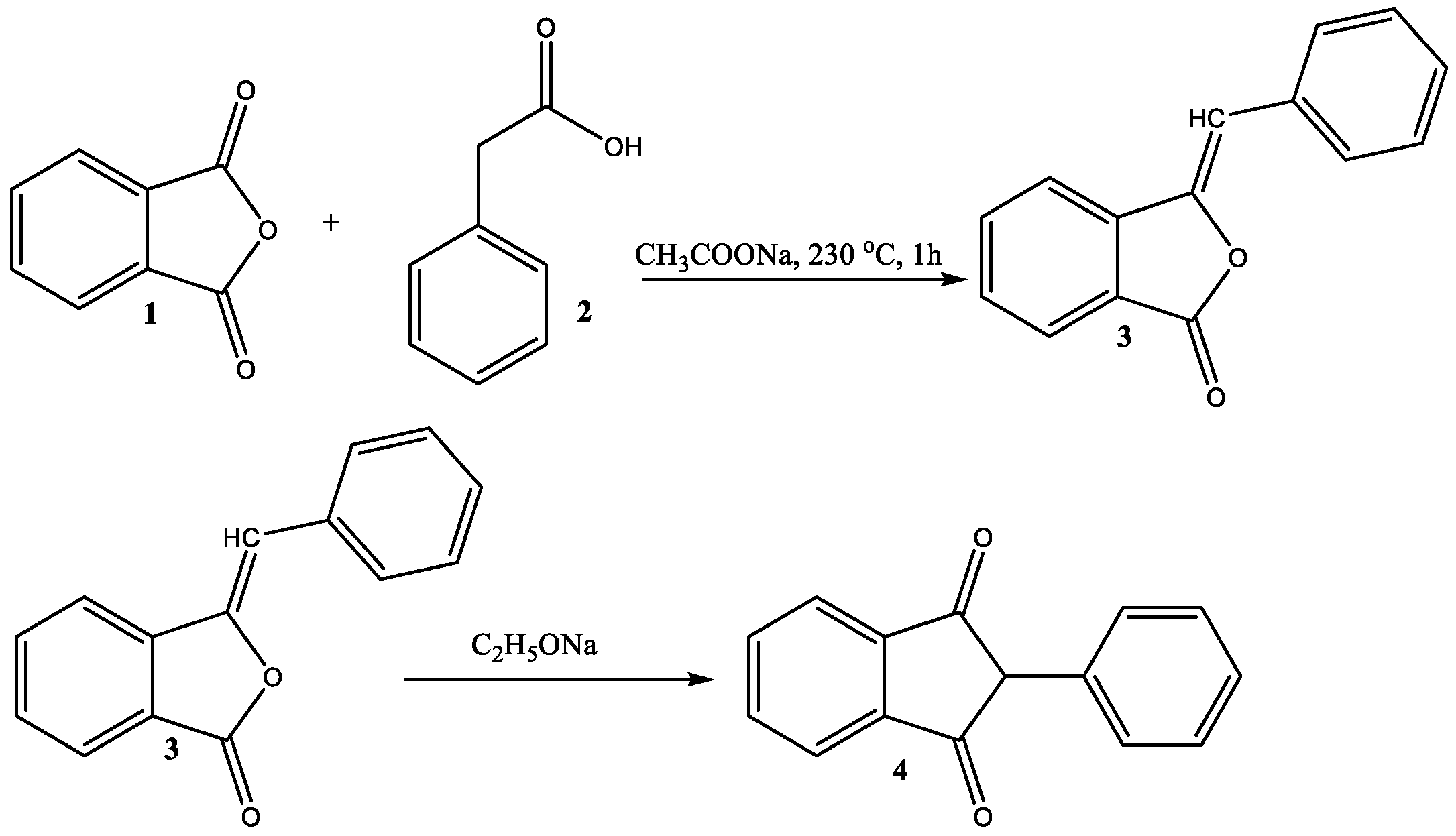
2.1.1. Synthesis of 2-Phenyl-1,3-Indandione
2.1.2. Synthesis of Galactose-Assisted PID-Loaded Ag NPs
2.2. Characterization of the Ag NPs. Analytical Techniques
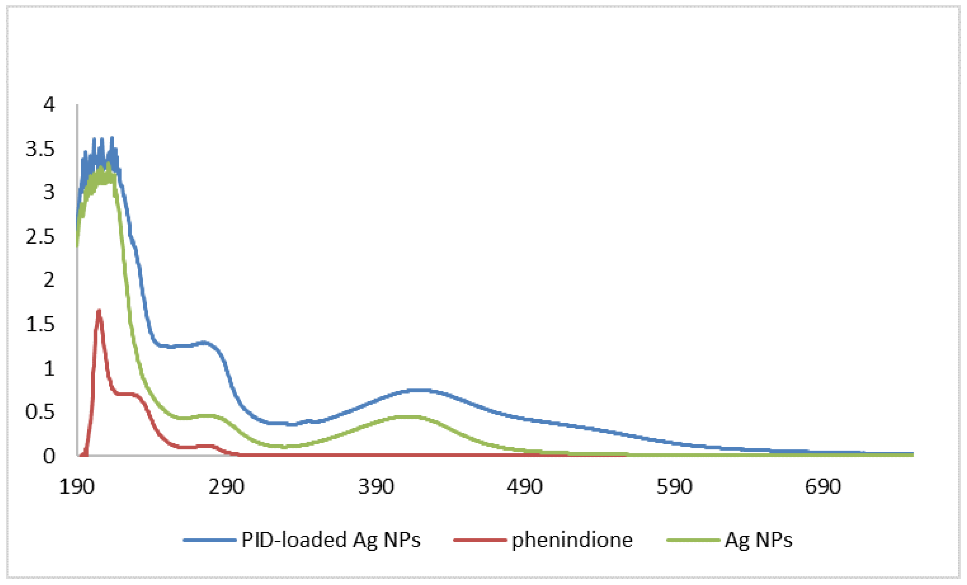
2.2.1. UV-Vis Spectra
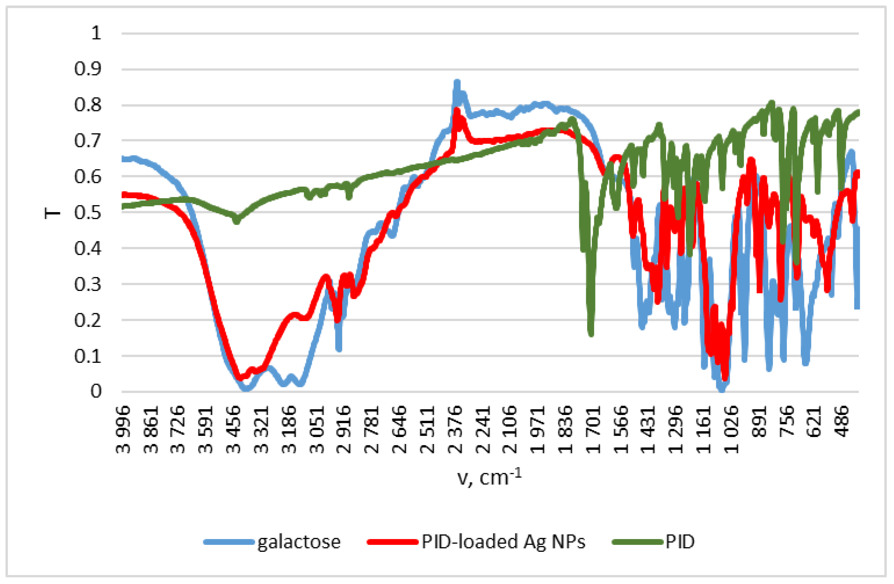
2.2.2. FTIR Spectra
2.2.3. spICP-MS
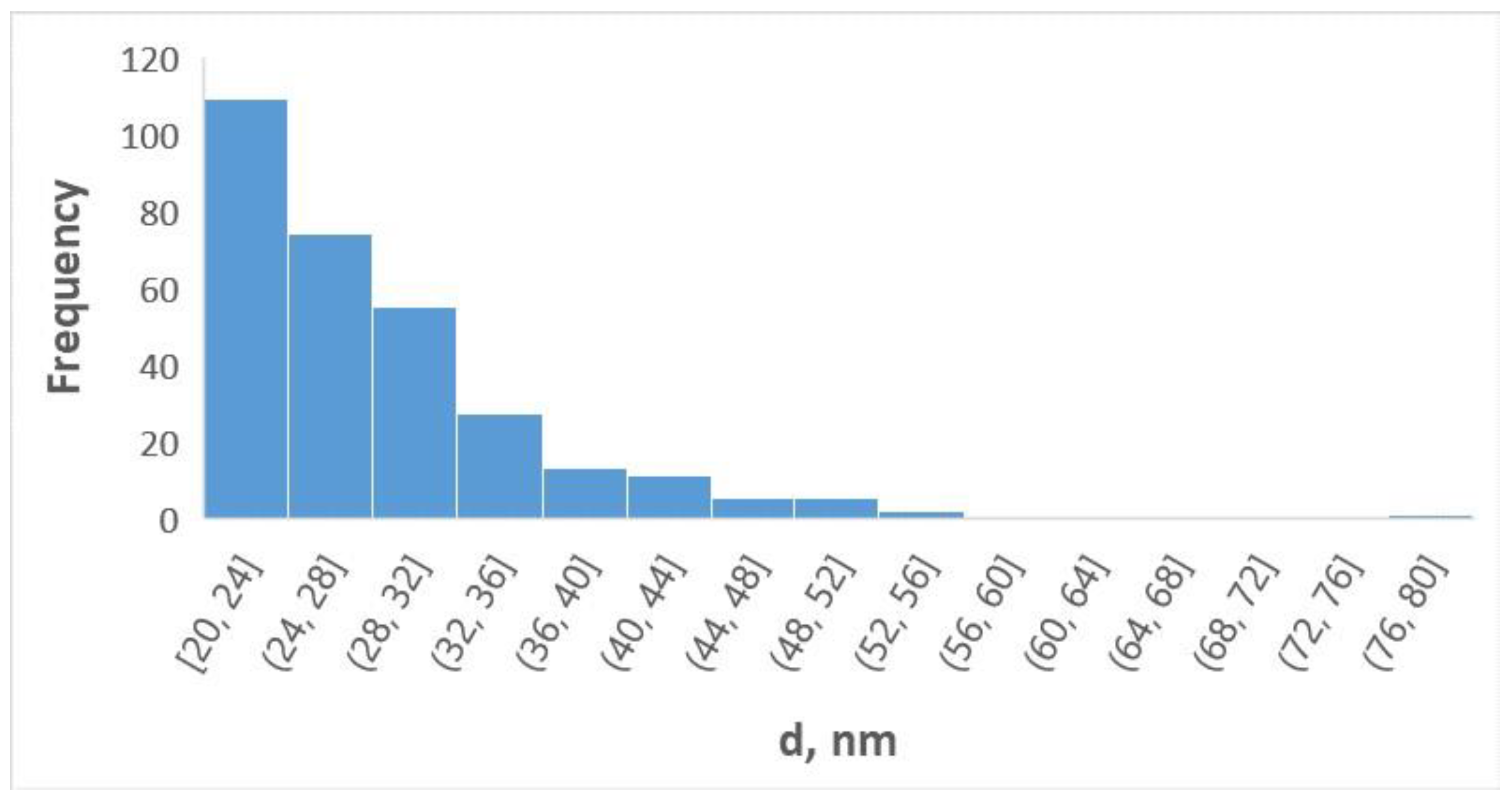
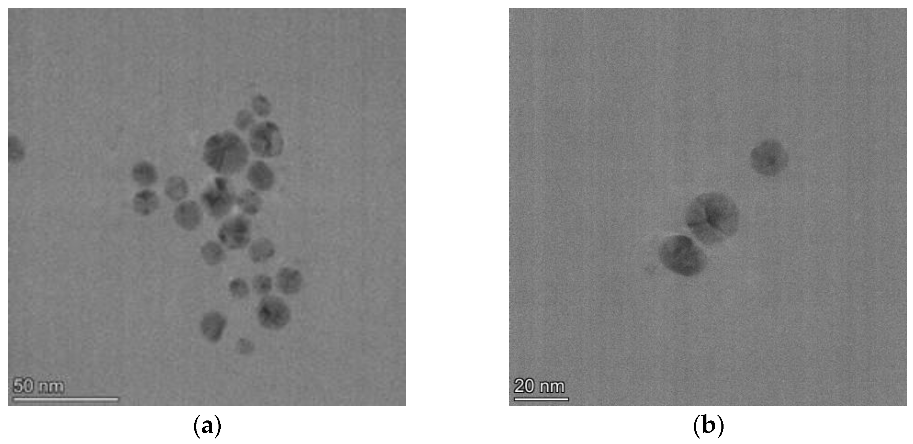
2.2.4. TEM
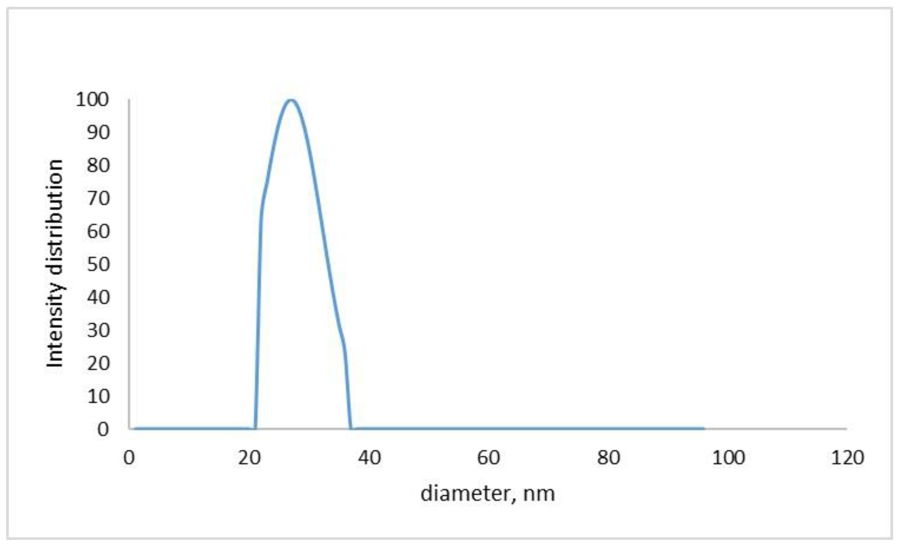
2.2.5. DLS and Zeta Potential
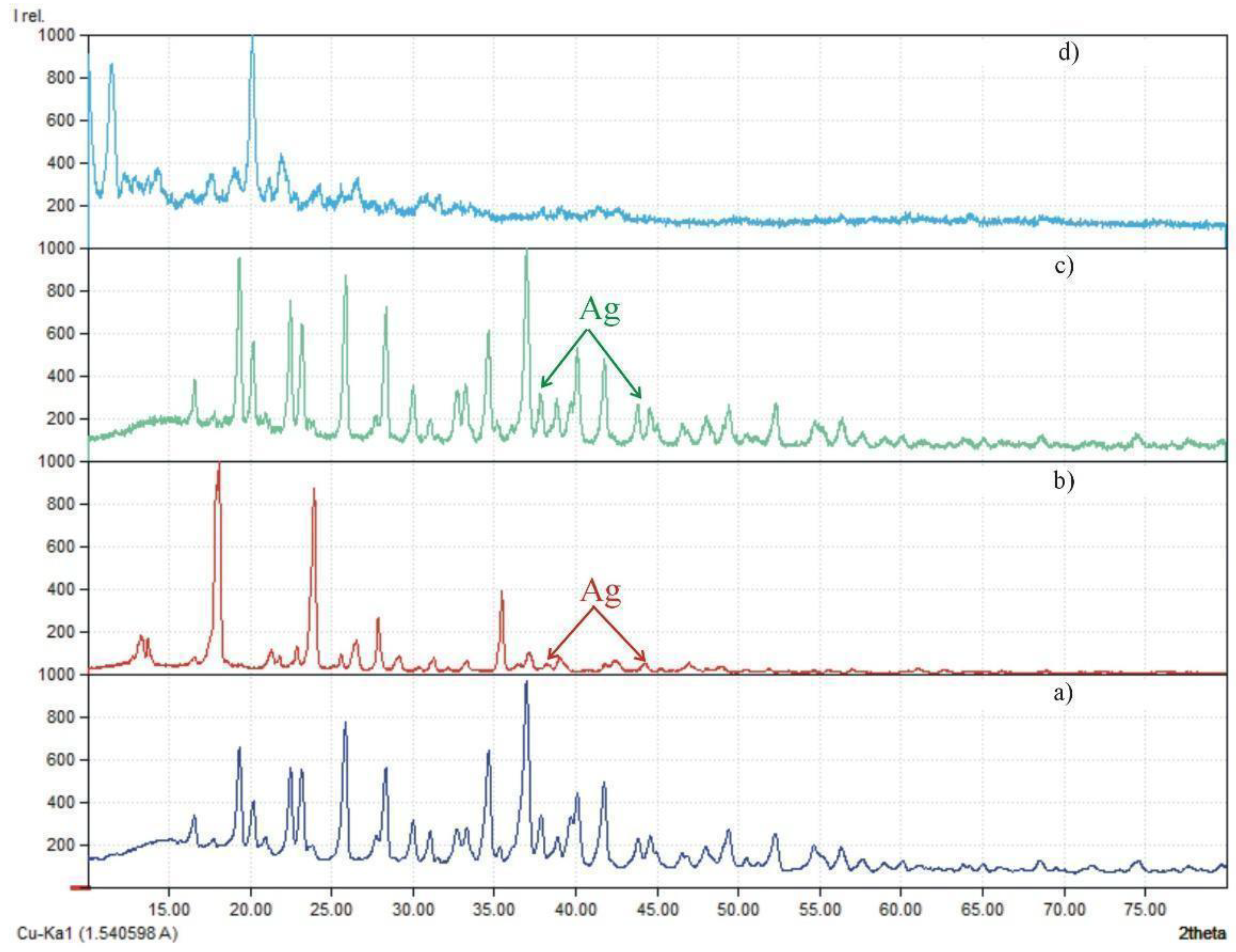
2.2.6. X-ray Diffraction (XRD)
2.3. In Vitro Drug Release
2.4. Computational Perspective
2.5. Microbiological Tests
2.6. Cytotoxic Activity
2.7. Anticoagulant Activity
2.8. Smooth Muscle Activity
2.8.1. Ex Vivo Experiments on Gastric Smooth Muscle Preparations (SMPs) from Rat Wistar
2.8.2. Method of Studying a Mechanical Activity of Isolated SM Preparation
2.9. Ethics Statement
2.10. Statistical Analysis
3. Results and Discussion
3.1. Physical Characterizations of Galactose-Assisted Drug-Loaded Ag NPs
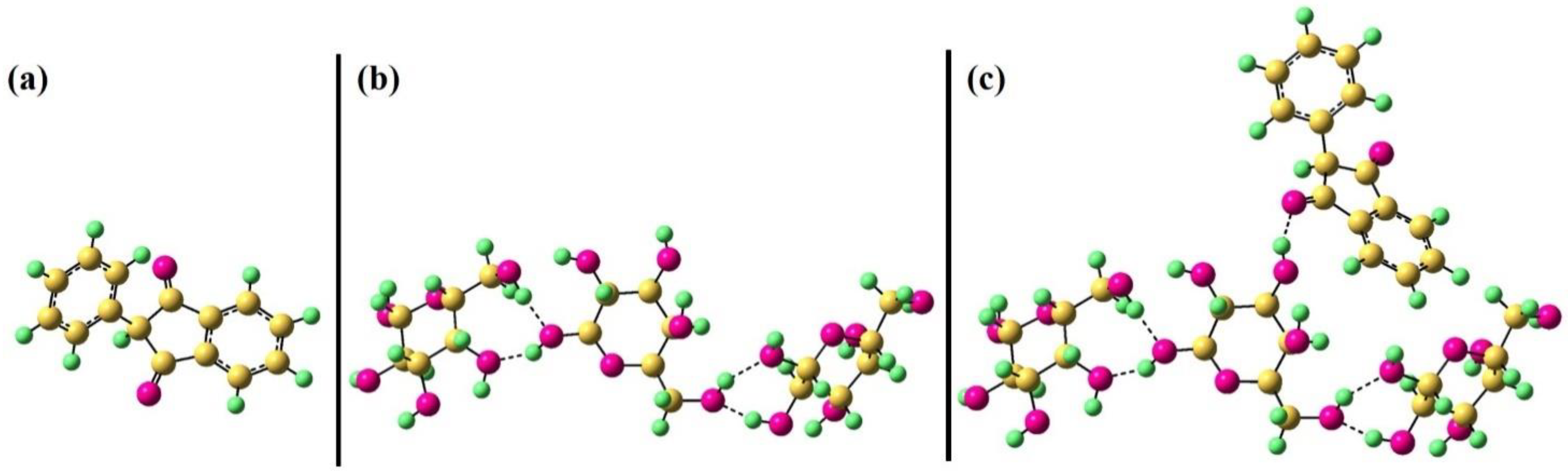
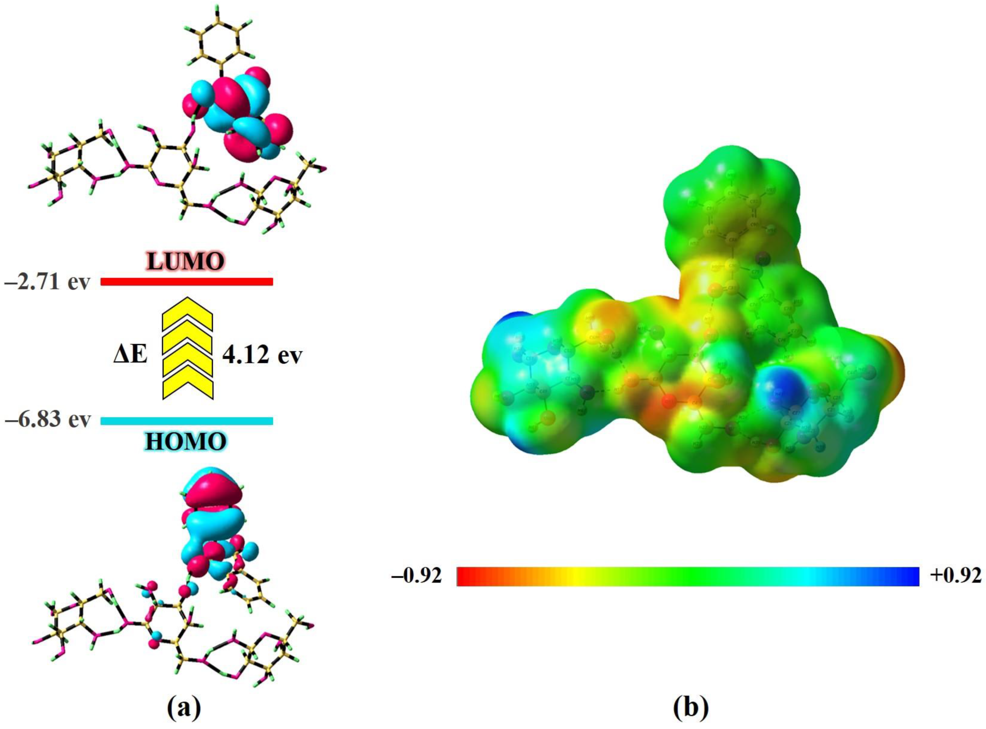
3.2. Phenindione Adsorption onto Galactose-Loaded Nanostructure by DFT
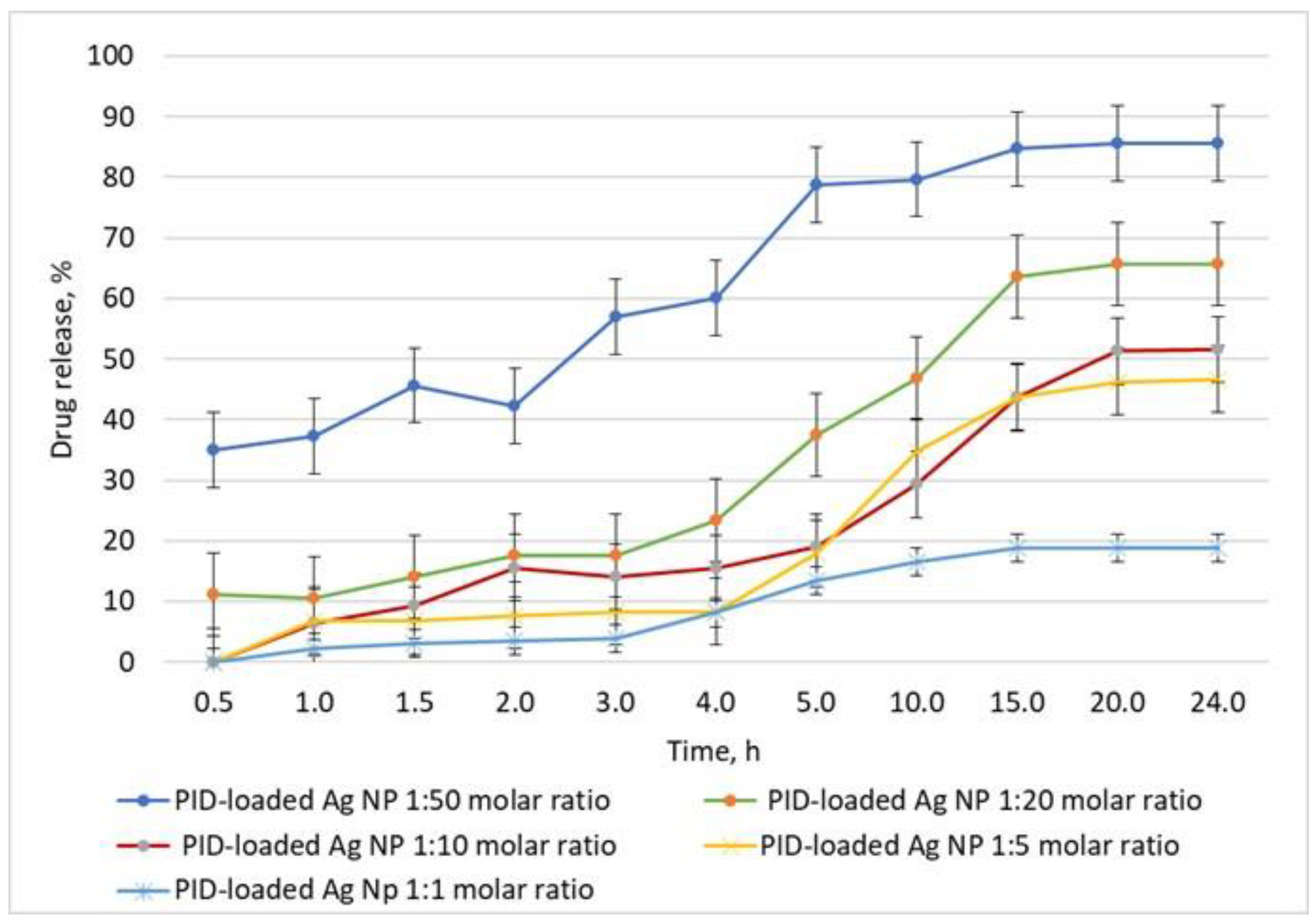
3.3. Parametric Drug Release Optimization
3.4. Antimicrobial Activity
3.5. Cytotoxic Activity
3.6. Anticoagulant Activity
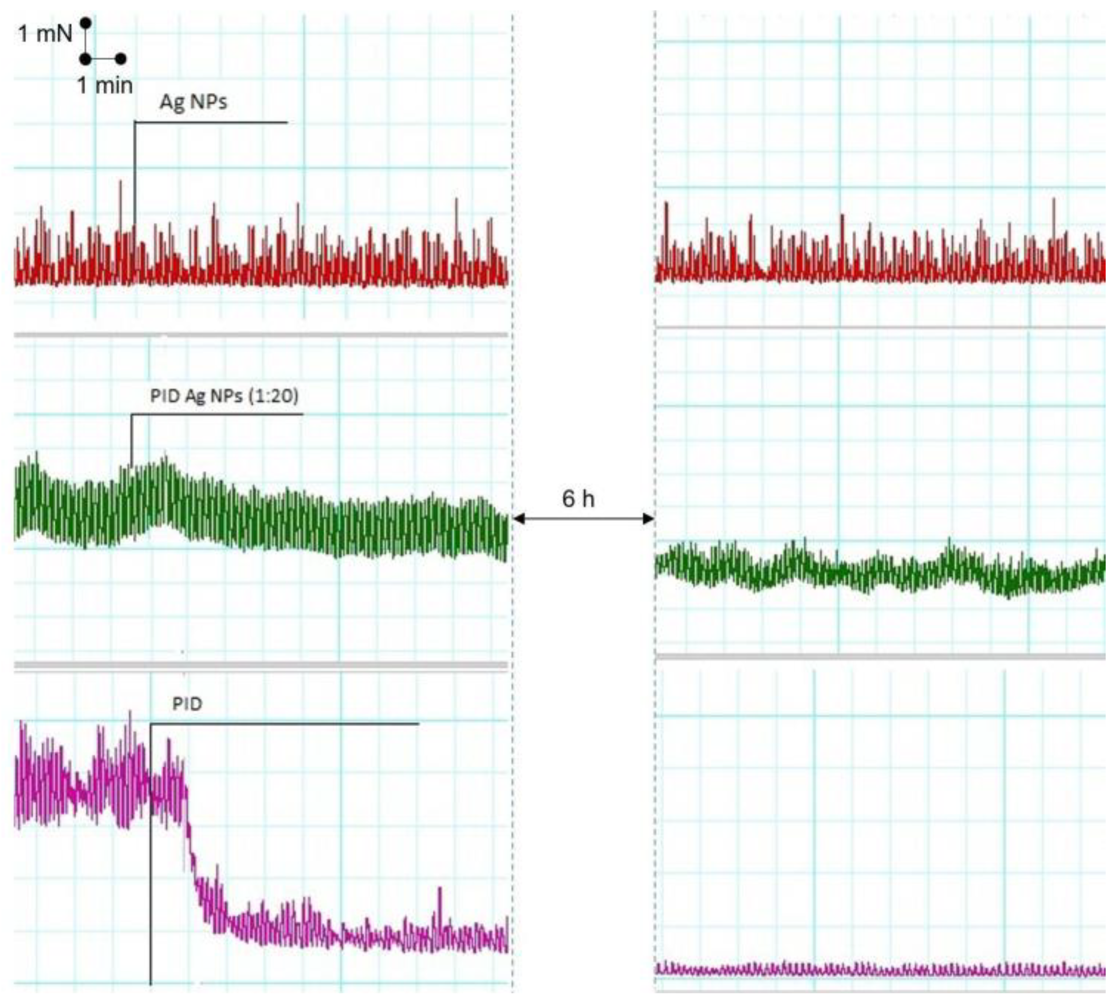
3.7. Ex Vivo Experiments on SM Activity
4. Conclusions
Supplementary Materials
Author Contributions
Funding
Institutional Review Board Statement
Informed Consent Statement
Data Availability Statement
Acknowledgments
Conflicts of Interest
References
- Vardanyan, R.; Hruby, V. Synthesis of Essential Drugs, 1st ed.; Elsevier: Amsterdam, The Netherlands, 2006; pp. 323–335. ISBN 9780080462127. [Google Scholar] [CrossRef]
- Kalishwaralal, K.; Deepak, V.; Pandian, S.B.R.K.; Kottaisamy, M.; Kanth, S.B.M.; Kartikeyan, B.; Gurunathan, S. Biosynthesis of Silver and Gold nanoparticles Using Brevibacterium casei. Colloids Surf. B 2010, 77, 257–262. [Google Scholar] [CrossRef] [PubMed]
- Shrivastava, S.; Bera, T.; Singh, S.K.; Singh, G.; Ramachandrarao, P.; Dash, D. Characterisation of antiplatelet properties of silver nanoparticles. ACS Nano 2009, 3, 1357–1364. [Google Scholar] [CrossRef] [PubMed]
- Kemp, M.M.; Kumar, A.; Mousa, S.; Park, T.J.; Ajayan, P.; Kubotera, N.; Mousa, S.A.; Linhardt, R.J. Synthesis of gold and silver nanoparticles stabilized with glycosaminoglycans having distinctive biological activities. Biomacromolecules 2009, 10, 589–595. [Google Scholar] [CrossRef] [PubMed]
- White, H.D. Oral antiplatelet therapy for atherothrombotic disease: Current evidence and new directions. Am. Heart J. 2011, 161, 450–461. [Google Scholar] [CrossRef]
- Latib, A.; Ielasi, A.; Ferri, L.; Chieffo, A.; Godino, C.; Carlino, M.; Montorfano, M.; Colombo, A. Aspirin intolerance and the need for dual antiplatelet therapy after stent implantation: A proposed alternative regimen. Int. J. Cardiol. 2013, 165, 444–447. [Google Scholar] [CrossRef]
- Reiter, R.A.; Jilma, B. Platelets and new antiplatelet drugs. Therapy 2005, 2, 465–502. [Google Scholar] [CrossRef]
- Ilas, J.; Jakopin, Z.; Borstnar, T.; Stegnar, M.; Kikelj, D. 3,4-Dihydro-2H-1,4-benzoxazine derivatives combining thrombin inhibitory and glycoprotein IIb/IIIa receptor antagonistic activity as a novel class of antithrombotic compounds with dual function. J. Med. Chem. 2008, 51, 5617–5629. [Google Scholar] [CrossRef]
- Chan, Y.C.; Valenti, D.; Mansfield, A.O.; Stansby, G. Warfarin induced skin necrosis. Br. J. Surg. 2000, 87, 266–272. [Google Scholar] [CrossRef]
- Liu, G.; Xu, J.; Chen, N.; Zhang, S.; Ding, Z.; Du, H. Synthesis of N6-alkyl(aryl)-2-alkyl(aryl) thioadenosines as antiplatelet agents. Eur. J. Med. Chem. 2012, 53, 114–123. [Google Scholar] [CrossRef]
- Griffin, J.P.; D’Arcy, P.F. A Manual of Adverse Drug Interactions, 5th ed.; Elsevier Science: Amsterdam, The Netherlands, 1997; pp. 111, 113–175, 177–231. ISBN 9780080525839. [Google Scholar]
- Pineo, G.F.; Hull, R.D. Chapter 43—Conventional Treatment of Deep Venous Thrombosis. In The Vein Book; Bergan, J.J., Ed.; Academic Press: Cambridge, MA, USA, 2007; pp. 381–394. [Google Scholar] [CrossRef]
- Merlob, P.; Habermann, J. Chapter 7—Anticoagulants, thrombocyte aggregation inhibitors and fibrinolytics. In Drugs During Pregnancy and Lactation, 3rd ed.; Schaefer, C., Peters, P., Miller, R.K., Eds.; Academic Press: Cambridge, MA, USA, 2015; pp. 725–729. [Google Scholar] [CrossRef]
- Pharmacology, B.J. Indanedione anticoagulants. In Me’ler’s Side Effects of Drugs, 16th ed.; Aronson, J.K., Ed.; Elsevier: Amsterdam, The Netherlands, 2016; pp. 50–51. [Google Scholar] [CrossRef]
- Vilums, M.; Heuberger, J.; Heitman, L.H.; Ijzerman, A.P. Indanes—Properties, Preparation, and Presence in Ligands for G Protein Coupled Receptors. Med. Res. Rev. 2015, 35, 1097–1126. [Google Scholar] [CrossRef]
- Mukhtar, A.; Shah, S.; Kanwal; Hameed, S.; Khan, K.M.; Khan, S.U.; Zaib, S.; Iqbal, J.; Parveen, S. Indane-1,3-diones: As Potential and Selective α-glucosidase Inhibitors, their Synthesis, in vitro and in silico Studies. Med. Chem. 2021, 17, 887–902. [Google Scholar] [CrossRef]
- Das, S. Annulations involving 2-arylidene-1,3-indanediones: Stereoselective synthesis of spiro- and fused scaffolds. New J. Chem. 2020, 44, 17148–17176. [Google Scholar] [CrossRef]
- Yellappa, S. An anti-Michael route for the synthesis of indole-spiro (indene-pyrrolidine) by 1,3-cycloaddition of azomethineylide with indole-derivatised ofelins. J. Heterocycl. Chem. 2020, 57, 1083–1089. [Google Scholar] [CrossRef]
- Abolhasani, H.; Zarghi, A.; Movahhed, T.K.; Abolhasani, A.; Daraei, B.; Dastmalchi, S. Design, synthesis and biological evaluation of novel indanone containing spiroisoxazoline derivativea with selective COX-2 inhibition as anticancer agents. Bioorg. Med. Chem. 2021, 32, 115960. [Google Scholar] [CrossRef]
- Ahmed, N. Synthetic advances in the indane natural products scaffolds as drug candidates: A review. Stud. Nat. Prod. Chem. 2016, 51, 383–434. [Google Scholar] [CrossRef]
- Prasher, P.; Sharma, M. Medicinal Chemistry of Indane and Its Analogues: A Mini Review. ChemistrySelect 2021, 6, 2658–2677. [Google Scholar] [CrossRef]
- Lee, J.Y.; Spicer, A.P. Hyaluronan: A multifunctionynthesizelton, stealth molecule. Curr. Opin. Cell Biol. 2000, 12, 581–586. [Google Scholar] [CrossRef]
- Raja, S.; Ramesh, V.; Thivaharan, V. Antibacterial and anticoagulant activity of silver nanoparticynthesizedised from a novel source–pods of Peltophorum pterocarpum. J. Ind. Eng. Chem. 2015, 29, 257–264. [Google Scholar] [CrossRef]
- Roy, K.; Srivastwa, A.; Ghosh, C. Anticoagulant, thrombolytic and antibacterial activities of Euphorbia acruensis latex-mediated bioengineered silver nanoparticles. Green Process. Synth. 2019, 8, 590–599. [Google Scholar] [CrossRef]
- Dakshayani, S.S.; Marulasiddeshwara, M.B.; Kumar, M.N.S.; Ramesh, G.; Kumar, P.R.; Devaraja, S.; Hosamani, R. Antimicrobial, anticoagulant and antiplatelet activities of green synthesized silver nanoparticles using Selaginella (Sanjeevini) plant extract. Int. J. Biol. Macromol. 2019, 131, 787–797. [Google Scholar] [CrossRef]
- Talank, N.; Morad, H.; Barabadi, B.; Mojab, F.; Amidi, S.; Kobarfard, F.; Mahjoub, M.A.; Jounaki, K.; Mohammadi, N.; Salehi, G.; et al. Bioengineering of green-synthesized silver nanoparticles: In vitro physicochemical, antibacterial, biofilm inhibitory, anticoagulant, and antioxidant performance. Talanta 2022, 243, 123374. [Google Scholar] [CrossRef] [PubMed]
- Asghar, M.A.; Yousuf, R.I.; Shoaib, M.H.; Asghar, M.A. Antibacterial, anticoagulant and cytotoxic evaluation of biocompatible nanocomposite of chitosan loaded green synthesized bioinspired silver nanoparticles. Int. J. Biol. Macromol. 2020, 160, 934–943. [Google Scholar] [CrossRef] [PubMed]
- Namasivayam, S.K.R.; Srinivasan, S.; Samrat, K.; Priyalakshmi, B.; Kumar, R.D.; Bharani, A.; Kumar, R.G.; Kavisri, M.; Moovendhan, M. Sustainable approach to manage the vulnerable rodents using eco-friendly green rodenticides formulation through nanotechnology principles—A review. Process Saf. Environ. Prot. 2023, 171, 591–606. [Google Scholar] [CrossRef]
- Douglass, M.; Garren, M.; Devine, R.; Mondal, A. Hitesh Handa, Bio-inspired hemocompatible surface modifications for biomedical applications. Prog. Mater. Sci. 2022, 130, 100997. [Google Scholar] [CrossRef]
- Barabadi, H.; Noqani, H.; Ashouri, F.; Prasad, A.; Jounaki, K.; Mobaraki, K.; Mohanta, Y.K.; Mostafavi, E. Nanobiotechnological approaches in anticoagulant therapy: The role of bioengineered silver and gold nanomaterials. Talanta 2023, 256, 124279. [Google Scholar] [CrossRef]
- Chardoli, A.; Karimi, N.; Fattahi, A. Biosynthesis, characterization, antimicrobial and cytotoxic effects of silver nanoparticles using Nigellaarvensis seed extract. Iran. J. Pharm. Res. 2017, 16, 1167–1175. [Google Scholar]
- Amin, Z.; Khashyarmanesh, Z.; Bazzaz, B.; Noghabi, Z. Does biosynthetic silver nanoparticles are more stable with lower toxicity than their synthetic counterparts? Iran. J. Pharm. Res. 2019, 18, 210–221. [Google Scholar]
- Salari, S.; Bahabadi, S.; Samzadeh-Kermani, A.; Yosefzaei, F. In-vitro evaluation of antioxidant and antibacterial potential of greensynthesized silver nanoparticles using prosopis farcta fruit extract. Iran. J. Pharm. Res. 2019, 18, 430–455. [Google Scholar]
- Yaqoob, A.A.; Ahmad, H.; Parveen, T.; Ahmad, A.; Oves, M.; Ismail, I.M.I.; Qari, H.A.; Umar, K.; Ibrahim, M.N.M. Recent advances in metal decorated nanomaterials and their various biological applications: A review. Front. Chem. 2020, 8, 341. [Google Scholar] [CrossRef]
- Barabadi, H.; Kobarfard, F.; Vahidi, H. Biosynthesis and Characterization of Biogenic Tellurium Nanoparticles by Using Penicillium chrysogenum PTCC 5031: A Novel Approach in Gold Biotechnology. Iran. J. Pharm. Res. 2018, 17, 87–97. [Google Scholar]
- Barabadi, H.; Honary, S.; Ebrahimi, P.; Alizadeh, A.; Naghibi, F.; Saravanan, M. Optimization of myco-synthesized silver nanoparticles by response surface methodology employing Box-Behnken design. Inorg. Nano Met. Chem. 2019, 49, 33–43. [Google Scholar] [CrossRef]
- De Matteis, V.; Rizzello, L.; Cascione, M.; Liatsi-Douvitsa, E.; Apriceno, A.; Rinaldi, R. Green Plasmonic Nanoparticles and Bio-Inspired Stimuli-Responsive Vesicles in Cancer Therapy Application. Nanomaterials 2020, 10, 1083. [Google Scholar] [CrossRef]
- Sharma, V.K.; Yngard, R.A.; Lin, Y. Silver nanoparticles: Green synthesis and their antimicrobial activities. Adv. Colloid. Interface Sci. 2009, 145, 83–96. [Google Scholar] [CrossRef] [PubMed]
- Panacek, A.; Kvítek, L.; Prucek, R.; Kolar, M.; Vecerova, R.; Pizúrova, N.; Sharma, V.; Nevecna, T.; Zboril, R. Silver colloid nanoparticles: Synthesis, characterization, and their antibacterial activity. J. Phys. Chem. B 2006, 110, 16248–16253. [Google Scholar] [CrossRef] [PubMed]
- Sabbagh, F.; Kiarostami, K.; Khatir, N.; Rezania, S.; Muhamad, I.; Hosseini, F. Effect of zinc content on structural, functional, morphological, and thermal properties of kappa-carrageenan/NaCMC nanocomposites. Polym. Test. 2021, 93, 106922. [Google Scholar] [CrossRef]
- Todorova, M.; Milusheva, M.; Kaynarova, L.; Georgieva, D.; Delchev, V.; Simeonova, S.; Pilicheva, B.; Nikolova, S. Drug-Loaded Silver Nanoparticles—A Tool for Delivery of a Mebeverine Precursor in Inflammatory Bowel Diseases Treatment. Biomedicines 2023, 11, 1593. [Google Scholar] [CrossRef]
- Kamble, S.; Bhosale, K.; Mohite, M.; Navale, S. Methods of Preparation of Nanoparticles. Int. Adv. Res. Sci. Commun. Technol. 2022, 2, 2581–9429. [Google Scholar] [CrossRef]
- Filippo, E.; Manno, D.; Serra, A. Self assembly and branching of sucrose stabilized silver nanoparticles by microwave assisted synthesis: From nanoparticles to branched nanowires structures. Colloids Surf. A Physicochem. Eng. Asp. 2009, 348, 205–211. [Google Scholar] [CrossRef]
- Filippo, E.; Serra, A.; Buccolieri, A.; Manno, D. Green synthesis of silver nanoparticles with sucrose and maltose: Morphological and structural characterization. J. Non-Cryst. Solids 2010, 356, 344–350. [Google Scholar] [CrossRef]
- Shervani, Z.; Yamamoto, Y. Carbohydrate-directed synthesis of silver and gold nanoparticles: Effect of the structure of carbohydrates and reducing agents on the size and morphology of the composites. Carbohydr. Res. 2011, 346, 651–658. [Google Scholar] [CrossRef]
- Ghiyasiyan-Arani, M.; Salavati-Niasari, M.; Masjedi-Arani, M.; Mazloom, F. An Easy Sonochemical Route for Synthesis, Characterization and Photocatalytic Performance of Nanosized FeVO 4 in the Presence of Aminoacids as green Capping Agents. J. Mater. Sci. Mater. Elect. 2018, 29, 474–485. [Google Scholar] [CrossRef]
- Caschera, D.; Toro, R.G.; Federici, F.; Montanari, R.; de Caro, T.; Al-Shemy, M.T.; Adel, A.M. Green Approach for the Fabrication of Silver-Oxidized Cellulose Nano-composite with Antibacterial Properties. Cellulose 2020, 27, 8059–8073. [Google Scholar] [CrossRef]
- Garza-Cervantes, J.A.; Mendiola-Garza, G.; de Melo, E.M.; Dugmore, T.I.; Matharu, A.S.; Morones-Ramirez, J.R. Antimicrobial Activity of a Silver-Microfibrillated Cellulose Biocomposite against Susceptible and Resistant Bacteria. Sci. Rep. 2020, 10, 7281. [Google Scholar] [CrossRef]
- Rather, R.A.; Sarwara, R.K.; Das, N.; Pal, B. Impact of Reducing and Capping Agents on Carbohydrates for the Growth of Ag and Cu Nanostructures and Their Antibacterial Activities. Particuology 2019, 43, 219–226. [Google Scholar] [CrossRef]
- Shanmuganathan, R.; Edison, T.N.J.I.; LewisOscar, F.; Kumar, P.; Shanmugam, S.; Pugazhendhi, A. Chitosan Nanopolymers: An Overview of Drug Delivery against Cancer. Int. J. Biol. Macromol. 2019, 130, 727–736. [Google Scholar] [CrossRef]
- Maiti, P.K.; Ghosh, A.; Parveen, R.; Saha, A.; Choudhury, M.G. Preparation of Carboxy-Methyl Cellulose-Capped Nanosilver Particles and Their Antimicrobial Evaluation by an Automated Device. Appl. Nanosci. 2019, 9, 105–111. [Google Scholar] [CrossRef]
- Duan, H.; Wang, D.; Li, Y. Green chemistry for nanoparticle synthesis. Chem. Soc. Rev. 2015, 44, 5778–5792. [Google Scholar] [CrossRef]
- Zeng, Q.; Shao, D.; Ji, W.; Li, J.; Chen, L.; Song, J.T. The nanotoxicity investigation of optical nanoparticles to cultured cells in vitro. Toxicol. Rep. 2014, 2, 137–144. [Google Scholar] [CrossRef]
- Dieckmann, W. Zur kenntnis der homo thalsaur. Chem. Ber. 1914, 47, 1439. [Google Scholar] [CrossRef]
- Pace, H.; Rogers, N.; Jarolimek, C.; Coleman, V.; Higgins, C.; Ranville, J. Determining Transport Efficiency for the Purpose of Counting and Sizing Nanoparticles via Single Particle Inductively Coupled Plasma Mass Spectrometry. Anal. Chem. 2011, 83, 9361–9369. [Google Scholar] [CrossRef]
- Frisch, M.; Trucks, G.; Schlegel, H.B.; Scuseria, G.E.; Robb, M.A.; Cheeseman, J.R.; Scalmani, G.; Barone, V.; Mennucci, B.; Petersson, G. Gaussian 09, Revision d. 01; Gaussian, Inc.: Wallingford, CT, USA, 2009; p. 201. [Google Scholar]
- Hadi, Z.; Milad, N.; Asal, Y.-S.; Hamedreza, J.; Saber, M.C.; Seyedeh, S.H. A DFT study on the therapeutic potential of carbon nanostructures as sensors and drug delivery carriers for curcumin molecule: NBO and QTAIM analyses. Colloids Surf. A Physicochem. Eng. Asp. 2022, 651, 129698. [Google Scholar] [CrossRef]
- Tumbarski, Y.; Deseva, I.; Mihaylova, D.; Stoyanova, M.; Krastev, L.; Nikolova, R.; Yanakieva, V.; Ivanov, I. Isolation, Characterization and Amino Acid Composition of a Bacteriocin Produced by Bacillus methylotrophicus Strain BM47. Food Technol. Biotechnol. 2018, 56, 546–552. [Google Scholar] [CrossRef]
- Clauss, A. Rapid physiological coagulation method in determination of fibrinogen. Acta Haematol. 1957, 17, 237–246. [Google Scholar] [CrossRef] [PubMed]
- Kennedy, D.; Orts-Gil, G.; Lai, C.-H.; Müller, L.; Haase, A.; Luch, A.; Seeberger, P. Carbohydrate functionalization of silver nanoparticles modulates cytotoxicity and cellular uptake. J. Nanobiotechnol. 2014, 12, 59. [Google Scholar] [CrossRef]
- Pryshchepa, O.; Pomastowski, P.; Buszewski, B. Silver nanoparticles: Synthesis, investigation techniques, and properties, Advances in Colloid and Interface Science. Adv. Colloid Interface Sci. 2020, 284, 102246. [Google Scholar] [CrossRef]
- Asif, M.; Yasmin, R.; Asif, R.; Ambreen, A.; Mustafa, M.; Umbreen, S. Green Synthesis of Silver Nanoparticles (AgNPs), Structural Characterization, and their Antibacterial Potential. Dose-Response 2022, 20, 15593258221088709. [Google Scholar] [CrossRef]
- De Leersnyder, I.; De Gelder, L.; Van Driessche, I.; Vermeir, P. Revealing the Importance of Aging, Environment, Size and Stabilization Mechanisms on the Stability of Metal Nanoparticles: A Case Study for Silver Nanoparticles in a Minimally Defined and Complex Undefined Bacterial Growth Medium. Nanomaterials 2019, 9, 1684. [Google Scholar] [CrossRef]
- Du, P.; Xu, Y.; Shi, Y.; Xu, Q.; Li, S.; Gao, M. Preparation and shape change of silver nanoparticles (AgNPs) loaded on the dialdehyde cellulose by in-situ synthesis method. Cellulose 2022, 29, 6831–6843. [Google Scholar] [CrossRef]
- Xavier, J.; Vincent, S.; Meder, F.; Vollmer, F. Advances in Optoplasmonic Sensors—Combining Optical Nano/Microcavities and Photonic Crystals with Plasmonic Nanostructures and Nanoparticles. Nanophotonics 2018, 7, 51807062. [Google Scholar] [CrossRef]
- Martinsson, E.; Otte, M.A.; Shahjamali, M.M.; Sepulveda, B.; Aili, D. Substrate Effect on the Refractive Index Sensitivity of Silver Nanoparticles. J. Phys. Chem. C 2014, 118, 24680–24687. [Google Scholar] [CrossRef]
- Arcas, A.S.; Jaramillo, L.; Costa, N.S.; Allil, R.C.S.B.; Werneck, M.M. Localized Surface Plasmon Resonance-Based Biosensor on Gold Nanoparticles for Taenia Solium Detection. Appl. Opt. 2021, 60, 8137–8144. [Google Scholar] [CrossRef] [PubMed]
- Raiche-Marcoux, G.; Loiseau, A.; Maranda, C.; Poliquin, A.; Boisselier, E. Parametric Drug Release Optimization of Anti-Inflammatory Drugs by Gold Nanoparticles for Topically Applied Ocular Therapy. Int. J. Mol. Sci. 2022, 23, 16191. [Google Scholar] [CrossRef] [PubMed]
- Zaheer, Z. Biogenic synthesis, optical, catalytic, and in vitro antimicrobial potential of Ag-nanoparticles prepared using Palm date fruit extract. J. Photochem. Photobiol. B Biol. 2018, 178, 584–592. [Google Scholar] [CrossRef] [PubMed]
- Jiang, P.; Zhou, J.J.; Li, R.; Gao, Y.; Sun, T.L.; Zhao, X.W.; Xiang, Y.J.; Xie, S.S. PVP-capped twinned gold plates from nanometer to micrometer. J. Nanoparticle Res. 2006, 8, 927. [Google Scholar] [CrossRef]
- Ezraa, L.; O’Della, Z.J.; Huia, J.; Riley, K.R. Emerging investigator series: Quantifying silver nanoparticle aggregation kinetics in realtime using particle impact voltammetry coupled with UV-vis spectroscopy. Environ. Sci. Nano. 2020, 7, 2509–2521. [Google Scholar] [CrossRef]
- AbuDalo, M.; Al-Mheidat, I.; Al-Shurafat, A.; Grinham, C.; Oyanedel-Craver, V. Synthesis of silver nanoparticles using a modified Tollens’ method in conjunction with phytochemicals and assessment of their antimicrobial activity. Peer J. 2019, 7, e641. [Google Scholar] [CrossRef]
- Ahmed, K.B.A.; Mohammed, A.S.; Anbazhagan, V. Interaction of sugar stabilized silver nanoparticles with the T-antigen specific lectin, jacalin from Artocarpus Integrifolia. Spectrochim. Acta Part A Mol. Biomol. Spectrosc. 2015, 145, 110–116. [Google Scholar] [CrossRef]
- Wiercigroch, E.; Szafraniec, E.; Czamara, K.; Pacia, M.Z.; Majzner, K.; Kochan, K.; Kaczor, A.; Baranska, M.; Malek, K. Raman and infrared spectroscopy of carbohydrates: A review. Spectrochim. Acta A Mol. Biomol. Spectrosc. 2017, 185, 317–335. [Google Scholar] [CrossRef]
- Wells, H.; Atalla, R. An investigation of the vibrational spectra of glucose, galactose, and mannose. J. Mol. Struct. 1990, 224, 385–424. [Google Scholar] [CrossRef]
- Sivakesava, S.; Irudayaraj, J. Prediction of inverted cane sugar adulteration of honey by Fourier transform infrared spectroscopy. J. Food Sci. 2001, 66, 972–978. [Google Scholar] [CrossRef]
- David, M.; Hategan, A.; Berghian-Grosan, C.; Magdas, D. The Development of Honey Recognition Models Based on the Association between ATR-IR Spectroscopy and Advanced Statistical Tools. Int. J. Mol. Sci. 2022, 23, 9977. [Google Scholar] [CrossRef]
- Iravani, S.; Korbekandi, H.; Mirmohammadi, S.; Zolfaghari, B. Synthesis of silver nanoparticles: Chemical, physical and biological methods. Res. Pharm. Sci. 2014, 9, 385–406. [Google Scholar]
- Islam, N. Green synthesis and biological activities of gold nanoparticles functionalized with Salix alba. Arab. J. Chem. 2019, 12, 2914–2925. [Google Scholar] [CrossRef]
- Nanomaterials in Plants, Algae, and Microorganisms: Concepts and Controversies, 1st ed.; Tripathi, D., Ahmad, P., Sharma, S., Chauhan, D., Dubey, N.K., Eds.; Academic Press: Cambridge, MA, USA, 2018. [Google Scholar]
- Younas, M.; Ahmad, M.A.; Jannat, F.T.; Ashfaq, T.; Ahmad, A. Chapter 18—Role of silver nanoparticles in multifunctional drug delivery. In Micro and Nano Technologies, Nanomedicine Manufacturing and Applications; Verpoort, F., Ahmad, I., Ahmad, A., Khan, A., Chee, C.Y., Eds.; Elsevier: Orlando, FL, USA, 2021; pp. 297–319. [Google Scholar] [CrossRef]
- Ivask, A. Toxicity mechanisms in Escherichia coli vary for silver nanoparticles and differ from ionic silver. ACS Nano 2014, 8, 374–386. [Google Scholar] [CrossRef]
- Stoehr, L.; Gonzalez, E.; Stampfl, A.; Casals, E.; Duschl, A.; Puntes, V.; Oostingh, G. Shape matters: Effects of silver nanospheres and wires on human alveolar epithelial cells. Part. Fibre Toxicol. 2011, 8, 36. [Google Scholar] [CrossRef]
- Huk, A.; Izak-Nau, E.; Reidy, B.; Boyles, M.; Duschl, A.; Lynch, I.; Dušinska, M. Is the toxic potential of nanosilver dependent on its size? Part. Fibre Toxicol. 2014, 11, 65. [Google Scholar] [CrossRef]
- Zhang, T.; Wang, L.; Chen, Q.; Chen, C. Cytotoxic potential of silver nanoparticles. Yonsei Med. J. 2014, 55, 283–291. [Google Scholar] [CrossRef]
- León-Silva, S.; Fernández-Luqueño, F.; López-Valdez, F. Silver Nanoparticles (AgNP) in the Environment: A Review of Potential Risks on Human and Environmental Health. Water Air Soil Pollut. 2016, 227, 306. [Google Scholar] [CrossRef]
- Sigalov, M.V. Keto–Enol Tautomerism of Phenindione and Its Derivatives: An NMR and Density Functional Theory (DFT) Reinvestigation. J. Phys. Chem. A 2015, 119, 1404–1414. [Google Scholar] [CrossRef]
- Mulvaney, P.; Liz-Marzan, L.; Giersig, M.; Ung, T.J. Silica encapsulation of quantum dots and metal clusters. Mater. Chem. 2000, 10, 1259–1270. [Google Scholar] [CrossRef]
- Philip, D. Biosynthesis of Au, Ag and Au-Ag nanoparticles using edible mushroom extract. Spectrochim. Acta A Mol. Biomol. Spectrosc. 2009, 73, 374–381. [Google Scholar] [CrossRef] [PubMed]
- Feizi-Dehnayebi, M.; Dehghanian, E.; Mansouri-Torshizi, H. A novel palladium (II) antitumor agent: Synthesis, characterization, DFT perspective, CT-DNA and BSA interaction studies via in-vitro and in-silico approaches. Spectrochim. Acta A Mol. Biomol. Spectrosc. 2021, 249, 119215. [Google Scholar] [CrossRef] [PubMed]
- Feizi-Dehnayebi, M.; Dehghanian, E.; Mansouri-Torshizi, H. Synthesis and characterization of Pd (II) antitumor complex, DFT calculation and DNA/BSA binding insight through the combined experimental and theoretical aspects. J. Mol. Struct. 2021, 1240, 130535. [Google Scholar] [CrossRef]
- Mantasha, I.; Shahid, M.; Kumar, M.; Ansari, A.; Akhtar, M.N.; AlDamen, M.A.; Song, Y.; Ahmad, M.; Khan, I.M. Exploring solvent dependent catecholase activity in transition metal complexes: An experimental and theoretical approach. New J. Chem. 2020, 44, 1371–1388. [Google Scholar] [CrossRef]
- Feizi-Dehnayebi, M.; Dehghanian, E.; Mansouri-Torshizi, H. Probing the biomolecular (DNA/BSA) interaction by new Pd (II) complex via in-depth experimental and computational perspectives: Synthesis, characterization, cytotoxicity, and DFT approach. J. Iran. Chem. Soc. 2022, 19, 3155–3175. [Google Scholar] [CrossRef]
- Feizi-Dehnayebi, M.; Dehghanian, E.; Mansouri-Torshizi, H. Biological activity of bis-(morpholineacetato) palladium (II) complex: Preparation, structural elucidation, cytotoxicity, DNA-/serum albumin-interaction, density functional theory, in-silico prediction and molecular modeling. Spectrochim. Acta A Mol. Biomol. Spectrosc. 2022, 281, 121543. [Google Scholar] [CrossRef]
- Feizi-Dehnayebi, M.; Dehghanian, E.; Mansouri-Torshizi, H. DNA/BSA binding affinity studies of new Pd (II) complex with SS and NN donor mixed ligands via experimental insight and molecular simulation: Preliminary antitumor activity, lipophilicity and DFT perspective. J. Mol. Liq. 2021, 344, 117853. [Google Scholar] [CrossRef]
- Séguy, L.; Groo, A.-C.; Malzert-Fréon, A. How nano-engineered delivery systems can help marketed and repurposed drugs in Alzheimer’s disease treatment? Drug Discov. Today 2022, 27, 1575–1589. [Google Scholar] [CrossRef]
- Prakash, P.; Gnanaprakasam, P.; Emmanuel, R.; Arokiyaraj, M.; Saravanan, M. Green synthesis of silver nanoparticles from leaf extract of Mimusops elengi, Linn. for enhanced antibacterial activity against multi drug resistant clinical isolates. Colloids Surf. B Biointerfaces 2013, 108, 255–259. [Google Scholar] [CrossRef]
- Solomon, D.; Gupta, N.; Mulla, N.S.; Shukla, S.; Guerrero, Y.A.; Gupta, V. Role of In Vitro Release Methods in Liposomal Formulation Development: Challenges and Regulatory Perspective. AAPSJ 2017, 19, 1669–1681. [Google Scholar] [CrossRef]
- Yu, M.; Yuan, W.; Li, D.; Schwendeman, A.; Schwendeman, S.P. Predicting Drug Release Kinetics from Nanocarriers inside Dialysis Bags. J. Control. Release 2019, 315, 23–30. [Google Scholar] [CrossRef]
- Kumar, B.; Jalodia, K.; Kumar, P.; Gautam, H.K. Recent Advances in Nanoparticle-Mediated Drug Delivery. J. Drug Deliv. Sci. Technol. 2017, 41, 260–268. [Google Scholar] [CrossRef]
- Sabbagh, F.; Khatir, N.M.; Kiarostami, K. Synthesis and Characterization of 𝒌-Carrageenan/PVA Nanocomposite Hydrogels in Combination with MgZnO Nanoparticles to Evaluate the Catechin Release. Polymers 2023, 15, 272. [Google Scholar] [CrossRef]
- Jain, A.S.; Pawar, P.S.; Sarkar, A.; Junnuthula, V.; Dyawanapelly, S. Bionanofactories for Green Synthesis of Silver Nanoparti-cles: Toward Antimicrobial Applications. Int. J. Mol. Sci. 2021, 22, 11993. [Google Scholar] [CrossRef]
- Guzman, M.; Arcos, M.; Dille, J.; Godet, S.; Rousse, C. Effect of the Concentration of NaBH4 and N2H4 as Reductant Agent on the Synthesis of Copper Oxide Nanoparticles and its Potential Antimicrobial Applications. Nano Biomed. Eng. 2018, 10, 392–405. [Google Scholar] [CrossRef]
- Narayanan, S.; Gupta, P.; Nazim, U.; Ali, M.; Karadkhelkar, N.; Ahmad, M.; Chen, Z.-S. Anti-cancer effect of Indanone-based thiazolyl hydrazone derivative on colon cancer cell lines. Int. J. Biochem. Cell Biol. 2019, 110, 21–28. [Google Scholar] [CrossRef]
- Chanda, D.; Bhushan, S.; Guru, S.K.; Shanker, K.; Wani, Z.A.; Rah, B.A.; Luqman, S.; Mondhe, D.M.; Pal, A.; Negi, A.S. Anticancer activity, toxicity and pharmacokinetic profile of an indanone derivative. Eur. J. Pharm. Sci. 2012, 47, 988–995. [Google Scholar] [CrossRef]
- Balasubramaniam, M.; Lakkaniga, N.R.; Dera, A.A.; Fayi, M.A.; Abohashrh, M.; Ahmad, I.; Chandramoorthy, H.C.; Nalini, G.; Rajagopalan, P. FCX-146, a potent allosteric inhibitor of Akt kinase in cancer cells: Lead optimization of the second-generation arylidene indanone scaffold. Biotechnol. Appl. Biochem. 2021, 68, 82–91. [Google Scholar] [CrossRef]
- Cohen, A.T.; Agnelli, G.; Anderson, F.A.; Arcelus, J.I.; Bergqvist, D.; Brecht, J.G. Venousthromboembolism (VTE) in Europe. The number of VTE events and associated morbidityand mortality. Thromb. Haemost. 2007, 98, 756–764. [Google Scholar] [PubMed]
- Heitt, J.A. The epidemiology of venous thromboembolism in the community. Arterioscler. Thromb. Vasc. Biol. 2008, 28, 370–372. [Google Scholar] [CrossRef]
- Biss, T.T.; Brandão, L.R.; Kahr, W.H.; Chan, A.K.; Williams, S. Clinical features andoutcome of pulmonary embolism in children. Brit. J. Haematol. 2008, 142, 808–818. [Google Scholar] [CrossRef] [PubMed]
- Ede, O.A.; Bindá, F.A.; Silva, A.M.; Costa, T.D.; Fernandes, M.C.; Fernandes, M.C. Risk factors andprophylaxis for venous thromboembolism in hospitals in the city of Manaus. J. Bras. Pneumol. 2009, 35, 114–121. [Google Scholar] [CrossRef]
- Santos Fernandes, C.J.S.; AlvesJúnior, J.L.; Gavilanes, F.; Prada, L.F.; Morinaga, L.K.; Rogerio, S. New anticoagulants for the treatment of venous thromboembolism. J. Bras. Pneumol. 2016, 42, 146–154. [Google Scholar] [CrossRef] [PubMed]
- Azeez, M.A.; Durodola, F.A.; Lateef, A.; Yekeen, T.A.; Adubi, A.O.; Oladipo, I.C.; Adebayo, E.A.; Badmus, J.A.; Abawulem, A.O. Green synthesized novel silver nanoparticles and their application as anticoagulant and thrombolytic agents: A perspective. IOP Conf. Ser. Mater. Sci. Eng. 2020, 805, 012043. [Google Scholar] [CrossRef]
- Quinn, A.; Bellamy, M. Chapter 53—Hemostasis and coagulation. In Foundations of Anesthesia, 2nd ed.; Hemmings, H.C., Hopkins, P.M., Eds.; Mosby: Maryland Heights, MO, USA, 2006; pp. 635–645. [Google Scholar] [CrossRef]
- Xiang, J.; Wang, H.; Ma, C.; Zhou, M.; Wu, Y.; Wang, L.; Guo, S.; Chen, T.; Shaw, C. Ex Vivo Smooth Muscle Pharmacological Effects of a Novel Bradykinin-Related Peptide, and Its Analogue, from Chinese Large Odorous Frog, Odorrana livida Skin Secretions. Toxins 2016, 8, 283. [Google Scholar] [CrossRef]
- Wright, D.; Sharma, P.; Ryu, M.H.; Rissé, P.A.; Ngo, M.; Maarsingh, H.; Koziol-White, C.; Jha, A.; Halayko, A.J.; West, A.R. Models to study airway smooth muscle contraction in vivo, ex vivo and in vitro: Implications in understanding asthma. Pulm. Pharmacol. Ther. 2013, 26, 24–36. [Google Scholar] [CrossRef]
- Liu, S.; Lin, Z. Vascular Smooth Muscle Cells Mechanosensitive Regulators and Vascular Remodeling. J. Vasc. Res. 2022, 59, 90–113. [Google Scholar] [CrossRef]
- Hashitani, H.; Brading, A.F.; Suzuki, H. Correlation between spontaneous electrical, calcium and mechanical activity in detrusor smooth muscle of the guinea-pig bladder. Br. J. Pharmacol. 2004, 141, 183–193. [Google Scholar] [CrossRef]
- Durnin, L.; Kwok, B.; Kukadia, P.; McAvera, R.; Corrigan, R.D.; Ward, S.M.; Zhang, Y.; Chen, Q.; Koh, S.D.; Sanders, K.M.; et al. An ex vivo bladder model with detrusor smooth muscle removed to analyze biologically active mediators released from the suburothelium. J. Physiol. 2019, 597, 1467–1485. [Google Scholar] [CrossRef]
- Tarhan, F.; Duman, N.Ç.; Özkulab, S.; Karaalp, A.; Cangüven, Ö. In vitro contractile responses of human detrusor smooth muscle to oxytocin: Does it really have effect? Aging Male 2020, 23, 1141–1145. [Google Scholar] [CrossRef]
- Upchurch, W.J.; Iaizzo, P.A. In vitro contractile studies within isolated tissue baths: Translational research from Visible Heart® Laboratories. Exp. Biol. Med. 2022, 247, 584–597. [Google Scholar] [CrossRef]
- Brozovich, F.V.; Nicholson, C.J.; Degen, C.V.; Gao, Y.Z.; Aggarwal, M.; Morgan, K.G. Mechanisms of Vascular Smooth Muscle Contraction. Pharmacol. Rev. 2016, 68, 476–532. [Google Scholar] [CrossRef]
- Jespersen, B.; Tykocki, N.R.; Watts, S.W.; Cobbett, P.J. Measurement of smooth muscle function in the isolated tissue bathapplications to pharmacology research. J. Vis. Exp. 2015, 95, 52324. [Google Scholar] [CrossRef]
- Ivanov, I.; Nikolova, S.; Aladjov, D.; Stefanova, I.; Zagorchev, P. Synthesis and Contractile Activity of Substituted 1,2,3,4-Tetrahydroisoquinolines. Molecules 2011, 16, 7019–7042. [Google Scholar] [CrossRef]
- Stefanova, I.; Argirova, M.; Krustev, A. Influence of model melanoidins on calciumdependent transport mechanisms in smooth muscle tissue. Mol. Nutr. Food Res. 2007, 51, 468–472. [Google Scholar] [CrossRef]
- Argirova, M.; Stefanova, I.; Krustev, A. New biological properties of coffee melanoidins. Food Funct. 2013, 4, 1204–1208. [Google Scholar] [CrossRef]
- Sagorchev, P.; Lukanov, J.; Beer, A.-M. Effects of 1.8-cineole (eucalyptol) on the spontaneous contractile activity of smooth muscles fibre. J. Med. Plants Res. 2015, 9, 486–493. [Google Scholar] [CrossRef]
- Beer, A.M.; Lukanov, J.; Sagorchev, P. Effect of Thymol on the spontaneous contractile activity of the smooth muscles. Phytomedicine 2007, 14, 65–69. [Google Scholar] [CrossRef]
- González, C.; Salazar-García, S.; Palestino, G.; Martínez-Cuevas, P.P.; Ramírez-Lee, M.A.; Jurado-Manzano, B.B.; Rosas-Hernández, H.; Gaytán-Pacheco, N.; Martel, G.; Espinosa-Tanguma, R.; et al. Effect of 45 nm silver nanoparticles (AgNPs) upon the smooth muscle of rat trachea: Role of nitric oxide. Toxicol. Lett. 2011, 207, 306–313. [Google Scholar] [CrossRef]
- Sheridan, H.; Lemon, S.; Frankish, N.; McArdle, P.; Higgins, T.; James, J.P.; Bhandari, P. Synthesis and antispasmodic activity of nature identical substituted indanes and analogues. Eur. J. Med. Chem. 1990, 25, 603–608. [Google Scholar] [CrossRef]


| Parameters | PID | Ag NPs | PID-Loaded Ag NPs |
|---|---|---|---|
| Eelec (eV) | −19,838.000 | −56,172.470 | −76,010.908 |
| Eads (eV) | - | - | −0.438 |
| EHOMO (eV) | −6.86 | −7.16 | −6.83 |
| ELUMO (eV) | −2.73 | −0.02 | −2.71 |
| ΔEgap (eV) | 4.13 | 7.14 | 4.12 |
| η | 2.06 | 3.57 | 2.06 |
| σ | 0.48 | 0.28 | 0.48 |
| χ | 4.79 | 3.59 | 4.77 |
| Pi | −4.79 | −3.59 | −4.77 |
| ω | 5.57 | 1.80 | 5.52 |
| Inhibition Zones, mm | ||
|---|---|---|
| Tested Microorganism | PID | PID-Loaded Ag NPs |
| Bacillus subtilis ATCC 6633 | 17 | 12 |
| Bacillus amyloliquefaciens 4BCL-YT | 25 | 13 |
| Staphylococcus aureus ATCC 25923 | 27 | 27 |
| Listeria monocytogenes NBIMCC 8632 | 22 | 15 |
| Enterococcus faecalis ATCC 29212 | 23 | 17 |
| Micrococcus luteus 2YC-YT | 30 | 20 |
| Salmonella enteritidis ATCC 13076 | 21 | 15 |
| Salmonella typhimurium NBIMCC 1672 | 25 | 13 |
| Klebsiella pneumoniae ATCC 13883 | 11 | - |
| Escherichia coli ATCC 25922 | 18 | 13 |
| Proteus vulgaris ATCC 6380 | 14 | 10 |
| Pseudomonas aeruginosa ATCC 9027 | 18 | 12 |
| Candida albicans NBIMCC 74 | 11 | - |
| Saccharomyces cerevisiae ATCC 9763 | 9 | - |
| Aspergillus niger ATCC 1015 | 11 | 13 |
| Aspergillus flavus | 9 | - |
| Penicillium chrysogenum | 13 | 10 |
| Rhizopus sp. | 11 | 8 |
| Fusarium moniliforme ATCC 38932 | 11 | 14 |
| Mucor sp. | 11 | - |
| Compound/Cell Line | CCL-1 | LAMA-84 | K-562 | T-24 |
|---|---|---|---|---|
| PID-loaded Ag NPs | 65.4 ± 8.3 | 25.3 ± 1.2 | 27 ± 3.2 | 53.2 ± 4.7 |
| Ag NPs | >200 | >300 | >300 | >300 |
| PID | >300 | >200 | >200 | >300 |
| PID-Loaded Ag NPs (Molar Ratio) | PT, s | APTT, s | Fibrinogen, g/L |
|---|---|---|---|
| 1:1 | 14.1 | 35 | 0.91 |
| 1:5 | 13.9 | 34.9 | 1.34 |
| 1:10 | 13.7 | 35.3 | 1.31 |
| 1:20 | 14.1 | 35.7 | 1.25 |
| 1:50 | no coagulation | 43.1 | 1.31 |
| reference | 8–10.8 | 28.2–31.4 | 1.91–2.87 |
| Compound/Parameter | Tonic Activity (mN) | Strength of The Contractile Reaction (mN) | Frequency (Number of Contractions/min) |
|---|---|---|---|
| SCA (initial reaction) | 1.81 ± 0.19 | 3.40 ± 0.19 | 4.97 ± 0.04 |
| Ag NPs | 1.69 ± 0.03 | 3.15 ± 0.16 | 4.93 ± 0.03 |
| SCA (initial reaction) | 1.89 ± 0.12 | 3.00 ± 0.18 | 4.80 ± 0.09 |
| PID-loaded Ag NPs (1:1) | 1.80 ± 0.07 | 2.88 ± 0.05 | 4.90 ± 0.07 |
| SCA (initial reaction) | 2.03 ± 0.15 | 3.64 ± 0.21 | 5.04 ± 0.05 |
| PID-loaded Ag NPs (1:5) | 2.07 ± 0.22 | 2.14 ± 0.11 | 5.09 ± 0.17 |
| SCA (initial reaction) | 1.99 ± 0.11 | 3.64 ± 0.21 | 5.03 ± 0.03 |
| PID-loaded Ag NPs (1:10) | 0.77 ± 0.09 * | 2.14 ± 0.11 * | 4.87 ± 0.11 |
| SCA (initial reaction) | 1.94 ± 0.07 | 2.44 ± 0.13 | 5.07 ± 0.10 |
| PID-loaded Ag NPs (1:20) | 0.91 ± 0.08 * | 0.98 ± 0.07 * | 4.90 ± 0.09 |
| SCA (initial reaction) | 1.67 ± 0.14 | 3.50 ± 1.21 | 4.88 ± 0.06 |
| PID-loaded Ag NPs (1:50) | 0.80 ± 0.14 * | 1.73 ± 0.16 * | 4.83 ± 0.08 |
| SCA (initial reaction) | 1.96 ± 0.09 | 2.42 ± 0.22 | 4.90 ± 0.04 |
| PID | 0.44 ± 0.07 * | 0.13 ± 0.03 * | 4.87 ± 0.03 |
Disclaimer/Publisher’s Note: The statements, opinions and data contained in all publications are solely those of the individual author(s) and contributor(s) and not of MDPI and/or the editor(s). MDPI and/or the editor(s) disclaim responsibility for any injury to people or property resulting from any ideas, methods, instructions or products referred to in the content. |
© 2023 by the authors. Licensee MDPI, Basel, Switzerland. This article is an open access article distributed under the terms and conditions of the Creative Commons Attribution (CC BY) license (https://creativecommons.org/licenses/by/4.0/).
Share and Cite
Nikolova, S.; Milusheva, M.; Gledacheva, V.; Feizi-Dehnayebi, M.; Kaynarova, L.; Georgieva, D.; Delchev, V.; Stefanova, I.; Tumbarski, Y.; Mihaylova, R.; et al. Drug-Delivery Silver Nanoparticles: A New Perspective for Phenindione as an Anticoagulant. Biomedicines 2023, 11, 2201. https://doi.org/10.3390/biomedicines11082201
Nikolova S, Milusheva M, Gledacheva V, Feizi-Dehnayebi M, Kaynarova L, Georgieva D, Delchev V, Stefanova I, Tumbarski Y, Mihaylova R, et al. Drug-Delivery Silver Nanoparticles: A New Perspective for Phenindione as an Anticoagulant. Biomedicines. 2023; 11(8):2201. https://doi.org/10.3390/biomedicines11082201
Chicago/Turabian StyleNikolova, Stoyanka, Miglena Milusheva, Vera Gledacheva, Mehran Feizi-Dehnayebi, Lidia Kaynarova, Deyana Georgieva, Vassil Delchev, Iliyana Stefanova, Yulian Tumbarski, Rositsa Mihaylova, and et al. 2023. "Drug-Delivery Silver Nanoparticles: A New Perspective for Phenindione as an Anticoagulant" Biomedicines 11, no. 8: 2201. https://doi.org/10.3390/biomedicines11082201
APA StyleNikolova, S., Milusheva, M., Gledacheva, V., Feizi-Dehnayebi, M., Kaynarova, L., Georgieva, D., Delchev, V., Stefanova, I., Tumbarski, Y., Mihaylova, R., Cherneva, E., Stoencheva, S., & Todorova, M. (2023). Drug-Delivery Silver Nanoparticles: A New Perspective for Phenindione as an Anticoagulant. Biomedicines, 11(8), 2201. https://doi.org/10.3390/biomedicines11082201