Abstract
The superior cerebellar artery (SCA) arises from the distal part of the basilar artery and passes by the oculomotor, trochlear, and trigeminal nerves. SCA is known to play a crucial role in the development of trigeminal neuralgia. However, due to its anatomical variability, it may also trigger other neurovascular compression (NVC), including hemifacial spasm, oculomotor nerve palsy, and ocular neuromyotonia. Additionally, it may be associated with ischemic syndromes and aneurysm development, highlighting its clinical significance. The most common anatomical variations of the SCA include duplication, a single vessel origin from the posterior cerebral artery (PCA), and a common trunk with PCA. Rarely observed variants include bifurcation and origin from the internal carotid artery. Certain anatomical variants such as early bifurcation and caudal course of duplicated SCA trunk may increase the risk of NVC. In this narrative review, we aimed to examine the impact of the anatomical variations of SCA on the NVCs based on papers published in Pubmed, Scopus, and Web of Science databases with a snowballing approach. Our review emphasizes the importance of a thorough understanding of the anatomical variability of SCA to optimize the management of patients with NVCs associated with this artery.
1. Introduction
1.1. Background on the Superior Cerebellar Artery
The superior cerebellar artery (SCA) is a branch of the distal part of the basilar artery (BA), passing in a posterolateral direction. During this course, the following segments are distinguished: anterior pontomesencephalic, lateral pontomesencephalic, cerebellomesencephalic, and cortical (see Figure 1). SCA provides blood supply for the pons, cerebral peduncle, the upper part of the cerebellum, and cerebellar peduncles [1,2].
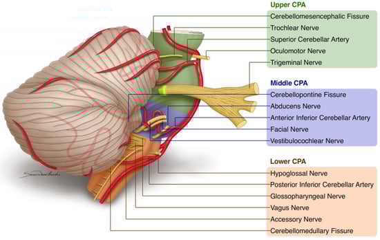
Figure 1.
Summary of basic knowledge about superior cerebellar artery. Legend: CPA—cerebellopontine angle.
1.2. Neurovascular Compression Syndromes
Neurovascular compression (NVC) syndromes are a group of disorders with a complex etiology. Numerous mechanisms have been proposed in their pathophysiology, but none have provided a sufficient explanation. The most likely chain of events involves the mechanical compression on the cranial nerve root entry/exit zone (REZ) by a pulsating vessel, leading to demyelination, disturbances in ion channels, and hyperexcitability of the nucleus [3,4,5,6,7,8,9]. Nevertheless, the mere presence of direct contact between these structures is insufficient for the diagnosis of NVC. The abnormal course of a vessel may also play a role in the development of these syndromes [10].
1.3. Importance of Understanding Anatomical Variations of the SCA
The cerebellopontine angle (CPA) can be divided into three parts: upper, middle, and lower (Figure 1) [11,12,13]. The upper CPA contains SCA and the cranial nerves: oculomotor, trochlear, and trigeminal. The middle CPA contains the anterior inferior cerebellar artery (AICA) and the cranial nerves: abducens, facial, and vestibulocochlear [14]. The lower CPA contains the posterior inferior cerebellar artery (PICA) and the cranial nerves: glossopharyngeal, vagus, accessory, and hypoglossal.
SCA, located in the upper CPA, is associated with trigeminal neuralgia (TN) and oculomotor compression syndromes such as oculomotor nerve palsy (ONP) and ocular neuromyotonia (NMT) [15,16,17,18,19]. However, it has also been described in conflict with the REZ of facial nerve leading to hemifacial spasm (HFS) [20]. These diverse clinical manifestations of NVC syndromes triggered by SCA highlight the importance of its anatomical variability [21].
SCA may be also associated with strokes and aneurysms [22,23]. SCA strokes are more frequently observed among patients with hypertension, cardiopathies, arrhythmias, hyperlipidemia, and diabetes, and those who smoke or use oral contraceptives [24]. Typical symptoms include nausea, vomiting, dizziness, tinnitus, headache, ataxia, and dysarthria [25]. SCA aneurysms account for less than 2% of intracranial aneurysms [26]. In a series of 36 SCA aneurysms treated by coiling, 65% presented with subarachnoid hemorrhage and 14% of patients had multiple intracranial aneurysms [26]. Interestingly, there were no episodes of rebleeding during the median clinical follow-up of 44.5 months (range 4–103 months, 118 patient-years) [26]. The only two cases of SCA aneurysm treated at our departments of neurosurgery in the last 10 years are provided in the Supplementary Material.
2. Anatomy of the Superior Cerebellar Artery
SCA typically originates as a single vessel from the distal part of BA [27,28,29]. After ~18.5 mm from the origin, near its maximal caudal descent, SCA bifurcates into two major trunks of similar diamaters: rostral and caudal [30]. The rostral trunk terminates by giving hemispheric, vermian, and perforating arteries. The caudal trunk provides blood supply through hemispheric, marginal, and perforating arteries.
Four segments can be distinguished on the SCA course: S1: anterior pontomesenphalic, S2: lateral pontomesenphalic, S2: cerebellomesoencephalic, and S4: cortical ones [31]. The anterior pontomesenphalic is the first SCA part passing inferior to the oculomotor nerve [28]. The lateral pontomesenphalic segment begins at the anterolateral margin of the brainstem and frequently dips caudally onto the lateral side of the upper pons [32]. Its caudal loop projects forward and may reach the oculomotor nerve at its REZ [30]. The trochlear nerve is frequently observed in the midportion of this segment. This segment passes into the cerebellomesencephalic segment at the anterior margin of the cerebellomesencephalic fissure [30]. In this segment, SCA branches enter the shallowest part of the fissure located posterosuperior to the REZ of the trigeminal nerve and course medial to the tentorial edge with its branches intertwined with the trochlear nerve [32]. The last, cortical segment, comprises branches located distally to the cerebellomesencephalic groove, which supply the cortex of the vermis and cerebellar hemisphere [30]. SCA provides blood supply for the pons, cerebral peduncle, the upper part of the cerebellum, and cerebellar peduncles [1,2,28].
3. Neurovascular Compression Syndromes Related to the Superior Cerebellar Artery
3.1. Trigeminal Neuralgia
TN patients may experience even 10–50 pain attacks per day [33,34,35]. A single attack may be described as electric-like, sharp, severe, and superficial pain. It can be accompanied by unintentional spasms of the facial muscles [36]. Therefore, TN significantly impacts a patient’s quality of life and may lead to psychological distress, anxiety, depression, and even suicide attempts [37,38,39,40]. Its prevalence is difficult to estimate and ranges from 0.03% to 0.3% [41,42,43,44]. It is higher in women over 40 years of age [44].
NVC represents the dominant cause of TN [45]. In the 1930s, Walter Dandy noted that the sensory roots of the trigeminal nerve are usually touched and/or modeled by an artery [46]. Several vessels may be responsible for NVC leading to TN (Table 1), primarily SCA (up to 90%) and anterior inferior cerebellar artery (AICA, 7–23.6%). The influence of their anatomical variants on the risk, prevalence, and severity of NVC is poorly understood. It was reported that tortuous and elongated SCA can increase the risk of TN [47]. In one MRI-based study duplicated SCAs were involved in TN [48]. The authors suggested that if one of the trunks takes a caudal course, duplication or early bifurcation of SCA may increase the risk of TN [48]. Rarely, TN is due to NVC with other vessels (including veins, e.g., superior petrosal venous complex [49] and arteries transfixing the sensory root of trigeminal nerve [50]) and/or arteriovenous malformation (AVM) arising from a dilated SCA, AICA, or posterior meningeal artery with venous drainage to the basal vein of Rosenthal, then posing a diagnostic challenge [51,52,53]. In these cases, SCA can act either as a compressing vessel or as a vessel supplying the AVM associated with TN [54,55,56].
Differential diagnosis of TN should encompass the following conditions: trauma [36], viral infections (e.g., SARS-CoV-2, Herpes Zoster) [57,58], multiple sclerosis [59], and intracranial malignancy [60]. In such cases, facial pain may be accompanied by other neurological symptoms as an effect of underlying pathology [61].
3.2. Oculomotor Compression Syndromes
The oculomotor nerve originates from the midbrain and passes laterally between the posterior cerebral artery (PCA) and SCA [62]. Both arteries provide their blood supply in the cisternal (initial) parts [63]. In further subchapters, we presented available information about NVCs among patients with oculomotor nerve palsy and ocular neuromyotonia. As only single cases of successful MVD in these patients were reported up to date, it is questionable if these entities are related to NVC.
3.2.1. Oculomotor Nerve Palsy
ONP may be due to NVC of the oculomotor nerve by SCA, mainly characterized by diplopia and ipsilateral ptosis [15,16,17,64,65]. Differential, and more common, diagnoses encompass aneurysms (especially those located in the BA tip, BA-SCA junction, posterior communicating artery, and cavernous segment of internal carotid artery), diabetes mellitus, trauma, brain tumors, infarction, and central nervous system infections [17,27]. A case of NVC of the oculomotor nerve between the atherosclerotic PCA and SCA was described [16]. The resolution of symptoms after microvascular decompression (MVD) indicates that in this case, NVC has triggered ONP. It is worth noting that in this patient, the MRI scan did not show evident NVC, and the diagnosis was confirmed intraoperatively [16]. One study described the NVC of the superomedial portion of the oculomotor nerve between the duplicated SCA and PICA. It was revealed on MRI and manifested by isolated pupillary dilation, but no information on treatment (including MVD) was provided [66]. Similarly, transient ONP due to NVC with flattening and elevation of the oculomotor nerve over a duplicated SCA and no other pathologies on imaging was described [15]. In this case, there was no information about treatment (including MVD).

Table 1.
Superior cerebellar artery as key vessel leading to NVC-related trigeminal neuralgia. Studies are arranged chronologically.
Table 1.
Superior cerebellar artery as key vessel leading to NVC-related trigeminal neuralgia. Studies are arranged chronologically.
| Anatomical Structure | Zorman & Wilson 1984 N = 118 [67] | Sindou et al., 1994 N = 322 [68] | Barker et al., 1996 N = 1204 [69] | Sindou et al., 2002 N = 579 [70] | Li et al., 2004 N = 62 [71] | Sekula et al., 2009 N = 14 [72] | Lorenzoni et al., 2012 N = 100 [4] |
|---|---|---|---|---|---|---|---|
| SCA | 83.3% | 90% | 75% | 88% | 58% | 57% | 71% |
| AICA | 15.5% | 23.6% | 10% | 25.1% | 38.7% | 7% | 11% |
| Others | PICA (1.1%) | BA (3.2%) | Small arteries (14%) Others ** | BA (3.5%) | PICA (12.9%) | Small arteries (14%) | BA (2%) VA (2%) |
Legend: AICA—anterior inferior cerebellar artery, BA—basilar artery, PICA—posterior inferior cerebellar artery, SCA—superior cerebellar artery, Small arteries—small diameter, unnamed arteries. **—VA (2%), PICA (1%), BA (1%), Labyrinthine artery (<1%).
3.2.2. Ocular Neuromyotonia
Ocular neuromyotonia (NMT) is another disorder potentially associated with NVC of the cisternal portion of SCA or less frequently PCA on the oculomotor nerve [18,19]. It is characterized by recurrent brief episodes of diplopia due to tonic extraocular muscle contraction and chronically damaged oculomotor nerve [73]. In such cases, typical MRI findings and symptom relief after MVD supports the diagnosis of NVC [18]. Based on MRI, the ocular NMT was also described as a result of NVC by elongated SCA [19].
Most of the literature base the diagnosis of NVC solely on radiological examinations, which carries the risk that the observed NVCs were nothing but incidental findings, not the proper cause of the symptoms [19,48,74]. The NVC hypothesis can be confirmed unequivocally only by the resolution of symptoms after MVD [18,19].
3.3. Hemifacial Spasm
HFS results from NVC of the facial nerve’s REZ and presents with intermittent twitching of muscles starting from the orbicularis oculi muscle [20,75]. Involuntary unilateral clonic and tonic movements of facial expression muscles may also occur [76,77]. Prevalence is approximately 10 per 100,000 [78,79]. The REZ of the facial nerve could be compressed by AICA, posterior inferior cerebellar artery (PICA), SCA, and vertebral artery (VA) [20,80,81,82]. Particularly, lateral deviation of VA increased the risk of HFS [20,80,81]. The exact role of SCA anatomical variants in HFS is not well established. Differential diagnosis should encompass: infections (e.g., otitis media, tubercular meningitis, COVID-19) [83,84], cerebellopontine angle tumors, brainstem damage, etc. [49,50,51].
4. Anatomical Variations of the Superior Cerebellar Artery
We reviewed studies that used various methods for the detection of the SCA anatomical variants (Table 2): anatomical dissection of cadaveric brain specimens [1,85,86,87,88,89,90], computed tomography angiography [91,92], and MRI angiography [48].
The reported variants included: (1) SCA duplication [1,48,85,86,88,89,90,91,92], (2) SCA originating as a single vessel from PCA [1,88,89,90,91,92], and (3) SCA creating a common trunk with PCA [1,48,87,89,90,91,92], (4) early SCA bifurcation [48,91], and (5) SCA originating from the internal carotid artery (ICA, Figure 2) [48].
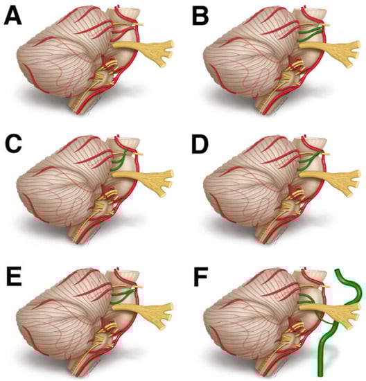
Figure 2.
The possible SCA course: (A) typical course, (B) SCA duplication, (C) single vessel origin from the posterior cerebral artery, (D) common trunk with the posterior cerebral artery, (E) bifurcation, (F) origin from the internal carotid artery. Note, that (D) is a type of (C), in which the P1 gives off the SCA and eventually narrows (we decided to distinguish this type as it was observed in previous papers).
Duplication: Duplication frequency varies from ~3% to ~30% [86,92]. The frequency of the left and right duplication is similar [48,86,92]. The bilateral variant was observed in 0.9–5% [1,86]. There was a single case of an 84-year-old woman with SCA duplication potentially associated with cerebellar infarction [23]. SCA triplication is sevenfold less frequent [90].
Other origination sites: SCA usually originates from the distal part of BA, however, it may also originate from the PCA or as a common trunk with PCA from BA. The first variant is observed in 1.88–15%, with bilateral occurrence in 1.2–5% of cases [1,90]. The second one was described in 1–5.93%, with bilateral occurrence in 0.7–1% of cases [87,92].
Early bifurcation: SCA commonly bifurcates into two major trunks, namely rostral and caudal, at a distance of 0.6–34.0 mm from its origin. When the bifurcation occurs at the proximal anterior pontomesencephalic segment, it is referred to as early bifurcation [48]. This variant has been reported in 3–9.4%, with bilateral occurrence in up to 0.9% of cases [48,91].
Table 2.
Prevalence of SCA anatomical variations. Studies are arranged chronologically.
Table 2.
Prevalence of SCA anatomical variations. Studies are arranged chronologically.
| Type of the Variation | Percentage Calculated Based on the Total Number of Patients | Percentage Calculated Based on the Total Number of SCAs | ||||||||
|---|---|---|---|---|---|---|---|---|---|---|
| Blackburn (1907) N = 220 [85] | Stopford (1916) N = 150 [86] | Caruso et al., (1991) N = 100 [87] | Uchino et al., (2003) N = 136 [48] | Pai et al., (2007) N = 25 [88] | Garcia-Gonzalez et al., (2012) N = 20 [1] | Pekcevik et al., (2013) N = 341 [91] | Krzyżewski et al., (2014) * N = 200 [92] | Ogeng’o et al., (2015) * N = 173 [89] | Kalaiyarasi and Chitra (2018) * N = 80 [90] | |
| Typical variant (SCA bilaterally originating as a single vessel from the BA) | N.D. | N.D. | N.D. | 82% | 80% | 35% | 70.7% | 89.43% | 72.1% | 75.6% |
| SCA originating from the PCA as a single vessel | N.D. | N.D. | N.D. | N.D. | 4% | 15% | 4.7% | 4.64% | 2% | 1.88% |
| SCA originating as a common trunk with PCA ** | N.D. | N.D. | 1% | 4.4% | N.D. | 40% | 5% | 5.93% | 2.5% | 3.13% |
| SCA originating from the ICA | N.D. | N.D. | N.D. | 0.7% | N.D. | N.D. | N.D. | N.D. | N.D. | N.D. |
| Duplication of SCA originated from BA | 3.61% | 31% | N.D. | 9.6% | 16% | 15% | 20.5% | 3.09% | 21.3% | 17.5% |
| SCA duplicated and originating from the PCA | N.D. | N.D. | N.D. | N.D. | N.D. | N.D. | 0.3% | N.D. | 2% | N.D. |
| Early bifurcation of the SCA | N.D. | N.D. | N.D. | 3% | N.D. | N.D. | 9.4% | N.D. | N.D. | N.D. |
| Absence of the SCA | N.D. | 0.67% | N.D. | N.D. | N.D. | N.D. | N.D. | 4% | N.D. | N.D. |
| Simultaneous duplication and common trunk of SCAs | N.D. | N.D. | N.D. | N.D. | N.D. | N.D. | 1.8% | N.D. | N.D. | N.D. |
Legend: ICA—internal carotid artery, N.D.—no data, PCA—posterior cerebral artery, SCA—superior cerebellar artery; *—these data are calculated regarding the total number of SCAs (not patients), **—a common trunk with PCA is an example of SCA originating from the PCA as a single vessel in which the P1 gives off the SCA and eventually narrows (we decided to distinguish this type as it was observed in previous papers).
Origin from the internal carotid artery is observed in less than 1% of cases, in which persistent trigeminal artery (PTA) supplies SCA [48]. This variant is known as Saltzman type II PTA. In such cases, the anterior pontomesencephalic segment is absent owing to the failure of fusion in the early embryonic stage [48].
5. The Approach of Better Visualization of Anatomical Variations in Diagnosis and Surgical Treatment
Proper patients qualification for treatment remains one of the most important steps of the diagnostic and therapeutic approach. The qualification for different invasive procedures (among others: peripheral trigeminal nerve branch procedures, percutaneous trigeminal rhizotomy, MVD, and stereoradiosurgery with gamma knife) for TN remains a subject of separate and still pending discussion and may be affected by the personal preferences of both patients and neurosurgeons. One of the leading indications for MVD in TN is the inability to achieve adequate medical control of TN trigeminal with ≥5 years of anticipated survival, without significant medical or surgical risk factors [93]. Furthermore, the knowledge of possible anatomical variants is crucial for achieving good surgical outcomes in SCA-related NVC syndromes, as confirmed by the relief of symptoms following MVD.
Neuroimaging enables the proper differential diagnosis in NVC patients. It allows us to rule out the entities such as brain tumors, aneurysms, arteriovenous malformations, and others. The success rate of MVD strongly depends on careful visualization of the nerve in its REZ during the surgery, not on radiological examination. Many NVCs, that can be observed intraoperatively are not seen in neuroimaging. On the other hand, visualization of contact between the nerve and vessel in the radiological examination without clinical symptoms is not sufficient for the diagnosis of NVCs. Therefore, the following subchapters discuss many issues of fairly limited direct clinical importance.
5.1. Magnetic Resonance Imaging
Meaney et al. used magnetic resonance angiography for NVC visualization in 1995 [94]. Several years later Naraghi et al. used the heavily T2-weighted sequences by constructive interference in steady-state [95]. Contemporary, the most applicable in the context of NVC detection are the combination of high-resolution 3D T2-weighted imaging with 3D time-of-flight angiography and 3D T1-weighted gadolinium-enhanced sequences [96].
As important as selecting the appropriate MR sequence is correct interpreting the imaging examination since, as has already been mentioned, mere visualization of neurovascular contact is not enough to confirm the presence of NVC [97]. Some neuroradiological features of such contact significantly increase the risk of symptomatic NVC. They include thinning of the nerve, arterial imprint, grooving of the nerve, or distorted course of the nerve [97]. On the other hand, it should be emphasized that the nerve itself can be displaced and changed by one vessel and the symptoms of NVC can be caused by another vessel impinging on the nerve in its REZ.
Diffusion Tensor Imaging and Diffusion-Weighted Imaging
Diffusion tensor imaging (DTI) and diffusion-weighted imaging (DWI) are forms of MR imaging that were investigated in patients with NVC [98,99]. The most commonly described alternations were observed in fractional anisotropy (FA) in DTI and apparent diffusion coefficient (ADC) [100,101]. It was reported that the reduction FA and increase in ADC had a close relationship to white matter tract degeneration. Such changes in mentioned parameter values are probably triggered by diffusivity averaging in all spatial directions caused by myelin deficiency [82]. Interestingly, both FA and ADC may be designated also in small anatomical areas. Thanks to this it is possible to evaluate NVCs consequences in these MRI protocols [102].
The usefulness of DTI in NVCs evaluation was mainly assessed on an example of TN [99,102,103,104,105,106]. The majority of studies reported that DTI can reveal alterations (as a sign of anisotropic microstructural changes) of nerve integrity on affected sites compared to non-affected sites and healthy controls [101,103,106]. Moreover, Willsey et al. found that the DTI examination allows for a diagnosis of TN subtypes (TN1 and TN2), thus facilitating patients’ selection for surgical procedures, and can be a prognostic marker following an intervention [105]. An interesting issue was raised in a prospective study on DTI usefulness evaluation performed by Lutz et al. [103]. They reported that in 3 symptomatic cases that were qualified for the MVD procedure preoperative high-resolution MRI did not reveal any NVC occurrence. Interestingly, after the surgery symptoms resolved. In reevaluation retrospectively also there were no NVCs in MRI. Lutz et al. revealed that in mentioned cases with negative MRI, the FA parameter was lower [103].
6. Future Directions and Conclusions
6.1. Importance of Further Studies in the Field
The SCA is frequently involved in symptomatic NVCs (40–90%). Its variability may be also a cause of HFS. The significance of NVC between SCA and the oculomotor nerve is poorly discussed in the literature. Nevertheless, it seems that in extremely rare cases mentioned NVC can be symptomatic and clinically manifested as ONP or oculomotor NMT. Up to date, little is known about SCA variability in the context of NVC. Further studies are required to analyze the influence of such variations on risk symptomatic NVCs development.
6.2. Conclusion and Clinical Points
6.2.1. Conclusion and Summary of Key Points
- Although SCA is a key vessel related to trigeminal neuralgia pathogenesis, the current state of knowledge about the link between SCA variability and NVC (especially TN) occurrence remains limited and required further investigation.
- Neuroimaging enables the proper differential diagnosis in NVC patients, i.e. ruling out brain tumors, aneurysms, arteriovenous malformations, and others.
- The success rate of MVD strongly depends on careful visualization of the nerve in its REZ at surgery not on radiological examination. Many NVCs, that can be observed intraoperatively are not seen in neuroimaging. Visualization of contact between nerve and vessel in the radiological examination without clinical symptoms does not suffice for the diagnosis of NVCs.
- Knowledge about possible anatomical variants of SCA may improve patients’ diagnosis, choosing an appropriate treatment strategy, and patients’ outcome surgical.
- A tortuous anterior pontomesencephalic segment of the SCA may lead to misdiagnosis as it may mimic arteriovenous malformation on imaging.
6.2.2. Clinical Points
- Typical pain in TN is characterized by sharp, electric shock-like paroxysmal lancinating pain, lasting a few seconds, typically triggered by sensory stimulation. In these cases, the neurologic exam remains normal, except for a mild sensory loss. A pain of another character situated in the trigeminal nerve innervation zone is known as atypical facial pain.
- Although the majority of the TN patients show a good response to carbamazepine, there are those (especially refractory cases) in whom microvascular decompression should be considered.
Supplementary Materials
The following supporting information can be downloaded at: https://www.mdpi.com/article/10.3390/biomedicines11072009/s1, Figure S1: The angiography with left superior cerebellar artery aneurysm embolization: (A) angiography revealed aneurysm (7 × 6 × 5 mm in size), (B,C) the aneurysm was further embolized with MicroPlex Coil System, Figure S2: The angiography with left superior cerebellar artery aneurysm embolization: (A) The angiography revealed aneurysm, (B,C) the aneurysm was embolized with MicroPlex Coil System.
Author Contributions
Conceptualization, B.M.S. and E.J.B.; investigation, M.M.; writing—original draft preparation, M.M., B.M.S. and E.J.B.; figure creating—S.W., B.M.S. and E.J.B.; writing—review and editing, F.F.K., M.M.P., D.K., S.W., M.R. and D.J.J. All authors have read and agreed to the published version of the manuscript.
Funding
This research received no external funding.
Data Availability Statement
Not applicable.
Acknowledgments
We would like to express our gratitude to Magdalena Kokosińska from the Information and Library Center of the Medical University of Lodz for her valuable assistance with accessing selected papers. Moreover, we would like to thank the authors whose studies were analyzed in the present review for their excellent work.
Conflicts of Interest
B.M.S. is Guest Editor of the Special Issue “Pathogenesis and Therapy of Neurovascular Compression Syndromes” in Biomedicines.
Abbreviations
| ADC | apparent diffusion coefficient |
| AICA | anterior inferior cerebellar artery |
| BA | basilar artery |
| DTI | diffusion tensor imaging |
| FA | fractional anisotropy |
| HFS | hemifacial spasm |
| MRI | magnetic resonance imaging |
| MVD | microvascular decompression |
| N.D. | no data |
| NMT | neuromyotonia |
| NVC | neurovascular compression |
| ONP | oculomotor nerve palsy |
| PCA | posterior cerebral artery |
| PICA | posterior inferior cerebellar artery |
| REZ | root entry/exit zone |
| SCA | superior cerebellar artery |
| VA | vertebral artery |
References
- Garcia-Gonzalez, U.; Cavalcanti, D.D.; Agrawal, A.; Spetzler, R.F.; Preul, M.C. Anatomical Study on the “Perforator-Free Zone”: Reconsidering the Proximal Superior Cerebellar Artery and Basilar Artery Perforators. Neurosurgery 2012, 70, 764–773. [Google Scholar] [CrossRef]
- Kim, S.J.; Lee, H.Y.; Lee, Y.H.; Lee, J.H.; Yang, J.H.; Lee, M.K.; Yang, H.J. Blood Supply by the Superior Cerebellar Artery and Posterior Inferior Cerebellar Artery to the Motor and Nonmotor Domains of the Human Dentate Nucleus. World Neurosurg. 2019, 122, e606–e611. [Google Scholar] [CrossRef] [PubMed]
- Szmyd, B.; Sołek, J.; Błaszczyk, M.; Jankowski, J.; Liberski, P.P.; Jaskólski, D.J.; Wysiadecki, G.; Karuga, F.F.; Gabryelska, A.; Sochal, M.; et al. The Underlying Pathogenesis of Neurovascular Compression Syndromes: A Systematic Review. Front. Mol. Neurosci. 2022, 15, 325. [Google Scholar] [CrossRef]
- Lorenzoni, J.; David, P.; Levivier, M. Patterns of Neurovascular Compression in Patients with Classic Trigeminal Neuralgia: A High-Resolution MRI-Based Study. Eur. J. Radiol. 2012, 81, 1851–1857. [Google Scholar] [CrossRef] [PubMed]
- Kuroki, A.; Møller, A.R. Facial Nerve Demyelination and Vascular Compression Are Both Needed to Induce Facial Hyperactivity: A Study in Rats. Acta Neurochir. 1994, 126, 149–157. [Google Scholar] [CrossRef] [PubMed]
- Møller, A.R.; Jannetta, P.J. Physiological Abnormalities in Hemifacial Spasm Studied during Microvascular Decompression Operations. Exp. Neurol. 1986, 93, 584–600. [Google Scholar] [CrossRef] [PubMed]
- Liu, M.X.; Zhong, J.; Xia, L.; Dou, N.N.; Li, S.T. IL-6 Contributes to Nav1.3 up-Regulation in Trigeminal Nerve Following Chronic Constriction Injury. Neurol. Res. 2020, 42, 504–514. [Google Scholar] [CrossRef]
- Mazzucchi, E.; Brinzeu, A.; Sindou, M. Arachnoiditis as an Outcome Factor for Microvascular Decompression in Classical Trigeminal Neuralgia. Acta Neurochir. 2019, 161, 1589–1598. [Google Scholar] [CrossRef]
- Farajzadeh, A.; Bathaie, S.Z.; Arabkheradmand, J.; Ghodsi, S.M.; Faghihzadeh, S. Different Pain States of Trigeminal Neuralgia Make Significant Changes in the Plasma Proteome and Some Biochemical Parameters: A Preliminary Cohort Study. J. Mol. Neurosci. 2018, 66, 524–534. [Google Scholar] [CrossRef]
- Karki, P.; Yamagami, M.; Takasaki, K.; Bohara, M.; Hosoyama, H.; Hanada, T.; Yamasaki, F.; Hanaya, R.; Arita, K. Microvascular Decompression in Patients Aged 30 Years or Younger. Asian J. Neurosurg. 2019, 14, 111–117. [Google Scholar] [CrossRef]
- Rhoton, A.L. The Cerebellopontine Angle and Posterior Fossa Cranial Nerves by the Retrosigmoid Approach. Neurosurgery 2000, 47, S93–S129. [Google Scholar] [CrossRef] [PubMed]
- Bobeff, E.J.; Wisniewski, K.; Jaskolski, D.J. Rzadkie Przypadki Guzów Kąta Mostowo-Móżdżkowego. Vertigoprofil 2019, 11, 11–15. [Google Scholar]
- Rhoton, A.L. Rhoton’s Cranial Anatomy and Surgical Approaches, 1st ed.; Oxford University Press: Oxford, UK, 2019. [Google Scholar]
- Rhoton, A.L. The Cerebellar Arteries. Neurosurgery 2000, 47, S29–S68. [Google Scholar] [CrossRef] [PubMed]
- Joshi, S.; Tee, W.W.H.; Franconi, C.; Prentice, D. Transient Oculomotor Nerve Palsy Due to Non-Aneurysmal Neurovascular Compression. J. Clin. Neurosci. 2017, 45, 136–137. [Google Scholar] [CrossRef]
- Suzuki, K.; Muroi, A.; Kujiraoka, Y.; Takano, S.; Matsumura, A. Oculomotor Palsy Treated by Microvascular Decompression. Surg. Neurol. 2008, 70, 210–212. [Google Scholar] [CrossRef]
- Raza, H.K.; Chen, H.; Chansysouphanthong, T.; Cui, G. The Aetiologies of the Unilateral Oculomotor Nerve Palsy: A Review of the Literature. Somatosens. Mot. Res. 2018, 35, 229–239. [Google Scholar] [CrossRef]
- Inoue, T.; Hirai, H.; Shimizu, T.; Tsuji, M.; Shima, A.; Suzuki, F.; Matsuda, M. Ocular Neuromyotonia Treated by Microvascular Decompression: Usefulness of Preoperative 3D Imaging: Case Report. J. Neurosurg. 2012, 117, 1166–1169. [Google Scholar] [CrossRef]
- Hashimoto, Y.; Hideyama, T.; Yamagami, A.; Sasaki, T.; Maekawa, R.; Shiio, Y. A Case of Ocular Neuromyotonia Caused by Neurovascular Compression of the Oculomotor Nerve by the Elongated Superior Cerebellar Artery. J. Neurol. 2016, 263, 1236–1238. [Google Scholar] [CrossRef]
- Sindou, M.P. Microvascular Decompression for Primary Hemifacial Spasm. Importance of Intraoperative Neurophysiological Monitoring. Acta Neurochir. 2005, 147, 1019–1026. [Google Scholar] [CrossRef]
- Matsushima, T. Microsurgical Anatomy and Surgery of the Posterior Cranial Fossa. Surgical Approaches and Procedures Based on Anatomical Study; Springer: Berlin, Germany, 2015. [Google Scholar]
- Padmavathi, G. Study of the Variations of Superior Cerebellar Artery in Human Cadavers. Int. J. Res. Med. Sci. 2014, 2, 699. [Google Scholar] [CrossRef]
- Porzionato, A.; Macchi, V.; Massaro, L.; Morra, A.; Sarasin, G.; Rambaldo, A.; De Caro, R. Double Origin of the Superior Cerebellar Artery Associated with Homolateral Haemorrhagic Infarction of Cerebellum. Folia Neuropathol. 2016, 54, 410–417. [Google Scholar] [CrossRef] [PubMed]
- Erdemoglu, A.K.; Duman, T. Superior Cerebellar Artery Territory Stroke. Acta Neurol. Scand. 1998, 98, 283–287. [Google Scholar] [CrossRef] [PubMed]
- Terao, S.I.; Sobue, G.; Izumi, M.; Miura, N.; Takeda, A.; Mitsuma, T. Infarction of Superior Cerebellar Artery Presenting as Cerebellar Symptoms. Stroke 1996, 27, 1679–1681. [Google Scholar] [CrossRef] [PubMed]
- Peluso, J.P.P.; Jan Van Rooij, W.; Sluzewski, M.; Beute, G.N. Superior Cerebellar Artery Aneurysms: Incidence, Clinical Presentation and Midterm Outcome of Endovascular Treatment. Neuroradiology 2007, 49, 747–751. [Google Scholar] [CrossRef] [PubMed]
- December, P. Trifurcation of Basilar Artery. Int. J. Anat. Var. 2013, 6, 199–200. [Google Scholar]
- Adam, B. Anatomia Człowieka; Państwowy Zakład Wydawnictw Lekarskich (PZWL): Warsaw, Poland, 2022. [Google Scholar]
- Tubbs, R.S.; Shoja, M.M.; Loukas, M. Bergman’s Comprehensive Encyclopedia of Human Anatomic Variation; Wiley-Blackwell: Hoboken, NJ, USA, 2016; ISBN 978-1-118-43035-4. [Google Scholar]
- Hardy, D.G.; Peace, D.A.; Rhoton, A.L. Microsurgical Anatomy of the Superior Cerebellar Artery. Neurosurgery 1980, 6, 10–28. [Google Scholar] [CrossRef]
- Ascanio, L.C.; Ogilvy, C.S.; Thomas, A.J.; Kicielinski, K.; Gupta, R.; Alturki, A.Y. Distal Parent Vessel Occlusion of 2 Superior Cerebellar Artery Fusiform Aneurysms: Report of 2 Cases and Literature Review. World Neurosurg. X 2019, 3, 100026. [Google Scholar] [CrossRef]
- Rodríguez-Hernández, A.; Rhoton, A.L.; Lawton, M.T. Segmental Anatomy of Cerebellar Arteries: A Proposed Nomenclature: Laboratory Investigation. J. Neurosurg. 2011, 115, 387–397. [Google Scholar] [CrossRef]
- Maarbjerg, S.; Gozalov, A.; Olesen, J.; Bendtsen, L. Trigeminal Neuralgia--a Prospective Systematic Study of Clinical Characteristics in 158 Patients. Headache 2014, 54, 1574–1582. [Google Scholar] [CrossRef]
- Mačianskytė, D.; Janužis, G.; Kubilius, R.; Adomaitienė, V.; Ščiupokas, A. Associations between Chronic Pain and Depressive Symptoms in Patients with Trigeminal Neuralgia. Medicina 2011, 47, 386–392. [Google Scholar] [CrossRef]
- Obermann, M.; Yoon, M.S.; Sensen, K.; Maschke, M.; Diener, H.C.; Katsarava, Z. Efficacy of Pregabalin in the Treatment of Trigeminal Neuralgia. Cephalalgia 2008, 28, 174–181. [Google Scholar] [CrossRef] [PubMed]
- Araya, E.I.; Claudino, R.F.; Piovesan, E.J.; Chichorro, J.G. Trigeminal Neuralgia: Basic and Clinical Aspects. Curr. Neuropharmacol. 2020, 18, 109–119. [Google Scholar] [CrossRef]
- Wu, T.H.; Hu, L.Y.; Lu, T.; Chen, P.M.; Chen, H.J.; Shen, C.C.; Wen, C.H. Risk of Psychiatric Disorders Following Trigeminal Neuralgia: A Nationwide Population-Based Retrospective Cohort Study. J. Headache Pain 2015, 16, 1–8. [Google Scholar] [CrossRef]
- Zakrzewska, J.M.; Wu, J.; Mon-Williams, M.; Phillips, N.; Pavitt, S.H. Evaluating the Impact of Trigeminal Neuralgia. Pain 2017, 158, 1166–1174. [Google Scholar] [CrossRef] [PubMed]
- Melek, L.; Smith, J.; Karamat, A.; Renton, T. Comparison of the Neuropathic Pain Symptoms and Psychosocial Impacts of Trigeminal Neuralgia and Painful Posttraumatic Trigeminal Neuropathy. J. Oral Facial Pain Headache 2019, 33, 77–88. [Google Scholar] [CrossRef] [PubMed]
- Chang, B.; Zhu, W.; Li, S. Effects of Depression and Anxiety on Microvascular Decompression Outcome for Trigeminal Neuralgia Patients. World Neurosurg. 2019, 128, e556–e561. [Google Scholar] [CrossRef]
- Katusic, S.; Beard, C.M.; Bergstralth, E.; Kurland, L.T. Incidence and Clinical Features of Trigeminal Neuralgia, Rochester, Minnesota, 1945–1984. Ann. Neurol. 1990, 27, 89–95. [Google Scholar] [CrossRef]
- Yoshimasu, F.; Kurland, L.T.; Elveback, L.R. Tic Douloureux in Rochester, Minnesota, 1945–1969. Neurology 1972, 22, 952–956. [Google Scholar] [CrossRef]
- Rozen, T.D. Trigeminal Neuralgia and Glossopharyngeal Neuralgia. Neurol. Clin. 2004, 22, 185–206. [Google Scholar] [CrossRef]
- De Toledo, I.P.; Conti Réus, J.; Fernandes, M.; Porporatti, A.L.; Peres, M.A.; Takaschima, A.; Linhares, M.N.; Guerra, E.; De Luca Canto, G. Prevalence of Trigeminal Neuralgia: A Systematic Review. J. Am. Dent. Assoc. 2016, 147, 570–576.e2. [Google Scholar] [CrossRef]
- Love, S.; Coakham, H.B. Trigeminal Neuralgia: Pathology and Pathogenesis. Brain 2001, 124, 2347–2360. [Google Scholar] [CrossRef] [PubMed]
- Eboli, P.; Stone, J.L.; Aydin, S.; Slavin, K.V. Historical Characterization of Trigeminal Neuralgia. Neurosurgery 2009, 64, 1183–1186. [Google Scholar] [CrossRef] [PubMed]
- Zheng, X.; Feng, B.; Hong, W.; Zhang, W.; Yang, M.; Tang, Y.; Zhong, J.; Hua, X.; Li, S. Management of Intraneural Vessels during Microvascular Decompression Surgery for Trigeminal Neuralgia. World Neurosurg. 2012, 77, 771–774. [Google Scholar] [CrossRef] [PubMed]
- Uchino, A.; Sawada, A.; Takase, Y.; Kudo, S. Variations of the Superior Cerebellar Artery: MR Angiographic Demonstration. Radiat. Med. 2003, 21, 235–238. [Google Scholar] [CrossRef]
- Wu, M.; Fu, X.; Ji, Y.; Ding, W.; Deng, D.; Wang, Y.; Jiang, X.; Niu, C. Microvascular Decompression for Classical Trigeminal Neuralgia Caused by Venous Compression: Novel Anatomic Classifications and Surgical Strategy. World Neurosurg. 2018, 113, e707–e713. [Google Scholar] [CrossRef]
- Tashiro, H.; Kondo, A.; Aoyama, I.; Nin, K.; Shimotake, K.; Nishioka, T.; Ikai, Y.; Takahashi, J. Trigeminal Neuralgia Caused by Compression from Arteries Transfixing the Nerve. J. Neurosurg. 1991, 75, 783–786. [Google Scholar] [CrossRef]
- Vanaclocha, V.; Herrera, J.M.; Martínez-Gómez, D.; Rivera-Paz, M.; Calabuig-Bayo, C.; Vanaclocha, L. Is There a Safe and Effective Way to Treat Trigeminal Neuralgia Associated with Vertebrobasilar Dolichoectasia? Presentation of 8 Cases and Literature Review. World Neurosurg. 2016, 96, 516–529. [Google Scholar] [CrossRef]
- Wróbel-Wiśniewska, G.; Jaskolski, D.J.; Zawirski, M. Neuralgia Nerwu Trójdzielnego Po Embolizacji Wewnatrz-Czaszkowego Oponowego Zniekształcenia Tetniczo-Zylnego. Opis Przypadku [Trigeminal Neuralgia Due to Lengthy AVM Treatment. A Case Report]. Neurol. Neurochir. Pol. 2003, 37, 259–263. [Google Scholar] [CrossRef]
- Li, Z.Y.; Liang, J.T.; Zhang, H.Q.; Bao, Y.H.; Ling, F. Trigeminal Neuralgia Caused by a Dilated Superior Cerebellar Artery and a Draining Vein of Cerebellar Arteriovenous Malformations: A Case Report and Review of the Literature. Acta Neurochir. 2017, 159, 689–694. [Google Scholar] [CrossRef]
- Athanasiou, T.C.; Nair, S.; Coakham, H.B.; Lewis, T.T. Arteriovenous Malformation Presenting with Trigeminal Neuralgia and Treated with Endovascular Coiling. Neurol. India 2005, 53, 247–248. [Google Scholar] [CrossRef]
- Ge, H.; Lv, X.; Jin, H.; He, H.; Li, Y. Role of Endovascular Embolization for Trigeminal Neuralgia Related to Cerebral Vascular Malformation. Interv. Neuroradiol. 2016, 22, 600–605. [Google Scholar] [CrossRef] [PubMed]
- Mineura, K.; Sasajima, H.; Itoh, Y.; Kowada, M.; Tomura, N.; Goto, K. Development of a Huge Varix Following Endovascular Embolization for Cerebellar Arteriovenous Malformation. A Case Report. Acta Radiol. 1998, 39, 189–192. [Google Scholar] [CrossRef] [PubMed]
- Molina-Gil, J.; González-Fernández, L.; García-Cabo, C. Trigeminal Neuralgia as the Sole Neurological Manifestation of COVID-19: A Case Report. Headache 2021, 61, 560–562. [Google Scholar] [CrossRef]
- Li, G.W.; Lan, Q.; Zhang, W.C. Clinical Characteristics and Treatment of Trigeminal Neuralgia Following Herpes Zoster. J. Craniofac. Surg. 2015, 26, e448–e451. [Google Scholar] [CrossRef]
- Di Stefano, G.; Maarbjerg, S.; Truini, A. Trigeminal Neuralgia Secondary to Multiple Sclerosis: From the Clinical Picture to the Treatment Options. J. Headache Pain 2019, 20, 1–10. [Google Scholar] [CrossRef] [PubMed]
- Wróbel-Wiśniewska, G.; Kasprzak, P.; Zawirski, M. Połowiczy Skurcz Twarzy Jako Poczatkowy Objaw Oponiaka Tylnej Jamy Czaszki [Hemifacial Spasm as an Early Symptom of Posterior Fossa Meningioma]. Neurol. Neurochir. Pol. 2003, 37, 459–463. [Google Scholar]
- Cruccu, G.; Finnerup, N.B.; Jensen, T.S.; Scholz, J.; Sindou, M.; Svensson, P.; Treede, R.D.; Zakrzewska, J.M.; Nurmikko, T. Trigeminal Neuralgia: New Classification and Diagnostic Grading for Practice and Research. Neurology 2016, 87, 220–228. [Google Scholar] [CrossRef]
- Marinković, S.; Gibo, H. The Neurovascular Relationships and the Blood Supply of the Oculomotor Nerve: The Microsurgical Anatomy of Its Cisternal Segment. Surg. Neurol. 1994, 42, 505–516. [Google Scholar] [CrossRef]
- Esmer, A.F.; Sen, T.; Comert, A.; Tuccar, E.; Karahan, S.T. The Neurovascular Relationships of the Oculomotor Nerve. Clin. Anat. 2011, 24, 583–589. [Google Scholar] [CrossRef]
- Hashimoto, M.; Ohtsuka, K.; Akiba, H.; Harada, K. Vascular Compression of the Oculomotor Nerve Disclosed by Thin-Slice Magnetic Resonance Imaging. Am. J. Ophthalmol. 1998, 125, 881–882. [Google Scholar] [CrossRef]
- Nakagawa, H.; Nakajima, S.; Nakajima, Y.; Furuta, Y.; Nishi, O.; Nishi, K. Bilateral Oculomotor Nerve Palsies Due to Posterior Cerebral Arterial Compression Relieved by Microvascular Decompression–Case Report. Neurol. Med. Chir. 1991, 31, 45–48. [Google Scholar] [CrossRef] [PubMed]
- Albayram, S.; Ozer, H.; Sarici, A.; Murphy, K.; Miller, N. Unilateral Mydriasis without Ophthalmoplegia—A Sign of Neurovascular Compression? Case Report. Neurosurgery 2006, 58, E582–E583. [Google Scholar] [CrossRef] [PubMed]
- Zorman, G.; Wilson, C.B. Outcome Following Microsurgical Vascular Decompression or Partial Sensory Rhizotomy in 125 Cases of Trigeminal Neuralgia. Neurology 1984, 34, 1362–1365. [Google Scholar] [CrossRef] [PubMed]
- Sindou, M.P.; Chiha, M.; Mertens, P. Anatomical Findings Observed during Microsurgical Approaches of the Cerebellopontine Angle for Vascular Decompression in Trigeminal Neuralgia (350 Cases). Stereotact. Funct. Neurosurg. 1994, 63, 203–207. [Google Scholar] [CrossRef]
- Barker, F.G.; Jannetta, P.J.; Bissonette, D.J.; Larkins, M.V.; Jho, H.D. The Long-Term Outcome of Microvascular Decompression for Trigeminal Neuralgia. N. Engl. J. Med. 1996, 334, 1077–1084. [Google Scholar] [CrossRef]
- Sindou, M.; Howeidy, T.; Acevedo, G. Anatomical Observations during Microvascular Decompression for Idiopathic Trigeminal Neuralgia (with Correlations between Topography of Pain and Site of the Neurovascular Conflict). Prospective Study in a Series of 579 Patients. Acta Neurochir. 2002, 144, 1–13. [Google Scholar] [CrossRef]
- Li, S.T.; Pan, Q.; Liu, N.; Shen, F.; Liu, Z.; Guan, Y.; Sindou, M.; Burchiel, K.J. Trigeminal Neuralgia: What Are the Important Factors for Good Operative Outcomes with Microvascular Decompression. Surg. Neurol. 2004, 62, 400–404. [Google Scholar] [CrossRef]
- Sekula, R.F.; Frederickson, A.M.; Jannetta, P.J.; Bhatia, S.; Quigley, M.R.; Abdel Aziz, K.M. Microvascular Decompression in Patients with Isolated Maxillary Division Trigeminal Neuralgia, with Particular Attention to Venous Pathology. Neurosurg. Focus 2009, 27, E10. [Google Scholar] [CrossRef]
- Soares-Dos-Reis, R.; Martins, A.I.; Brás, A.; Matos, A.; Bento, C.; Lemos, J. Ocular Neuromyotonia. Pract. Neurol. 2018, 18, 389–390. [Google Scholar] [CrossRef]
- Tanrikulu, L.; Scholz, T.; Nikoubashman, O.; Wiesmann, M.; Clusmann, H. Preoperative MRI in Neurovascular Compression Syndromes and Its Role for Microsurgical Considerations. Clin. Neurol. Neurosurg. 2015, 129, 17–20. [Google Scholar] [CrossRef]
- Colosimo, C.; Bologna, M.; Lamberti, S.; Avanzino, L.; Marinelli, L.; Fabbrini, G.; Abbruzzese, G.; Defazio, G.; Berardelli, A. A Comparative Study of Primary and Secondary Hemifacial Spasm. Arch. Neurol. 2006, 63, 441–444. [Google Scholar] [CrossRef] [PubMed]
- Chaudhry, N.; Srivastava, A.; Joshi, L. Hemifacial Spasm: The Past, Present and Future. J. Neurol. Sci. 2015, 356, 27–31. [Google Scholar] [CrossRef] [PubMed]
- Abbruzzese, G.; Berardelli, A.; Defazio, G. Hemifacial Spasm. Handb. Clin. Neurol. 2011, 100, 675–680. [Google Scholar] [CrossRef] [PubMed]
- Auger, R.G.; Whisnant, J.P. Hemifacial Spasm in Rochester and Olmsted County, Minnesota, 1960 to 1984. Arch. Neurol. 1990, 47, 1233–1234. [Google Scholar] [CrossRef]
- Nilsen, B.; Le, K.D.; Dietrichs, E. Prevalence of Hemifacial Spasm in Oslo, Norway. Neurology 2004, 63, 1532–1533. [Google Scholar] [CrossRef]
- Yan, X.; Gu, J.; Quan, J.; Zhang, X.; Zhou, X.; Qu, J.; Zhou, L. Anatomical Deviations of Vertebral Artery in Hemifacial Spasm: A Quantitative Study. Surg. Radiol. Anat. 2021, 43, 291–299. [Google Scholar] [CrossRef]
- Guan, H.X.; Zhu, J.; Zhong, J. Correlation between Idiopathic Hemifacial Spasm and the MRI Characteristics of the Vertebral Artery. J. Clin. Neurosci. 2011, 18, 528–530. [Google Scholar] [CrossRef]
- Greenberg, M. Functional Neurosurgery and Stereotactic Neurosurgery. In Handbook of Neurosurgery; Stuttgart: New York, NY, USA; New Delhi, India; Thieme: Rio de Janeiro, Brazil, 2020. [Google Scholar]
- Tambasco, N.; Filidei, M.; Nigro, P.; Parnetti, L.; Simoni, S. Botulinum Toxin for the Treatment of Hemifacial Spasm: An Update on Clinical Studies. Toxins 2021, 13, 881. [Google Scholar] [CrossRef]
- Vanaparthy, R.; Malayala, S.V.; Balla, M. COVID-19-Induced Vestibular Neuritis, Hemi-Facial Spasms and Raynaud’s Phenomenon: A Case Report. Cureus 2020, 12, e11752. [Google Scholar] [CrossRef]
- Blackburn, I.W. Anomalies of the Encephalic Arteries among the Insane. A Study of the Arteries at the Base of the Encephallon in Two Hundred and Twenty Consecutive Cases of Mental Disease, with Special Reference to Anomalies of the Circle of Willis. J. Comp. Neurol. Psychol. 1907, 17, 493–517. [Google Scholar] [CrossRef]
- Stopford, J.S. The Arteries of the Pons and Medulla Oblongata. J. Anat. Physiol. 1916, 50, 131–164. [Google Scholar] [CrossRef] [PubMed]
- Caruso, G.; Vincentelli, F.; Rabehanta, P.; Giudicelli, G.; Grisoli, F. Anomalies of the P1 Segment of the Posterior Cerebral Artery: Early Bifurcation or Duplication, Fenestration, Common Trunk with the Superior Cerebellar Artery. Acta Neurochir. 1991, 109, 66–71. [Google Scholar] [CrossRef] [PubMed]
- Pai, B.; Varma, R.; Kulkarni, R.; Nirmala, S.; Manjunath, L.; Rakshith, S. Microsurgical Anatomy of the Posterior Circulation. Neurol. India 2007, 55, 31–41. [Google Scholar] [CrossRef] [PubMed]
- Ogeng’o, J.; Elbusaidy, H.; Sinkeet, S.; Olabu, B.; Mwachaka, P.; Inyimili, M. Variant Origin of the Superior Cerebellar Artery in a Black Kenyan Population. Eur. J. Anat. 2023, 19, 287–290. [Google Scholar]
- Kalaiyarasi, S.; Chitra, P.S. A Study of Variations in the Origin of Superior Cerebellar Artery and the Dimensions of Its Proximal Segment. Int. J. Anat. Res. 2018, 6, 4849–4853. [Google Scholar] [CrossRef]
- Pekcevik, Y.; Pekcevik, R. Variations of the Cerebellar Arteries at CT Angiography. Surg. Radiol. Anat. 2014, 36, 455–461. [Google Scholar] [CrossRef]
- Krzyzewski, R.M.; Stachura, M.K.; Stachura, A.M.; Rybus, J.; Tomaszewski, K.A.; Klimek-Piotrowska, W.; Brzegowy, P.; Urbanik, A.; Walocha, J.A. Variations and Morphometric Analysis of the Proximal Segment of the Superior Cerebellar Artery. Neurol. Neurochir. Pol. 2014, 48, 229–235. [Google Scholar] [CrossRef]
- Greenberg, M.S. Handbook of Neurosurgery: 98.6 Neurovascular Compression Syndromes. Thieme: Stuttgart, Germany, 2023. [Google Scholar]
- Meaney, J.F.M.; Eldridge, P.R.; Dunn, L.T.; Nixon, T.E.; Whitehouse, G.H.; Miles, J.B. Demonstration of Neurovascular Compression in Trigeminal Neuralgia with Magnetic Resonance Imaging. Comparison with Surgical Findings in 52 Consecutive Operative Cases. J. Neurosurg. 1995, 83, 799–805. [Google Scholar] [CrossRef]
- Naraghi, R.; Hastreiter, P.; Tomandl, B.; Bonk, A.; Huk, W.; Fahlbusch, R. Three-Dimensional Visualization of Neurovascular Relationships in the Posterior Fossa: Technique and Clinical Application. J. Neurosurg. 2004, 100, 1025–1035. [Google Scholar] [CrossRef]
- Haller, S.; Etienne, L.; Kövari, E.; Varoquaux, A.D.; Urbach, H.; Becker, M. Imaging of Neurovascular Compression Syndromes: Trigeminal Neuralgia, Hemifacial Spasm, Vestibular Paroxysmia, and Glossopharyngeal Neuralgia. AJNR. Am. J. Neuroradiol. 2016, 37, 1384–1392. [Google Scholar] [CrossRef]
- Maurya, V.; Sreedhar, C.M.; Khera, A.; Bhatia, M.; Sharma, V. Trigeminal Neuralgia: When Does Neurovascular Contact Turn into a Conflict? Med. J. Armed Forces India 2019, 75, 134–139. [Google Scholar] [CrossRef] [PubMed]
- Lope-Piedrafita, S. Diffusion Tensor Imaging (DTI). Methods Mol. Biol. 2018, 1718, 103–116. [Google Scholar] [CrossRef] [PubMed]
- Herweh, C.; Kress, B.; Rasche, D.; Tronnier, V.; Tröger, J.; Sartor, K.; Stippich, C. Loss of Anisotropy in Trigeminal Neuralgia Revealed by Diffusion Tensor Imaging. Neurology 2007, 68, 776–778. [Google Scholar] [CrossRef] [PubMed]
- Basser, P.J.; Pierpaoli, C. Microstructural and Physiological Features of Tissues Elucidated by Quantitative-Diffusion-Tensor MRI. 1996. J. Magn. Reson. 2011, 213, 560–570. [Google Scholar] [CrossRef]
- Lummel, N.; Mehrkens, J.H.; Linn, J.; Buchholz, G.; Stahl, R.; Bochmann, K.; Brückmann, H.; Lutz, J. Diffusion Tensor Imaging of the Trigeminal Nerve in Patients with Trigeminal Neuralgia Due to Multiple Sclerosis. Neuroradiology 2015, 57, 259–267. [Google Scholar] [CrossRef]
- Desouza, D.D.; Hodaie, M.; Davis, K.D. Abnormal Trigeminal Nerve Microstructure and Brain White Matter in Idiopathic Trigeminal Neuralgia. Pain 2014, 155, 37–44. [Google Scholar] [CrossRef]
- Lutz, J.; Thon, N.; Stahl, R.; Lummel, N.; Tonn, J.C.; Linn, J.; Mehrkens, J.H. Microstructural Alterations in Trigeminal Neuralgia Determined by Diffusion Tensor Imaging Are Independent of Symptom Duration, Severity, and Type of Neurovascular Conflict. J. Neurosurg. 2016, 124, 823–830. [Google Scholar] [CrossRef]
- Fujiwara, S.; Sasaki, M.; Wada, T.; Kudo, K.; Hirooka, R.; Ishigaki, D.; Nishikawa, Y.; Ono, A.; Yamaguchi, M.; Ogasawara, K. High-Resolution Diffusion Tensor Imaging for the Detection of Diffusion Abnormalities in the Trigeminal Nerves of Patients with Trigeminal Neuralgia Caused by Neurovascular Compression. J. Neuroimaging 2011, 21, e102–e108. [Google Scholar] [CrossRef]
- Willsey, M.S.; Collins, K.L.; Conrad, E.C.; Chubb, H.A.; Patil, P.G. Diffusion Tensor Imaging Reveals Microstructural Differences between Subtypes of Trigeminal Neuralgia. J. Neurosurg. 2019, 133, 573–579. [Google Scholar] [CrossRef]
- Chen, J.; Guo, Z.Y.; Liang, Q.Z.; Liao, H.Y.; Su, W.R.; Chen, C.X.; Fu, S.X.; Han, X.J. Structural Abnormalities of Trigeminal Root with Neurovascular Compression Revealed by High Resolution Diffusion Tensor Imaging. Asian Pac. J. Trop. Med. 2012, 5, 749–752. [Google Scholar] [CrossRef]
Disclaimer/Publisher’s Note: The statements, opinions and data contained in all publications are solely those of the individual author(s) and contributor(s) and not of MDPI and/or the editor(s). MDPI and/or the editor(s) disclaim responsibility for any injury to people or property resulting from any ideas, methods, instructions or products referred to in the content. |
© 2023 by the authors. Licensee MDPI, Basel, Switzerland. This article is an open access article distributed under the terms and conditions of the Creative Commons Attribution (CC BY) license (https://creativecommons.org/licenses/by/4.0/).