
A Novel MDM2-Binding Chalcone Induces Apoptosis of Oral Squamous Cell Carcinoma

 , , ,
, , ,  , , and
Abstract
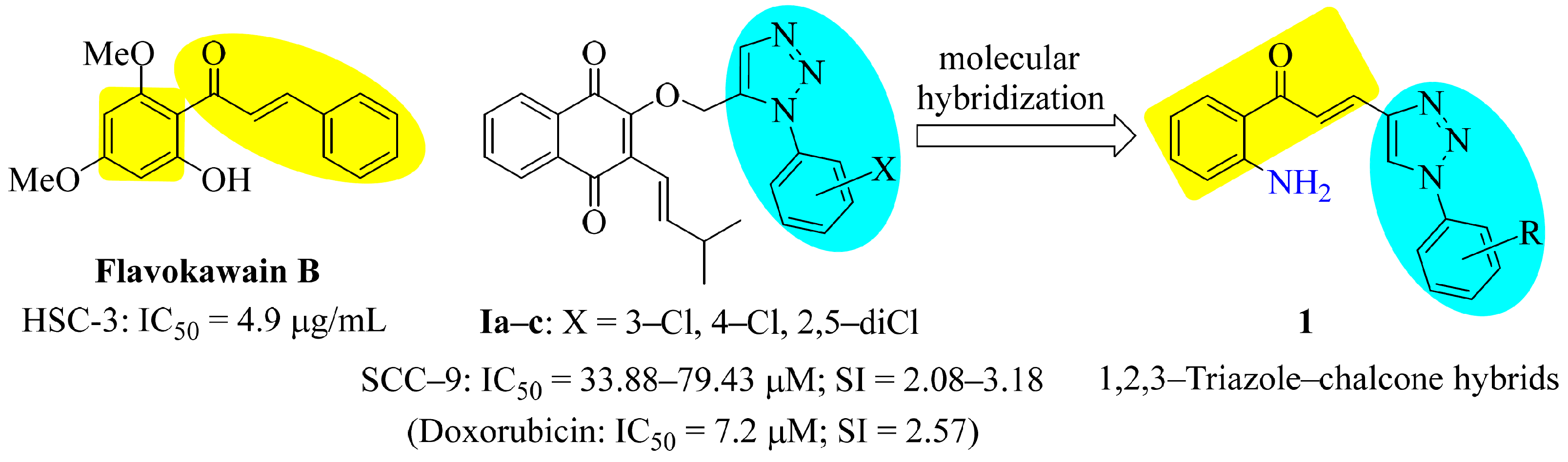
1. Introduction
2. Materials and Methods
2.1. Chemistry
2.1.1. General Remarks
2.1.2. General Procedure for Production of 1,2,3-Triazole Alcohols
Ethyl 4-(4-(hydroxymethyl)-1H-1,2,3-triazol-1-yl)benzoate
(1-(2,6-Dimethylphenyl)-1H-1,2,3-triazol-4-yl)methanol
2.1.3. General Procedure for Production of Compounds 2e and 2f
Ethyl 4-(4-Formyl-1H-1,2,3-triazol-1-yl)benzoate (2e)
1-(2,6-Dimethylphenyl)-1H-1,2,3-triazole-4-carbaldehyde (2f)
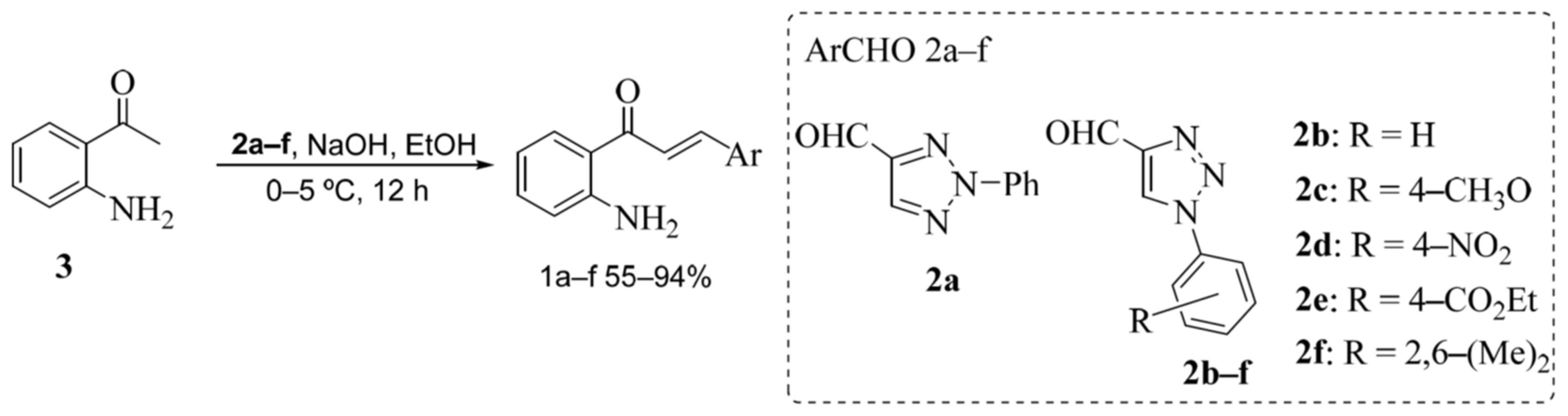
2.1.4. General Procedure for Production of Compounds 1a–f
(E)-1-(2-Aminophenyl)-3-(2-phenyl-2H-1,2,3-triazol-4-yl)prop-2-en-1-one (1a)
(E)-1-(2-Aminophenyl)-3-(1-phenyl-1H-1,2,3-triazol-4-yl)prop-2-en-1-one (1b)
(E)-1-(2-Aminophenyl)-3-(1-(4-methoxyphenyl)-1H-1,2,3-triazol-4-yl)prop-2-en-1-one (1c)
(E)-1-(2-Aminophenyl)-3-(1-(4-nitrophenyl)-1H-1,2,3-triazol-4-yl)prop-2-en-1-one (1d)
Ethyl(E)-4-(4-(3-(2-aminophenyl)-3-oxoprop-1-en-1-yl)-1H-1,2,3-triazol-1-yl)benzoate (1e)
(E)-1-(2-Aminophenyl)-3-(1-(2,6-dimethylphenyl)-1H-1,2,3-triazol-4-yl)prop-2-en-1-one (1f)
2.2. Biological Assays
2.2.1. Cells and Reagents
2.2.2. Cell Viability Assay (Cytotoxicity)
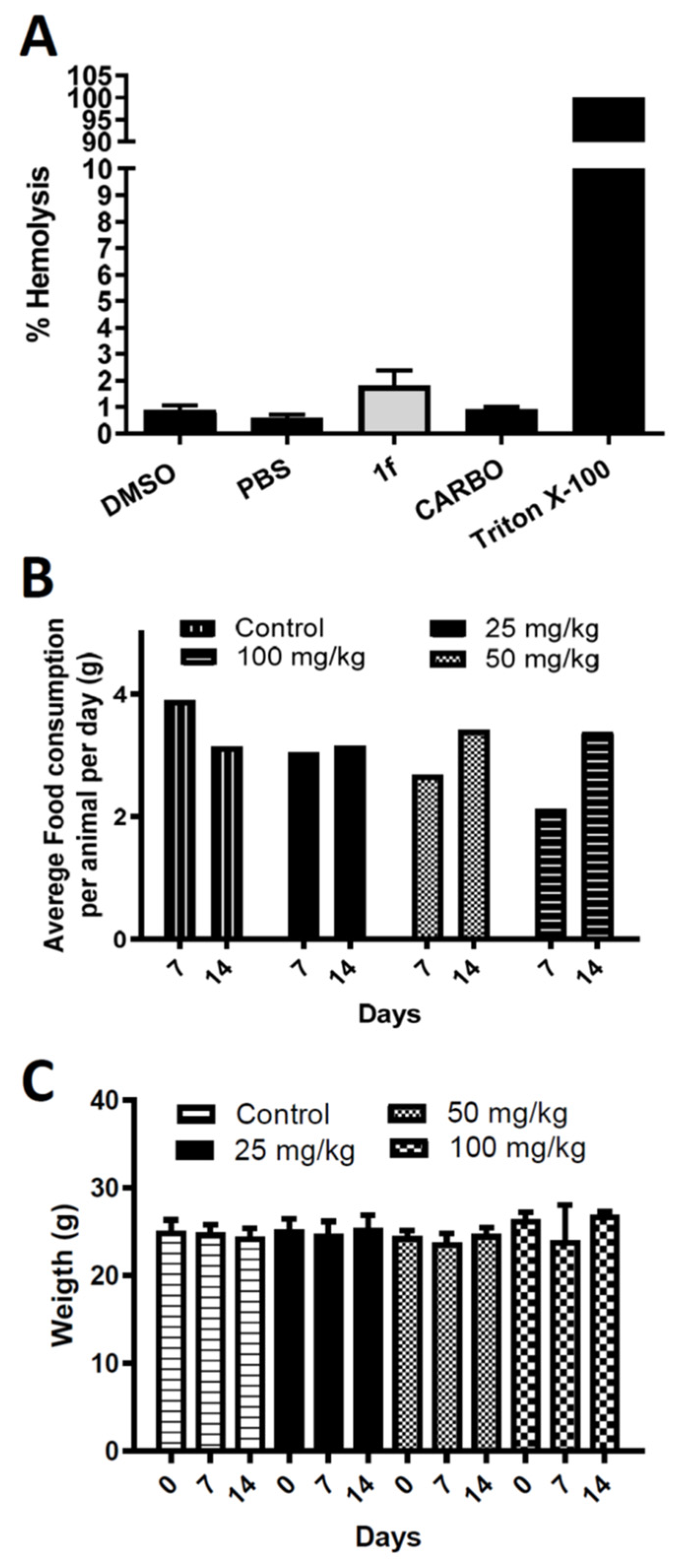
2.2.3. Hemolysis Assay
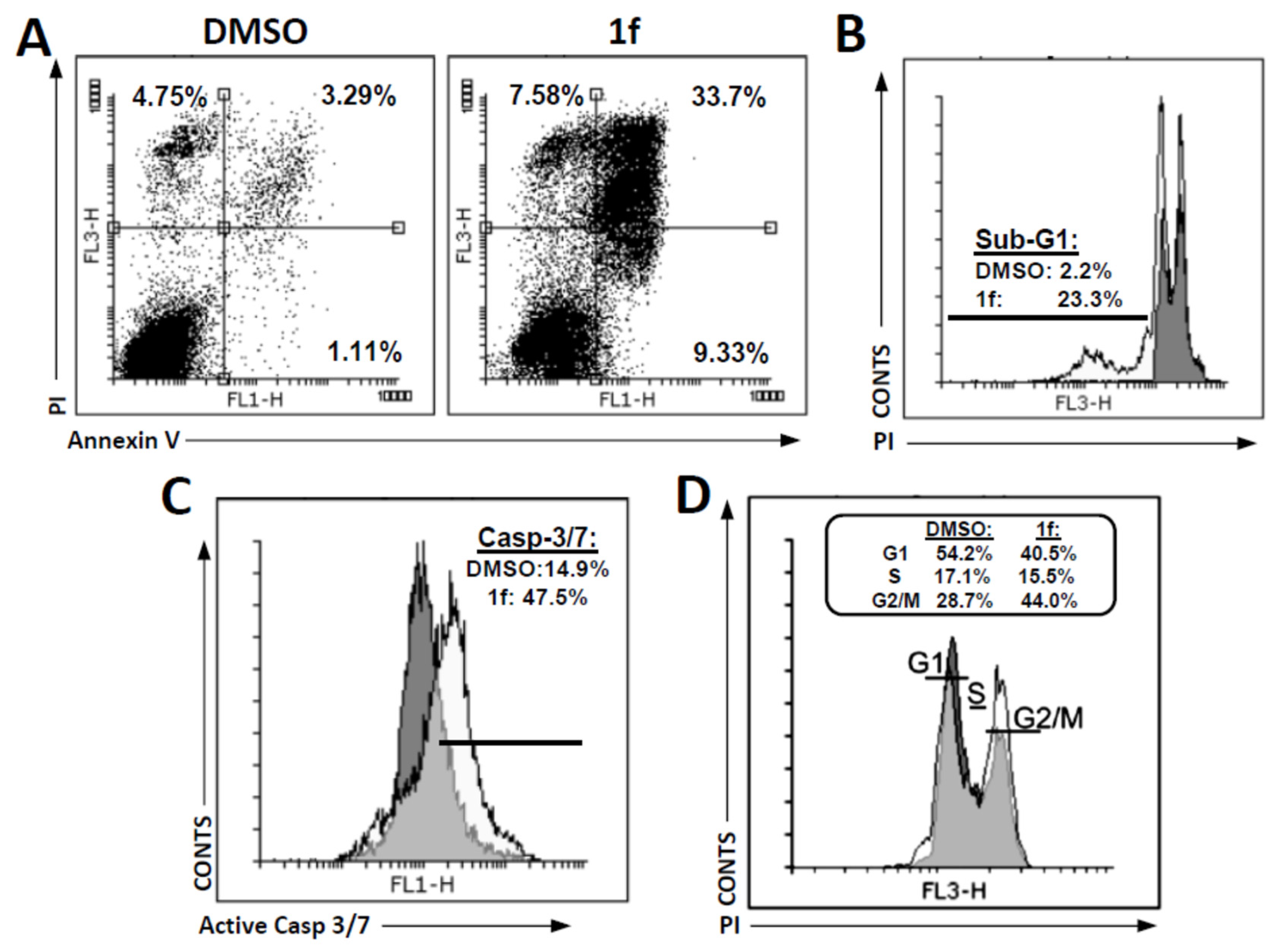
2.2.4. Cell Cycle and SubG1 Analysis
2.2.5. Apoptosis Analysis
2.2.6. Statistical Analysis, IC50 Calculation
2.2.7. In Vivo Acute Toxicity Study
2.3. In Silico Studies
2.3.1. Prediction of Toxicity and Pharmacokinetic Properties
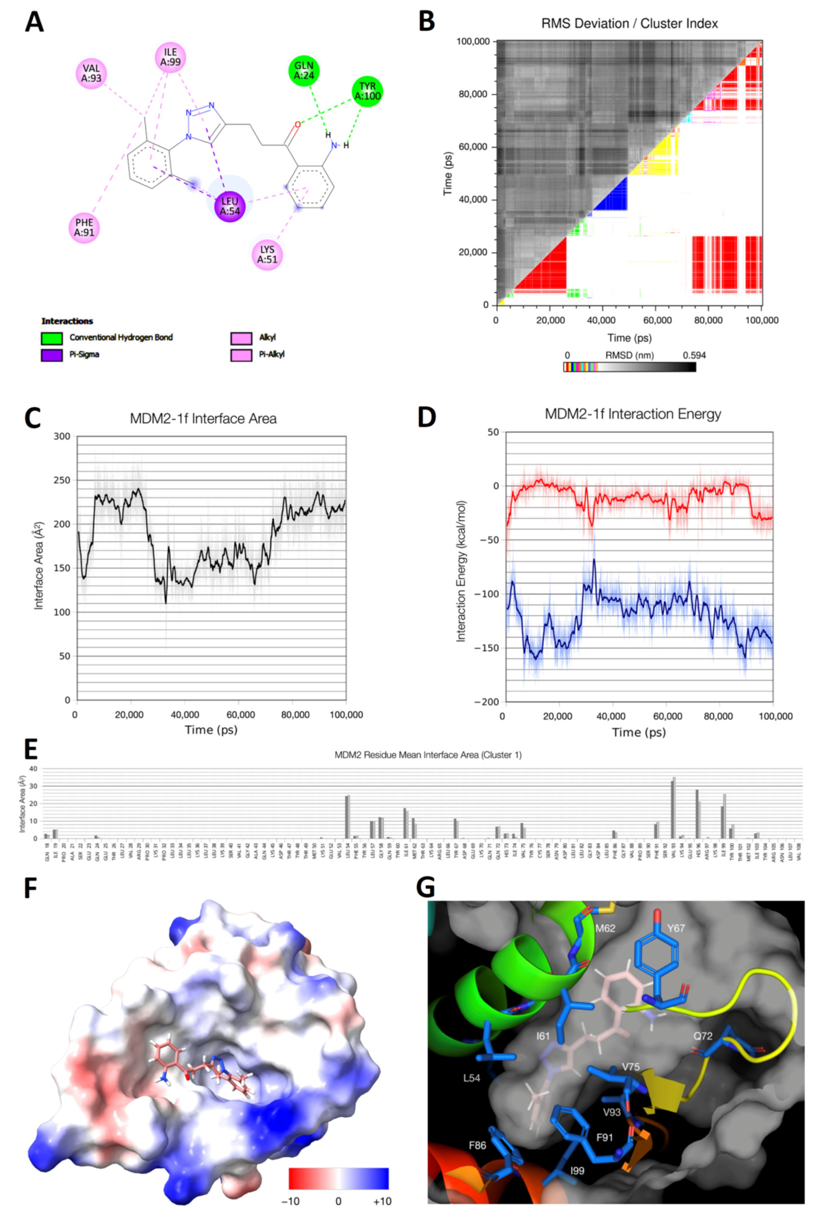
2.3.2. In Silico Docking Studies
2.3.3. Molecular Dynamics Calculation
3. Results and Discussion
3.1. Chemistry
3.2. Biological Assays
3.2.1. Cytotoxicity, Selectivity, and Hemolytic and Toxic Potential of New Chalcones
3.2.2. Prediction of Anticancer Target of 1f by Molecular Docking and Modeling
3.2.3. Prediction of Toxicity and Pharmacokinetic Properties of Compound 1f
3.2.4. Cell Death Investigation
4. Conclusions
Supplementary Materials
Author Contributions
Funding
Institutional Review Board Statement
Informed Consent Statement
Data Availability Statement
Acknowledgments
Conflicts of Interest
References
- Siegel, R.L.; Miller, K.D.; Jemal, A. Cancer statistics, 2019. CA Cancer J. Clin. 2019, 69, 7–34. [Google Scholar] [CrossRef] [PubMed]
- Sung, H.; Ferlay, J.; Siegel, R.L.; Laversanne, M.; Soerjomataram, I.; Jemal, A.; Bray, F. Global Cancer Statistics 2020: GLOBOCAN Estimates of Incidence and Mortality Worldwide for 36 Cancers in 185 Countries. CA Cancer J. Clin. 2021, 71, 209–249. [Google Scholar] [CrossRef] [PubMed]
- Chi, A.C.; Day, T.A.; Neville, B.W. Oral cavity and oropharyngeal squamous cell carcinoma-an update. CA Cancer J. Clin. 2015, 65, 401–421. [Google Scholar] [CrossRef] [PubMed]
- Chai, A.W.Y.; Lim, K.P.; Cheong, S.C. Translational genomics and recent advances in oral squamous cell carcinoma. Semin. Cancer Biol. 2020, 61, 71–83. [Google Scholar] [CrossRef]
- Li, C.C.; Shen, Z.; Bavarian, R.; Yang, F.; Bhattacharya, A. Oral Cancer: Genetics and the Role of Precision Medicine. Dent. Clin. N. Am. 2018, 62, 29–46. [Google Scholar] [CrossRef]
- Society, A.C. Treating Oral Cavity and Oropharyngeal Cancer. 2023. Available online: https://www.cancer.org/cancer/types/oral-cavity-and-oropharyngeal-cancer/treating.html (accessed on 5 May 2023).
- Güneri, P.; Epstein, J.B. Late stage diagnosis of oral cancer: Components and possible solutions. Oral Oncol. 2014, 50, 1131–1136. [Google Scholar] [CrossRef]
- Cragg, G.M.; Newman, D.J. Plants as a source of anti-cancer agents. J. Ethnopharmacol. 2005, 100, 72–79. [Google Scholar] [CrossRef] [PubMed]
- Hashem, S.; Ali, T.A.; Akhtar, S.; Nisar, S.; Sageena, G.; Ali, S.; Al-Mannai, S.; Therachiyil, L.; Mir, R.; Elfaki, I.; et al. Targeting cancer signaling pathways by natural products: Exploring promising anti-cancer agents. Biomed. Pharmacother. 2022, 150, 113054. [Google Scholar] [CrossRef] [PubMed]
- Guo, M.; Jin, J.; Zhao, D.; Rong, Z.; Cao, L.-Q.; Li, A.-H.; Sun, X.-Y.; Jia, L.-Y.; Wang, Y.-D.; Huang, L.; et al. Research Advances on Anti-Cancer Natural Products. Front. Oncol. 2022, 12, 866154. [Google Scholar] [CrossRef] [PubMed]
- Sharma, P.; Malhi, D.S.; Sohal, H.S. Biological potencies of chalcones in medicinal chemistry. Mater. Today Proc. 2022, 68, 899–904. [Google Scholar] [CrossRef]
- de Souza, P.S.; Bibá, G.C.C.; Melo, E.D.D.N.; Muzitano, M.F. Chalcones against the hallmarks of cancer: A mini-review. Nat Prod Res. 2022, 36, 4809–4826. [Google Scholar] [CrossRef]
- WalyEldeen, A.A.; Sabet, S.; El-Shorbagy, H.M.; Abdelhamid, I.A.; Ibrahim, S.A. Chalcones: Promising therapeutic agents targeting key players and signaling pathways regulating the hallmarks of cancer. Chem. Interact. 2023, 369. [Google Scholar] [CrossRef]
- Hseu, Y.C.; Lee, M.S.; Wu, C.R.; Cho, H.J.; Lin, K.Y.; Lai, G.H.; Wang, S.Y.; Kuo, Y.H.; Kumar, K.J.; Yang, H.L. The chalcone flavokawain B induces G2/M cell-cycle arrest and apoptosis in human oral carcinoma HSC-3 cells through the intracellular ROS generation and downregulation of the Akt/p38 MAPK signaling pathway. J. Agric. Food Chem. 2012, 60, 2385–2397. [Google Scholar] [CrossRef]
- Seo, J.-H.; Choi, H.W.; Oh, H.-N.; Lee, M.-H.; Kim, E.; Yoon, G.; Cho, S.-S.; Park, S.-M.; Cho, Y.S.; Chae, J.; et al. Licochalcone D directly targets JAK2 to induced apoptosis in human oral squamous cell carcinoma. J. Cell. Physiol. 2018, 234, 1780–1793. [Google Scholar] [CrossRef]
- Oh, H.-N.; Oh, K.B.; Lee, M.-H.; Seo, J.-H.; Kim, E.; Yoon, G.; Cho, S.-S.; Cho, Y.S.; Choi, H.W.; Chae, J.-I.; et al. JAK2 regulation by licochalcone H inhibits the cell growth and induces apoptosis in oral squamous cell carcinoma. Phytomedicine 2019, 52, 60–69. [Google Scholar] [CrossRef]
- Hao, Y.; Zhang, C.; Sun, Y.; Xu, H. Licochalcone A inhibits cell proliferation, migration, and invasion through regulating the PI3K/AKT signaling pathway in oral squamous cell carcinoma. OncoTargets Ther. 2019, 12, 4427–4435. [Google Scholar] [CrossRef] [PubMed]
- Gao, F.; Huang, G.; Xiao, J. Chalcone hybrids as potential anticancer agents: Current development, mechanism of action, and structure-activity relationship. Med. Res. Rev. 2020, 40, 2049–2084. [Google Scholar] [CrossRef] [PubMed]
- Ouyang, Y.; Li, J.; Chen, X.; Fu, X.; Sun, S.; Wu, Q. Chalcone Derivatives: Role in Anticancer Therapy. Biomolecules 2021, 11, 894. [Google Scholar] [CrossRef] [PubMed]
- Constantinescu, T.; Lungu, C.N. Anticancer Activity of Natural and Synthetic Chalcones. Int. J. Mol. Sci. 2021, 22, 11306. [Google Scholar] [CrossRef]
- Pinheiro, S.; Pessoa, J.C.; Pinheiro, E.M.C.; Muri, E.M.F.; Filho, E.V.; Loureiro, L.B.; Freitas, M.C.R.; Silva, C.M.D., Jr.; Fiorot, R.G.; Carneiro, J.W.M.; et al. 2H-1,2,3-Triazole-chalcones as novel cytotoxic agents against prostate cancer. Bioorg. Med. Chem. Lett. 2020, 30, 127454. [Google Scholar] [CrossRef] [PubMed]
- Gurrapu, N.; Kumar, E.P.; Kolluri, P.K.; Putta, S.; Sivan, S.K.; Subhashini, N. Synthesis, biological evaluation and molecular docking studies of novel 1,2,3-triazole tethered chalcone hybrids as potential anticancer agents. J. Mol. Struct. 2020, 1217, 128356. [Google Scholar] [CrossRef]
- Othman, E.M.; Fayed, E.A.; Husseiny, E.M.; Abulkhair, H.S. Apoptosis induction, PARP-1 inhibition, and cell cycle analysis of leukemia cancer cells treated with novel synthetic 1,2,3-triazole-chalcone conjugates. Bioorganic Chem. 2022, 123, 105762. [Google Scholar] [CrossRef] [PubMed]
- Macedo, A.L.; da Silva, D.P.; Moreira, D.L.; de Queiroz, L.N.; Vasconcelos, T.; Araujo, G.F.; Kaplan, M.A.C.; Pereira, S.S.; de Almeida, E.C.; Valverde, A.L.; et al. Cytotoxicity and selectiveness of Brazilian Piper species towards oral carcinoma cells. Biomed. Pharmacother. 2018, 110, 342–352. [Google Scholar] [CrossRef] [PubMed]
- Da Fonseca, A.C.C.; de Queiroz, L.N.; Felisberto, J.S.; Ramos, Y.J.; Marques, A.M.; Wermelinger, G.F.; Pontes, B.; de Lima Moreira, D.; Robbs, B.K. Cytotoxic effect of pure compounds from Piper rivinoides Kunth against oral squamous cell carcinoma. Nat. Prod. Res. 2021, 35, 6163–6167. [Google Scholar] [CrossRef]
- Machado, T.Q.; Felisberto, J.R.S.; Guimarães, E.F.; Queiroz, G.A.; Fonseca, A.C.C.D.; Ramos, Y.J.; Marques, A.M.; Moreira, D.L.; Robbs, B.K. Apoptotic effect of beta-pinene on oral squamous cell carcinoma as one of the major compounds from essential oil of medicinal plant Piper rivinoides Kunth. Nat. Prod. Res. 2022, 36, 1636–1640. [Google Scholar] [CrossRef]
- De Queiroz, L.N.; Da Fonseca, A.C.C.; Wermelinger, G.F.; da Silva, D.P.D.; Pascoal, A.; Sawaya, A.; de Almeida, E.C.P.; do Amaral, B.S.; de Lima Moreira, D.; Robbs, B.K. New substances of Equisetum hyemale L. extracts and their in vivo antitumoral effect against oral squamous cell carcinoma. J. Ethnopharmacol. 2023, 303, 116043. [Google Scholar] [CrossRef]
- Zorzanelli, B.C.; de Queiroz, L.N.; Santos, R.M.; Menezes, L.M.; Gomes, F.C.; Ferreira, V.F.; Silva, F.D.C.D.; Robbs, B.K. Potential cytotoxic and selective effect of new benzo[b]xanthenes against oral squamous cell carcinoma. Future Med. Chem. 2018, 10, 1141–1157. [Google Scholar] [CrossRef] [PubMed]
- Cavalcanti Chipoline, I.; Carolina Carvalho da Fonseca, A.; Ribeiro Machado da Costa, G.; Pereira de Souza, M.; Won-Held Rabelo, V.; de Queiroz, L.N.; Luiz Ferraz de Souza, T.; Cardozo Paes de Almeida, E.; Alvarez Abreu, P.; Pontes, B.; et al. Molecular mechanism of action of new 1,4-naphthoquinones tethered to 1,2,3-1H-triazoles with cytotoxic and selective effect against oral squamous cell carcinoma. Bioorg. Chem. 2020, 101, 103984. [Google Scholar] [CrossRef]
- Zorzanelli, B.C.; Ouverney, G.; Pauli, F.P.; da Fonseca, A.C.C.; de Almeida, E.C.P.; de Carvalho, D.G.; Possik, P.A.; Rabelo, V.W.-H.; Abreu, P.A.; Pontes, B.; et al. Pro-Apoptotic Antitumoral Effect of Novel Acridine-Core Naphthoquinone Compounds against Oral Squamous Cell Carcinoma. Molecules 2022, 27, 5148. [Google Scholar] [CrossRef]
- Borges, A.A.; de Souza, M.P.; da Fonseca, A.C.C.; Wermelinger, G.F.; Ribeiro, R.C.B.; Amaral, A.A.P.; de Carvalho, C.J.C.; Abreu, L.S.; de Queiroz, L.N.; de Almeida, E.C.P.; et al. Chemoselective Synthesis of Mannich Adducts from 1,4-Naphthoquinones and Profile as Autophagic Inducers in Oral Squamous Cell Carcinoma. Molecules 2022, 28, 309. [Google Scholar] [CrossRef] [PubMed]
- Kozłowska, J.; Potaniec, B.; Baczyńska, D.; Żarowska, B.; Anioł, M. Synthesis and Biological Evaluation of Novel Aminochalcones as Potential Anticancer and Antimicrobial Agents. Molecules 2019, 24, 4129. [Google Scholar] [CrossRef] [PubMed]
- Santos, M.B.; Pinhanelli, V.C.; Garcia, M.A.; Silva, G.; Baek, S.J.; França, S.C.; Fachin, A.L.; Marins, M.; Regasini, L.O. Antiproliferative and pro-apoptotic activities of 2′- and 4′-aminochalcones against tumor canine cells. Eur. J. Med. Chem. 2017, 138, 884–889. [Google Scholar] [CrossRef]
- Silva, R.H.N.; Machado, T.Q.; da Fonseca, A.C.C.; Tejera, E.; Perez-Castillo, Y.; Robbs, B.K.; de Sousa, D.P. Molecular Modeling and In Vitro Evaluation of Piplartine Analogs against Oral Squamous Cell Carcinoma. Molecules 2023, 28, 1675. [Google Scholar] [CrossRef]
- Faget, D.V.; Lucena, P.I.; Robbs, B.K.; Viola, J.P.B. NFAT1 C-Terminal Domains Are Necessary but Not Sufficient for Inducing Cell Death. PLoS ONE 2012, 7, e47868. [Google Scholar] [CrossRef] [PubMed]
- Parasuraman, S. Toxicological screening. J. Pharmacol. Pharmacother. 2011, 2, 74–79. [Google Scholar] [PubMed]
- Daina, A.; Michielin, O.; Zoete, V. SwissADME: A free web tool to evaluate pharmacokinetics, drug-likeness and medicinal chemistry friendliness of small molecules. Sci. Rep. 2017, 7, 42717. [Google Scholar] [CrossRef]
- Lipinski, C.A.; Lombardo, F.; Dominy, B.W.; Feeney, P.J. Experimental and computational approaches to estimate solubility and permeability in drug discovery and development settings. Adv. Drug Deliv. Rev. 2001, 46, 3–26. [Google Scholar] [CrossRef]
- Anil, B.; Riedinger, C.; Endicott, J.A.; Noble, M.E.M. The structure of an MDM2–Nutlin-3a complex solved by the use of a validated MDM2 surface-entropy reduction mutant. Acta Crystallogr. Sect. D Biol. Crystallogr. 2013, 69, 1358–1366. [Google Scholar] [CrossRef] [PubMed]
- Hanwell, M.D.; Curtis, D.E.; Lonie, D.C.; Vandermeersch, T.; Zurek, E.; Hutchison, G.R. Avogadro: An advanced semantic chemical editor, visualization, and analysis platform. J. Cheminform. 2012, 4, 17. [Google Scholar] [CrossRef] [PubMed]
- Schuttelkopf, A.W.; van Aalten, D.M. PRODRG: A tool for high-throughput crystallography of protein-ligand complexes. Acta Crystallogr. D Biol. Crystallogr. 2004, 60, 1355–1363. [Google Scholar] [CrossRef]
- Morris, G.M.; Huey, R.; Lindstrom, W.; Sanner, M.F.; Belew, R.K.; Goodsell, D.S.; Olson, A.J. AutoDock4 and AutoDockTools4: Automated docking with selective receptor flexibility. J. Comput. Chem. 2009, 30, 2785–2791. [Google Scholar] [CrossRef]
- Berendsen, H.J.C.; Van Der Spoel, D.; Van Drunen, R. GROMACS: A message-passing parallel molecular dynamics implementation. Comput. Phys. Commun. 1995, 91, 43–56. [Google Scholar] [CrossRef]
- Yabe, M.; Mori, K.; Ueda, K.; Takeda, M. Development of PolyParGen Software to Facilitate the Determination of Molecular Dynamics Simulation Parameters for Polymers. J. Comput. Chem. Jpn. Int. Ed. 2019, 5, 2018-0034. [Google Scholar] [CrossRef]
- Hornak, V.; Abel, R.; Okur, A.; Strockbine, B.; Roitberg, A.; Simmerling, C. Comparison of multiple Amber force fields and development of improved protein backbone parameters. Proteins Struct. Funct. Bioinform. 2006, 65, 712–725. [Google Scholar] [CrossRef] [PubMed]
- Connolly, M.L. Analytical molecular surface calculation. J. Appl. Crystallogr. 1983, 16, 548–558. [Google Scholar] [CrossRef]
- Pettersen, E.F.; Goddard, T.D.; Huang, C.C.; Meng, E.C.; Couch, G.S.; Croll, T.I.; Morris, J.H.; Ferrin, T.E. UCSF ChimeraX: Structure visualization for researchers, educators, and developers. Protein Sci. 2021, 30, 70–82. [Google Scholar] [CrossRef]
- Schrödinger, L.; DeLano, W. PyMOL. 2020. Available online: http://www.pymol.org/pymol (accessed on 5 May 2023).
- Goud, G.L.; Ramesh, S.; Ashok, D.; Reddy, V.P.; Yogeeswari, P.; Sriram, D.; Saikrishna, B.; Manga, V. Design, synthesis, molecular-docking and antimycobacterial evaluation of some novel 1,2,3-triazolyl xanthenones. MedChemComm 2017, 8, 559–570. [Google Scholar] [CrossRef]
- Dai, Z.C.; Chen, Y.F.; Zhang, M.; Li, S.K.; Yang, T.T.; Shen, L.; Wang, J.X.; Qian, S.S.; Zhu, H.L.; Ye, Y.H. Synthesis and antifungal activity of 1,2,3-triazole phenylhydrazone derivatives. Org. Biomol. Chem. 2015, 13, 477–486. [Google Scholar] [CrossRef] [PubMed]
- Basri, D.F.; Alamin, Z.A.; Chan, K.M. Assessment of cytotoxicity and genotoxicity of stem bark extracts from Canarium odontophyllum Miq. (dabai) against HCT 116 human colorectal cancer cell line. BMC Complement. Altern. Med. 2016, 16, 36. [Google Scholar] [CrossRef] [PubMed]
- Hoque Apu, E.; Akram, S.U.; Rissanen, J.; Wan, H.; Salo, T. Desmoglein 3—Influence on oral carcinoma cell migration and invasion. Exp. Cell Res. 2018, 370, 353–364. [Google Scholar] [CrossRef] [PubMed]
- Salo, T.; Sutinen, M.; Hoque Apu, E.; Sundquist, E.; Cervigne, N.K.; de Oliveira, C.E.; Akram, S.U.; Ohlmeier, S.; Suomi, F.; Eklund, L.; et al. A novel human leiomyoma tissue derived matrix for cell culture studies. BMC Cancer 2015, 15, 981. [Google Scholar] [CrossRef]
- Mahapatra, D.K.; Bharti, S.K.; Asati, V. Anti-cancer chalcones: Structural and molecular target perspectives. Eur. J. Med. Chem. 2015, 98, 69–114. [Google Scholar] [CrossRef]
- Stoll, R.; Renner, C.; Hansen, S.; Palme, S.; Klein, C.; Belling, A.; Zeslawski, W.; Kamionka, M.; Rehm, T.; Muhlhahn, P.; et al. Chalcone derivatives antagonize interactions between the human oncoprotein MDM2 and p53. Biochemistry 2001, 40, 336–344. [Google Scholar] [CrossRef] [PubMed]
- Moll, U.M.; Petrenko, O. The MDM2-p53 Interaction. Mol. Cancer Res. 2003, 1, 1001–1008. [Google Scholar] [PubMed]
- Haupt, Y.; Maya, R.; Kazaz, A.; Oren, M. Mdm2 promotes the rapid degradation of p53. Nature 1997, 387, 296–299. [Google Scholar] [CrossRef]
- Matsumura, T.; Yoshihama, Y.; Kimura, T.; Shintani, S.; Alcalde, R.E. p53 and MDM2 expression in oral squamous cell carcinoma. Oncology 1996, 53, 308–312. [Google Scholar] [CrossRef]
- Vassilev, L.T.; Vu, B.T.; Graves, B.; Carvajal, D.; Podlaski, F.; Filipovic, Z.; Kong, N.; Kammlott, U.; Lukacs, C.; Klein, C.; et al. In vivo activation of the p53 pathway by small-molecule antagonists of MDM2. Science 2004, 303, 844–848. [Google Scholar] [CrossRef]
- Leao, M.; Soares, J.; Gomes, S.; Raimundo, L.; Ramos, H.; Bessa, C.; Queiroz, G.; Domingos, S.; Pinto, M.; Inga, A.; et al. Enhanced cytotoxicity of prenylated chalcone against tumour cells via disruption of the p53-MDM2 interaction. Life Sci. 2015, 142, 60–65. [Google Scholar] [CrossRef]
- Wade, M.; Wahl, G.M. Targeting Mdm2 and Mdmx in cancer therapy: Better living through medicinal chemistry? Mol. Cancer Res. MCR 2009, 7, 1–11. [Google Scholar] [CrossRef] [PubMed]
- Tovar, C.; Rosinski, J.; Filipovic, Z.; Higgins, B.; Kolinsky, K.; Hilton, H.; Zhao, X.; Vu, B.T.; Qing, W.; Packman, K.; et al. Small-molecule MDM2 antagonists reveal aberrant p53 signaling in cancer: Implications for therapy. Proc. Natl. Acad. Sci. USA 2006, 103, 1888–1893. [Google Scholar] [CrossRef]
- Issaeva, N.; Bozko, P.; Enge, M.; Protopopova, M.; Verhoef, L.G.; Masucci, M.; Pramanik, A.; Selivanova, G. Small molecule RITA binds to p53, blocks p53-HDM-2 interaction and activates p53 function in tumors. Nat. Med. 2004, 10, 1321–1328. [Google Scholar] [CrossRef]
- Wu, F.; Zhou, Y.; Li, L.; Shen, X.; Chen, G.; Wang, X.; Liang, X.; Tan, M.; Huang, Z. Computational Approaches in Preclinical Studies on Drug Discovery and Development. Front. Chem. 2020, 8, 726. [Google Scholar] [CrossRef]
- Hodos, R.A.; Kidd, B.A.; Shameer, K.; Readhead, B.P.; Dudley, J.T. In silico methods for drug repurposing and pharmacology. Wiley interdisciplinary reviews. Syst. Biol. Med. 2016, 8, 186–210. [Google Scholar]
- Palm, K.; Stenberg, P.; Luthman, K.; Artursson, P. Polar molecular surface properties predict the intestinal absorption of drugs in humans. Pharm. Res. 1997, 14, 568–571. [Google Scholar] [CrossRef] [PubMed]
- Alrushaid, S.; Sayre, C.L.; Yanez, J.A.; Forrest, M.L.; Senadheera, S.N.; Burczynski, F.J.; Lobenberg, R.; Davies, N.M. Pharmacokinetic and Toxicodynamic Characterization of a Novel Doxorubicin Derivative. Pharmaceutics 2017, 9, 35. [Google Scholar] [CrossRef]
- Oguri, S.; Sakakibara, T.; Mase, H.; Shimizu, T.; Ishikawa, K.; Kimura, K.; Smyth, R.D. Clinical pharmacokinetics of carboplatin. J. Clin. Pharmacol. 1988, 28, 208–215. [Google Scholar] [CrossRef] [PubMed]
- Hamaguchi, K.; Godwin, A.K.; Yakushiji, M.; O’Dwyer, P.J.; Ozols, R.F.; Hamilton, T.C. Cross-resistance to diverse drugs is associated with primary cisplatin resistance in ovarian cancer cell lines. Cancer Res. 1993, 53, 7. [Google Scholar]
- Mansilla, S.; Llovera, L.; Portugal, J. Chemotherapeutic targeting of cell death pathways. Anti Cancer Agents Med. Chem. 2012, 12, 226–238. [Google Scholar] [CrossRef] [PubMed]
- Bertheloot, D.; Latz, E.; Franklin, B.S. Necroptosis, pyroptosis and apoptosis: An intricate game of cell death. Cell. Mol. Immunol. 2021, 18, 1106–1121. [Google Scholar] [CrossRef]
- Vaseva, A.V.; Marchenko, N.D.; Moll, U.M. The transcription-independent mitochondrial p53 program is a major contributor to nutlin-induced apoptosis in tumor cells. Cell Cycle 2009, 8, 1711–1719. [Google Scholar] [CrossRef] [PubMed]
- Rigatti, M.J.; Verma, R.; Belinsky, G.S.; Rosenberg, D.W.; Giardina, C. Pharmacological inhibition of Mdm2 triggers growth arrest and promotes DNA breakage in mouse colon tumors and human colon cancer cells. Mol. Carcinog. 2012, 51, 363–378. [Google Scholar] [CrossRef] [PubMed]
- Yi, H.; Yan, X.; Luo, Q.; Yuan, L.; Li, B.; Pan, W.; Zhang, L.; Chen, H.; Wang, J.; Zhang, Y.; et al. A novel small molecule inhibitor of MDM2-p53 (APG-115) enhances radiosensitivity of gastric adenocarcinoma. J. Exp. Clin. Cancer Res. CR 2018, 37, 97. [Google Scholar] [CrossRef] [PubMed]
- Secchiero, P.; Bosco, R.; Celeghini, C.; Zauli, G. Recent advances in the therapeutic perspectives of Nutlin-3. Curr. Pharm. Des. 2011, 17, 569–577. [Google Scholar] [CrossRef] [PubMed]
- Hsu, Y.L.; Kuo, P.L.; Tzeng, W.S.; Lin, C.C. Chalcone inhibits the proliferation of human breast cancer cell by blocking cell cycle progression and inducing apoptosis. Food Chem. Toxicol. Int. J. Publ. Br. Ind. Biol. Res. Assoc. 2006, 44, 704–713. [Google Scholar] [CrossRef] [PubMed]
- Ramirez-Tagle, R.; Escobar, C.A.; Romero, V.; Montorfano, I.; Armisen, R.; Borgna, V.; Jeldes, E.; Pizarro, L.; Simon, F.; Echeverria, C. Chalcone-Induced Apoptosis through Caspase-Dependent Intrinsic Pathways in Human Hepatocellular Carcinoma Cells. Int. J. Mol. Sci. 2016, 17, 260. [Google Scholar] [CrossRef]


| Compounds | SCC9—Oral Cancer | |
|---|---|---|
| IC50 (µM) | S.D. | |
| 1a | 9.95 | 0.04 |
| 1b | 12.72 | 0.05 |
| 1c | 9.32 | 0.03 |
| 1d | N.D. | N.D. |
| 1e | N.D. | N.D. |
| 1f | 3.87 | 0.06 |
| Carboplatin | 155.67 | 0.07 |
| Doxorubicin | 2.99 | 0.06 |
| Oral Tumor Cells | Primary Gingival Fibroblast (HGF) | Average S.I. | |||||||||||
|---|---|---|---|---|---|---|---|---|---|---|---|---|---|
| Compound | SCC9 | SCC25 | SCC4 | Average (IC50) | |||||||||
| IC50 | S.D. | S.I. | IC50 | S.D. | S.I. | IC50 | S.D. | S.I. | IC50 | S.D. | |||
| 1b | 12.72 | 0.05 | 2.80 | 12.53 | 0.02 | 2.85 | 12.62 | 0.02 | 2.83 | 12.62 | 35.66 | 0.03 | 2.82 |
| 1f | 3.87 | 0.06 | 7.63 | 5.03 | 0.03 | 5.88 | 4.73 | 0.02 | 6.25 | 4.54 | 29.56 | 0.06 | 6.51 |
| Carboplatin | 155.67 | 0.05 | 2.15 | 190.85 | 0.04 | 1.75 | 148.04 | 0.04 | 2.26 | 164.63 | 334.54 | 0.04 | 2.03 |
| Doxorubicin | 2.99 | 0.06 | 1.16 | 0.90 | 0.07 | 3.86 | 0.57 | 0.09 | 6.09 | 1.49 | 3.47 | 0.25 | 2.34 |
| compound 1f | |||
|---|---|---|---|
| Tumor Cell Type | IC50 | S.D. | S.I. |
| HCT-116 (colon cancer) | 3.99 | 0.058 | 7.41 |
| HT29 (adenocarcinoma) | 3.92 | 0.076 | 7.55 |
| HEP2G (hepatocarcinoma) | 39.38 | 0.108 | 2.56 |
| Compounds | cLogP | nON | nOH/NH | MW | Lipinski’s Violations a | TPSA (Å2) | Oral Bioavailability | P-Glycoprotein Inhibitor | P-Glycoprotein Substrate |
|---|---|---|---|---|---|---|---|---|---|
| 1f | 1.78 | 5 | 3 | 364 | 0 | 114.26 | +0.67 | −0.54 | −0.72 |
| butlin-3a | 3.94 | 5 | 2 | 583.51 | 1 | 83.14 | +0.53 | +0.90 | +0.82 |
| Doxorubicin | −2.10 | 12 | 6 | 543 | 3 | 206.1 | −0.91 | −0.92 | +0.95 |
| Carboplatin | −1.79 | 6 | 4 | 371 | 0 | 126.6 | −0.60 | −0.99 | −0.99 |
| SCC9 | HT29 | Fibroblast (HGF) | ||||||
|---|---|---|---|---|---|---|---|---|
| Compound | IC50 | S.D. | S.I. | IC50 | S.D. | S.I. | IC50 | S.D. |
| 1f | 3.88 | 0.06 | 7.63 | 3.92 | 0.76 | 7.55 | 29.56 | 0.06 |
| nutlin-3a | 17.99 | 0.043 | 2.72 | 6.79 | 0.084 | 7.22 | 49.06 | 0.05 |
Disclaimer/Publisher’s Note: The statements, opinions and data contained in all publications are solely those of the individual author(s) and contributor(s) and not of MDPI and/or the editor(s). MDPI and/or the editor(s) disclaim responsibility for any injury to people or property resulting from any ideas, methods, instructions or products referred to in the content. |
© 2023 by the authors. Licensee MDPI, Basel, Switzerland. This article is an open access article distributed under the terms and conditions of the Creative Commons Attribution (CC BY) license (https://creativecommons.org/licenses/by/4.0/).
Share and Cite
Wermelinger, G.F.; Rubini, L.; da Fonseca, A.C.C.; Ouverney, G.; de Oliveira, R.P.R.F.; de Souza, A.S.; Forezi, L.S.M.; Limaverde-Sousa, G.; Pinheiro, S.; Robbs, B.K. A Novel MDM2-Binding Chalcone Induces Apoptosis of Oral Squamous Cell Carcinoma. Biomedicines 2023, 11, 1711. https://doi.org/10.3390/biomedicines11061711
Wermelinger GF, Rubini L, da Fonseca ACC, Ouverney G, de Oliveira RPRF, de Souza AS, Forezi LSM, Limaverde-Sousa G, Pinheiro S, Robbs BK. A Novel MDM2-Binding Chalcone Induces Apoptosis of Oral Squamous Cell Carcinoma. Biomedicines. 2023; 11(6):1711. https://doi.org/10.3390/biomedicines11061711
Chicago/Turabian StyleWermelinger, Guilherme Freimann, Lucas Rubini, Anna Carolina Carvalho da Fonseca, Gabriel Ouverney, Rafael P. R. F. de Oliveira, Acácio S. de Souza, Luana S. M. Forezi, Gabriel Limaverde-Sousa, Sergio Pinheiro, and Bruno Kaufmann Robbs. 2023. "A Novel MDM2-Binding Chalcone Induces Apoptosis of Oral Squamous Cell Carcinoma" Biomedicines 11, no. 6: 1711. https://doi.org/10.3390/biomedicines11061711
APA StyleWermelinger, G. F., Rubini, L., da Fonseca, A. C. C., Ouverney, G., de Oliveira, R. P. R. F., de Souza, A. S., Forezi, L. S. M., Limaverde-Sousa, G., Pinheiro, S., & Robbs, B. K. (2023). A Novel MDM2-Binding Chalcone Induces Apoptosis of Oral Squamous Cell Carcinoma. Biomedicines, 11(6), 1711. https://doi.org/10.3390/biomedicines11061711