Article

Implication of the PTN/RPTPβ/ζ Signaling Pathway in Acute Ethanol Neuroinflammation in Both Sexes: A Comparative Study with LPS

by
María Rodríguez-Zapata
1,
Milagros Galán-Llario
1,
Héctor Cañeque-Rufo
1,2,
Julio Sevillano
2,
María Gracia Sánchez-Alonso
2,
José M. Zapico
2,
Marcel Ferrer-Alcón
3,
María Uribarri
3,
Beatriz de Pascual-Teresa
2,
María del Pilar Ramos-Álvarez
2,
Gonzalo Herradón
1,4,
Carmen Pérez-García
1,4 and
Esther Gramage
1,4,*
1
Departamento de Ciencias Farmacéuticas y de la Salud, Facultad de Farmacia, Universidad San Pablo-CEU, CEU Universities, Urbanización Montepríncipe, Boadilla del Monte, 28660 Madrid, Spain
2
Departamento de Química y Bioquímica, Facultad de Farmacia, Universidad San Pablo-CEU, CEU Universities, Urbanización Montepríncipe, Boadilla del Monte, 28660 Madrid, Spain
3
BRAINco Biopharma, S.L., Bizkaia Technology Park, Zamudio, 48170 Vizcaya, Spain
4
Instituto de Estudios de las Adicciones, Universidad San Pablo-CEU, CEU Universities, Urbanización Montepríncipe, Boadilla del Monte, 28660 Madrid, Spain
*
Author to whom correspondence should be addressed.
Biomedicines 2023, 11(5), 1318; https://doi.org/10.3390/biomedicines11051318
Submission received: 31 December 2022 / Revised: 16 March 2023 / Accepted: 26 April 2023 / Published: 28 April 2023
(This article belongs to the Special Issue Biological Aspects of Drug Addiction 2.0)

Abstract

:
Binge drinking during adolescence increases the risk of alcohol use disorder, possibly by involving alterations of neuroimmune responses. Pleiotrophin (PTN) is a cytokine that inhibits Receptor Protein Tyrosine Phosphatase (RPTP) β/ζ. PTN and MY10, an RPTPβ/ζ pharmacological inhibitor, modulate ethanol behavioral and microglial responses in adult mice. Now, to study the contribution of endogenous PTN and the implication of its receptor RPTPβ/ζ in the neuroinflammatory response in the prefrontal cortex (PFC) after acute ethanol exposure in adolescence, we used MY10 (60 mg/kg) treatment and mice with transgenic PTN overexpression in the brain. Cytokine levels by X-MAP technology and gene expression of neuroinflammatory markers were determined 18 h after ethanol administration (6 g/kg) and compared with determinations performed 18 h after LPS administration (5 g/kg). Our data indicate that Ccl2, Il6, and Tnfa play important roles as mediators of PTN modulatory actions on the effects of ethanol in the adolescent PFC. The data suggest PTN and RPTPβ/ζ as targets to differentially modulate neuroinflammation in different contexts. In this regard, we identified for the first time important sex differences that affect the ability of the PTN/RPTPβ/ζ signaling pathway to modulate ethanol and LPS actions in the adolescent mouse brain.

1. Introduction

Binge drinking is a common practice worldwide and contributes to an increased risk of developing alcohol use disorder (AUD). In 2020, 61.1 million people were binge alcohol (ethanol) users [1]. Specifically, in Spain, up to 70.5% of adolescents reported ethanol consumption during the last 12 months. It is important to note that gender differences regarding the prevalence of consumption and the prevalence of binge drinking have been reported, both higher in adolescent girls than in boys [2]. Adolescence is a critical period for consumption because the brain is more vulnerable to the damaging effects of ethanol [3,4]. Evidence obtained from experimental animals has shown that binge-like ethanol exposure during adolescence impairs prefrontal cortex (PFC) maturation and hippocampal neurogenesis and induces synaptic and myelin disarrangements [5,6].
Different studies suggest the role of neuroinflammation in the pathophysiology of excessive ethanol consumption [7,8]. However, the molecular mechanisms of ethanol actions in the adolescent brain remain elusive. It is important to note that females seem to be remarkably vulnerable to ethanol damage [5,9,10], and more susceptible than males to the neurodevelopmental dysfunction associated with heavy ethanol use in adolescence [11].
Ethanol activates the neuroimmune system and the signaling pathway of toll-like receptor 4 (TLR4) in glial cells and contributes to the release of cytokines and proinflammatory mediators [5,12]. Among others, Interleukin 6 (IL6), Interleukin 1β (IL1β), and tumor necrosis factor α (TNFα) are key elements of the proinflammatory response to ethanol [13,14,15]. In addition, monocyte chemoattractant protein 1 (MCP1), one of the most potent microglia chemokines, which is produced by many other central nervous system (CNS) cell types, has emerged as an important factor that regulates neuroinflammation in different contexts [16,17]. Acute ethanol treatment increases brain mRNA levels of Il6, Il1β, Tnfa, and Ccl2 (MCP1) proinflammatory genes in mice. However, these changes together with microglial alterations seem to be region-specific and sensitive to timing [9,14,15,18].
The genetic elimination of TLR4, whose most relevant ligand is lipopolysaccharide (LPS), modulates neuroinflammation and other ethanol-deleterious effects in the mouse brain [5,19]. In addition, during acute ethanol intoxication, an association between elevated cytokine/chemokine levels and the upregulation of TLR4 mRNA levels in total blood was found exclusively in females, pointing out important sex differences in the immune response to ethanol [11]. Moreover, recent studies have shown an increase in blood LPS levels after ethanol binge drinking, suggesting a connection between the release of the endotoxin through systemic circulation and the neuroinflammation induced by the drug [20]. However, as ethanol itself can trigger the TLR4 response in microglia [21,22], further studies are needed to clarify the specific contribution of ethanol and the release of LPS in this neuroinflammatory process.
Pleiotrophin (PTN) is a cytokine that plays a key role in CNS repair and the differentiation of neurons. It is part of the Ptn/Mdk developmental gene family, sharing over 50% identity in amino acid sequence with Midkine (MK) and hence many functions [23]. Interestingly, PTN is upregulated in the mouse brain after the administration of ethanol [24,25] and MK expression is higher in the frontal cortex of human alcoholics [26]. In fact, both cytokines are involved in the modulation of the neuroimmune response and neuroinflammation induced by different drugs of abuse [27]. In addition, we demonstrated that in Ptn knockout mice (Ptn−/−), the conditioning effects of ethanol are significantly increased, whereas the transgenic overexpression of Ptn in the mouse brain (Ptn-Tg) blocks the rewarding effects of this drug [25]. This evidence suggests that PTN/MK may regulate ethanol effects through the modulation of neuroinflammation.
PTN and MK exert their actions through different receptors [27]. One of them, the Receptor Protein Tyrosine Phosphatase β/ζ (RPTPβ/ζ, also known as PTPRZ1) is mainly expressed in the CNS in important areas for ethanol effects, such as the PFC, amygdala, or hippocampus [28]. Accordingly, RPTPβ/ζ is one of the most relevant receptors in the modulation of PTN effects in neuroinflammation [27]. The binding of PTN to RPTPβ/ζ induces its dimerization, and thus inactivates its phosphatase activity [29], increasing the phosphorylation levels of the RPTPβ/ζ substrates [30,31,32]. Some of them are known to play important roles in ethanol consumption such as anaplastic lymphoma kinase (ALK) [33] and Fyn kinase [34]. ALK has also been identified as a PTN receptor [35]. On the other hand, it has been proven that Fyn kinase participates in the triggered signaling pathway in the dorsal striatum during ethanol exposure. Moreover, this kinase plays an important role in the molecular mechanisms implicated in ethanol self-administration [23,36]. Interestingly, Fyn kinase activation participates in LPS signaling through TLR4 and in the consequent neuroinflammation process [37]. On the other hand, the pharmacological inhibition of ALK reduces ethanol binge-drinking behavior in rodents [33]. The specific contribution of the PTN/RPTPβ/ζ signaling pathway in the ethanol-induced neuroimmune response remains unclear.
Recently, we designed and synthesized a blood–brain barrier (BBB) permeable selective inhibitor of RPTPβ/ζ, named MY10. This compound has shown similar biological activities in vivo to PTN, the physiological inhibitory ligand of RPTPβ/ζ. MY10 interacts with the intracellular domain PD1 of the receptor and inhibits its tyrosine phosphatase activity, thus mimicking the inhibitory action of PTN [38]. The administration of MY10 in mice has been demonstrated to reduce ethanol consumption and to block ethanol-reinforcing actions [39,40,41], an effect also found in mice with Ptn overexpression in the brain (Ptn-Tg mice) [25]. Accordingly, RPTPβ/ζ inhibition by MY10 replicates PTN actions, increasing LPS-induced microglial response (Iba1 immunoreactivity after 7.5 mg/kg of LPS) in the PFC of adult mice [42,43]. Recently, we demonstrated that the pharmacological inhibition of the phosphatase activity of RPTPβ/ζ fully prevented the loss of hippocampal neurogenesis induced by acute ethanol exposure [44]. Moreover, that work suggested an important role of RPTPβ/ζ function in ethanol-induced immune responses in the hippocampus in a sex-dependent manner. Therefore, the aim of the present study is to elucidate the implication of the PTN/RPTPβ/ζ signaling pathway in ethanol-induced neuroinflammation in the PFC of adolescent male and female mice. In addition, we aimed to explore the possible similarities with the LPS-induced neuroinflammatory process.
Our results suggest that the PTN/RPTPβ/ζ signaling pathway differently participates in the neuroinflammatory processes induced by ethanol and LPS. Moreover, possible differences in RPTPβ/ζ function in neuroinflammation in males and females are suggested.

2. Materials and Methods

2.1. Animals

Ptn-Tg mice on a C57BL/6J background were generated by pronuclear injection as previously described [25]. The acceptor vector contained the regulatory regions responsible for tissue-specific expression of the Thy-1 gene, which drives neuron-specific expression of transgenes [45,46]. Ptn-specific overexpression in different brain areas of adult mice, including a ~2–3-fold upregulation in the prefrontal cortex (PFC) and a 20% increase in PTN protein levels in the striatum, was established by quantitative real-time polymerase chain reaction (qRT-PCR), in situ hybridization, and by Western blot [25,42,47].
Male and female C57BL/6J (wild type, Ptn+/+) and Ptn-Tg mice of 5–6 weeks of age (15–22 g) were used. Mice were housed under controlled environmental conditions (22 ± 1 °C and a 12-h light/12-h dark cycle) with free access to food and water. All the animals used in this study were maintained in accordance with the ARRIVE guidelines and with the European Union Laboratory Animal Care Rules (2010/63/EU directive), and protocols were approved by the Animal Research Committee of USP-CEU (authorization reference: PROEX 76.0/20).

2.2. Treatments

The RPTPβ/ζ inhibitor MY10 was synthesized as previously described [37]. Female and male Ptn+/+ mice were administered with 60 mg/kg MY10 or vehicle (Veh; 10% dehydrated ethanol, 20% polysorbate 80, 70% PEG-300) by oral gavage in a volume of 0.1 mL one hour before a single i.p. injection of ethanol (6 g/kg, 25% v/v), LPS (5 mg/kg, 10 mL/kg) or saline (control). MY10 treatment was based on previous studies with this compound, in which we demonstrated that this dose and route of administration reduces ethanol consumption, prevents ethanol-induced loss of hippocampal neurogenesis, and provides good bioavailability and BBB penetration in mice [38,39,41,44]. The dose of ethanol was chosen to achieve in mice Blood Ethanol Concentrations (BECs) equivalent to those found in human adolescents after binge alcohol drinking (peak blood concentration, 700–750 mg/dL) [48,49]. All animals were fully recovered at the time of sacrifice, 18 h after ethanol administration.
To study the effect of the endogenous overexpression of PTN, Ptn-Tg mice were compared with Ptn+/+ with the same treatment. On the other hand, the effect of the inhibition of RPTPβ/ζ by MY10 treatment was evaluated only in Ptn+/+ mice. To minimize the number of animals used in the study and apply the 3Rs principles, the same control mice were used in both sets of experiments. For that reason, all the mice used in the study received an oral administration of the vehicle independently of the genotype (except for Ptn+/+ mice treated with MY10).
Mice were decapitated 18 h after ethanol, LPS, or saline administration and PFC were rapidly removed and frozen in dry ice and stored at −80 °C until the procedures. To quantify protein levels of cytokines, ethanol-treated mice and their controls were used (n = 3–7/group/sex). For tissular mRNA expression analysis, ethanol- and LPS-treated mice and their controls were used (n = 5–6/group/sex).

2.3. Cytokine Levels

Approximately 5 mg of PFC were homogenized in 100 μL of homogenization buffer (0.05% Tween 20 and protease inhibitor cocktail (Thermo Fisher Scientific Inc., Waltham, MA, USA) in PBS, pH 7.2). Samples were processed as previously described [43] and the total protein content of each sample was measured using the BCA protein assay kit (Thermo Fisher Scientific Inc., Waltham, MA, USA). Levels of IL1β, IL6, MCP1, and TNFα were measured by multiplex luminometry (Beadlyte mouse multiplex cytokine detection system, MHSTCMAG-70K, Merck Millipore, Spain) according to the manufacturer’s description.

2.4. Quantitative Real-Time PCR

Total RNA from PFC was isolated using the Total RNA Isolation Kit (Nzytech, Lisbon, Portugal). First-strand cDNA was synthesized using the first-strand cDNA Synthesis Kit (Nzytech). Quantitative real-time PCR analysis was performed using the SYBR green method (Quantimix Easy kit, Biotools, Madrid, Spain) in a CFX96 Real-Time System (Bio-Rad, Hercules, CA, USA). The relative expression of each gene, in brain tissue, was normalized using Rpl37. The primer sequences are shown in Table 1.

2.5. Statistical Analysis

All statistical analyses were performed using Graphpad Prism version 8 (San Diego, CA, USA). Data are presented as mean ± standard error of the mean (SEM).
To elucidate the role of endogenous PTN, Ptn-Tg mice were compared with wild type (Ptn+/+) mice, and the results were analyzed using a three-way ANOVA, considering treatment, genotype, and sex as variables. To better discern the effect of the treatment and of the genotype, when a significant interaction between treatment and genotype variables was found and the absence of a sex effect was confirmed, we performed a two-way ANOVA, excluding the sex factor. When three-way ANOVA revealed that sex and/or treatment variables were significant, it was properly represented in the graph. No further comparisons were performed in those cases. For detailed results of these statistical analyses see Supplementary Tables S2 and S4.
The implication of the RPTPβ/ζ signaling pathway was assessed through the pharmacological inhibition of the receptor with MY10. In this case, data were analyzed using a two-way ANOVA, having as variables treatment and sex (Tables S3 and S5).
To comparatively study the expression of RPTPβ/ζ signaling pathway components in ethanol and LPS models, males and females were separately analyzed. Two-way ANOVA was used when results from Ptn-Tg mice were included in the analysis (considering treatment and genotype as variables), and one-way ANOVA was performed when only different treatments were tested in wild type (Ptn+/+) mice (Table S6).
All references to statistical significance made to the three-way, two-way, and one-way ANOVA test’s individual factors or their interaction are shown in Supplementary Tables S1–S6. In the figures, only significant differences revealed by post-hoc comparisons with Bonferroni’s post-hoc test are represented.

3. Results

3.1. Ptn Overexpression Attenuates Acute Ethanol-Induced MCP1 Increment in the PFC of Male and Female Mice

To explore the implication of the overexpression of endogenous PTN (Ptn-Tg mice) and the specific role of RPTPβ/ζ (MY10 treatment) in the ethanol-induced neuroinflammatory process, cytokine levels were measured through multiplex luminometry in the PFC of adolescent mice 18 h after a single injection of ethanol (6 g/kg, i.p.) (Figure 1). An effect of the genotype for all the variables, together with an effect of the interaction sex x genotype for IL1B and TNFα was found (Table S1). No remarkable changes were observed in the protein levels of IL1β (Figure 1a,b) and TNFα (Figure 1g,h) after ethanol administration. However, acute ethanol tended to increase IL6 levels in the PFC of mice from all the experimental groups (Figure 1c,d). ANOVA revealed that treatment with ethanol significantly affected the PFC levels of IL6 in Ptn-Tg comparisons (Figure 1c and Table S1a) and in MY10-treated groups (Figure 1d and Table S1c). In addition, three-way ANOVA showed a significant effect of both genotype and treatment on MCP1 (Figure 1e) levels in PFC and a significant interaction between variables (Table S1a,b). An ethanol-induced increase in MCP1 in Ptn+/+ mice was prevented in Ptn-Tg mice (Figure 1e). Interestingly, this effect tends to be replicated in Ptn+/+ male mice when RPTP is blocked by MY10, but seems to be sex-dependent, as it was not observed in females (Figure 1f).

3.2. Ptn Overexpression Potentiates the Increases of Il6 and Ccl2 mRNA Levels in Female Mice and Blocks the Upregulation of Tnfa Expression in Males after Acute Ethanol Exposure

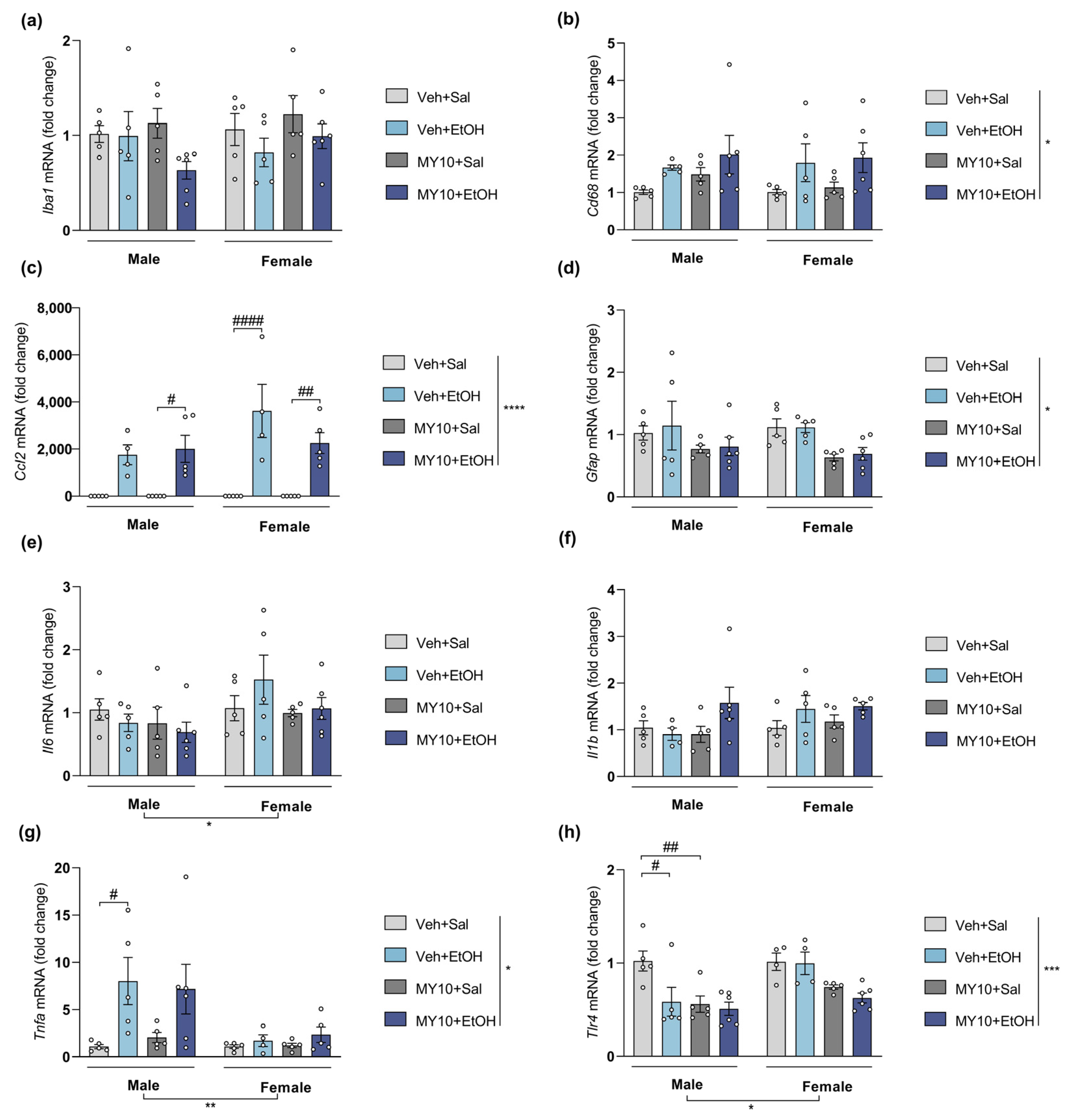
First, the effect of Ptn overexpression was studied by comparing the actions of ethanol on Ptn+/+ and Ptn-Tg adolescent mice (Figure 2). Three-way ANOVA revealed that ethanol treatment significantly affected mRNA levels of the microglia markers Iba1 (Figure 2a) and Cd68 (Figure 2b), as well as the proinflammatory marker Il1b (Figure 2f). However, ethanol did not modify Gfap levels of expression (Figure 2d), and only a significant effect of genotype was found. Detailed results of the statistical analysis are included in Table S2. In addition, the mRNA level of Il1b was significantly affected by sex. Adolescent female mice showed higher levels of Il1b than males (Figure 2f). A similar effect was found when Tlr4 levels of expression were measured: significant differences were found regarding sex, genotype, and the interaction of both factors (Figure 2h and Table S2). We also found that prefrontal Ccl2 (MCP1) and Il6 levels are significantly increased in female Ptn-Tg mice after ethanol treatment when compared to ethanol-treated female Ptn+/+ mice and to ethanol-treated male Ptn-Tg mice (Figure 2c,e). Additionally, in male Ptn+/+ mice, Tnfa was found overexpressed after ethanol exposure, although this effect was fully prevented in Ptn-Tg mice. Very surprisingly, the levels of Tnfa mRNA were unaltered by ethanol administration or genotype in adolescent female mice (Figure 2g). To better understand these differences, correlations between the expression levels of all the factors and Ptn expression were analyzed in ethanol-treated Ptn+/+ and Ptn-Tg mice. Interestingly, Ptn and Tnfa mRNA levels significantly (p = 0.032) and positively correlate only in male Ptn+/+ receiving ethanol (Figure S1), which may contribute to the important difference in this experimental group compared with the others regarding Tnfa mRNA levels (Figure 2h). On the other side, a significant (p = 0.009) positive correlation was found between Ptn and Gfap mRNA levels in the PFC of adolescent female ethanol-treated Ptn+/+ mice (Figure S1), suggesting the participation of astrocytes in the sex differences observed after ethanol exposure.
We also evaluated whether MY10 administration modulates the expression of genes involved in neuroinflammation in the PFC of adolescent Ptn+/+ mice after a single injection of ethanol. We found that the pharmacological inhibition of RPTPβ/ζ by MY10 did not modulate the expression of these markers in the adolescent PFC after acute ethanol administration (Figure 3, statistical analysis in Table S3). It is interesting to note that Ptn overexpression decreased prefrontal levels of Gfap in male and female adolescent brains regardless of the treatment received (Figure 2d). Here, we show that the pharmacological inhibition of RPTPβ/ζ with MY10 mimics these PTN actions (Figure 3d), suggesting that the PTN/RPTPβ/ζ signaling pathway modulates mRNA expression of the astrocytic marker Gfap in the PFC during adolescence.

3.3. Effect of Ptn Overexpression and Pharmacological Inhibition of RPTPβ/ζ with MY10 on the Expression of Neuroinflammatory Markers after LPS Administration

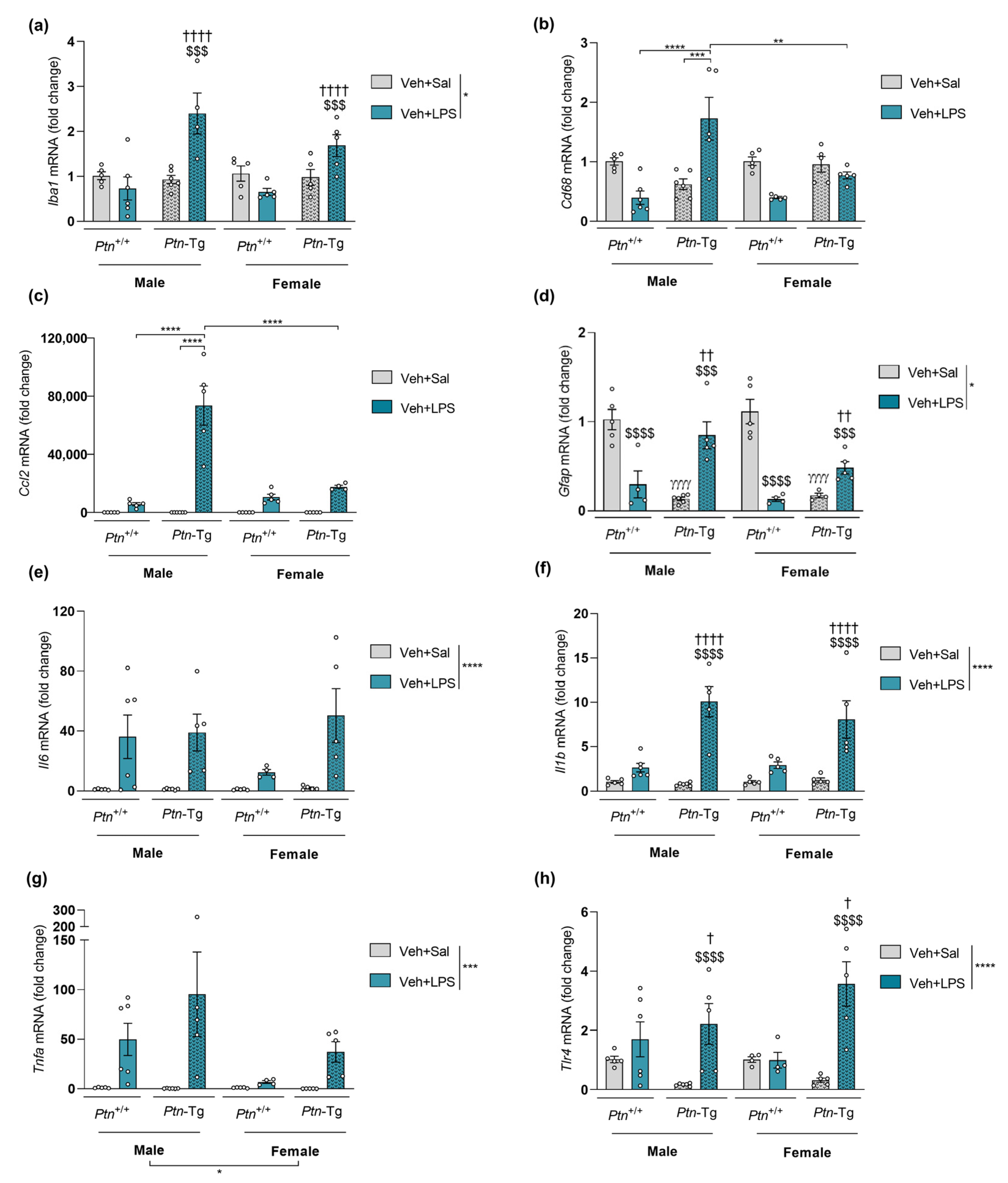
To compare with ethanol effects, we studied in Ptn+/+ and Ptn-Tg adolescent mice the gene expression of the same neuroinflammatory markers after a general neuroinflammatory stimulus, LPS (Figure 4). Three-way ANOVA revealed that LPS treatment significantly affected mRNA levels of Il6 (Figure 4e) and Tnfa (Figure 4g), with the latter also being affected by sex (the detailed statistical results are shown in Table S4a). However, Ptn overexpression did not seem to modify the levels of expression of these proinflammatory markers. LPS treatment and genotype significantly affected the mRNA levels of microglial marker Iba1 (Figure 4a), astrocytic marker Gfap (Figure 4d), cytokine Il1b (Figure 4e) and Tlr4 (Figure 4h), and a significant interaction between variables was revealed (Table S4a, b). Moreover, Ptn overexpression induced a significant increase in mRNA levels of Iba1 and Il1b in the PFC of LPS-treated mice in both sexes (Figure 4a,e). On the contrary, we found that only male Ptn-Tg mice treated with LPS showed significantly increased prefrontal levels of Cd68 (Figure 4b) and Ccl2 (Figure 4c). Specifically, genotype differences were found in Cd68 mRNA levels analysis, while significant differences in Ccl2 mRNA levels were found regarding treatment, sex, genotype, and the combinations of these variables (Table S4a). Interestingly, acute LPS exposure induced a decrease in Gfap levels in Ptn+/+ mice. However, in Ptn-Tg mice, significantly enhanced levels were found after LPS treatment (Figure 4d). It needs to be noted that basal levels (saline-treated mice) of this marker were found to be significantly diminished in Ptn-Tg mice.
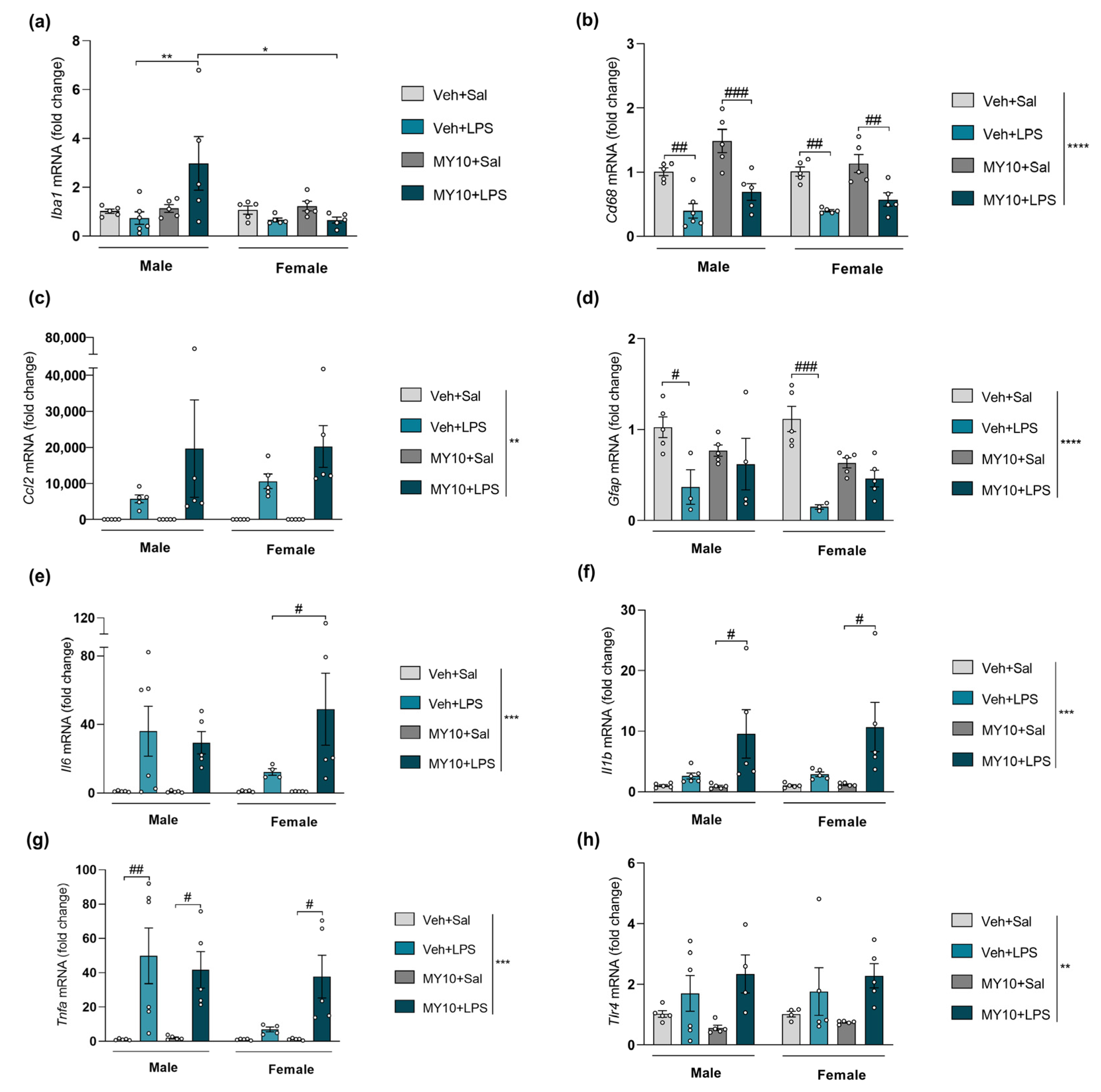
We also studied the involvement of RPTPβ/ζ in the neuroinflammation induced by LPS in the PFC of Ptn+/+ adolescent mice (Figure 5). The inhibition of RPTPβ/ζ by MY10 did not revert the changes in mRNA levels of Cd68 (Figure 5b), Ccl2 (Figure 5c), and Tlr4 (Figure 5h) induced by LPS. Post-hoc comparisons revealed that LPS treatment only upregulates Iba1 in males when the RPTPβ/ζ inhibitor, MY10, was present (Figure 5a). The decrease in Gfap mRNA levels observed in Ptn+/+ mice was absent when RPTPβ/ζ was inhibited by MY10 (Figure 5d). In addition, LPS only induced a significant upregulation of Il1b when RPTPβ/ζ was inhibited (Figure 5f) in a sex-independent manner. Interestingly, Il1b expression seems to follow the same tendency in this case as in Ptn-Tg (Figure 4f). Regarding changes in Il6 (Figure 5e) and Tnfa (Figure 5g) levels, LPS treatment produced a milder effect in female mice. However, when RPTPβ/ζ is inhibited by MY10, the mRNA levels of Il6 and Tnfa in the PFC of female mice are significantly increased to similar levels to the ones observed in LPS-treated males (for a detailed statistical analysis, see Table S5).

3.4. Comparative Study of PTN/RPTPβ/ζ Signaling Pathway after Ethanol and LPS Administration

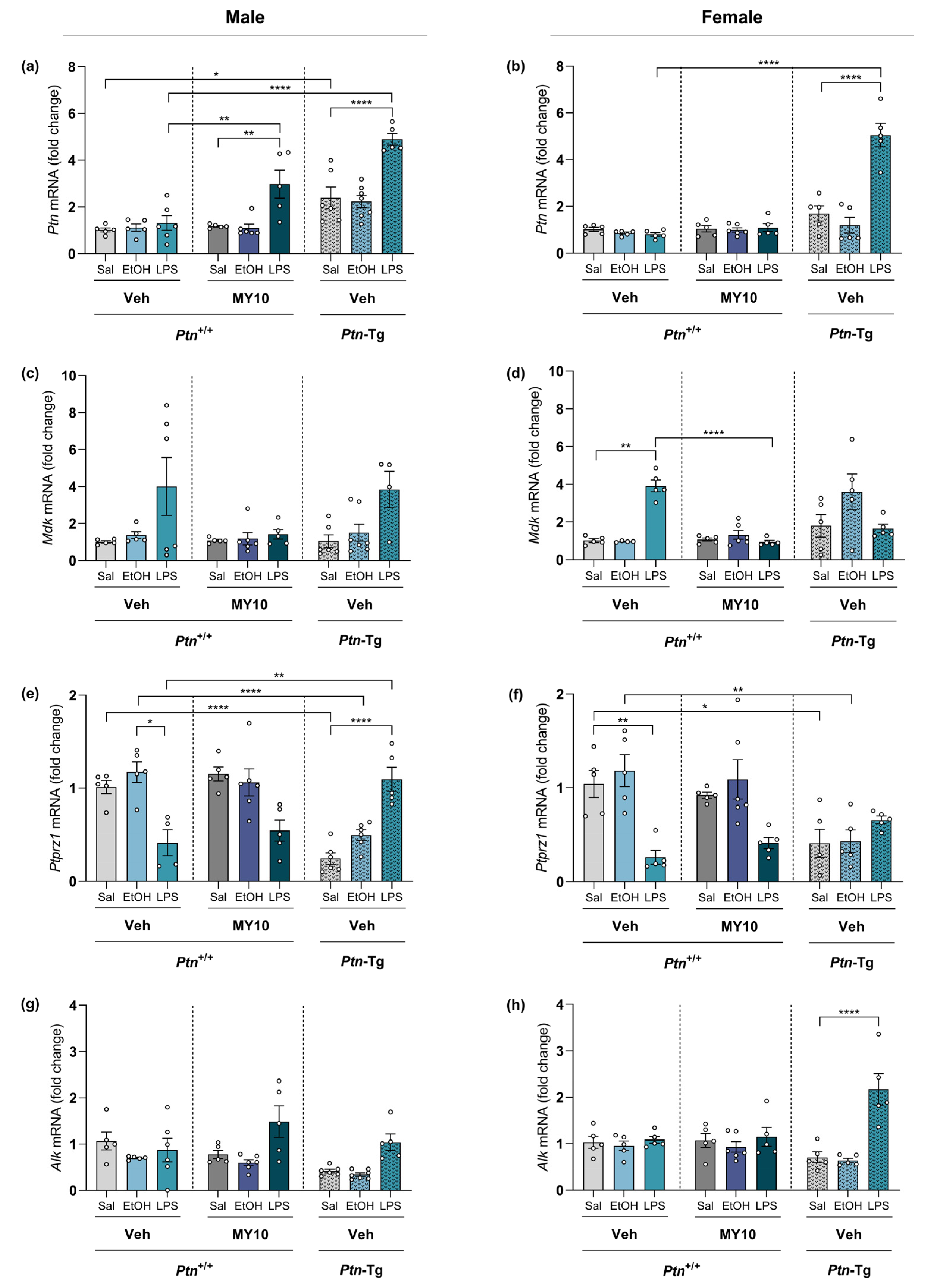
To shed some light on the possible molecular mechanisms underlying ethanol and LPS-induced neuroinflammatory processes, we analyzed the expression of Ptn, Mdk, Ptprz1, and Alk in the PFC of adolescent mice after a single injection of ethanol (6 g/kg, i.p.) or LPS (5 mg/kg, i.p.) (Figure 6). Ptn levels were significantly increased after LPS acute exposure in Ptn-Tg mice of both sexes. Similarly, LPS induced the upregulation of Ptn mRNA levels in males treated with MY10. Interestingly, this upregulation was not observed in females (Figure 6a, b; statistical analysis in Table S6). In contrast, acute ethanol administration did not alter Ptn levels. We found that LPS induces an upregulation of Mdk levels in wild type mice, an effect that was prevented by treatment with MY10 (Figure 6c,d). As it happened with Ptn expression, ethanol treatment did not regulate Mdk expression. LPS reduced Ptprz1 expression levels in wild type mice, an effect that was not apparently regulated by MY10 (Figure 6e,f). In addition, we found that Ptn overexpression led to a significant downregulation of Ptprz1 in control (vehicle-saline) and ethanol-treated mice (Figure 6e,f). Interestingly, LPS prevented this downregulation of Ptprz1 only in Ptn-Tg male mice (Figure 6e,f). We also measured mRNA levels of expression of Alk, a key downstream element of the RPTPβ/ζ pathway, to compare ethanol and LPS responses. Only an increase in Alk expression was found in LPS-treated female Ptn-Tg (Figure 6g,h). As ALK plays an important role in ethanol consumption [33] and could contribute to sex differences in the ethanol-induced response in the PFC [50], we aimed to explore the participation of Alk in these sex differences. To that, the group of mice included in ethanol experiments were also analyzed together, comparing males and females. In that case, an interesting effect of sex was found, both when Ptn-Tg mice were compared to Ptn+/+ and when the role of MY10 was studied in Ptn+/+ mice (Figure S2). Thus, ALK appears as a potential candidate to better understand sex differences in ethanol response.

4. Discussion

Binge drinking constitutes a substantial risk for the adolescent immature brain. Ethanol-induced neuroinflammation during adolescence impairs PFC maturation and hippocampal neurogenesis and induces synaptic and myelin disarrangements [3,4,5,6]. Recently, we demonstrated that the hippocampal neurogenic loss induced by ethanol during adolescence was fully prevented when RPTPβ/ζ was inhibited by MY10 [44]. Glial morphological changes did not fully explain these actions of MY10 on the hippocampus or PFC; however, a possible modulation of neuroinflammation by RPTPβ/ζ in a sex-dependent manner cannot be ruled out. Thus, we aimed to shed some light on the role of the PTN/RPTPβ/ζ signaling pathway in the effects of ethanol related to neuroinflammation in the PFC, one of the more vulnerable regions in the adolescent brain [5,6], and to confirm if this participation is sex-affected.
Ethanol-induced neuroinflammation is still controversial. Some hallmarks of the neuroinflammatory process are absent after ethanol consumption, while other reports strongly support the importance of neuroinflammation in ethanol actions [8,51]. Accordingly, we found that levels of IL1β and TNFα are not modified in PFC 18 h after acute ethanol, while this treatment significantly upregulates IL6 and MCP1 protein levels. Although neither Ptn overexpression nor RPTPβ/ζ inhibition by MY10 seems to alter the increase in IL6 levels induced by ethanol, the overexpression of the RPTPβ/ζ endogenous inhibitor PTN blocks the ethanol-induced increase in MCP1 levels (a summary of these effects on cytokine protein levels is presented in Table 2). Interestingly, this action of PTN overexpression does not seem to be uniquely mediated by RPTPβ/ζ since the treatment with MY10 does not alter MCP1 levels in response to ethanol in a significant manner.
Ethanol treatment induced significant effects on Iba1 and Cd68 mRNA expression, which is consistent with results obtained by others using a different ethanol route of administration (intragastrical, i.g.) [18]. Thus, intraperitoneal ethanol administration replicates altered microglial gene expression induced by an acute i.g. ethanol dose, suggesting that ethanol-induced prefrontal microglial changes are independent from gut disruptions in adolescent mice. No significant effects of Ptn overexpression or RPTPβ/ζ pharmacological inhibition were detected on gene expression of microglial markers Iba1 and Cd68 (a summary of PTN and MY10 effects on gene expression is presented in Table 3). It is important to note that other authors have reported sex differences in microglial states in healthy brains and in pathological conditions, such as stress or neurodegeneration [52,53,54]. However, in our experimental conditions, ethanol effects on prefrontal Iba1 mRNA levels are independent of sex. This correlates with the absence of sex differences in prefrontal Iba1 immunoreactivity after ethanol injury in adolescent mice [44].
The astrocytic response is proposed as a link between neuroinflammation processes induced by different stimuli. Here, we show that Ptn overexpression decreases prefrontal levels of Gfap in both sexes. The pharmacological inhibition of RPTPβ/ζ with MY10 mimics these PTN effects, suggesting an important role of astrocytes in the actions of the PTN/RPTPβ/ζ signaling pathway after ethanol exposure during adolescence. Interestingly, increased mRNA and protein level of GFAP has been reported only in female mice after ethanol drinking [9,50,55]. However, in our experimental conditions, Gfap prefrontal levels were not altered after ethanol administration. A limitation of this study is that gene expression is only measured at one timepoint, which may underlie this apparent discrepancy, together with other experimental conditions such as ethanol route of administration or the use of other astrocytic markers. The evidence supports the need to perform further studies to deeply dissect the participation of both PTN/RPTPβ/ζ cascade and astrocytic response in the acute ethanol-induced immune response and in the sex differences here identified.
Ethanol is known to increase the levels of proinflammatory cytokine TNFα via the participation of microglia [18,56]. Interestingly, this increment was fully prevented in ethanol-treated Ptn-Tg male mice, suggesting a role of PTN in the modulation of ethanol-induced changes in Tnfa expression. However, this effect is not replicated when RPTPβ/ζ is blocked by MY10, suggesting that this PTN action may be mediated by other receptors. Surprisingly, Tnfa levels were not altered in female mice after ethanol acute exposure and only a positive correlation with Ptn levels was found in males, evidencing different molecular mechanisms underlaying ethanol-induced neuroinflammation in the brain of female and male mice.
In contrast to the protein determinations performed, we found that the expression of Il6 and Ccl2 (MCP1) is significantly increased by ethanol in female Ptn-Tg mice, suggesting different actions of PTN in the PFC of adolescent male and female mice. Post-transcriptional modifications may be responsible for the differences in the levels of expression between protein and gene. It is worth noting that cytokines have short half-lives in both plasma and tissue [14].
On the other hand, Il1b expression does not seem to be significantly modulated by PTN or MY10. Ethanol withdrawal increases the expression of Tnfa, Il1b, and Ccl2 whereas the expression of Il6 is increased within the initial hours of acute intoxication [14,18,51,57]. However, Il1b prefrontal levels were not altered 18 h after intraperitoneal ethanol administration. It is important to note that this proinflammatory cytokine contributes to aberrant inflammation and has been found to be increased in the human alcoholic brain [51]. Furthermore, IL1β signaling is involved in the expression of other key elements of ethanol-induced neuroinflammation in the mouse brain such as Il6, Ccl2, and Tnfa [57,58]. Interestingly, microglia depletion does not counteract ethanol-induced increases of Il1b, Il6, and Ccl2 [18], suggesting that other brain cell types may be the primary source of these proinflammatory markers. Neurons express Ccl2 [59] and astrocytes have been shown to produce IL6 and IL1β and express Ccl2 [14,18,60,61]. PTN could play an important role in these cells as Ptn upregulation modulates the expression of Il6 and Ccl2 and tend to increase Il1b prefrontal levels in adolescent females after acute ethanol treatment. PTN is known to be expressed in different cell types, including neurons and astrocytes [62], suggesting the actions of this neurotrophic factor in these cells may underlie the regulation of ethanol-induced changes in the levels of Ccl2, Il6, and Il1b in the PFC of Ptn-Tg mice. However, RPTPβ/ζ does not seem to be the main receptor mediating PTN actions because the inhibition of this receptor with MY10 does not significantly modulate ethanol-induced changes in the expression of these inflammatory markers.
Different studies support the role of the immune TLR4 response in ethanol-induced neuroinflammation and its detrimental effects in adult and adolescent mice [5,9,10,19,63]. Ethanol is known to contribute to gut leakiness and increase serum levels of LPS [56,64,65]. In fact, recent studies point to a connection between the release of LPS through systemic circulation and the neuroinflammation induced by ethanol [13,20,56]. In the present work, acute LPS exposure increases Iba1 and Cd68 mRNA levels in adolescent Ptn-Tg male mice, suggesting an enhancing effect of PTN on microglial responses. In addition, RPTPβ/ζ inhibition with MY10 potentiates Iba1, not Cd68, and mRNA levels increase after LPS, which is aligned with previous results obtained in adult mice exposed to LPS [42,43]. The data indicate an important role of the PTN/RPTPβ/ζ signaling pathway in LPS-induced overexpression of Iba1 in the PFC and suggest Ptn overexpression effects on LPS-induced increase in Cd68 levels are mediated through different PTN receptors. Very interestingly, these alterations were not replicated in female mice, indicating a sex-dependent contribution of PTN and RPTPβ/ζ to LPS-induced microglial responses.
We observed LPS-induced increases in the levels of prefrontal expression of Ccl2, Il6, Il1b, and Tnfa, confirming previous studies by others [17,66]. Interestingly, Ccl2 (MCP1) expression is dramatically increased in adolescent male mice exposed to LPS when Ptn is overexpressed. MCP1 is a key element in the activation of immune cells and production of inflammatory mediators in the brain during endotoxemia and can be produced by different cell types including microglia, astrocytes, and neurons [17,59,60]. Previous studies revealed that Ptn overexpression potentiates striatal astrocytosis and microglial activation after different stimuli, including LPS, in adult mice [27,43,67]. Another important factor involved in the effects of LPS, Il1b [66] is highly increased after LPS when Ptn is overexpressed or RPTPβ/ζ is inhibited with MY10 in both sexes. It is remarkable that the increase in Il6 and Tnfa mRNA levels induced by LPS is less pronounced in females than males; however, this sex-dependent effect of LPS is counteracted when female mice are administered with MY10. Taken together, the data presented here show that the PTN/RPTPβ/ζ axis highly regulates LPS-induced neuroinflammation and suggests this pathway may be a target for the modulation of neuroinflammation in the adolescent brain.
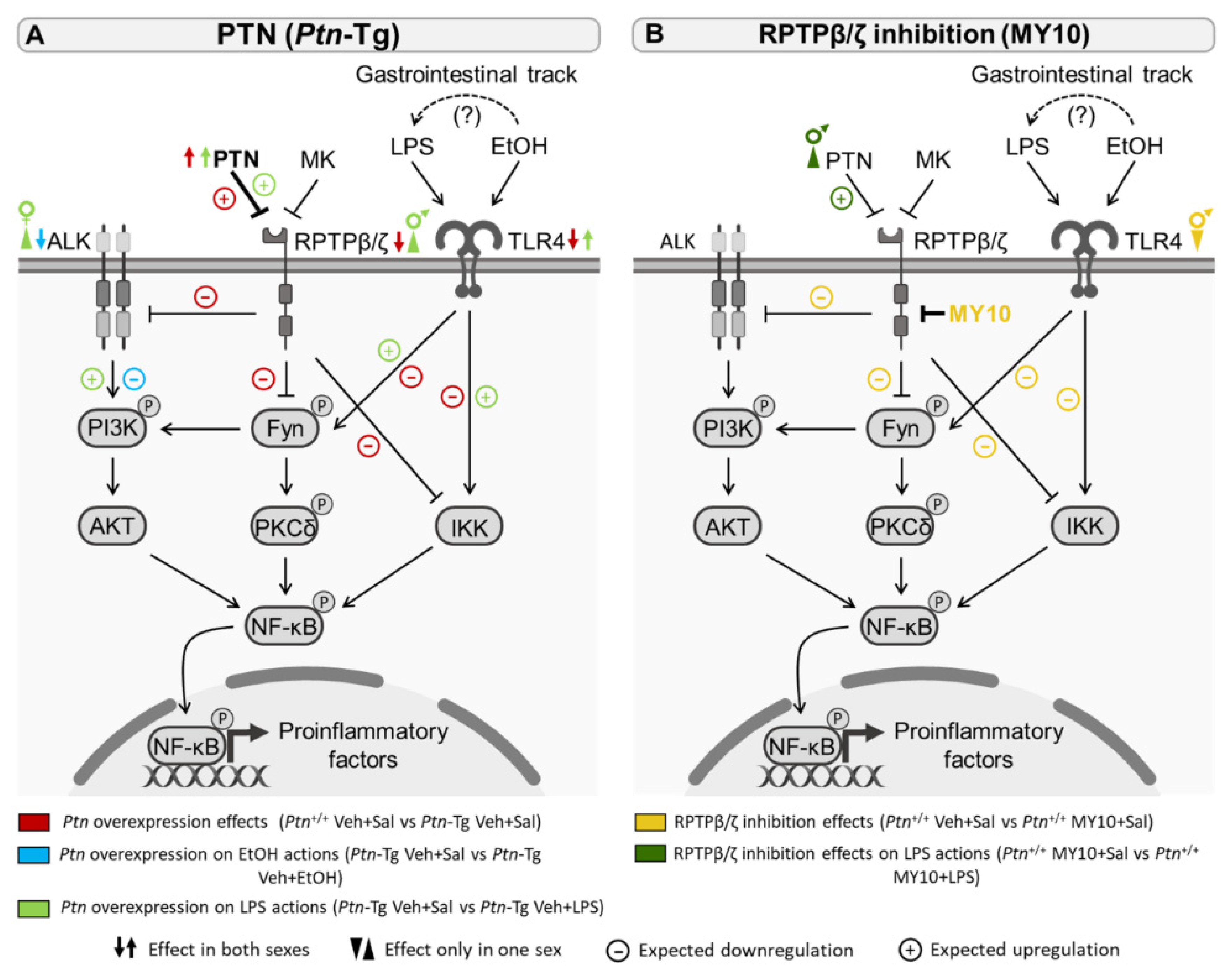
Ethanol- and LPS-induced activation of TLR4 triggers intracellular signaling pathways that increase the transcription of different inflammatory cytokines and chemokines via NF-kB (Figure 7), including IL6, IL1β, and MCP1 [5,42,57,68]. TLR4 is expressed in microglia and other cell types, such as astrocytes. In fact, this receptor plays a crucial role in ethanol-induced immune response in astrocytes [18,68] and astrogliosis is detected in postmortem alcoholic human brains [69,70]. In addition, LPS induces astrocytosis in the mouse PFC [41,42], suggesting that the astrocytic response could be a link between neuroinflammatory processes induced by different stimuli. Moreover, it should be considered in the modulation of the levels of proinflammatory markers such as Il6, Il1b, and Ccl2, which cannot be explained by the microglial response [18,41,42].
Astrocytes were identified as MK-producing cells [71], and MK expression was increased in astrocytes in the prefrontal cortex of human alcoholics [26]. Ethanol exposure induced MK expression in neuronal cells, and it was found that MK contributed to the activation of ALK signaling in response to ethanol [72]. In the present work, ethanol exposure did not alter Ptn, Mdk, Ptprz1, or Alk expression in the PFC of adolescent mice. In contrast, Mdk levels were upregulated after LPS exposure, whereas the expression of its receptor Ptprz1 was decreased, probably as a compensatory mechanism, like that observed in mice overexpressing the other endogenous ligand of this receptor, Ptn. Importantly, the administration of MY10 prevented an LPS-induced increase in Mdk. In contrast, pharmacological inhibition of RPTPβ/ζ with MY10 caused a significant upregulation of Ptn only in the PFC of male mice, whereas transgenic Ptn overexpression further potentiated the upregulation of Ptn expression in both males and females treated with LPS (Figure 7). These results are intriguing because the RPTPβ/ζ signaling pathway can be regulated both by MK and PTN, through the modulation of tyrosine phosphorylation of substrates of RPTPβ/ζ which, in turn, are known regulators of neuroinflammation. Activation through tyrosine phosphorylation of Fyn kinase, a substrate of RPTPβ/ζ, mediates LPS-induced activation of the NFκB pathway, suggesting that PTN/RPTPβ/ζ regulates proinflammatory cascades [27,37]. Accordingly, cerebral overexpression of Ptn potentiates LPS-induced microglial activation and neuroinflammation, whereas LPS-induced astrocytosis was blocked in the PFC of Ptn−/− and Mdk−/− mice [41,43]. It is necessary to note that the main findings of this manuscript are based only on changes in gene expression. It seems essential to study in detail possible differences in the expression of genes and proteins to fully understand the role of the PTN/RPTPβ/ζ signaling pathway in ethanol- and LPS-induced neuroinflammation.

5. Conclusions

Proinflammatory markers Ccl2, Il6, and Il1b seem to have an important role in the PTN modulation of ethanol effects during adolescence. Our data suggest that not all the PTN actions on the neuroinflammation induced by ethanol are mediated by RPTPβ/ζ. In addition, the contribution of PTN and RPTPβ/ζ to the changes in microglial gene marker expression after ethanol and LPS exposure during adolescence shows differences that strongly suggest the implication of more cell types in these PTN/RPTPβ/ζ effects. Remarkably, important sex differences regarding ethanol and LPS actions during adolescence, and their PTN/RPTPβ/ζ modulation, are reported.

Supplementary Materials

The following supporting information can be downloaded at: https://www.mdpi.com/article/10.3390/biomedicines11051318/s1, Figure S1: Significant correlations are established between Tnfa, Gfap and Ptn mRNA level after ethanol acute exposure in the prefrontal cortex (PFC) of Ptn+/+ male mice and female mice; Figure S2: Effect of genetic overexpression of Ptn and RPTPβ/ζ inhibition on ethanol-induced Alk mRNA expression changes; Table S1: Statistical data of protein expression analysis after ethanol treatment; Table S2: Statistical data of mRNA expression analysis after ethanol treatment; Table S3: Statistical data of mRNA expression analysis after ethanol treatment; Table S4: Statistical data of mRNA expression analysis after LPS treatment; Table S5: Statistical data of mRNA expression analysis after LPS treatment; Table S6: Statistical data of mRNA expression analysis after ethanol or LPS treatment.

Author Contributions

Conceptualization, G.H., C.P.-G. and E.G.; Data curation, M.R.-Z., M.G.-L. and E.G.; Formal analysis, J.S. and M.G.S.-A.; Funding acquisition, B.d.P.-T., M.d.P.R.-Á. and G.H.; Methodology, M.R.-Z., M.G.-L., H.C.-R. and J.M.Z.; Resources, M.F.-A., M.U., B.d.P.-T. and M.d.P.R.-Á.; Supervision, G.H.; Writing—original draft, M.R.-Z. and E.G.; Writing—review & editing, J.S., G.H., C.P.-G. and E.G. All authors have read and agreed to the published version of the manuscript.

Funding

This work was supported by the National Plan on Drug abuse, Ministerio de Sanidad of Spain (MSSSI, grant PNSD2019I015 to G.H.), ISCIII-Redes de Investigación Cooperativa Orientadas a Resultados en Salud (RICORS), Red de Investigación en Atención Primaria de Adicciones (RIAPAd; grant RD21/0009/0013 to G.H.), Ministry of Science, Innovation and Universities of Spain (MICINN) funds (Ref. RTI2018-095615-B-I00 to G.H. and M.d.P.R.-Á.), and Fundación Universitaria San Pablo CEU – Banco Santander (FUSPBS-PPC06_2017 to E.G.). M.R.-Z. was supported by Consejería de Educación, Juventud y Deporte of Community of Madrid and by European Social Fund.

Institutional Review Board Statement

The animal study protocol was approved by the Animal Research Committee of USP-CEU (authorization reference: PROEX 76.0/20).

Informed Consent Statement

Not applicable.

Data Availability Statement

The data presented in this study are available on reasonable request from the corresponding author.

Acknowledgments

We thank our colleagues in the animal facility in the Universidad San Pablo-CEU.

Conflicts of Interest

The authors declare no conflict of interest.

References

  1. NSDUH. National Survey of Drug Use and Health. Available online: https://www.samhsa.gov/data/release/2020-national-survey-drug-use-and-health-nsduh-releases (accessed on 30 December 2022).
  2. Observatorio Español de las Drogas y las Adicciones. Monografía alcohol 2021. Consumo Y Consecuencias 2021, 109. [Google Scholar]
  3. Broadwater, M.A.; Liu, W.; Crews, F.T.; Spear, L.P. Persistent loss of hippocampal neurogenesis and increased cell death following adolescent, but not adult, chronic ethanol exposure. Dev. Neurosci. 2014, 36, 297–305. [Google Scholar] [CrossRef] [PubMed]
  4. Pascual, M.; Blanco, A.M.; Cauli, O.; Miñarro, J.; Guerri, C. Intermittent ethanol exposure induces inflammatory brain damage and causes long-term behavioural alterations in adolescent rats. Eur. J. Neurosci. 2007, 25, 541–550. [Google Scholar] [CrossRef] [PubMed]
  5. Montesinos, J.; Pascual, M.; Pla, A.; Maldonado, C.; Rodríguez-Arias, M.; Miñarro, J.; Guerri, C. TLR4 elimination prevents synaptic and myelin alterations and long-term cognitive dysfunctions in adolescent mice with intermittent ethanol treatment. Brain Behav. Immun. 2015, 45, 233–244. [Google Scholar] [CrossRef] [PubMed]
  6. Vetreno, R.P.; Crews, F.T. Binge ethanol exposure during adolescence leads to a persistent loss of neurogenesis in the dorsal and ventral hippocampus that is associated with impaired adult cognitive functioning. Front. Neurosci. 2015, 9, 35. [Google Scholar] [CrossRef] [PubMed]
  7. Pascual, M.; López-Hidalgo, R.; Montagud-Romero, S.; Ureña-Peralta, J.R.; Rodríguez-Arias, M.; Guerri, C. Role of mTOR-regulated autophagy in spine pruning defects and memory impairments induced by binge-like ethanol treatment in adolescent mice. Brain Pathol. 2021, 31, 174–188. [Google Scholar] [CrossRef]
  8. Robinson, G.; Most, D.; Ferguson, L.B.; Mayfield, J.; Harris, R.A.; Blednov, Y.A. Neuroimmune pathways in alcohol consumption: Evidence from behavioral and genetic studies in rodents and humans. Int. Rev. Neurobiol. 2014, 118, 13–39. [Google Scholar] [CrossRef]
  9. Alfonso-Loeches, S.; Pascual, M.; Guerri, C. Gender differences in alcohol-induced neurotoxicity and brain damage. Toxicology 2013, 311, 27–34. [Google Scholar] [CrossRef]
  10. Montesinos, J.; Pascual, M.; Rodríguez-Arias, M.; Miñarro, J.; Guerri, C. Involvement of TLR4 in the long-term epigenetic changes, rewarding and anxiety effects induced by intermittent ethanol treatment in adolescence. Brain Behav. Immun. 2016, 53, 159–171. [Google Scholar] [CrossRef]
  11. Pascual, M.; Montesinos, J.; Marcos, M.; Torres, J.L.; Costa-Alba, P.; García-García, F.; Laso, F.J.; Guerri, C. Gender differences in the inflammatory cytokine and chemokine profiles induced by binge ethanol drinking in adolescence. Addict. Biol. 2017, 22, 1829–1841. [Google Scholar] [CrossRef]
  12. Pascual, M.; Montesinos, J.; Guerri, C. Role of the innate immune system in the neuropathological consequences induced by adolescent binge drinking. J. Neurosci. Res. 2018, 96, 765–780. [Google Scholar] [CrossRef]
  13. Crews, F.T.; Lawrimore, C.J.; Walter, T.J.; Coleman, L.G., Jr. The role of neuroimmune signaling in alcoholism. Neuropharmacology 2017, 122, 56–73. [Google Scholar] [CrossRef]
  14. Gano, A.; Mondello, J.E.; Doremus-Fitzwater, T.L.; Deak, T. Rapid alterations in neuroimmune gene expression after acute ethanol: Timecourse, sex differences and sensitivity to cranial surgery. J. Neuroimmunol. 2019, 337, 577083. [Google Scholar] [CrossRef]
  15. Vore, A.S.; Deak, T. Alcohol, inflammation, and blood-brain barrier function in health and disease across development. Int. Rev. Neurobiol. 2022, 161, 209–249. [Google Scholar] [CrossRef]
  16. Singh, S.; Anshita, D.; Ravichandiran, V. MCP-1: Function, regulation, and involvement in disease. Int. Immunopharmacol. 2021, 101, 107598. [Google Scholar] [CrossRef]
  17. Thompson, W.L.; Karpus, W.J.; Van Eldik, L.J. MCP-1-deficient mice show reduced neuroinflammatory responses and increased peripheral inflammatory responses to peripheral endotoxin insult. J. Neuroinflamm. 2008, 5, 35. [Google Scholar] [CrossRef]
  18. Walter, T.J.; Crews, F.T. Microglial depletion alters the brain neuroimmune response to acute binge ethanol withdrawal. J. Neuroinflamm. 2017, 14, 86. [Google Scholar] [CrossRef]
  19. Kelley, K.W.; Dantzer, R. Alcoholism and inflammation: Neuroimmunology of behavioral and mood disorders. Brain Behav. Immun. 2011, 25 (Suppl. S1), S13–S20. [Google Scholar] [CrossRef]
  20. Bala, S.; Marcos, M.; Gattu, A.; Catalano, D.; Szabo, G. Acute binge drinking increases serum endotoxin and bacterial DNA levels in healthy individuals. PLoS ONE 2014, 9, e96864. [Google Scholar] [CrossRef]
  21. Fernandez-Lizarbe, S.; Pascual, M.; Gascon, M.S.; Blanco, A.; Guerri, C. Lipid rafts regulate ethanol-induced activation of TLR4 signaling in murine macrophages. Mol. Immunol. 2008, 45, 2007–2016. [Google Scholar] [CrossRef]
  22. Fernandez-Lizarbe, S.; Pascual, M.; Guerri, C. Critical role of TLR4 response in the activation of microglia induced by ethanol. J. Immunol. 2009, 183, 4733–4744. [Google Scholar] [CrossRef] [PubMed]
  23. Gramage, E.; Herradón, G. Connecting Parkinson’s disease and drug addiction: Common players reveal unexpected disease connections and novel therapeutic approaches. Curr. Pharm. Des. 2011, 17, 449–461. [Google Scholar] [CrossRef] [PubMed]
  24. Herradón, G.; Pérez-García, C. Targeting midkine and pleiotrophin signalling pathways in addiction and neurodegenerative disorders: Recent progress and perspectives. Br. J. Pharmacol. 2014, 171, 837–848. [Google Scholar] [CrossRef] [PubMed]
  25. Vicente-Rodríguez, M.; Pérez-García, C.; Ferrer-Alcón, M.; Uribarri, M.; Sánchez-Alonso, M.G.; Ramos, M.P.; Herradón, G. Pleiotrophin differentially regulates the rewarding and sedative effects of ethanol. J. Neurochem. 2014, 131, 688–695. [Google Scholar] [CrossRef] [PubMed]
  26. Flatscher-Bader, T.; Wilce, P.A. Impact of alcohol abuse on protein expression of midkine and excitatory amino acid transporter 1 in the human prefrontal cortex. Alcohol. Clin. Exp. Res. 2008, 32, 1849–1858. [Google Scholar] [CrossRef]
  27. Herradon, G.; Ramos-Alvarez, M.P.; Gramage, E. Connecting Metainflammation and Neuroinflammation through the PTN-MK-RPTPβ/ζ Axis: Relevance in Therapeutic Development. Front. Pharmacol. 2019, 10, 377. [Google Scholar] [CrossRef]
  28. Cressant, A.; Dubreuil, V.; Kong, J.; Kranz, T.M.; Lazarini, F.; Launay, J.M.; Callebert, J.; Sap, J.; Malaspina, D.; Granon, S.; et al. Loss-of-function of PTPR γ and ζ, observed in sporadic schizophrenia, causes brain region-specific deregulation of monoamine levels and altered behavior in mice. Psychopharmacology 2017, 234, 575–587. [Google Scholar] [CrossRef]
  29. Meng, K.; Rodriguez-Peña, A.; Dimitrov, T.; Chen, W.; Yamin, M.; Noda, M.; Deuel, T.F. Pleiotrophin signals increased tyrosine phosphorylation of beta beta-catenin through inactivation of the intrinsic catalytic activity of the receptor-type protein tyrosine phosphatase beta/zeta. Proc. Natl. Acad. Sci. USA 2000, 97, 2603–2608. [Google Scholar] [CrossRef]
  30. Pariser, H.; Ezquerra, L.; Herradon, G.; Perez-Pinera, P.; Deuel, T.F. Fyn is a downstream target of the pleiotrophin/receptor protein tyrosine phosphatase beta/zeta-signaling pathway: Regulation of tyrosine phosphorylation of Fyn by pleiotrophin. Biochem. Biophys. Res. Commun. 2005, 332, 664–669. [Google Scholar] [CrossRef]
  31. Pariser, H.; Herradon, G.; Ezquerra, L.; Perez-Pinera, P.; Deuel, T.F. Pleiotrophin regulates serine phosphorylation and the cellular distribution of beta-adducin through activation of protein kinase C. Proc. Natl. Acad. Sci. USA 2005, 102, 12407–12412. [Google Scholar] [CrossRef]
  32. Pariser, H.; Perez-Pinera, P.; Ezquerra, L.; Herradon, G.; Deuel, T.F. Pleiotrophin stimulates tyrosine phosphorylation of beta-adducin through inactivation of the transmembrane receptor protein tyrosine phosphatase beta/zeta. Biochem. Biophys. Res. Commun. 2005, 335, 232–239. [Google Scholar] [CrossRef]
  33. Dutton, J.W., 3rd; Chen, H.; You, C.; Brodie, M.S.; Lasek, A.W. Anaplastic lymphoma kinase regulates binge-like drinking and dopamine receptor sensitivity in the ventral tegmental area. Addict. Biol. 2017, 22, 665–678. [Google Scholar] [CrossRef]
  34. Ron, D.; Berger, A. Targeting the intracellular signaling “STOP” and “GO” pathways for the treatment of alcohol use disorders. Psychopharmacology 2018, 235, 1727–1743. [Google Scholar] [CrossRef]
  35. Stoica, G.E.; Kuo, A.; Aigner, A.; Sunitha, I.; Souttou, B.; Malerczyk, C.; Caughey, D.J.; Wen, D.; Karavanov, A.; Riegel, A.T.; et al. Identification of anaplastic lymphoma kinase as a receptor for the growth factor pleiotrophin. J. Biol. Chem. 2001, 276, 16772–16779. [Google Scholar] [CrossRef]
  36. Wang, J.; Carnicella, S.; Phamluong, K.; Jeanblanc, J.; Ronesi, J.A.; Chaudhri, N.; Janak, P.H.; Lovinger, D.M.; Ron, D. Ethanol induces long-term facilitation of NR2B-NMDA receptor activity in the dorsal striatum: Implications for alcohol drinking behavior. J. Neurosci. Off. J. Soc. Neurosci. 2007, 27, 3593–3602. [Google Scholar] [CrossRef]
  37. Panicker, N.; Saminathan, H.; Jin, H.; Neal, M.; Harischandra, D.S.; Gordon, R.; Kanthasamy, K.; Lawana, V.; Sarkar, S.; Luo, J.; et al. Fyn Kinase Regulates Microglial Neuroinflammatory Responses in Cell Culture and Animal Models of Parkinson’s Disease. J. Neurosci. Off. J. Soc. Neurosci. 2015, 35, 10058–10077. [Google Scholar] [CrossRef]
  38. Pastor, M.; Fernández-Calle, R.; Di Geronimo, B.; Vicente-Rodríguez, M.; Zapico, J.M.; Gramage, E.; Coderch, C.; Pérez-García, C.; Lasek, A.W.; Puchades-Carrasco, L.; et al. Development of inhibitors of receptor protein tyrosine phosphatase β/ζ (PTPRZ1) as candidates for CNS disorders. Eur. J. Med. Chem. 2018, 144, 318–329. [Google Scholar] [CrossRef]
  39. Calleja-Conde, J.; Fernández-Calle, R.; Zapico, J.M.; Ramos, A.; de Pascual-Teresa, B.; Bühler, K.M.; Echeverry-Alzate, V.; Giné, E.; Rodríguez de Fonseca, F.; López-Moreno, J.A.; et al. Inhibition of Receptor Protein Tyrosine Phosphatase β/ζ Reduces Alcohol Intake in Rats. Alcohol. Clin. Exp. Res. 2020, 44, 1037–1045. [Google Scholar] [CrossRef]
  40. Fernández-Calle, R.; Gramage, E.; Zapico, J.M.; de Pascual-Teresa, B.; Ramos, A.; Herradón, G. Inhibition of RPTPβ/ζ blocks ethanol-induced conditioned place preference in pleiotrophin knockout mice. Behav. Brain Res. 2019, 369, 111933. [Google Scholar] [CrossRef]
  41. Fernández-Calle, R.; Vicente-Rodríguez, M.; Pastor, M.; Gramage, E.; Di Geronimo, B.; Zapico, J.M.; Coderch, C.; Pérez-García, C.; Lasek, A.W.; de Pascual-Teresa, B.; et al. Pharmacological inhibition of Receptor Protein Tyrosine Phosphatase β/ζ (PTPRZ1) modulates behavioral responses to ethanol. Neuropharmacology 2018, 137, 86–95. [Google Scholar] [CrossRef]
  42. Fernández-Calle, R.; Galán-Llario, M.; Gramage, E.; Zapatería, B.; Vicente-Rodríguez, M.; Zapico, J.M.; de Pascual-Teresa, B.; Ramos, A.; Ramos-Álvarez, M.P.; Uribarri, M.; et al. Role of RPTPβ/ζ in neuroinflammation and microglia-neuron communication. Sci. Rep. 2020, 10, 20259. [Google Scholar] [CrossRef] [PubMed]
  43. Fernández-Calle, R.; Vicente-Rodríguez, M.; Gramage, E.; Pita, J.; Pérez-García, C.; Ferrer-Alcón, M.; Uribarri, M.; Ramos, M.P.; Herradón, G. Pleiotrophin regulates microglia-mediated neuroinflammation. J. Neuroinflamm. 2017, 14, 46. [Google Scholar] [CrossRef] [PubMed]
  44. Galán-Llario, M.; Rodríguez-Zapata, M.; Gramage, E.; Vicente-Rodríguez, M.; Fontán-Baselga, T.; Ovejero-Benito, M.C.; Pérez-García, C.; Carrasco, J.; Moreno-Herradón, M.; Sevillano, J.; et al. Receptor protein tyrosine phosphatase β/ζ regulates loss of neurogenesis in the mouse hippocampus following adolescent acute ethanol exposure. Neurotoxicology 2023, 94, 98–107. [Google Scholar] [CrossRef] [PubMed]
  45. Aigner, L.; Arber, S.; Kapfhammer, J.P.; Laux, T.; Schneider, C.; Botteri, F.; Brenner, H.R.; Caroni, P. Overexpression of the neural growth-associated protein GAP-43 induces nerve sprouting in the adult nervous system of transgenic mice. Cell 1995, 83, 269–278. [Google Scholar] [CrossRef]
  46. Caroni, P. Overexpression of growth-associated proteins in the neurons of adult transgenic mice. J. Neurosci. Methods 1997, 71, 3–9. [Google Scholar] [CrossRef]
  47. Vicente-Rodríguez, M.; Herradón, G.; Ferrer-Alcón, M.; Uribarri, M.; Pérez-García, C. Chronic Cocaine Use Causes Changes in the Striatal Proteome Depending on the Endogenous Expression of Pleiotrophin. Chem. Res. Toxicol. 2015, 28, 1443–1454. [Google Scholar] [CrossRef]
  48. Pruett, S.; Tan, W.; Howell, G.E., 3rd; Nanduri, B. Dosage scaling of alcohol in binge exposure models in mice: An empirical assessment of the relationship between dose, alcohol exposure, and peak blood concentrations in humans and mice. Alcohol 2020, 89, 9–17. [Google Scholar] [CrossRef]
  49. Wiener, S.W.; Olmedo, R.; Howland, M.; Nelson, L.; Hoffman, R. Ethanol elimination kinetics following massive ingestion in an ethanol naive child. Hum. Exp. Toxicol. 2013, 32, 775–777. [Google Scholar] [CrossRef]
  50. Hamada, K.; Ferguson, L.B.; Mayfield, R.D.; Krishnan, H.R.; Maienschein-Cline, M.; Lasek, A.W. Binge-like ethanol drinking activates anaplastic lymphoma kinase signaling and increases the expression of STAT3 target genes in the mouse hippocampus and prefrontal cortex. Genes Brain Behav. 2021, 20, e12729. [Google Scholar] [CrossRef]
  51. Coleman, L.G., Jr.; Crews, F.T. Innate Immune Signaling and Alcohol Use Disorders. Handb. Exp. Pharmacol. 2018, 248, 369–396. [Google Scholar] [CrossRef]
  52. Bilbo, S.D.; Smith, S.H.; Schwarz, J.M. A lifespan approach to neuroinflammatory and cognitive disorders: A critical role for glia. J. Neuroimmune Pharmacol. Off. J. Soc. NeuroImmune Pharmacol. 2012, 7, 24–41. [Google Scholar] [CrossRef]
  53. Bollinger, J.L. Uncovering microglial pathways driving sex-specific neurobiological effects in stress and depression. Brain Behav. Immun.-Health 2021, 16, 100320. [Google Scholar] [CrossRef]
  54. Posillico, C.K. Three’s Company: Neuroimmune activation, sex, and memory at the tripartite synapse. Brain Behav. Immun.-Health 2021, 16, 100326. [Google Scholar] [CrossRef]
  55. Wilhelm, C.J.; Hashimoto, J.G.; Roberts, M.L.; Bloom, S.H.; Andrew, M.R.; Wiren, K.M. Astrocyte Dysfunction Induced by Alcohol in Females but Not Males. Brain Pathol. 2016, 26, 433–451. [Google Scholar] [CrossRef]
  56. Crews, F.T.; Bechara, R.; Brown, L.A.; Guidot, D.M.; Mandrekar, P.; Oak, S.; Qin, L.; Szabo, G.; Wheeler, M.; Zou, J. Cytokines and alcohol. Alcohol. Clin. Exp. Res. 2006, 30, 720–730. [Google Scholar] [CrossRef]
  57. Lippai, D.; Bala, S.; Petrasek, J.; Csak, T.; Levin, I.; Kurt-Jones, E.A.; Szabo, G. Alcohol-induced IL-1β in the brain is mediated by NLRP3/ASC inflammasome activation that amplifies neuroinflammation. J. Leukoc. Biol. 2013, 94, 171–182. [Google Scholar] [CrossRef]
  58. Layé, S.; Gheusi, G.; Cremona, S.; Combe, C.; Kelley, K.; Dantzer, R.; Parnet, P. Endogenous brain IL-1 mediates LPS-induced anorexia and hypothalamic cytokine expression. Am. J. Physiol. Regul. Integr. Comp. Physiol. 2000, 279, R93–R98. [Google Scholar] [CrossRef]
  59. Yang, G.; Meng, Y.; Li, W.; Yong, Y.; Fan, Z.; Ding, H.; Wei, Y.; Luo, J.; Ke, Z.J. Neuronal MCP-1 mediates microglia recruitment and neurodegeneration induced by the mild impairment of oxidative metabolism. Brain Pathol. 2011, 21, 279–297. [Google Scholar] [CrossRef]
  60. Babcock, A.A.; Kuziel, W.A.; Rivest, S.; Owens, T. Chemokine expression by glial cells directs leukocytes to sites of axonal injury in the CNS. J. Neurosci. Off. J. Soc. Neurosci. 2003, 23, 7922–7930. [Google Scholar] [CrossRef]
  61. Lau, L.T.; Yu, A.C. Astrocytes produce and release interleukin-1, interleukin-6, tumor necrosis factor alpha and interferon-gamma following traumatic and metabolic injury. J. Neurotrauma 2001, 18, 351–359. [Google Scholar] [CrossRef]
  62. Kadomatsu, K. The midkine family in cancer, inflammation and neural development. Nagoya J. Med. Sci. 2005, 67, 71–82. [Google Scholar] [PubMed]
  63. Montesinos, J.; Gil, A.; Guerri, C. Nalmefene Prevents Alcohol-Induced Neuroinflammation and Alcohol Drinking Preference in Adolescent Female Mice: Role of TLR4. Alcohol. Clin. Exp. Res. 2017, 41, 1257–1270. [Google Scholar] [CrossRef] [PubMed]
  64. Adachi, Y.; Moore, L.E.; Bradford, B.U.; Gao, W.; Thurman, R.G. Antibiotics prevent liver injury in rats following long-term exposure to ethanol. Gastroenterology 1995, 108, 218–224. [Google Scholar] [CrossRef] [PubMed]
  65. Wang, H.J.; Zakhari, S.; Jung, M.K. Alcohol, inflammation, and gut-liver-brain interactions in tissue damage and disease development. World J. Gastroenterol. 2010, 16, 1304–1313. [Google Scholar] [CrossRef]
  66. D’Mello, C.; Swain, M.G. Immune-to-Brain Communication Pathways in Inflammation-Associated Sickness and Depression. Curr. Top. Behav. Neurosci. 2017, 31, 73–94. [Google Scholar] [CrossRef]
  67. Vicente-Rodríguez, M.; Rojo Gonzalez, L.; Gramage, E.; Fernández-Calle, R.; Chen, Y.; Pérez-García, C.; Ferrer-Alcón, M.; Uribarri, M.; Bailey, A.; Herradón, G. Pleiotrophin overexpression regulates amphetamine-induced reward and striatal dopaminergic denervation without changing the expression of dopamine D1 and D2 receptors: Implications for neuroinflammation. Eur. Neuropsychopharmacol. J. Eur. Coll. Neuropsychopharmacol. 2016, 26, 1794–1805. [Google Scholar] [CrossRef]
  68. Alfonso-Loeches, S.; Pascual-Lucas, M.; Blanco, A.M.; Sanchez-Vera, I.; Guerri, C. Pivotal role of TLR4 receptors in alcohol-induced neuroinflammation and brain damage. J. Neurosci. Off. J. Soc. Neurosci. 2010, 30, 8285–8295. [Google Scholar] [CrossRef]
  69. He, J.; Crews, F.T. Increased MCP-1 and microglia in various regions of the human alcoholic brain. Exp. Neurol. 2008, 210, 349–358. [Google Scholar] [CrossRef]
  70. Koga, M.; Shimono, M.; Tateishi, J.; Ikuta, F. Chronic alcoholism: Report of an autopsy case and comparison with pseudoulegyric type of hepatocerebral degeneration. Folia Psychiatr. Neurol. Jpn. 1978, 32, 141–148. [Google Scholar] [CrossRef]
  71. Mochizuki, R.; Takeda, A.; Sato, N.; Kimpara, T.; Onodera, H.; Itoyama, Y.; Muramatsu, T. Induction of midkine expression in reactive astrocytes following rat transient forebrain ischemia. Exp. Neurol. 1998, 149, 73–78. [Google Scholar] [CrossRef]
  72. He, D.; Chen, H.; Muramatsu, H.; Lasek, A.W. Ethanol activates midkine and anaplastic lymphoma kinase signaling in neuroblastoma cells and in the brain. J. Neurochem. 2015, 135, 508–521. [Google Scholar] [CrossRef]
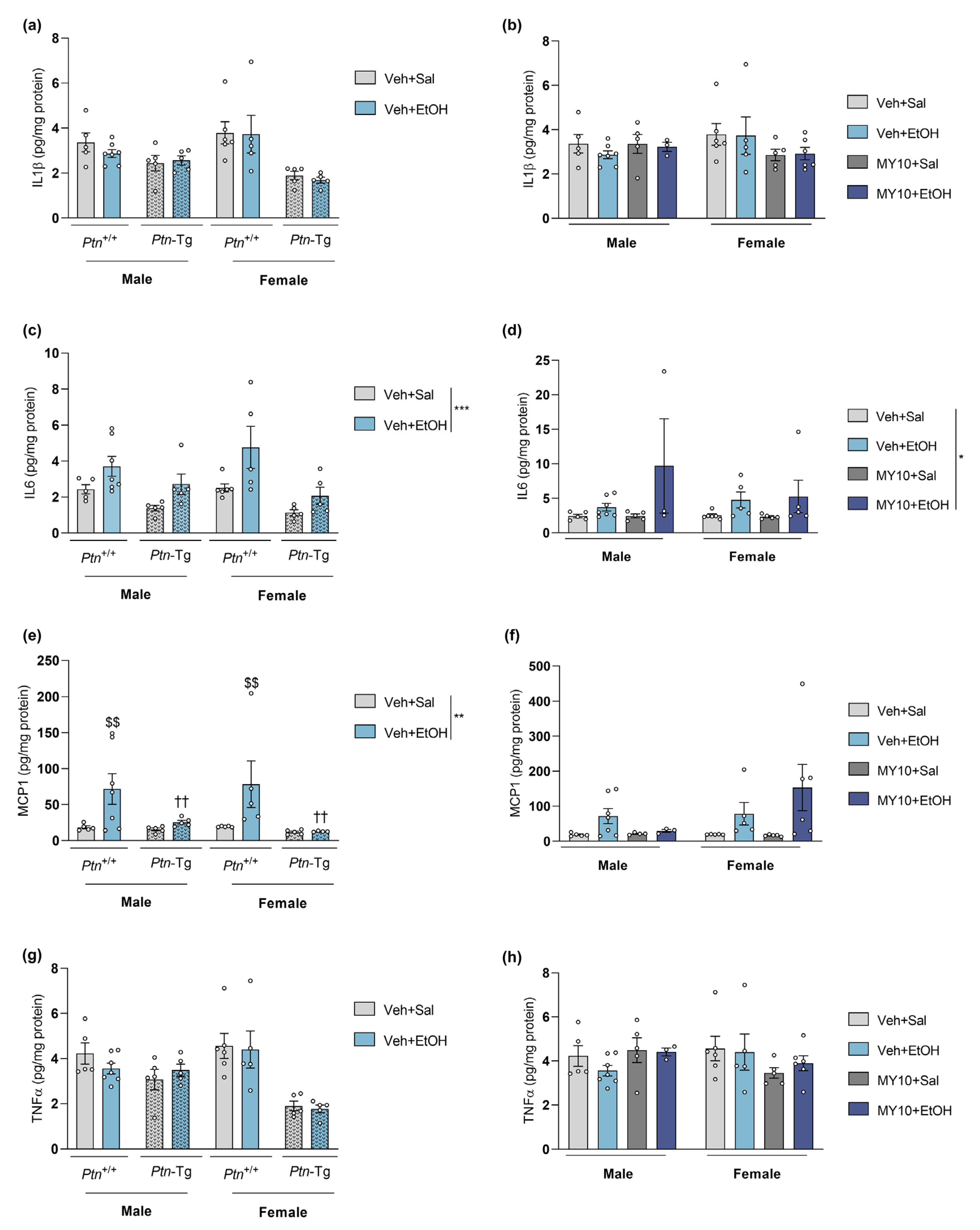
Figure 1. Cytokine expression in the PFC of adolescent male and female mice after acute ethanol administration. Graphs show data from a genetic Ptn overexpression model (Ptn-Tg vs. Ptn+/+ mice; a,c,e,g) and data from Ptn+/+ mice with RPTPβ/ζ inhibition by MY10 (b,d,f,h). Protein levels of (a,b) IL1β: Interleukin 1β; (c,d) IL6: Interleukin 6; (e,f) MCP1: monocyte chemoattractant protein 1 and (g,h) TNFα: tumor necrosis factor α were measured using multiplex luminometry. Three and two-way ANOVA analysis was performed, when appropriate. Data are presented as mean ± SEM (n = 3–7/group). * p < 0.05, ** p < 0.01, *** p < 0.001. If appropriate, only genotype and treatment factors were considered. $$ p < 0.01 vs. vehicle-saline within the same genotype, excluding sex factor. †† p < 0.01 vs. Ptn+/+ vehicle-ethanol, excluding sex factor.
Biomedicines 11 01318 g001
Figure 2. Effect of genetic overexpression of Ptn on ethanol-induced gene expression changes: (a) Iba1 (ionized calcium-binding adapter molecule 1) mRNA; (b) Cd68 (cluster of differentiation factor 68) mRNA; (c) Ccl2 (C-C motif chemokine 2) mRNA; (d) Gfap (glial fibrillary acidic protein) mRNA; (e) Il6 (Interleukin 6) mRNA; (f) Il1b (Interleukin 1 Beta) mRNA; (g) Tnfa (tumor necrosis factor alpha) mRNA; (h) Tlr4 (toll-like receptor 4) in the PFC of adolescent Ptn+/+ and Ptn-Tg male and female mice 18 h after a single dose of ethanol (6 g/kg, i.p.). Three-way ANOVA analysis was performed. Data are presented as mean ± SEM (n = 5–6/group). * p < 0.05; ** p < 0.01; *** p < 0.001; **** p < 0.0001.
Biomedicines 11 01318 g002
Figure 3. Effect of RPTPβ/ζ inhibition on ethanol-induced gene expression changes: (a) Iba1 (ionized calcium-binding adapter molecule 1) mRNA; (b) Cd68 (cluster of differentiation factor 68) mRNA; (c) Ccl2 (C-C motif chemokine 2) mRNA; (d) Gfap (glial fibrillary acidic protein) mRNA; (e) Il6 (Interleukin 6) mRNA; (f) Il1b (Interleukin 1 Beta) mRNA; (g) Tnfa (tumor necrosis factor alpha) mRNA; (h) Tlr4 (toll-like receptor 4) in the PFC of adolescent Ptn+/+ male and female mice 18 h after treatment with a single dose of ethanol (6 g/kg, i.p.). One-hour prior, mice were administered vehicle (Veh) or MY10 (60 mg/kg, i.g.). Two-way ANOVA analysis was performed. Data are presented as mean ± SEM (n = 5–6/group). * p < 0.05; ** p < 0.01; *** p < 0.001 **** p < 0.0001. # p < 0.05, ## p < 0.01; #### p < 0.0001 for differences between treatments within the same sex.
Biomedicines 11 01318 g003
Figure 4. Effect of genetic overexpression of Ptn on LPS-induced gene expression changes: (a) Iba1 (ionized calcium-binding adapter molecule 1) mRNA; (b) Cd68 (cluster of differentiation factor 68) mRNA; (c) Ccl2 (C-C motif chemokine 2) mRNA; (d) Gfap (glial fibrillary acidic protein) mRNA; (e) Il6 (Interleukin 6) mRNA; (f) Il1b (Interleukin 1 Beta) mRNA; (g) Tnfa (tumor necrosis factor alpha); (h) Tlr4 (toll-like receptor 4) mRNA levels in the PFC of adolescent Ptn+/+ and Ptn-Tg male and female mice 18 h after treatment with a single dose of LPS (5 mg/kg, i.p.). Three-way ANOVA analysis was performed. Data are presented as mean ± SEM (n = 5–6/group). * p < 0.05; ** p < 0.01, *** p < 0.001, **** p < 0.0001. If appropriate only genotype and treatment factors were considered. $$$ p < 0.001; $$$$ p < 0.0001 vs. vehicle-saline within the same genotype, excluding sex factor. † p < 0.05; †† p < 0.01; †††† p < 0.0001 vs. Ptn+/+ vehicle-LPS, excluding sex factor. γγγγ p < 0.0001 vs. vehicle-Sal, excluding sex factor.
Biomedicines 11 01318 g004
Figure 5. Effect of RPTPβ/ζ inhibition on LPS-induced gene expression changes: (a) Iba1 (ionized calcium-binding adapter molecule 1) mRNA; (b) Cd68 (cluster of differentiation factor 68) mRNA; (c) Mcp1 (C-C motif chemokine 2) mRNA; (d) Gfap (glial fibrillary acidic protein) mRNA; (e) Il6 (Interleukin 6) mRNA; (f) Il1b (Interleukin 1 Beta) mRNA; (g) Tnfa (tumor necrosis factor alpha) (h) Tlr4 (toll-like receptor 4) mRNA in the PFC of adolescent Ptn+/+ male and female mice 18 h after treatment with a single dose of LPS (5 mg/kg, i.p.). One-hour prior, mice were administered vehicle (Veh) or MY10 (60 mg/kg, i.g.). Two-way ANOVA analysis was performed. Data are presented as mean ± SEM (n = 5–6/group). * p < 0.05; ** p < 0.01, *** p < 0.001, ****p < 0.0001; # p < 0.05, ## p < 0.01, ### p < 0.001 for differences between treatments within the same sex.
Biomedicines 11 01318 g005
Figure 6. Modulation of PTN/MK/RPTPβζ signaling pathway after acute ethanol or LPS treatment. As the same control mice are used, each graph comparatively shows the effect of ethanol or LPS in two different sets: comparisons between Ptn+/+ and Ptn-Tg mice, and comparisons of Ptn+/+ mice treated with MY10 (or vehicle, Veh). Ptn (a,b), Mdk (c,d), Ptprz1 (e,f), and Alk (g,h) mRNA levels are shown in the PFC of male (left) and female (right) adolescent mice. Data are presented as mean ± SEM (n = 3–7/group). * p < 0.05; ** p < 0.01; **** p < 0.0001.
Biomedicines 11 01318 g006
Figure 7. Proposed mechanisms of the effect of Ptn overexpression (A) and the pharmacological inhibition of RPTPβ/ζ with MY10 (B) on some of the principal signaling pathways implicated in ethanol and/or LPS acute response in adolescent mice. The different significant effects found are discernible by color; red: Ptn overexpression effects (Ptn+/+ Veh+Sal vs. Ptn-Tg Veh+Sal); light green: Ptn overexpression on LPS actions (Ptn-Tg Veh+Sal vs. Ptn-Tg Veh+LPS); blue: Ptn overexpression on EtOH actions (Ptn-Tg Veh+Sal vs. Ptn-Tg Veh+EtOH); yellow: RPTPβ/ζ inhibition effects (Ptn+/+ MY10+Sal vs. Ptn+/+ Veh+Sal), dark green: RPTPβ/ζ inhibition effects on LPS actions (Ptn+/+ MY10+Sal vs. Ptn+/+ MY10+LPS). Symbols are combined with the colors. ↑↓ for significant effect found in both sexes; ▲▼ for effect found only in one sex; Biomedicines 11 01318 i001 for expected downregulation and Biomedicines 11 01318 i002 for expected upregulation.
Biomedicines 11 01318 g007
Table 1. Primer sets used for qPCR analysis. Alk: Anaplastic lymphoma kinase; Ccl2: Chemokine (C-C motif) ligand 2; Cd68: cluster of differentiation factor 68; Gfap: glial fibrillary acidic protein; Iba1: ionized calcium-binding adapter molecule 1; Il1b: Interleukin 1 beta; Il6: Interleukin 6; Mdk: Midkine; Ptn: Pleiotrophin; Ptprz1: Protein Tyrosine Phosphatase receptor type Z1; Rpl37: Ribosomal protein RPL37; Tlr4: toll-like receptor 4; Tnfa: tumor necrosis factor α.
GenePrimer ForwardPrimer Reverse
Alk5′-TAAAGACGCTGCCAGAAGT-3′5′-GGTGGTTGAATTTGCTGATG-3′
Ccl25′-GGCTCAGCCAGATGCAGTTAA-3′5′-CCTACTCATTGGGATCATCTTGCT-3′
Cd685′-TGGCGGTGGAATACAATGTG-3′5′-GATGAATTCTGCGCCATGAA-3′
Gfap5′-AACAACCTGGCTGCGTAT-3′5′-CTGCCTCGTATTGAGTGC-3′
Iba15′-GTCCTTGAAGCGAATGCTGG-3′5′-CATTCTCAAGATGGCAGATC-3′
Il1b5′-GCTGAAAGCTCTCCACCTCA-3′5′-AGGCCACAGGTATTTTGTCG-3′
Il65′-TAGTCCTTCCTACCCCAATTTCC-3′5′-TTGGTCCTTAGCCACTCCTTC-3′
Mdk5′-TGATGGGAGCACTGGCAC-3′5′-CATTGTACCGCGCCTTCTT-3′
Ptn5′-TTGGGGAGAATGTGACCTCAATAC-3′5′-GGCTTGGAGATGGTGACAGTTTTC-3′
Ptprz15′-CTACACAGGAGCACTAAATC-3′5′-CTGTTTTCCCAGTGTTGTGA-3′
Rpl375′-ACCGCAGATTCAGACATGGATT-3′5′-AGCGTAGGATCCCAGAGCAA-3′
Tlr45′-TAGGACTCTGATCATGGCACTG-3′5′-GGAACTACCTCTATGCAGGGAT-3′
Tnfa5′-AGGCACTCCCCCAAAAGATG-3′5′-TGAGGGTCTGGGCCATAGAA-3′
Table 2. Overview of the effect of PTN genetic overexpression (Ptn-Tg) and RPTPβ/ζ inhibition (treatment with MY10) on protein level changes induced by ethanol. Data represented in Figure 1 are summarized. ns: no significant changes; (-): significant decrease; grey: both sexes.
ProteinEthanol
PTN (Ptn-Tg)RPTPβ/ζ Inhibition (MY10)
IL1β (Figure 1a,b)nsns
IL6 (Figure 1c,d)nsns
MCP1 (Figure 1e,f)(-)ns
TNFα (Figure 1g,h)nsns
Table 3. Overview of the effect of PTN genetic overexpression (Ptn-Tg) and RPTPβ/ζ inhibition (treatment with MY10) on mRNA expression changes induced by ethanol (left) or LPS (right). Data represented in Figure 2, Figure 3, Figure 4 and Figure 5 are summarized. ns: no significant changes; (+): significant increase; (-): significant decrease; blue: males; orange: females; grey: both sexes.
mRNA ExpressionEthanolLPS
PTN (Ptn-Tg)RPTPβ/ζ Inhibition (MY10)PTN (Ptn-Tg)RPTPβ/ζ Inhibition (MY10)
Iba1 (Figure 2, Figure 3, Figure 4 and Figure 5a)nsns(+)(+)
Cd68 (Figure 2, Figure 3, Figure 4 and Figure 5b)nsns(+)ns
Ccl2 (Figure 2, Figure 3, Figure 4 and Figure 5c)(+)ns(+)ns
Gfap (Figure 2, Figure 3, Figure 4 and Figure 5d)nsns(+)ns
Il6 (Figure 2, Figure 3, Figure 4 and Figure 5e)(+)nsns(+)
Il1b (Figure 2, Figure 3, Figure 4 and Figure 5f)nsns(+)(+)
Tnfa (Figure 2, Figure 3, Figure 4 and Figure 5g)(-)nsns(+)
Tlr4 (Figure 2, Figure 3, Figure 4 and Figure 5h)nsns(+)ns
Disclaimer/Publisher’s Note: The statements, opinions and data contained in all publications are solely those of the individual author(s) and contributor(s) and not of MDPI and/or the editor(s). MDPI and/or the editor(s) disclaim responsibility for any injury to people or property resulting from any ideas, methods, instructions or products referred to in the content.