
Parkinsonism Is Associated with Altered SMA-Basal Ganglia Structural and Functional Connectivity in Frontotemporal Degeneration
 ,
,  , , ,
, , ,  ,
,  ,
,
Abstract
:1. Introduction
2. Materials and Methods
2.1. Participants
2.2. Clinical and Neuropsychological Assessment
2.3. MRI Data Acquisition
- -
- High-resolution three-dimensional (3D) T1-weighted (T1-3D) MPRAGE sequence (repetition time (TR) = 2400 ms, echo time (TE) = 2.12 ms, inversion time (TI) = 1000 ms, flip angle = 8°, field of view (FOV) = 256 mm, matrix = 256 × 256, 176 sagittal slices 1-mm thick, no gap);
- -
- Diffusion tensor imaging (DTI) single-shot, echo-planar, spin-echo sequence with 10 interspersed volumes of b = 0 (b0) and 64 gradient directions, TR = 4600 ms, TE = 78 ms, multiband acceleration factor = 2, monopolar diffusion scheme, FOV = 192 mm, matrix = 96 × 96, b = 1000 s/mm2, 72 contiguous axial 2-mm thick slices;
- -
- Blood oxygen level-dependent (BOLD) single-shot echo-planar imaging (TR = 3000 ms, TE = 30 ms, flip angle = 89°, FOV = 192 mm, 64 × 64 matrix, 50 contiguous axial 3-mm thick slices, 140 volumes, voxel size = 3 mm3), with all patients instructed to close their eyes and remain awake during the resting-state functional MRI acquisitions;
- -
- Dual turbo spin-echo, proton density (PD) and T2-weighted images (TR = 3320 ms, TE1 = 10 ms, TE2 = 103 ms, FOV = 220 mm, matrix = 384 × 384, 25 axial slices 4-mm thick, 30% gap);
- -
- High-resolution 3D fluid-attenuated inversion recovery (FLAIR) sequence (TR = 6000 ms, TE = 395 ms, TI = 2100 ms, FOV = 256 mm, matrix = 256 × 256, 176 sagittal slices 1-mm thick, no gap).
- -
- Two expert radiologists (PP and CG, with more than 20 and 10 years of experience, respectively) examined all MRIs to assess the presence of T2 and T2 FLAIR white matter hyperintensities (WMH). The amount of WMH was quantified using the four-stage Fazekas visual rating scale (Fazekas 0–1 = no to mild WMH, Fazekas 2 = moderate WMH, Fazekas 3 = severe WMH) [32].
2.4. MRI Analysis
2.4.1. Data Preprocessing
2.4.2. Cortical Thickness
2.4.3. Basal Ganglia and Thalamus Volumetry
2.4.4. Selection of Regions of Interest (ROIs)
2.4.5. Structural Connectivity—Tractography
2.4.6. Functional Connectivity—ROI-to-ROI Correlation Analyses
2.5. Statistical Analyses
3. Results
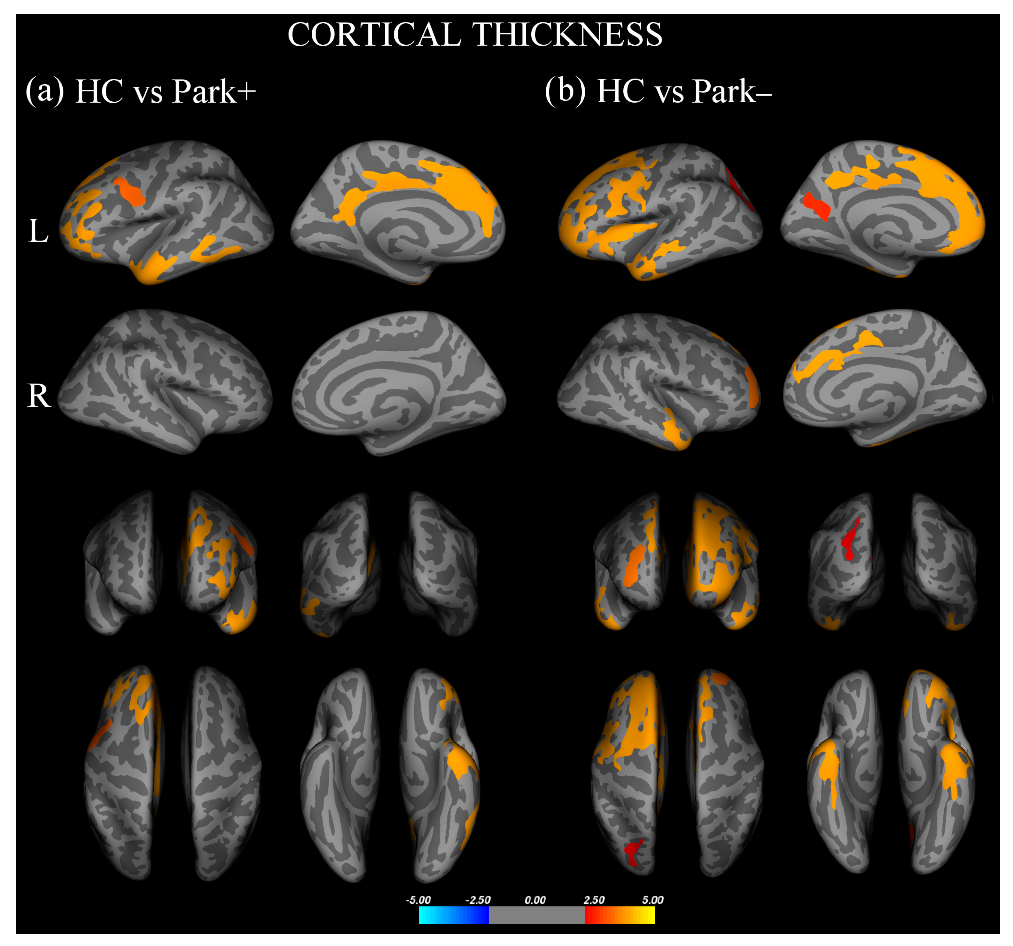
3.1. Cortical Thickness and Basal Ganglia/Thalamic Volumetry
3.2. Structural Connectivity
3.3. Functional Connectivity
3.4. Correlation Analyses
4. Discussion
4.1. Cortical Thickness in FTD
4.2. Striatal Degeneration in FTD with Parkinsonism
4.3. Reduced SMA-Basal Ganglia Structural and Functional Connectivity in FTD with Parkinsonism
4.4. Study Strengths and Limitations
5. Conclusions
Supplementary Materials
Author Contributions
Funding
Institutional Review Board Statement
Informed Consent Statement
Data Availability Statement
Conflicts of Interest
References
- Seltman, R.E.; Matthews, B.R. Frontotemporal Lobar Degeneration: Epidemiology, Pathology, Diagnosis and Management. CNS Drugs 2012, 26, 841–870. [Google Scholar] [CrossRef] [PubMed]
- Perry, D.C.; Miller, B.L. Frontotemporal Dementia. Semin. Neurol. 2013, 33, 336–341. [Google Scholar] [CrossRef] [PubMed]
- Convery, R.; Mead, S.; Rohrer, J.D. Review: Clinical, Genetic and Neuroimaging Features of Frontotemporal Dementia. Neuropathol. Appl. Neurobiol. 2019, 45, 6–18. [Google Scholar] [CrossRef] [PubMed]
- Bang, J.; Spina, S.; Miller, B.L. Frontotemporal Dementia. Lancet 2015, 386, 1672–1682. [Google Scholar] [CrossRef]
- Baizabal-Carvallo, J.F.; Jankovic, J. Parkinsonism, Movement Disorders and Genetics in Frontotemporal Dementia. Nat. Rev. Neurol. 2016, 12, 175–185. [Google Scholar] [CrossRef]
- Park, H.K.; Park, K.H.; Yoon, B.; Lee, J.-H.; Choi, S.H.; Joung, J.H.; Yoon, S.J.; Kim, B.C.; Kim, S.H.; Kim, E.-J.; et al. Clinical Characteristics of Parkinsonism in Frontotemporal Dementia According to Subtypes. J. Neurol. Sci. 2017, 372, 51–56. [Google Scholar] [CrossRef]
- Surguchov, A. Biomarkers in Parkinson’s Disease. In Neurodegenerative Diseases Biomarkers: Towards Translating Research to Clinical Practice; Peplow, P.V., Martinez, B., Gennarelli, T.A., Eds.; Neuromethods; Springer: New York, NY, USA, 2022; pp. 155–180. ISBN 978-1-07-161712-0. [Google Scholar]
- Ishii, K.; Sakamoto, S.; Sasaki, M.; Kitagaki, H.; Yamaji, S.; Hashimoto, M.; Imamura, T.; Shimomura, T.; Hirono, N.; Mori, E. Cerebral Glucose Metabolism in Patients with Frontotemporal Dementia. J. Nucl. Med. 1998, 39, 1875–1878. [Google Scholar]
- Jeong, Y.; Cho, S.S.; Park, J.M.; Kang, S.J.; Lee, J.S.; Kang, E.; Na, D.L.; Kim, S.E. 18F-FDG PET Findings in Frontotemporal Dementia: An SPM Analysis of 29 Patients. J. Nucl. Med. 2005, 46, 233–239. [Google Scholar]
- Di Stasio, F.; Suppa, A.; Fabbrini, A.; Marsili, L.; Asci, F.; Conte, A.; Trebbastoni, A.; De Lena, C.; Berardelli, A. Parkinsonism Is Associated with Altered Primary Motor Cortex Plasticity in Frontotemporal Dementia–Primary Progressive Aphasia Variant. Neurobiol. Aging 2018, 69, 230–238. [Google Scholar] [CrossRef]
- Kim, E.J.; Rabinovici, G.D.; Seeley, W.W.; Halabi, C.; Shu, H.; Weiner, M.W.; DeArmond, S.J.; Trojanowski, J.Q.; Gorno-Tempini, M.L.; Miller, B.L.; et al. Patterns of MRI Atrophy in Tau Positive and Ubiquitin Positive Frontotemporal Lobar Degeneration. J. Neurol. Neurosurg. Psych. 2007, 78, 1375–1378. [Google Scholar] [CrossRef]
- Halabi, C.; Halabi, A.; Dean, D.L.; Wang, P.-N.; Boxer, A.L.; Trojanowski, J.Q.; Dearmond, S.J.; Miller, B.L.; Kramer, J.H.; Seeley, W.W. Patterns of Striatal Degeneration in Frontotemporal Dementia. Alzheimer Dis. Assoc. Disord. 2013, 27, 74–83. [Google Scholar] [CrossRef] [PubMed]
- Jakabek, D.; Power, B.D.; Macfarlane, M.D.; Walterfang, M.; Velakoulis, D.; van Westen, D.; Lätt, J.; Nilsson, M.; Looi, J.C.L.; Santillo, A.F. Regional Structural Hypo- and Hyperconnectivity of Frontal-Striatal and Frontal-Thalamic Pathways in Behavioral Variant Frontotemporal Dementia. Hum. Brain Mapp. 2018, 39, 4083–4093. [Google Scholar] [CrossRef] [PubMed]
- Bocchetta, M.; Malpetti, M.; Todd, E.G.; Rowe, J.B.; Rohrer, J.D. Looking beneath the Surface: The Importance of Subcortical Structures in Frontotemporal Dementia. Brain Commun. 2021, 3, fcab158. [Google Scholar] [CrossRef] [PubMed]
- Haslinger, B.; Erhard, P.; Kämpfe, N.; Boecker, H.; Rummeny, E.; Schwaiger, M.; Conrad, B.; Ceballos-Baumann, A.O. Event-Related Functional Magnetic Resonance Imaging in Parkinson’s Disease before and after Levodopa. Brain 2001, 124, 558–570. [Google Scholar] [CrossRef]
- Buhmann, C.; Glauche, V.; Stürenburg, H.J.; Oechsner, M.; Weiller, C.; Büchel, C. Pharmacologically Modulated FMRI--Cortical Responsiveness to Levodopa in Drug-Naive Hemiparkinsonian Patients. Brain A J. Neurol. 2003, 126, 451–461. [Google Scholar] [CrossRef] [PubMed]
- Chung, J.W.; Burciu, R.G.; Ofori, E.; Coombes, S.A.; Christou, E.A.; Okun, M.S.; Hess, C.W.; Vaillancourt, D.E. Beta-Band Oscillations in the Supplementary Motor Cortex Are Modulated by Levodopa and Associated with Functional Activity in the Basal Ganglia. Neuroimage Clin. 2018, 19, 559–571. [Google Scholar] [CrossRef]
- Pietracupa, S.; Suppa, A.; Upadhyay, N.; Giannì, C.; Grillea, G.; Leodori, G.; Modugno, N.; Di Biasio, F.; Zampogna, A.; Colonnese, C.; et al. Freezing of Gait in Parkinson’s Disease: Gray and White Matter Abnormalities. J. Neurol. 2018, 265, 52–62. [Google Scholar] [CrossRef]
- Wu, T.; Wang, L.; Chen, Y.; Zhao, C.; Li, K.; Chan, P. Changes of Functional Connectivity of the Motor Network in the Resting State in Parkinson’s Disease. Neurosci. Lett. 2009, 460, 6–10. [Google Scholar] [CrossRef]
- Tessitore, A.; Cirillo, M.; De Micco, R. Functional Connectivity Signatures of Parkinson’s Disease. J. Parkinsons Dis. 2019, 9, 637–652. [Google Scholar] [CrossRef]
- Gorno-Tempini, M.L.; Hillis, A.E.; Weintraub, S.; Kertesz, A.; Mendez, M.; Cappa, S.F.; Ogar, J.M.; Rohrer, J.D.; Black, S.; Boeve, B.F.; et al. Classification of Primary Progressive Aphasia and Its Variants. Neurology 2011, 76, 1006–1014. [Google Scholar] [CrossRef]
- Rascovsky, K.; Hodges, J.R.; Knopman, D.; Mendez, M.F.; Kramer, J.H.; Neuhaus, J.; van Swieten, J.C.; Seelaar, H.; Dopper, E.G.P.; Onyike, C.U.; et al. Sensitivity of Revised Diagnostic Criteria for the Behavioural Variant of Frontotemporal Dementia. Brain 2011, 134, 2456–2477. [Google Scholar] [CrossRef] [PubMed]
- Meeter, L.H.; Kaat, L.D.; Rohrer, J.D.; van Swieten, J.C. Imaging and Fluid Biomarkers in Frontotemporal Dementia. Nat. Rev. Neurol. 2017, 13, 406–419. [Google Scholar] [CrossRef] [PubMed]
- Postuma, R.B.; Berg, D.; Stern, M.; Poewe, W.; Olanow, C.W.; Oertel, W.; Obeso, J.; Marek, K.; Litvan, I.; Lang, A.E.; et al. MDS Clinical Diagnostic Criteria for Parkinson’s Disease. Mov. Disord. 2015, 30, 1591–1601. [Google Scholar] [CrossRef] [PubMed]
- Antonini, A.; Abbruzzese, G.; Ferini-Strambi, L.; Tilley, B.; Huang, J.; Stebbins, G.T.; Goetz, C.G.; Barone, P.; MDS-UPDRS Italian Validation Study Group; Bandettini di Poggio, M.; et al. Validation of the Italian Version of the Movement Disorder Society--Unified Parkinson’s Disease Rating Scale. Neurol. Sci. 2013, 34, 683–687. [Google Scholar] [CrossRef]
- Folstein, M.F.; Folstein, S.E.; McHugh, P.R. Mini-Mental State. J. Psychiatr. Res. 1975, 12, 189–198. [Google Scholar] [CrossRef]
- Dubois, B.; Slachevsky, A.; Litvan, I.; Pillon, B. The FAB: A Frontal Assessment Battery at Bedside. Neurology 2000, 55, 1621–1626. [Google Scholar] [CrossRef]
- Reitan, R.M.; Wolfson, D. The Halstead-Reitan Neuropsychological Test Battery: Theory and Clinical Interpretation; Reitan Neuropsychology; Neuropsychology Press: Tucson, AZ, USA, 1985. [Google Scholar]
- Novelli, G.; Papagno, C.; Capitani, E.; Laiacona, M.; Vallar, G.; Cappa, S.F. Tre test clinici di ricerca e produzione lessicale. Taratura su soggetti normali. Arch. Di Psicol. Neurol. E Psichiatr. 1986, 47, 477–506. [Google Scholar]
- Spinnler, H. Standardizzazione e Taratura Italiana Di Test Neuropsicologici. Ital. J. Neurol. Sci. 1987, 6, 21–120. [Google Scholar]
- Morris, J.C. The Clinical Dementia Rating (CDR): Current Version and Scoring Rules. Neurology 1993, 43, 2412. [Google Scholar] [CrossRef]
- Fazekas, F.; Chawluk, J.B.; Alavi, A.; Hurtig, H.I.; Zimmerman, R.A. MR Signal Abnormalities at 1.5 T in Alzheimer’s Dementia and Normal Aging. AJR Am. J. Roentgenol. 1987, 149, 351–356. [Google Scholar] [CrossRef]
- Esteban, O.; Markiewicz, C.J.; Blair, R.W.; Moodie, C.A.; Isik, A.I.; Erramuzpe, A.; Kent, J.D.; Goncalves, M.; DuPre, E.; Snyder, M.; et al. FMRIPrep: A Robust Preprocessing Pipeline for Functional MRI. Nat. Methods 2019, 16, 111–116. [Google Scholar] [CrossRef] [PubMed]
- Esteban, O. FMRIPrep: A Robust Preprocessing Pipeline for Functional MRI, version 20.1.1. Zenodo 2020. [Google Scholar] [CrossRef]
- Gorgolewski, K.; Burns, C.D.; Madison, C.; Clark, D.; Halchenko, Y.O.; Waskom, M.L.; Ghosh, S.S. Nipype: A Flexible, Lightweight and Extensible Neuroimaging Data Processing Framework in Python. Front. Neuroinform. 2011, 5–13. [Google Scholar] [CrossRef] [PubMed]
- Gorgolewski, K. Nipype Software. Zenodo 2018. [Google Scholar] [CrossRef]
- Smith, S.M.; Jenkinson, M.; Woolrich, M.W.; Beckmann, C.F.; Behrens, T.E.J.; Johansen-Berg, H.; Bannister, P.R.; De Luca, M.; Drobnjak, I.; Flitney, D.E.; et al. Advances in Functional and Structural MR Image Analysis and Implementation as FSL. NeuroImage 2004, 23, S208–S219. [Google Scholar] [CrossRef]
- Leichnetz, G.R.; Astruc, J. The Course of Some Prefrontal Corticofugals to the Pallidum, Substantia Innominata, and Amygdaloid Complex in Monkeys. Exp. Neurol. 1977, 54, 104–109. [Google Scholar] [CrossRef]
- Naito, A.; Kita, H. The Cortico-Pallidal Projection in the Rat: An Anterograde Tracing Study with Biotinylated Dextran Amine. Brain Res. 1994, 653, 251–257. [Google Scholar] [CrossRef]
- Stephenson-Jones, M.; Kardamakis, A.A.; Robertson, B.; Grillner, S. Independent Circuits in the Basal Ganglia for the Evaluation and Selection of Actions. PNAS 2013, 110, E3670–E3679. [Google Scholar] [CrossRef]
- Milardi, D.; Gaeta, M.; Marino, S.; Arrigo, A.; Vaccarino, G.; Mormina, E.; Rizzo, G.; Milazzo, C.; Finocchio, G.; Baglieri, A.; et al. Basal Ganglia Network by Constrained Spherical Deconvolution: A Possible Cortico-Pallidal Pathway? Mov. Disord. 2015, 30, 342–349. [Google Scholar] [CrossRef]
- Grewal, S.S.; Holanda, V.M.; Middlebrooks, E.H. Corticopallidal Connectome of the Globus Pallidus Externus in Humans: An Exploratory Study of Structural Connectivity Using Probabilistic Diffusion Tractography. Am. J. Neuroradiol. 2018, 39, 2120–2125. [Google Scholar] [CrossRef]
- Cacciola, A.; Milardi, D.; Bertino, S.; Basile, G.A.; Calamuneri, A.; Chillemi, G.; Rizzo, G.; Anastasi, G.; Quartarone, A. Structural Connectivity-Based Topography of the Human Globus Pallidus: Implications for Therapeutic Targeting in Movement Disorders. Mov. Disord. 2019, 34, 987–996. [Google Scholar] [CrossRef] [PubMed]
- Fan, L.; Li, H.; Zhuo, J.; Zhang, Y.; Wang, J.; Chen, L.; Yang, Z.; Chu, C.; Xie, S.; Laird, A.R.; et al. The Human Brainnetome Atlas: A New Brain Atlas Based on Connectional Architecture. Cereb. Cortex 2016, 26, 3508–3526. [Google Scholar] [CrossRef] [PubMed]
- Jbabdi, S.; Woolrich, M.W.; Andersson, J.L.R.; Behrens, T.E.J. A Bayesian Framework for Global Tractography. Neuroimage 2007, 37, 116–129. [Google Scholar] [CrossRef]
- Gschwind, M.; Pourtois, G.; Schwartz, S.; Van De Ville, D.; Vuilleumier, P. White-Matter Connectivity between Face-Responsive Regions in the Human Brain. Cereb. Cortex 2012, 22, 1564–1576. [Google Scholar] [CrossRef]
- Javad, F.; Warren, J.D.; Micallef, C.; Thornton, J.S.; Golay, X.; Yousry, T.; Mancini, L. Auditory Tracts Identified with Combined FMRI and Diffusion Tractography. Neuroimage 2014, 84, 562–574. [Google Scholar] [CrossRef]
- Beaulieu, C.; Does, M.D.; Snyder, R.E.; Allen, P.S. Changes in Water Diffusion Due to Wallerian Degeneration in Peripheral Nerve. Magn. Reason. Med. 1996, 36, 627–631. [Google Scholar] [CrossRef]
- Sen, P.N.; Basser, P.J. A Model for Diffusion in White Matter in the Brain. Biophys. J. 2005, 89, 2927–2938. [Google Scholar] [CrossRef]
- Ma, K.K.Y.; Lin, S.; Mok, V.C.T. Neuroimaging in Vascular Parkinsonism. Curr. Neurol. Neurosci. Rep. 2019, 19, 102. [Google Scholar] [CrossRef]
- Hartikainen, P.; Räsänen, J.; Julkunen, V.; Niskanen, E.; Hallikainen, M.; Kivipelto, M.; Vanninen, R.; Remes, A.M.; Soininen, H. Cortical Thickness in Frontotemporal Dementia, Mild Cognitive Impairment, and Alzheimer’s Disease. J. Alzheimers Dis. 2012, 30, 857–874. [Google Scholar] [CrossRef]
- Nicastro, N.; Malpetti, M.; Cope, T.E.; Bevan-Jones, W.R.; Mak, E.; Passamonti, L.; Rowe, J.B.; O’Brien, J.T. Cortical Complexity Analyses and Their Cognitive Correlate in Alzheimer’s Disease and Frontotemporal Dementia. J. Alzheimers Dis. 2020, 76, 331–340. [Google Scholar] [CrossRef]
- Rohrer, J.D.; Warren, J.D.; Modat, M.; Ridgway, G.R.; Douiri, A.; Rossor, M.N.; Ourselin, S.; Fox, N.C. Patterns of Cortical Thinning in the Language Variants of Frontotemporal Lobar Degeneration. Neurology 2009, 72, 1562–1569. [Google Scholar] [CrossRef] [PubMed]
- Garibotto, V.; Borroni, B.; Agosti, C.; Premi, E.; Alberici, A.; Eickhoff, S.B.; Brambati, S.M.; Bellelli, G.; Gasparotti, R.; Perani, D.; et al. Subcortical and Deep Cortical Atrophy in Frontotemporal Lobar Degeneration. Neurobiol. Aging 2011, 32, 875–884. [Google Scholar] [CrossRef] [PubMed]
- Möller, C.; Dieleman, N.; van der Flier, W.M.; Versteeg, A.; Pijnenburg, Y.; Scheltens, P.; Barkhof, F.; Vrenken, H. More Atrophy of Deep Gray Matter Structures in Frontotemporal Dementia Compared to Alzheimer’s Disease. J. Alzheimers Dis. 2015, 44, 635–647. [Google Scholar] [CrossRef] [PubMed]
- Chow, T.W.; Izenberg, A.; Binns, M.A.; Freedman, M.; Stuss, D.T.; Scott, C.J.M.; Ramirez, J.; Black, S.E. Magnetic Resonance Imaging in Frontotemporal Dementia Shows Subcortical Atrophy. DEM 2008, 26, 79–88. [Google Scholar] [CrossRef]
- Möller, C.; Hafkemeijer, A.; Pijnenburg, Y.A.L.; Rombouts, S.A.R.B.; van der Grond, J.; Dopper, E.; van Swieten, J.; Versteeg, A.; Pouwels, P.J.W.; Barkhof, F.; et al. Joint Assessment of White Matter Integrity, Cortical and Subcortical Atrophy to Distinguish AD from Behavioral Variant FTD: A Two-Center Study. Neuroimage Clin. 2015, 9, 418–429. [Google Scholar] [CrossRef]
- Murley, A.G.; Rowe, J.B. Neurotransmitter Deficits from Frontotemporal Lobar Degeneration. Brain 2018, 141, 1263–1285. [Google Scholar] [CrossRef]
- Bocchetta, M.; Gordon, E.; Cardoso, M.J.; Modat, M.; Ourselin, S.; Warren, J.D.; Rohrer, J.D. Thalamic Atrophy in Frontotemporal Dementia—Not Just a C9orf72 Problem. Neuroimage Clin. 2018, 18, 675–681. [Google Scholar] [CrossRef]
- Hodges, J.R.; Davies, R.R.; Xuereb, J.H.; Casey, B.; Broe, M.; Bak, T.H.; Kril, J.J.; Halliday, G.M. Clinicopathological Correlates in Frontotemporal Dementia. Ann. Neurol. 2004, 56, 399–406. [Google Scholar] [CrossRef]
- Chu, M.; Wu, L.; Liu, L.; Nan, H.; Jiang, D.; Wang, Y.; Rosa Neto, P. Clinical, Genetic, and Pathological Features of Very Early Onset Frontotemporal Lobe Degeneration: A Systematic Review. Curr. Alzheimer Res. 2022. [Google Scholar] [CrossRef]
- Jbabdi, S.; Johansen-Berg, H. Tractography: Where Do We Go from Here? Brain Connect. 2011, 1, 169–183. [Google Scholar] [CrossRef]


| HC (N = 30) | FTD (N = 30) | p * | Park− (N = 18) | Park+ (N = 12) | p * | |
|---|---|---|---|---|---|---|
| Demographic/clinical features | ||||||
| Age | 68.2 ± 6.4 | 70.1 ± 7.4 | ns | 68.1 ± 7.8 | 73.3 ± 5.8 | ns |
| Female/male, n | 9/21 | 9/21 | ns | 7/11 | 2/10 | ns |
| Disease duration, y | - | 3.8 ± 1.9 | - | 3.7 ± 1.9 | 4.1 ± 1.8 | ns |
| FTD-subtype (nfv-PPA/bv-FTD), n | - | 19/11 | - | 10/8 | 9/3 | ns |
| Neuropsychological scores | ||||||
| CDR-FTD | - | 6.4 ± 3.4 | - | 6.5 ± 3.3 | 6.3 ± 3.7 | ns |
| MMSE | - | 20.6 ± 7.1 | - | 21.2 ± 7.3 | 19.7 ± 7.1 | ns |
| FAB | - | 10.8 ± 4.3 | - | 11.7 ± 4.1 | 9.2 ± 4.2 | ns |
| TMT A, sec | - | 142.3 ± 99.1 | - | 135.5 ± 104.8 | 155.5 ± 91.7 | ns |
| TMT B, sec | - | 224.5 ± 90.5 | - | 196.7 ± 96.9 | 276.9 ± 46.0 | ns |
| VSF | - | 20.4 ± 11.4 | - | 21.3 ± 11.8 | 18.8 ± 10.9 | ns |
| VPF | - | 14.0 ± 12.1 | - | 14.9 ± 13.0 | 12.1 ± 10.6 | ns |
| MDS-UPDRS-III § | - | 17.8 ± 10.4 § | - | - | 17.8 ± 10.4 | - |
| HC | Park− | Park+ | p * | H | Post hoc | |
|---|---|---|---|---|---|---|
| L global cortical thickness (mm) | 2.361 ± 0.08 | 2.217 ± 0.12 | 2.204 ± 0.12 | <0.001 | 21.02 | HC-Park− p < 0.001 |
| HC-Park+ p = 0.001 | ||||||
| Park−Park+ ns | ||||||
| R global cortical thickness (mm) | 2.356 ± 0.08 | 2.279 ± 0.11 | 2.252 ± 0.09 | 0.003 | 10.95 | HC-Park− p = 0.038 |
| HC-Park+ p = 0.007 | ||||||
| Park−Park+ ns | ||||||
| L global cortical volume | 0.1344 ± 0.0093 | 0.1195 ± 0.0146 | 0.1197 ± 0.0167 | <0.001 | 17.07 | HC-Park− p = 0.001 |
| HC-Park+ p = 0.009 | ||||||
| Park−Park+ ns | ||||||
| R global cortical volume | 0.1377 ± 0.0079 | 0.1278 ± 0.0123 | 0.1261 ± 0.0140 | <0.001 | 13.01 | HC-Park− p = 0.006 |
| HC-Park+ p = 0.016 | ||||||
| Park− Park+ ns | ||||||
| L putamen, fraction | 0.0028 ± 0.0003 | 0.0025 ± 0.0006 | 0.0023 ± 0.0005 | 0.010 | 9.22 | HC-Park− ns |
| HC-Park+ p = 0.016 | ||||||
| Park− Park+ ns | ||||||
| R putamen, fraction | 0.0029 ± 0.0002 | 0.0027 ± 0.0004 | 0.0025 ± 0.0004 | 0.004 | 10.86 | HC-Park− ns |
| HC-Park+ p = 0.004 | ||||||
| Park− Park+ ns | ||||||
| L globus pallidus, fraction | 0.0012 ± 0.0001 | 0.0011 ± 0.0002 | 0.0010 ± 0.0002 | ns | - | - |
| R globus pallidus, fraction | 0.0012 ± 0.0002 | 0.0011 ± 0.0001 | 0.0011 ± 0.0002 | ns | - | - |
| L thalamic fraction, fraction | 0.0046 ± 0.0005 | 0.0041 ± 0.0005 | 0.0041 ± 0.0005 | <0.001 | 17.10 | HC-Park− p = 0.002 |
| HC-Park+ p = 0.003 | ||||||
| Park−Park+ ns | ||||||
| R thalamic fraction, fraction | 0.0045 ± 0.0003 | 0.0043 ± 0.0005 | 0.0041 ± 0.0004 | 0.009 | 9.33 | HC-Park− ns |
| HC-Park+ p = 0.007 | ||||||
| Park− Park+ ns |
| HC | Park− | Park+ | p * | H | Post Hoc | |
|---|---|---|---|---|---|---|
| WM Tracts–FA | ||||||
| SMA-putamen | 0.478 ± 0.04 | 0.454 ± 0.05 | 0.433 ± 0.03 | 0.003 | 11.31 | HC-Park− ns |
| HC-Park+ p = 0.003 | ||||||
| Park–Park+ ns | ||||||
| SMA-pallidus | 0.500 ± 0.04 | 0.481 ± 0.05 | 0.455 ± 0.02 | 0.006 | 10.36 | HC-Park− ns |
| HC-Park+ p = 0.004 | ||||||
| Park–Park+ ns | ||||||
| SMA-thalamus | 0.504 ± 0.04 | 0.487 ± 0.05 | 0.462 ± 0.03 | 0.012 | 8.93 | HC-Park− ns |
| HC-Park+ p = 0.008 | ||||||
| Park–Park+ ns | ||||||
| M1-putamen | 0.496 ± 0.05 | 0.472 ± 0.05 | 0.452 ± 0.03 | ns | - | - |
| M1-pallidus | 0.518 ± 0.05 | 0.497 ± 0.05 | 0.478 ± 0.03 | ns | - | - |
| M1-thalamus | 0.519 ± 0.05 | 0.510 ± 0.05 | 0.492 ± 0.04 | ns | - | - |
| ROI pairs RSFC–r (Z-transformed) | ||||||
| SMA-putamen | 0.689 ± 0.25 | 0.714 ± 0.21 | 0.465 ± 0.19 | 0.012 | 8.76 | HC-Park− ns |
| HC-Park+ p = 0.017 | ||||||
| Park—Park+ p = 0.027 | ||||||
| SMA-pallidus | 0.492 ± 0.19 | 0.485 ± 0.17 | 0.346 ± 0.19 | ns | - | - |
| SMA-thalamus | 0.665 ± 0.24 | 0.793 ± 0.29 | 0.604 ± 0.36 | ns | - | - |
| M1-putamen | 0.638 ± 0.29 | 0.698 ± 0.22 | 0.510 ± 0.33 | ns | - | - |
| M1-pallidus | 0.465 ± 0.23 | 0.487 ± 0.16 | 0.348 ± 0.21 | ns | - | - |
| M1-thalamus | 0.717 ± 0.31 | 0.828 ± 0.31 | 0.626 ± 0.36 | ns | - | - |
Disclaimer/Publisher’s Note: The statements, opinions and data contained in all publications are solely those of the individual author(s) and contributor(s) and not of MDPI and/or the editor(s). MDPI and/or the editor(s) disclaim responsibility for any injury to people or property resulting from any ideas, methods, instructions or products referred to in the content. |
© 2023 by the authors. Licensee MDPI, Basel, Switzerland. This article is an open access article distributed under the terms and conditions of the Creative Commons Attribution (CC BY) license (https://creativecommons.org/licenses/by/4.0/).
Share and Cite
Piervincenzi, C.; Suppa, A.; Petsas, N.; Fabbrini, A.; Trebbastoni, A.; Asci, F.; Giannì, C.; Berardelli, A.; Pantano, P. Parkinsonism Is Associated with Altered SMA-Basal Ganglia Structural and Functional Connectivity in Frontotemporal Degeneration. Biomedicines 2023, 11, 522. https://doi.org/10.3390/biomedicines11020522
Piervincenzi C, Suppa A, Petsas N, Fabbrini A, Trebbastoni A, Asci F, Giannì C, Berardelli A, Pantano P. Parkinsonism Is Associated with Altered SMA-Basal Ganglia Structural and Functional Connectivity in Frontotemporal Degeneration. Biomedicines. 2023; 11(2):522. https://doi.org/10.3390/biomedicines11020522
Chicago/Turabian StylePiervincenzi, Claudia, Antonio Suppa, Nikolaos Petsas, Andrea Fabbrini, Alessandro Trebbastoni, Francesco Asci, Costanza Giannì, Alfredo Berardelli, and Patrizia Pantano. 2023. "Parkinsonism Is Associated with Altered SMA-Basal Ganglia Structural and Functional Connectivity in Frontotemporal Degeneration" Biomedicines 11, no. 2: 522. https://doi.org/10.3390/biomedicines11020522
APA StylePiervincenzi, C., Suppa, A., Petsas, N., Fabbrini, A., Trebbastoni, A., Asci, F., Giannì, C., Berardelli, A., & Pantano, P. (2023). Parkinsonism Is Associated with Altered SMA-Basal Ganglia Structural and Functional Connectivity in Frontotemporal Degeneration. Biomedicines, 11(2), 522. https://doi.org/10.3390/biomedicines11020522