
Is Pulmonary Involvement a Distinct Phenotype of Post-COVID-19?
 , ,
, ,  ,
,  ,
,  and
and
Abstract
:1. Introduction
2. Materials and Methods
3. Results
4. Discussion
5. Conclusions
Author Contributions
Funding
Institutional Review Board Statement
Informed Consent Statement
Data Availability Statement
Acknowledgments
Conflicts of Interest
References
- Casas-Rojo, J.M.; Antón-Santos, J.M.; Millán-Núñez-Cortés, J.; Lumbreras-Bermejo, C.; Ramos-Rincón, J.M.; Roy-Vallejo, E.; Artero-Mora, A.; Arnalich-Fernández, F.; García-Bruñén, J.M.; Vargas-Núñez, J.A.; et al. Clinical characteristics of patients hospitalized with COVID-19 in Spain: Results from the SEMI-COVID-19 Registry. Rev. Clin. Esp. 2020, 220, 480–494. [Google Scholar] [CrossRef] [PubMed]
- Cascella, M.; Rajnik, M.; Aleem, A.; Dulebohn, S.C.; Di Napoli, R. Features, Evaluation, and Treatment of Coronavirus (COVID-19); StatPearls Publishing: Treasure Island, FL, USA, 2023. Available online: http://www.ncbi.nlm.nih.gov/books/NBK554776/ (accessed on 23 April 2023).
- Hedberg, P.; Granath, F.; Bruchfeld, J.; Askling, J.; Sjöholm, D.; Fored, M.; Färnert, A.; Naucler, P. Post COVID-19 condition diagnosis: A population-based cohort study of occurrence, associated factors, and healthcare use by severity of acute infection. J. Intern. Med. 2023, 293, 246–258. [Google Scholar] [CrossRef] [PubMed]
- Alkodaymi, M.S.; Omrani, O.A.; Fawzy, N.A.; Shaar, B.A.; Almamlouk, R.; Riaz, M.; Obeidat, M.; Obeidat, Y.; Gerberi, D.; Taha, R.M.; et al. Prevalence of post-acute COVID-19 syndrome symptoms at different follow-up periods: A systematic review and meta-analysis. Clin. Microbiol. Infect. 2022, 28, 657–666. [Google Scholar] [CrossRef] [PubMed]
- Selvakumar, J.; Havdal, L.B.; Drevvatne, M.; Brodwall, E.M.; Lund Berven, L.; Stiansen-Sonerud, T.; Einvik, G.; Leegaard, T.M.; Tjade, T.; Michelsen, A.E.; et al. Prevalence and Characteristics Associated with Post–COVID-19 Condition Among Nonhospitalized Adolescents and Young Adults. JAMA Netw. Open. 2023, 6, e235763. [Google Scholar] [CrossRef]
- Fernández-de-Las-Peñas, C.; Rodríguez-Jiménez, J.; Cancela-Cilleruelo, I.; Guerrero-Peral, A.; Martín-Guerrero, J.D.; García-Azorín, D.; Cornejo-Mazzuchelli, A.; Hernández-Barrera, V.; Pellicer-Valero, O.J. Post-COVID-19 Symptoms 2 Years after SARS-CoV-2 Infection among Hospitalized vs Nonhospitalized Patients. JAMA Netw. Open. 2022, 5, e2242106. [Google Scholar] [CrossRef] [PubMed]
- Global Burden of Disease Long COVID Collaborators; Wulf Hanson, S.; Abbafati, C.; Aerts, J.G.; Al-Aly, Z.; Ashbaugh, C.; Ballouz, T.; Blyuss, O.; Bobkova, P.; Bonsel, G.; et al. Estimated Global Proportions of Individuals with Persistent Fatigue, Cognitive, and Respiratory Symptom Clusters Following Symptomatic COVID-19 in 2020 and 2021. JAMA 2022, 328, 1604–1615. [Google Scholar] [PubMed]
- Conti, V.; Corbi, G.; Sabbatino, F.; De Pascale, D.; Sellitto, C.; Stefanelli, B.; Bertini, N.; De Simone, M.; Liguori, L.; Di Paola, I.; et al. Long COVID: Clinical Framing, Biomarkers, and Therapeutic Approaches. J. Pers. Med. 2023, 13, 334. [Google Scholar] [CrossRef] [PubMed]
- Post COVID-19 Condition (Long COVID). Available online: https://www.who.int/europe/news-room/fact-sheets/item/post-covid-19-condition (accessed on 25 April 2023).
- Castanares-Zapatero, D.; Chalon, P.; Kohn, L.; Dauvrin, M.; Detollenaere, J.; Maertens de Noordhout, C.; Primus-de Jong, C.; Cleemput, I.; Van den Heede, K. Pathophysiology and mechanism of long COVID: A comprehensive review. Ann. Med. 2022, 54, 1473–1487. [Google Scholar] [CrossRef]
- Machado, M.; Valerio, M.; Álvarez-Uría, A.; Olmedo, M.; Veintimilla, C.; Padilla, B.; De la Villa, S.; Guinea, J.; Escribano, P.; Ruiz-Serrano, M.J.; et al. Invasive pulmonary aspergillosis in the COVID-19 era: An expected new entity. Mycoses 2021, 64, 132–143. [Google Scholar] [CrossRef]
- Xu, R.; Zhou, Y.; Cai, L.; Wang, L.; Han, J.; Yang, X.; Chen, J.; Chen, J.; Ma, C.; Shen, L. Co-reactivation of the human herpesvirus alpha subfamily (herpes simplex virus-1 and varicella zoster virus) in a critically ill patient with COVID-19. Br. J. Dermatol. 2020, 183, 1145–1147. [Google Scholar] [CrossRef]
- Rodríguez-Tajes, S.; Miralpeix, A.; Costa, J.; López-Suñé, E.; Laguno, M.; Pocurull, A.; Lens, S.; Mariño, Z.; Forns, X. Low risk of hepatitis B reactivation in patients with severe COVID-19 who receive immunosuppressive therapy. J. Viral Hepat. 2021, 28, 89–94. [Google Scholar] [CrossRef] [PubMed]
- Arghir, I.; Ion, I.; Dantes, E.; Fildan, A.-P.; Tofolean, D.; Cambrea, S.; Babu, L.; Arghir, O.-C. Severe Covid-19 Associated with Advanced Forms of Pulmonary Tb Disease in an Endemic Area of Romania. Chest 2022, 161, A342. [Google Scholar] [CrossRef]
- Chong, W.H.; Neu, K.P. Incidence, diagnosis and outcomes of COVID-19-associated pulmonary aspergillosis (CAPA): A systematic review. J. Hosp. Infect. 2021, 113, 115–129. [Google Scholar] [CrossRef] [PubMed]
- Martinelli, N.; Rigoni, A.M.; De Marchi, S.; Osti, N.; Donini, M.; Montagnana, M.; Castagna, A.; Pattini, P.; Udali, S.; De Franceschi, L.; et al. High Plasma Levels of Activated Factor VII-Antithrombin Complex Point to Increased Tissue Factor Expression in Patients with SARS-CoV-2 Pneumonia: A Potential Link with COVID-19 Prothrombotic Diathesis. Diagnostics 2022, 12, 2792. [Google Scholar] [CrossRef] [PubMed]
- Ackermann, M.; Verleden, S.E.; Kuehnel, M.; Haverich, A.; Welte, T.; Laenger, F.; Vanstapel, A.; Werlein, C.; Stark, H.; Tzankov, A.; et al. Pulmonary Vascular Endothelialitis, Thrombosis, and Angiogenesis in Covid-19. N. Engl. J. Med. 2020, 383, 120–128. [Google Scholar] [CrossRef]
- Song, E.; Zhang, C.; Israelow, B.; Lu-Culligan, A.; Prado, A.V.; Skriabine, S.; Lu, P.; Weizman, O.-E.; Liu, F.; Dai, Y.; et al. Neuroinvasion of SARS-CoV-2 in human and mouse brain. J. Exp. Med. 2021, 218, e20202135. [Google Scholar] [CrossRef]
- Ryabkova, V.A.; Gavrilova, N.Y.; Fedotkina, T.V.; Churilov, L.P.; Shoenfeld, Y. Myalgic Encephalomyelitis/Chronic Fatigue Syndrome and Post-COVID Syndrome: A Common. Neuroimmune Ground? Diagnostics 2023, 13, 66. [Google Scholar] [CrossRef]
- Lavi, Y.; Vojdani, A.; Halpert, G.; Sharif, K.; Ostrinski, Y.; Zyskind, I.; Lattin, M.T.; Zimmerman, J.; Silverberg, J.I.; Rosenberg, A.Z.; et al. Dysregulated Levels of Circulating Autoantibodies against Neuronal and Nervous System Autoantigens in COVID-19 Patients. Diagnostics 2023, 13, 687. [Google Scholar] [CrossRef]
- Kouranloo, K.; Dey, M.; Elwell, H.; Nune, A. A systematic review of the incidence, management and prognosis of new-onset autoimmune connective tissue diseases after COVID-19. Rheumatol. Int. 2023, 43, 1221–1243. [Google Scholar] [CrossRef]
- Normatov, M.G.; Karev, V.E.; Kolobov, A.V.; Mayevskaya, V.A.; Ryabkova, V.A.; Utekhin, V.J.; Churilov, L.P. Post-COVID Endocrine Disorders: Putative Role of Molecular Mimicry and Some Pathomorphological Correlates. Diagnostics 2023, 13, 522. [Google Scholar] [CrossRef]
- Mehandru, S.; Merad, M. Pathological sequelae of long-haul COVID. Nat. Immunol. 2022, 23, 194–202. [Google Scholar] [CrossRef] [PubMed]
- Overview|COVID-19 Rapid Guideline: Managing the Long-Term Effects of COVID-19|Guidance|NICE. Published December 18, 2020. Available online: https://www.nice.org.uk/guidance/ng188 (accessed on 22 September 2023).
- Choi, B.C.K. Slopes of a Receiver Operating Characteristic Curve and Likelihood Ratios for a Diagnostic Test. Am. J. Epidemiol. 1998, 148, 1127–1132. [Google Scholar] [CrossRef] [PubMed]
- Salamanna, F.; Veronesi, F.; Martini, L.; Landini, M.P.; Fini, M. Post-COVID-19 Syndrome: The Persistent Symptoms at the Post-viral Stage of the Disease. A Systematic Review of the Current Data. Front. Med. 2021, 8, 653516. [Google Scholar] [CrossRef] [PubMed]
- McGroder, C.F.; Zhang, D.; Choudhury, M.A.; Salvatore, M.M.; D’Souza, B.M.; Hoffman, E.A.; Wei, Y.; Baldwin, M.R.; Garcia, C.K. Pulmonary fibrosis 4 months after COVID-19 is associated with severity of illness and blood leucocyte telomere length. Thorax 2021, 76, 1242–1245. [Google Scholar] [CrossRef] [PubMed]
- Durstenfeld, M.S.; Peluso, M.J.; Peyser, N.D.; Lin, F.; Knight, S.J.; Djibo, A.; Khatib, R.; Kitzman, H.; O’Brien, E.; Williams, N.; et al. Factors Associated with Long COVID Symptoms in an Online Cohort Study. Open Forum Infect. Dis. 2023, 10, ofad047. [Google Scholar] [CrossRef]
- Jassat, W.; Mudara, C.; Vika, C.; Welch, R.; Arendse, T.; Dryden, M.; Blumberg, L.; Mayet, N.; Tempia, S.; Parker, A.; et al. A cohort study of post-COVID-19 condition across the Beta, Delta, and Omicron waves in South Africa: 6-month follow-up of hospitalized and nonhospitalized participants. Int. J. Infect. Dis. 2023, 128, 102–111. [Google Scholar] [CrossRef]
- Fernández-de-Las-Peñas, C.; Martín-Guerrero, J.D.; Pellicer-Valero, Ó.J.; Navarro-Pardo, E.; Gómez-Mayordomo, V.; Cuadrado, M.L.; Arias-Navalón, J.A.; Cigarán-Méndez, M.; Hernández-Barrera, V.; Arendt-Nielsen, L. Female Sex Is a Risk Factor Associated with Long-Term Post-COVID Related-Symptoms but Not with COVID-19 Symptoms: The LONG-COVID-EXP-CM Multicenter Study. J. Clin. Med. 2022, 11, 413. [Google Scholar] [CrossRef]
- Soh, H.S.; Cho, B. Long COVID-19 and Health-Related Quality of Life of Mild Cases in Korea: 3-Months Follow-up of a Single Community Treatment Center. J. Korean Med. Sci. 2022, 37, e326. [Google Scholar] [CrossRef]
- Blomberg, B.; Mohn, K.G.-I.; Brokstad, K.A.; Zhou, F.; Linchausen, D.W.; Hansen, B.-A.; Lartey, S.; Onyango, T.B.; Kuwelker, K.; Sævik, M.; et al. Long COVID in a prospective cohort of home-isolated patients. Nat. Med. 2021, 27, 1607–1613. [Google Scholar] [CrossRef]
- Munker, D.; Veit, T.; Barton, J.; Mertsch, P.; Mümmler, C.; Osterman, A.; Khatamzas, E.; Barnikel, M.; Hellmuth, J.C.; Münchhoff, M.; et al. Pulmonary function impairment of asymptomatic and persistently symptomatic patients 4 months after COVID-19 according to disease severity. Infection 2021, 50, 157–168. [Google Scholar] [CrossRef]
- Clark, N.; Fan, V.S.; Slatore, C.G.; Locke, E.; Whitson, H.E.; Nici, L.; Thielke, S.M. Dyspnea and Pain Frequently Co-occur among Medicare Managed Care Recipients. Ann. Am. Thorac. Soc. 2014, 11, 890–897. [Google Scholar] [CrossRef]
- McNicholas, W.T.; Hansson, D.; Schiza, S.; Grote, L. Sleep in chronic respiratory disease: COPD and hypoventilation disorders. Eur. Respir. Rev. 2019, 28, 190064. [Google Scholar] [CrossRef] [PubMed]
- Salari, N.; Hosseinian-Far, A.; Jalali, R.; Vaisi-Raygani, A.; Rasoulpoor, S.; Mohammadi, M.; Rasoulpoor, S.; Khaledi-Paveh, B. Prevalence of stress, anxiety, depression among the general population during the COVID-19 pandemic: A systematic review and meta-analysis. Glob. Health 2020, 16, 57. [Google Scholar] [CrossRef] [PubMed]
- Premraj, L.; Kannapadi, N.V.; Briggs, J.; Seal, S.M.; Battaglini, D.; Fanning, J.; Suen, J.; Robba, C.; Fraser, J.; Cho, S.-M. Mid and long-term neurological and neuropsychiatric manifestations of post-COVID-19 syndrome: A meta-analysis. J. Neurol. Sci. 2022, 434, 120162. [Google Scholar] [CrossRef] [PubMed]
- Antoniou, K.M.; Vasarmidi, E.; Russell, A.-M.; Andrejak, C.; Crestani, B.; Delcroix, M.; Dinh-Xuan, A.T.; Poletti, V.; Sverzellati, N.; Vitacca, M.; et al. European Respiratory Society Statement on Long COVID-19 Follow-Up. Eur. Respir. J. 2022, 60, 2102174. Available online: https://erj.ersjournals.com/content/early/2022/02/03/13993003.02174-2021 (accessed on 6 June 2022). [CrossRef] [PubMed]
- Bartczak, K.T.; Milkowska-Dymanowska, J.; Piotrowski, W.J.; Bialas, A.J. The utility of telemedicine in managing patients after COVID-19. Sci Rep. 2022, 12, 21392. [Google Scholar] [CrossRef] [PubMed]
- The RECOVERY Collaborative Group. Dexamethasone in Hospitalized Patients with Covid-19. N. Engl. J. Med. 2021, 384, 693–704. [Google Scholar] [CrossRef]
- Peng, M.; He, J.; Xue, Y.; Yang, X.; Liu, S.; Gong, Z. Role of Hypertension on the Severity of COVID-19, A Review. J. Cardiovasc. Pharmacol. 2021, 78, e648. [Google Scholar] [CrossRef]
- Shibata, S.; Kobayashi, K.; Tanaka, M.; Asayama, K.; Yamamoto, E.; Nakagami, H.; Hoshide, S.; Kishi, T.; Matsumoto, C.; Mogi, M.; et al. COVID-19 pandemic and hypertension: An updated report from the Japanese Society of Hypertension project team on COVID-19. Hypertens. Res. 2023, 46, 589–600. [Google Scholar] [CrossRef]
- Guan, W.-J.; Liang, W.-H.; Zhao, Y.; Liang, H.-R.; Chen, Z.-S.; Li, Y.-M.; Liu, X.-Q.; Chen, R.-C.; Tang, C.-L.; Wang, T.; et al. Comorbidity and its impact on 1590 patients with COVID-19 in China: A nationwide analysis. Eur. Respir. J. 2020, 55, 2000547. [Google Scholar] [CrossRef]
- Nevola, R.; Criscuolo, L.; Beccia, D.; Delle Femine, A.; Ruocco, R.; Imbriani, S.; Alfano, M.; Villani, A.; Russo, A.; Perillo, P.; et al. Impact of chronic liver disease on SARS-CoV-2 infection outcomes: Roles of stage, etiology and vaccination. World J. Gastroenterol. 2023, 29, 800–814. [Google Scholar] [CrossRef] [PubMed]
- Singh, S.; Khan, A. Clinical Characteristics and Outcomes of Coronavirus Disease 2019 Among Patients with Preexisting Liver Disease in the United States: A Multicenter Research Network Study. Gastroenterology 2020, 159, 768–771.e3. [Google Scholar] [CrossRef] [PubMed]
- Mallet, V.; Beeker, N.; Bouam, S.; Sogni, P.; Pol, S. Prognosis of French COVID-19 patients with chronic liver disease: A national retrospective cohort study for 2020. J. Hepatol. 2021, 75, 848–855. [Google Scholar] [CrossRef] [PubMed]
- Middleton, P.; Hsu, C.; Lythgoe, M.P. Clinical outcomes in COVID-19 and cirrhosis: A systematic review and meta-analysis of observational studies. BMJ Open Gastroenterol. 2021, 8, e000739. [Google Scholar] [CrossRef] [PubMed]
- Solomon, J.J.; Heyman, B.; Ko, J.P.; Condos, R.; Lynch, D.A. CT of Postacute Lung Complications of COVID-19. Radiology 2021, 301, E383–E395. [Google Scholar] [CrossRef]
- Van Gassel, R.J.J.; Bels, J.L.M.; Raafs, A.; van Bussel, B.C.T.; van de Poll, M.C.G.; Simons, S.O.; van der Meer, L.W.L.; Gietema, H.A.; Posthuma, R.; van Santen, S. High Prevalence of Pulmonary Sequelae at 3 Months after Hospital Discharge in Mechanically Ventilated Survivors of COVID-19. Am. J. Respir. Crit. Care Med. 2021, 203, 371–374. [Google Scholar] [CrossRef]
- Fabbri, L.; Moss, S.; Khan, F.; Chi, W.; Xia, J.; Robinson, K.A.; Smyth, A.; Jenkins, G.; Stewart, I. Post-viral parenchymal lung disease following COVID-19 and viral pneumonitis hospitalisation: A systematic review and meta-analysis. medRxiv 2021, 2021.03.15.21253593. Available online: https://www.medrxiv.org/content/10.1101/2021.03.15.21253593v2 (accessed on 13 April 2023).
- Li, F.; Zhang, X.; Comellas, A.P.; Hoffman, E.A.; Yang, T.; Lin, C.-L. Contrastive learning and subtyping of post-COVID-19 lung computed tomography images. Front. Physiol. 2022, 13, 999263. [Google Scholar] [CrossRef]
- Mancini, D.M.; Brunjes, D.L.; Lala, A.; Trivieri, M.G.; Contreras, J.P.; Natelson, B.H. Use of Cardiopulmonary Stress Testing for Patients with Unexplained Dyspnea Post-Coronavirus Disease. JACC Heart Fail. 2021, 9, 927–937. [Google Scholar] [CrossRef]
- Maamar, M.; Artime, A.; Pariente, E.; Fierro, P.; Ruiz, Y.; Gutiérrez, S.; Tobalina, M.; Díaz-Salazar, S.; Ramos, C.; Olmos, J.M.; et al. Post-COVID-19 syndrome, low-grade inflammation and inflammatory markers: A cross-sectional study. Curr. Med. Res. Opin. 2022, 38, 901–909. [Google Scholar] [CrossRef]
- Townsend, L.; Dyer, A.H.; Jones, K.; Dunne, J.; Mooney, A.; Gaffney, F.; O’Connor, L.; Leavy, D.; O’Brien, K.; Dowds, J.; et al. Persistent fatigue following SARS-CoV-2 infection is common and independent of severity of initial infection. PLoS ONE 2020, 15, e0240784. [Google Scholar] [CrossRef]
- Hendren, N.S.; Drazner, M.H.; Bozkurt, B.; Cooper, L.T. Description and Proposed Management of the Acute COVID-19 Cardiovascular Syndrome. Circulation 2020, 141, 1903–1914. [Google Scholar] [CrossRef] [PubMed]
- García-Abellán, J.; Fernández, M.; Padilla, S.; García, J.A.; Agulló, V.; Lozano, V.; Ena, N.; García-Sánchez, L.; Gutiérrez, F.; Masiá, M. Immunologic phenotype of patients with long-COVID syndrome of 1-year duration. Front. Immunol. 2022, 13, 920627. [Google Scholar] [CrossRef] [PubMed]
- Sonnweber, T.; Tymoszuk, P.; Sahanic, S.; Boehm, A.; Pizzini, A.; Luger, A.; Schwabl, C.; Nairz, M.; Grubwieser, P.; Kurz, K.; et al. Investigating phenotypes of pulmonary COVID-19 recovery: A longitudinal observational prospective multicenter trial. eLife 2022, 11, e72500. [Google Scholar] [CrossRef] [PubMed]

| Parameter | Value |
|---|---|
| Age, years, median (IQR) | 60 (50–67) |
| Males, n (%) | 155 (54.96%) |
| BMI, kg/m2, median (IQR) | 28.09 (25.61–31.65) |
| General health assessment, 0–100 numeric scale | 70 (50–80) |
| Hypertension, n (%) | 136 (48.23%) |
| Obesity, n (%) | 69 (24.47%) |
| Diabetes, n (%) | 52 (18.44%) |
| Asthma/COPD, n (%) | 38 (13.48%) |
| Heart failure, n (%) | 34 (12.06%) |
| Kidney failure, n (%) | 6 (2.12%) |
| Liver failure, n (%) | 7 (2.48%) |
| History of arterial thromboembolic events, n (%) | 30 (10.64%; 14 myocardial infarctions; 16 stroke) |
| History of neoplasm, n (%) | 21 (7.45%) |
| Obstructive sleep apnea, n (%) | 1 (0.004%) |
| Hypothyroidism, n (%) | 14 (4.96%) |
| Invasive ventilation, n (%) | 2 (0.01) |
| Non-invasive ventilation, n (%) | 2 (0.01) |
| Oxygen therapy, n (%) | 114 (40.43%) |
| Corticosteroid, n (%) | 107 (37.94%) |
| Remdesivir, n (%) | 16 (5.67%) |
| Tocilizumab, n (%) | 6 (2.12%) |
| Fresh frozen plasma, n (%) | 20 (7.09%) |
| Antibiotic, n (%) | 131 (46.45%) |
| Parameter | Mean (SD) or Median (IQR) | Pulmonary | Non-Pulmonary | p-Value |
|---|---|---|---|---|
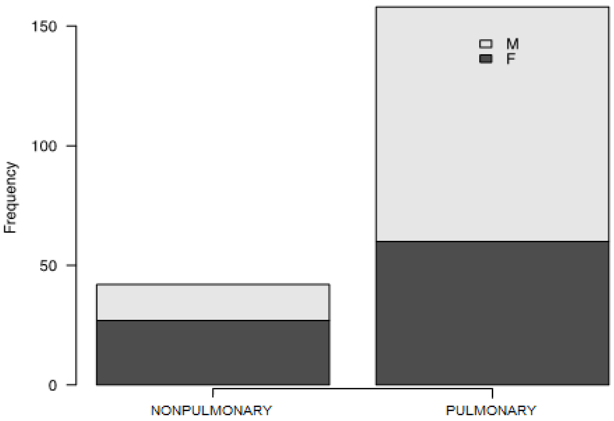
| Males, % | 56.5% | 49% | 7.5% | 0.002 |
| Age, years | 57.26 (12.20) | 58.87 (11.71) | 51.21 (12.26) | <0.001 |
| Pack-years | 18 (10–25) | 20 (10–30) | 10 (7–20) | 0.044 |
| Spontaneous saturation, % | 97 (95–98) | 97 (95–98) | 98 (97–99) | 0.025 |
| Dyspnea during acute phase, 1–5 numeric scale | 3 (1–4) | 3 (2–4.25) | 2 (1–3) | 0.000 |
| Duration of dyspnea, days | 12 (6–20) | 14 (7–29) | 10 (4–13) | 0.023 |
| Duration of myalgia, days | 9 (5–14.25) | 10 (6–15) | 7 (5–10) | 0.041 |
| Dyspnea during assessment, 1–5 numeric scale | 1 (0–2) | 1 (0–2) | 0 (0–1) | 0.000 |
| Hospitalization length, days | 11 (7–16) | 12 (7.75–17) | 8.5 (6.75–11.25) | 0.027 |
| CT control score, points | 1 (1–2) | 2 (1–3) | 0 (0–0) | 0.000 |
| FEV1, % | 89 (79–98) | 88 (78–96) | 92.5 (82.75–99) | 0.052 |
| FVC, % | 87 (78–97) | 87 (77–97) | 89.5 (84–101.25) | 0.042 |
| RV, % | 99 (85–113) | 96 (79–109.75) | 111 (97.5–124) | 0.000 |
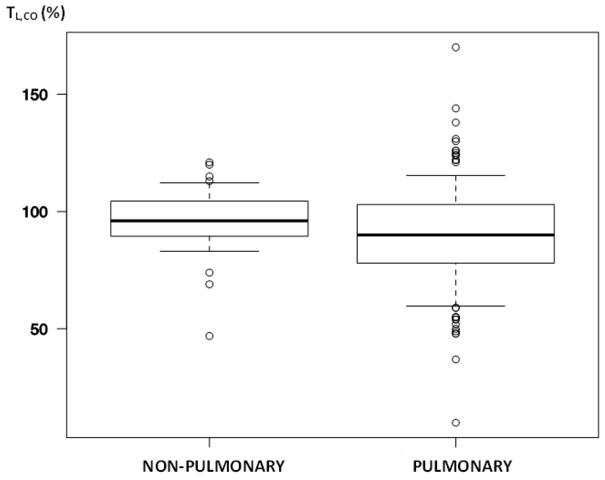
| TL,CO, % | 92 (80–103) | 90 (78.25–103) | 96 (89.5–104.5) | 0.022 |
| TLC, % | 97.79 (14.37) | 95.91 (14.12) | 104.44 (13.38) | <0.001 |
| Cardiac troponin, ng/L | 6 (3–9) | 7 (4–12) | 3.5 (3–5) | 0.003 |
| D-Dimer, ng/mL | 358 (248–598) | 365 (265.5–619) | 286 (182–469.75) | 0.004 |
| WBCs, G/L | 6.8 (6–8.025) | 6.9 (6–8.1) | 6.35 (5.65–7.225) | 0.032 |
| Immature cells, G/L | 0.02 (0.01–0.04) | 0.02 (0.01–0.04) | 0.01 (0.01–0.02) | 0.002 |
| Monocytes, G/L | 0.6 (0.5–0.7) | 0.6 (0.5–0.7) | 0.5 (0.4–0.6) | 0.002 |
| RDW-CV, %CV | 13.4 (12.8–14.4) | 13.4 (12.9–14.525) | 12.9 (12.4–13.65) | 0.004 |
| RDW-SD, fl | 43.9 (41.675–47.875) | 44.9 (42.025–48.425) | 42.8 (41.2–44.95) | 0.008 |
| Creatinine, mg/dL | 0.82 (0.71–1) | 0.86 (0.7375–1.0225) | 0.75 (0.635–0.875) | 0.004 |
| Coefficient | Estimate | OR | 95% CI | p-Value |
|---|---|---|---|---|
| Intercept | −5.22 | 0.005 | 0.0005–0.07 | <0.001 |
| Age | 0.07 | 1.07 | 1.03–1.11 | <0.001 |
| Male sex | 1.75 | 5.73 | 2.23–14.7 | <0.001 |
| Low TL,CO | 1.56 | 4.77 | 1.18–19.2 | 0.03 |
| Severity of dyspnea during COVID-19 | 0.58 | 1.78 | 1.34–2.37 | <0.001 |
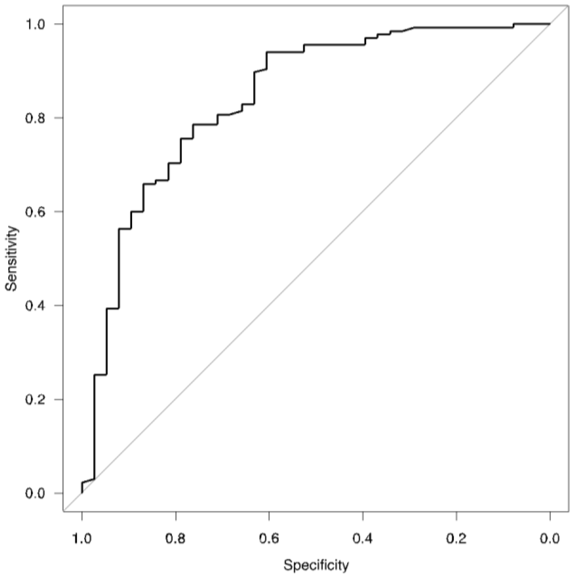
| AUROC | 0.842 | - | 0.77–0.92 | - |
Disclaimer/Publisher’s Note: The statements, opinions and data contained in all publications are solely those of the individual author(s) and contributor(s) and not of MDPI and/or the editor(s). MDPI and/or the editor(s) disclaim responsibility for any injury to people or property resulting from any ideas, methods, instructions or products referred to in the content. |
© 2023 by the authors. Licensee MDPI, Basel, Switzerland. This article is an open access article distributed under the terms and conditions of the Creative Commons Attribution (CC BY) license (https://creativecommons.org/licenses/by/4.0/).
Share and Cite
Bartczak, K.T.; Miłkowska-Dymanowska, J.; Pietrusińska, M.; Kumor-Kisielewska, A.; Stańczyk, A.; Majewski, S.; Piotrowski, W.J.; Lipiński, C.; Wawrocki, S.; Białas, A.J. Is Pulmonary Involvement a Distinct Phenotype of Post-COVID-19? Biomedicines 2023, 11, 2694. https://doi.org/10.3390/biomedicines11102694
Bartczak KT, Miłkowska-Dymanowska J, Pietrusińska M, Kumor-Kisielewska A, Stańczyk A, Majewski S, Piotrowski WJ, Lipiński C, Wawrocki S, Białas AJ. Is Pulmonary Involvement a Distinct Phenotype of Post-COVID-19? Biomedicines. 2023; 11(10):2694. https://doi.org/10.3390/biomedicines11102694
Chicago/Turabian StyleBartczak, Krystian T., Joanna Miłkowska-Dymanowska, Małgorzata Pietrusińska, Anna Kumor-Kisielewska, Adam Stańczyk, Sebastian Majewski, Wojciech J. Piotrowski, Cezary Lipiński, Sebastian Wawrocki, and Adam J. Białas. 2023. "Is Pulmonary Involvement a Distinct Phenotype of Post-COVID-19?" Biomedicines 11, no. 10: 2694. https://doi.org/10.3390/biomedicines11102694
APA StyleBartczak, K. T., Miłkowska-Dymanowska, J., Pietrusińska, M., Kumor-Kisielewska, A., Stańczyk, A., Majewski, S., Piotrowski, W. J., Lipiński, C., Wawrocki, S., & Białas, A. J. (2023). Is Pulmonary Involvement a Distinct Phenotype of Post-COVID-19? Biomedicines, 11(10), 2694. https://doi.org/10.3390/biomedicines11102694