Article

Pancreatic Pathological Changes in Murine Toxoplasmosis and Possible Association with Diabetes Mellitus

by
Asmaa M. El-kady
1,*,
Amal M. Alzahrani
2,
Hayam Elshazly
3,4,
Eman Abdullah Alshehri
5,
Majed H. Wakid
6,7,
Hattan S. Gattan
6,7,
Wafa Abdullah I. Al-Megrin
8,
Mashael S. Alfaifi
9,
Khalil Mohamed
9,
Waheeb Alharbi
10,
Hatem A. Elshabrawy
11,* and
Salwa S. Younis
12
1
Department of Medical Parasitology, Faculty of Medicine, South Valley University, Qena 83523, Egypt
2
Department of Biology, Faculty of Sciences & Arts in Almandaq, Al Baha University, Al Baha 65779, Saudi Arabia
3
Department of Biology, Faculty of Sciences-Scientific Departments, Qassim University, Buraidah 52571, Saudi Arabia
4
Department of Zoology, Faculty of Science, Beni-Suef University, Beni Suef 62521, Egypt
5
Department of Zoology, College of Science, King Saud University, Riyadh 11362, Saudi Arabia
6
Department of Medical Laboratory Technology, Faculty of Applied Medical Sciences, King Abdulaziz University, Jeddah 21589, Saudi Arabia
7
Special Infectious Agents Unit, King Fahd Medical Research Center, Jeddah 21589, Saudi Arabia
8
Department of Biology, College of Science, Princess Nourah bint Abdulrahman University, P.O. Box 84428, Riyadh 11671, Saudi Arabia
9
Department of Epidemiology, Faculty of Public Health and Health Informatics, Umm Al-Qura University, Mecca 21961, Saudi Arabia
10
Department of Physiology, Faculty of Medicine, Umm Al-Qura University, Mecca 21961, Saudi Arabia
11
Department of Molecular and Cellular Biology, College of Osteopathic Medicine, Sam Houston State University, Conroe, TX 77304, USA
12
Departments of Medical Parasitology, Faculty of Medicine, Alexandria University, Alexandria 21131, Egypt
*
Authors to whom correspondence should be addressed.
Biomedicines 2023, 11(1), 18; https://doi.org/10.3390/biomedicines11010018
Submission received: 2 November 2022 / Revised: 7 December 2022 / Accepted: 16 December 2022 / Published: 22 December 2022

Abstract

:
Background: Previous studies have reported involvement of Toxoplasma gondii (T. gondii) infections in the pathogenesis of some autoimmune diseases, such as polymyositis, rheumatoid arthritis, autoimmune thyroiditis, and Crohn’s disease. However, data on the association between T. gondii infections and Type 1 diabetes mellitus (T1DM) are still controversial. Therefore, in the present study, we aimed to investigate the pancreatic pathological changes in mouse models with acute and chronic toxoplasmosis and their association with T1DM. Materials and Methods: Three groups (10 mice each) of male Swiss Albino mice were used. One group of mice was left uninfected, whereas the second and third groups were infected with the acute virulent T. gondii RH strain and the chronic less virulent Me49 T. gondii strain, respectively. T. gondii-induced pancreatic pathological changes were evaluated by histopathological examination of pancreatic tissues. Moreover, the expression of insulin, levels of caspase-3, and the pancreatic infiltration of CD8+ T cells were evaluated using immunohistochemical staining. Results: Pancreatic tissues of T. gondii-infected animals showed significant pathological alterations and variable degrees of insulitis. Mice with acute toxoplasmosis exhibited marked enlargement and reduced numbers of islets of Langerhans. However, mice with chronic toxoplasmosis showed considerable reduction in size and number of islets of Langerhans. Moreover, insulin staining revealed significant reduction in β cell numbers, whereas caspase-3 staining showed induced apoptosis in islets of Langerhans of acute toxoplasmosis and chronic toxoplasmosis mice compared to uninfected mice. We detected infiltration of CD8+ T cells only in islets of Langerhans of mice with chronic toxoplasmosis. Conclusions: Acute and chronic toxoplasmosis mice displayed marked pancreatic pathological changes with reduced numbers of islets of Langerhans and insulin-producing-β cells. Since damage of β cells of islets of Langerhans is associated with the development of T1DM, our findings may support a link between T. gondii infections and the development of T1DM.

1. Introduction

Toxoplasma gondii (T. gondii) is an obligate intracellular opportunistic parasite that infects almost all mammals, including humans [1,2]. Infections with T. gondii are usually asymptomatic in immunocompetent individuals; however, T. gondii can cause serious disease immunocompromised individuals and pregnant women [3,4,5,6]. Humans can acquire infection through ingestion of tissue cysts in meat, food and water contaminated with oocysts from infected cats, and congenitally from infected mothers [7].
It has been well documented that Th1 cell-mediated and humoral immune responses develop following T. gondii infection [8]. Although these immune responses are required for the host defense against T. gondii, excessive inflammatory response damages the host tissues [9]. Additionally, T. gondii infection could result in autoantibodies production, which may further potentiate tissue damage [10].
Previous clinical studies have associated anti-Toxoplasma antibodies with several autoimmune diseases (AID) including polymyositis [11], rheumatoid arthritis (RA) [12,13,14], autoimmune thyroid diseases [11,15,16], Crohn’s disease [17], anti-phospholipid syndrome [18], Wegener’s granulomatosis [19] and autoimmune bullous diseases [20]. However, experimental studies reported conflicting findings on the association between AID and T. gondii infection [21,22,23].
Type 1 diabetes mellitus (T1DM) is an autoimmune disorder that results from the destruction of β cells of islets of Langerhans in the pancreas by antigen-specific T lymphocytes [24]. To date, 0.3% of the US population have type 1 diabetes (1.6 million per 330 million US residents) [25]. The pathological characteristic of T1DM is insulitis, which is defined by autoreactive CD4+ and CD8+ T-cell infiltration of insulin-producing-β cells of islets of Langerhans [26,27,28]. Similar to other autoimmune diseases, genetic and environmental factors, such as infectious agents, are involved in the development of T1DM [27,29].
Data on the association between T. gondii infection and T1DM are still controversial [30]. Several studies have reported that diabetic patients have a high seroprevalence of toxoplasmosis antibodies [30,31,32,33,34,35,36]. On the other hand, the ability of T. gondii to infiltrate and proliferate inside pancreatic cells has been linked to an increased risk of developing diabetes [36,37].
In the present study, we evaluated the pancreatic pathological changes in mice infected with acute virulent and chronic less virulent T. gondii strains. Our findings demonstrated insulitis and marked reduction in numbers of β cells of islets of Langerhans in pancreatic tissues of infected animals associated with apoptotic cell death. Therefore, our data may provide a possible link between T. gondii infections and development of T1DM.

2. Materials and Methods

2.1. Animal Experiment

The present study was conducted at the Department of Medical Parasitology, Faculty of Medicine, Alexandria University, Egypt.
Four to six week old laboratory bred male Swiss Albino mice, weighing 20–25 g, were used in our experiment. Mice were housed in well-ventilated cages and were provided with water and fed on regular pellet meals. All mice groups were kept under strict light cycles (12 h light/12 h dark cycle) and a temperature of 25 ± 2 °C). Stool examination was performed for three successive days to rule out any parasitic infections using the formol-ether concentration method [38] and modified Ziehl–Neelsen technique [39]. Mice were divided into three groups (10 mice each), which included the uninfected control group, acute toxoplasmosis group (infected intraperitoneally with 1 × 10⁴ tachyzoites of the virulent RH HXGPRT (-) strain of T. gondii tachyzoites/mouse, and chronic toxoplasmosis group (infected orally with 10 cysts/mouse of the Me49 less virulent strain of T. gondii). Mice in the acute toxoplasmosis group were sacrificed 5 days post-infection (PI) [36], whereas mice in the chronic toxoplasmosis group were sacrificed 60 days PI [36].

2.1.1. Infection with Me49 Strain of T. gondii

Ten mice were orally infected with cysts of Me49 strain (10 cysts/mouse) obtained from brain homogenate of infected mice, 8 weeks PI [40,41]. Briefly, the brain homogenate was prepared by homogenizing each infected brain in 1 mL saline using a tissue homogenizer (Wheaton, IL, USA). Cysts were microscopically counted using hemocytometer under 400× magnification. The brain suspension was then diluted to a concentration of 100 cysts/mL and each mouse was infected with 0.1 mL containing 10 cysts [41].

2.1.2. Infection with Virulent RH Strain of T. gondii

Tachyzoites of the virulent RH strain of T. gondii were obtained by serial intraperitoneal passages in Swiss Albino mice. Briefly, tachyzoites were obtained by flushing the peritoneal cavity with Phosphate Buffered Saline (PBS) 5 days post infection [42]. The peritoneal fluid was centrifuged at 200× g for 5 min at room temperature, to remove peritoneal cells and cellular debris. The supernatant was then centrifuged for 10 min at 800× g [43], and the residue containing the tachyzoites was counted by hemocytometer then diluted in PBS to the concentration of 1 × 10⁵/mL. Each mouse was infected with 1 × 10⁴ in 0.1 mL PBS [44].

2.2. Histopathological Examination

2.2.1. Hematoxylin and Eosin Staining

Pancreas was isolated from animals of all groups, fixed in 10% formalin, dehydrated in ascending concentrations of ethanol, and embedded in paraffin. Sections of 3 µm thickness were prepared and stained with hematoxylin and eosin (H&E) stain and then images were taken and examined by an independent pathologist. ImageJ scanner and viewer software were used to scan slides and process images (LOCI, University of Wisconsin, Madison, WI, US).
Pancreatic tissue sections were examined for the degree of necrosis and inflammatory cell infiltration. In addition, the size, number, and the morphology of islets of Langerhans/mouse were examined in 3 random high power fields (HPF; 400x) and mean was calculated/group. In addition, the presence of insulitis was evaluated and graded on a scale 0–4 as previously described: islets devoid of any mononuclear cells = 0; minimum focal islet infiltrate = 1+; peri-islet infiltrate of <25% of islet circumference = 2+; peri-islet infiltration and <50% intra-islet area = 3+; intra-islet infiltration >50% of islet area = 4+ [45].

2.2.2. Immunohistochemistry

Pancreatic tissues were examined for expression of insulin, levels of caspase-3, and infiltration of CD8 T cells. Briefly, 4 µm thick paraffin pancreatic tissue sections were de-paraffinized in xylene for 20 min, rehydrated with descending ethanol concentrations and then rinsed in distilled water. Sections were placed in citrate buffer (pH 6.0) and heated in microwave for epitope retrieval. Endogenous peroxidases were then blocked by incubating in 0.6% H2O2 for 10 min. Tissues sections were washed twice with PBS then treated with superblock and incubated overnight at room temperature with the following primary antibodies: Anti-insulin rabbit monoclonal antibody (Catalog no.: A19066, ABclonal, Woburn, MA 01801, USA,), anti-caspase-3 rabbit polyclonal antibody (Catalog no.: A11953, ABclonal, Woburn, MA 01801, USA) and anti-CD8 alpha rabbit monoclonal antibody (Catalog no.: 50389-R309, Sino Biological US Inc. Houston, TX 77074, USA). Sections were washed twice with PBS containing 0.05% Tween-20 (PBS-T) and then incubated with Mouse/Rabbit ImmunoDetector DAB HRP for 1 h at room temperature (Catalog No.: BSB 0003, BIO SB, Santa Barbara, CA 93117, USA.). Finally, slides were washed with PBS-T and incubated with 0.05% DAB and 0.01% H2O2 for 3 min. The sections were then counterstained with hematoxylin for 1 min, dehydrated in increasing concentrations of ethanol (70%, 80%, 90%, and 100%), and cleared in xylene for 5 min. Finally, all slides were mounted with DPX, cover-slipped, and imaged at 400× magnification using an Olympus light microscope equipped with a digital camera (Olympus, Japan, BX53).
Cells with reaction to insulin, CD8 and caspase-3 antibodies were considered positive. Semi-quantitative analysis of positive-stained tissue sections was performed through modified Allred scoring system guidelines [46]. Positive cells were counted in three pancreatic islets in three different HPFs/mouse (400×) and the mean number was calculated/group.

2.3. Statistical Analysis

Statistical analysis was performed with Statistical Package for the Social Sciences (SPSS 21). Differences between the study groups were calculated using repeated measures ANOVA test by the LSD post hoc test. Differences were considered statistically significant at p < 0.05.

3. Results

3.1. Pancreatic Tissues of Mice with Acute and Chronic Toxoplasmosis Demonstrated Marked Insulitis Characterized by Reduced Number and Abnormal Size of Islets of Langerhans

Examination of H & E-stained pancreatic tissue sections of uninfected mice showed uniform, rounded islets of Langerhans within pancreatic acini with no evidence of necrosis or inflammation (Figure 1A, black arrows). In contrast, significant pathological changes were detected in the pancreatic tissue sections of acute and chronic T. gondii-infected mice. Mice infected with acute virulent RH strain showed grade 2 insulitis with inflammatory cell infiltration (Figure 1B, black arrows), edema (Figure 1B, arrow heads), and areas of necrosis (Figure 1B, red arrows). Moreover, islets of Langerhans were significantly enlarged compared to uninfected mice (Figure 1B,D; p = 0.001). However, pancreatic tissue sections of chronic Me49 strain-infected mice showed grade 1 insulitis and a significant reduction in the size of islets of Langerhans compared to uninfected mice (Figure 1C,D; p = 0.001).
Islets of Langerhans were characterized by chronic inflammatory cellular infiltrate (Figure 1C, black arrows) and fibrous-like substance (Figure 1C, red arrow). Interestingly, islets of Langerhans of mice with chronic toxoplasmosis were significantly smaller than those in mice with acute toxoplasmosis (Figure 1D; p = 0.003).
Our findings also demonstrate that both T. gondii-acute and chronic infections significantly reduced the number of islets of Langerhans compared to uninfected animals (Figure 1E; p = 0.018 and 0.021 for acute and chronic infections, respectively). However, there was no statistically significant difference in the number of islets of Langerhans between acute and chronic T. gondii infections.

3.2. Islets of Langerhans of Mice with Chronic Toxoplasmosis Are Infiltrated with CD8+ T Cells

Next, we used immunohistochemical staining to examine infiltration of CD8+ T cells into islets of Langerhans. Our results showed that uninfected (Figure 2A) and acute toxoplasmosis mice (Figure 2B) were negative for CD8+ T cell infiltration in islets of Langerhans. On the other hand, islets of Langerhans of mice with chronic toxoplasmosis were infiltrated with CD8+ T cells (Figure 2C, black arrows).

3.3. Acute and Chronic Toxoplasmosis Are Characterized by Lower Number of β Cells in Islets of Langerhans

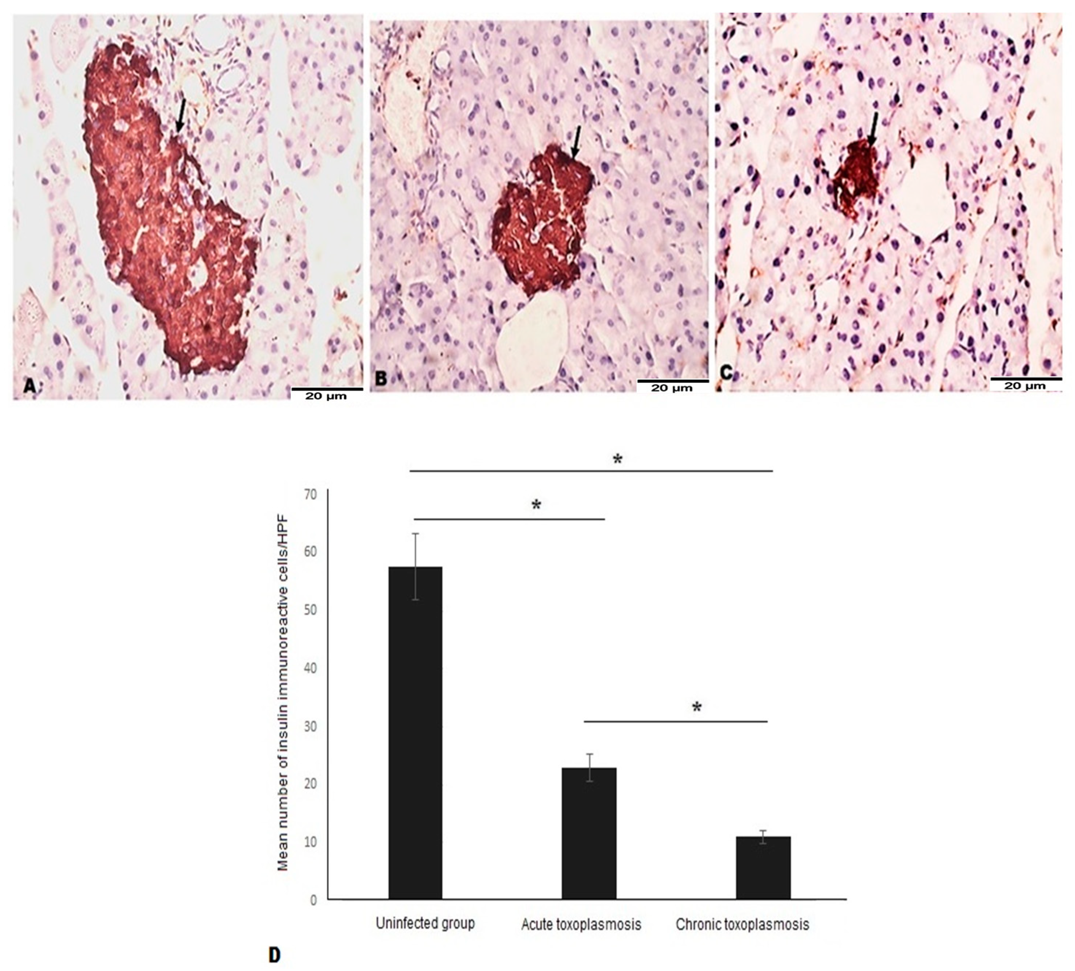
Next, we aimed to examine the effect of insulitis on the number of β cells of islets of Langerhans. Immunohistochemical staining of insulin in pancreatic tissue sections revealed higher number of β cells in islets of Langerhans of uninfected mice (Figure 3A, black arrow) compared to T. gondii-acute (Figure 3B, black arrow) and chronically (Figure 3C, black arrow) infected mice.
The mean number of β cells were significantly lower in acute and chronic T. gondii-infected mice compared to uninfected mice (Figure 3D, p = 0.003). In comparison to the chronic toxoplasmosis group, islets of Langerhans of mice with acute toxoplasmosis had statistically significant higher number of β cells (Figure 3D; p = 0.021).

3.4. Acute and Chronic Toxoplasmosis Are Associated with Apoptotic Cell Death of Islets of Langerhans

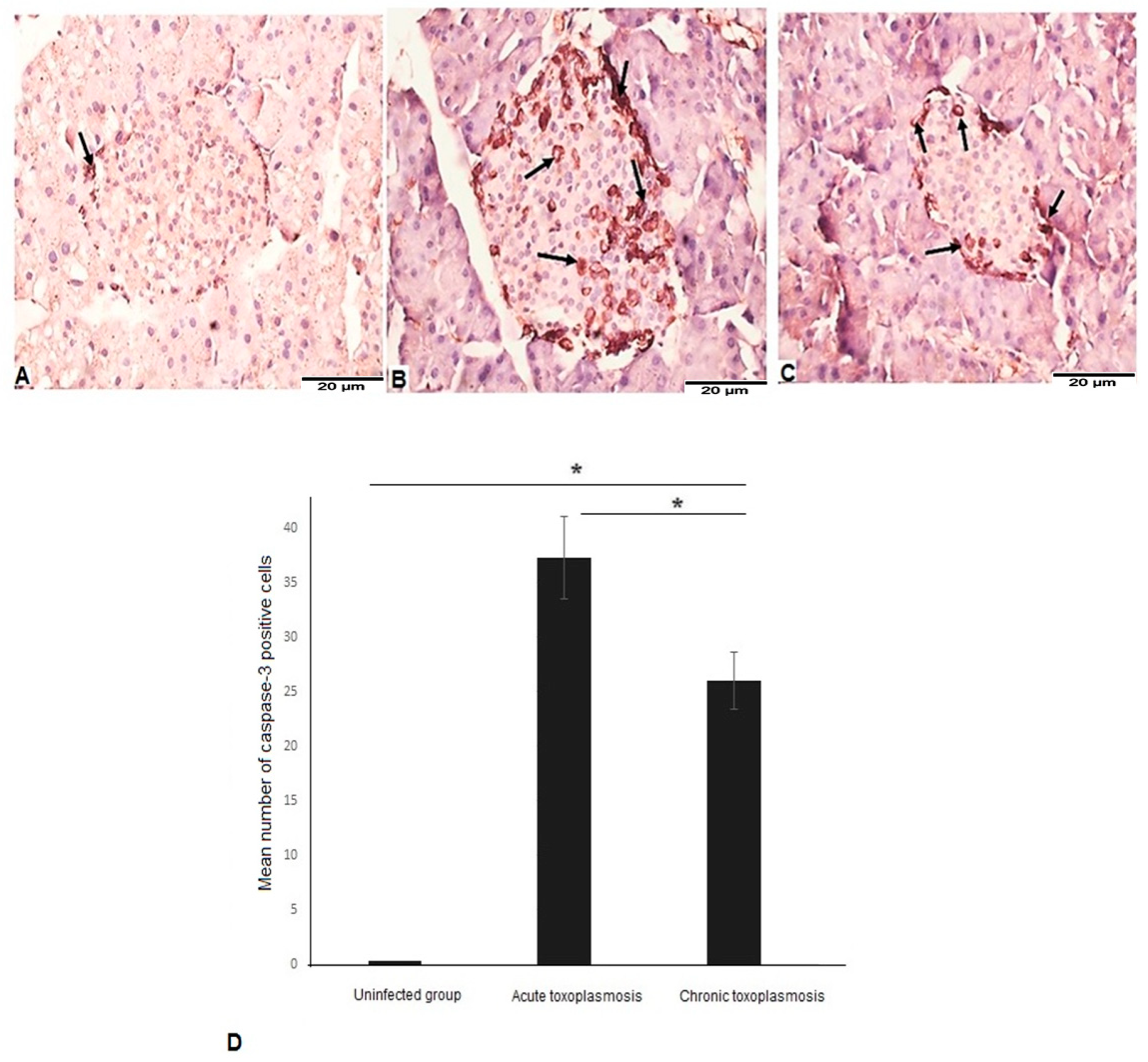
Apoptosis of β cells of islets of Langerhans has been previously shown to be associated with the development of T1DM [36]. To examine the apoptotic cell death in the islets of Langerhans in mice with acute and chronic toxoplasmosis, we performed immunohistochemical staining of capsase-3 in pancreatic tissue sections. We detected low levels of caspase-3 in islets of Langerhans of uninfected mice (Figure 4A). Pancreatic tissue sections of mice with acute (Figure 4B) and chronic (Figure 4C) toxoplasmosis showed elevated levels and significantly higher numbers of caspase-3-positive cells (apoptotic cells) in islets of Langerhans (Figure 4D; p = 0.001), compared to uninfected animals. Interestingly, the number of caspase-3-positive cells were significantly higher in acute toxoplasmosis than the chronic toxoplasmosis group (Figure 4D; p = 0.015).

4. Discussion

Diabetes mellitus (DM) is one of the most common endocrine disorders [47]. It has been postulated that several environmental factors including infectious agents can trigger the onset of the disease [27,29]. The causal link between toxoplasmosis and diabetes is still controversial, and they were thought to be predisposed to each other depending on which developed first [48]. Chronic toxoplasmosis was suggested to play a role in the pathogenesis of type 2 diabetes mellitus (T2D) due to the correlation between the state of insulin resistance in T2D and the elevated levels of circulating inflammatory cytokines including IL-2,4; IL-6,5; IL-12,6; TNF; and IFN [48]. Some reports suggested that T. gondii infection can cause T1DM due to detection of tachyzoites and bradyzoites in the pancreatic tissues of experimentally infected animals [49]. However, the particular mechanism through which chronic toxoplasmosis could lead to the development of T1D has not yet been elucidated, to our knowledge.
In the present study, we examined the pancreatic tissues of mice infected with acute and chronic T. gondii strains to assess the effect of T. gondii infection on the pancreas. Significant T. gondii-induced pathological changes were detected in both toxoplasmosis groups. The most important finding was insulitis, which was demonstrated in RH and Me49 T. gondii-infected animals. It is well documented that insulitis is the main pathological finding in T1DM and individuals who develop insulitis will eventually progress to T1DM [50,51,52,53]. In agreement with our report, T. gondii-induced insulitis has been reported by Nassief Beshay et al. [36]. It has been postulated that cytotoxic CD8+T cells are the most predominant immune cells that infiltrate islets of Langerhans of pancreas causing insulitis and destruction of the insulin-producing-β cells of islets of Langerhans [50,51]. Given that T. gondii is an intracellular pathogen, CD8+ T-lymphocytes, which identify and eliminate intracellular infections in cells infected with viral, bacterial, and parasitic organisms, are anticipated to play a major part in the immune response against this parasite [52]. Our results showed the infiltration of CD8+ T cells into the islets of Langerhans in the chronic toxoplasmosis group. This may be explained by the ability of Me49 strain to induce a pro-inflammatory response characterized by exacerbated Th1 response [8]. This finding is consistent with insulitis and autoimmune destruction of β cells of islets of Langerhans in cases of chronic toxoplasmosis, and possible progression to T1DM [53]. On the contrary, the pancreatic tissues of RH infected mice showed no infiltration with CD8+ T cells, which may be explained by that RH strain mostly causing acute death of mice in a short time. Additionally, it has been documented that RH T. gondii dampens the Th1-type immune response [8].
It was interesting that chronic in the toxoplasmosis group showed deposition of fibrous-like material within the islets of Langerhans. The deposition of fibrous-like material has been reported in one study among the pathological alterations induced by streptozotocin, a chemical commonly used in induction of T1DM [54].
It is well documented that reduction of the number and size of the islets are hallmarks of T1DM pathology and this reduction is associated with subsequent reduction of insulin secretion. [55,56]. Our findings showed that number of islets of Langerhans were significantly lower in acute and the chronic toxoplasmosis groups compared to uninfected mice. However, acute toxoplasmosis mice had enlargement islets of Langerhans due to edema and infiltration of leukocytes. On the contrary, the chronic toxoplasmosis group showed marked reduction of the size of the Islets of Langerhans. No statistically significant difference in the number of Islets of Langerhans was observed between the RH and Me49-infected animals. Similar observations were previously reported in pancreatic tissues of Me49 T.gondii-infected mice [36]. Moreover, reduced size and number of islets were observed in diabetic mice in which alloxan and streptozotocin were used to induce T1DM [54,57,58,59,60].
Several studies have suggested that apoptosis of β cells of islets of Langerhans is a critical step in the development of T1DM [61]. In line with a study by Nassief Beshay et al. [36], we demonstrated a significantly higher expression of caspase-3 and evident apoptosis in the islets of Langerhans in Me49 T. gondii-infected mice compared to the uninfected control group. The role of apoptosis of β cells of islets of Langerhans in induction of T1DM is supported by a study, which showed that caspase-3 deficient mice were protected from acquiring diabetes in a multiple-low-dose streptozotocin autoimmune diabetes paradigm [61].
We found that the number of insulin-producing β cells was significantly lower in the acute and the chronic toxoplasmosis groups (as demonstrated by the lower number of anti-insulin immunoreactive cells) compared to uninfected group. This finding is supported by previous results obtained by Nassief Beshay et al. [36]. Similarly, Ahmadi et al. found a decreased number of insulin immunoreactive cells in the pancreatic islets of diabetic rats [62]. This reduction in number of β cells of islets of Langerhans could be attributed to the observed apoptosis and reduction of the size of the islets.
Based on the results of previous studies, both RH and Me49 T. gondii strains have been isolated from human patients [63,64]. So, in the present study we aimed to assess the effect of both strains on the pancreas. However, we do acknowledge some limitations in the present work. The major limitation is the difference between both strains in the route of infection, pathogenicity and the survival of both groups of infected mice and subsequently the difference in follow up times.

5. Conclusions

The development of T1DM involves genetic and environmental factors that trigger an autoimmune response that leads to destruction of β cells of islets of Langerhans [65]. In our study, we showed that acute and chronic T. gondii-infected mice demonstrated insulitis, reduced number of islets of Langerhans and the insulin-producing β cells, and increased apoptosis of cells within islets of Langerhans. However, unlike acute RH strain-infected mice, islets of Langerhans of chronic Me49 strain-infected mice were infiltrated with CD8+ T cells, which were previously associated with chronic toxoplasmosis and development of T1DM [36]. To the best of our knowledge, this is the first study showing the pathological effect of acute RH strain of T. gondii on the pancreas, but more studies are needed to explain the mechanism of such changes. We believe that the observed pathological changes in pancreatic tissues of acute and chronic toxoplasmosis mice groups may explain the association of T. gondii infections with the development of T1DM.
To further corroborate the link between toxoplasmosis and the development of T1DM, more prospective and retrospective cohort studies are needed on the incidence of T1DM among T. gondii seropositive patients. The establishment of implication of T. gondii infection in T1DM development could help with diabetes risk prediction, early therapeutic intervention, and potential utility of T. gondii therapeutics for prevention of T1DM.

Author Contributions

Conceptualization, A.M.E.-k., H.A.E. and S.S.Y.; experimental design and methodology, A.M.E.-k., A.M.A., H.E., E.A.A., M.H.W., H.S.G., M.S.A., W.A.I.A.-M., K.M., H.A.E. and S.S.Y.; writing-original drafts, A.M.E.-k. and H.A.E.; data analysis, A.M.E.-k., A.M.A., H.E., E.A.A., M.H.W., H.S.G., W.A.I.A.-M., M.S.A., K.M., W.A., H.A.E. and S.S.Y.; writing—review and editing, A.M.E.-k., W.A. and H.A.E.; investigation, A.M.E.-k. and H.A.E.; supervision, A.M.E.-k. and H.A.E.; project administration, A.M.E.-k. and H.A.E.; critical revisions and writing, A.M.E.-k., H.A.E., W.A. and M.H.W. All authors have read and agreed to the published version of the manuscript.

Funding

This research received no external funding.

Institutional Review Board Statement

This study was conducted according to the guidelines of the Declaration of Helsinki and approved by the Research Ethics Committee of the Faculty of Medicine, Alexandria University, Egypt (Protocol code: 0305385). Animal care was done according to the NIH Guide for care and use of laboratory animals.

Informed Consent Statement

Not applicable.

Data Availability Statement

Not applicable.

Acknowledgments

Princess Nourah bint Abdulrahman University Researchers Supporting Project (PNURSP2023R39), Princess Nourah bint Abdulrahman University, Riyadh, Saudi Arabia.

Conflicts of Interest

The authors declare no conflict of interest.

References

  1. Webster, J.P.; Dubey, J.P. Toxoplasmosis of animals and humans. Parasites Vectors 2010, 3, 112. [Google Scholar] [CrossRef] [Green Version]
  2. Dubey, J.P. Advances in the life cycle of Toxoplasma gondii. Int. J. Parasitol. 1998, 28, 1019–1024. [Google Scholar] [CrossRef] [PubMed] [Green Version]
  3. Sutterland, A.L.; Fond, G.; Kuin, A.; Koeter, M.W.J.; Lutter, R.; van Gool, T.; Yolken, R.; Szoke, A.; Leboyer, M.; de Haan, L. Beyond the association. Toxoplasma gondii in schizophrenia, bipolar disorder, and addiction: Systematic review and meta-analysis. Acta Psychiatr. Scand. 2015, 132, 161–179. [Google Scholar] [CrossRef] [PubMed]
  4. Ahmadpour, E.; Daryani, A.; Sharif, M.; Sarvi, S.; Aarabi, M.; Mizani, A.; Rahimi, M.T.; Shokri, A. Toxoplasmosis in immunocompromised patients in Iran: A systematic review and meta-analysis. J. Infect. Dev. Ctries. 2014, 8, 1503–1510. [Google Scholar] [CrossRef] [PubMed]
  5. Galvan-Ramírez, M.D.L.L.; Troyo-Sanroman, R.; Roman, S.; Bernal-Redondo, R.; Castellanos, J.L.V. Prevalence of Toxoplasma infection in Mexican newborns and children: A systematic review from 1954 to 2009. ISRN Pediatr. 2012, 2012, 501216. [Google Scholar] [CrossRef] [PubMed] [Green Version]
  6. Foroutan-Rad, M.; Khademvatan, S.; Majidiani, H.; Aryamand, S.; Rahim, F.; Malehi, A.S. Seroprevalence of Toxoplasma gondii in the Iranian pregnant women: A systematic review and meta-analysis. Acta Trop. 2016, 158, 160–169. [Google Scholar] [CrossRef]
  7. Tenter, A.M.; Heckeroth, A.R.; Weiss, L.M. Toxoplasma gondii: From animals to humans. Int. J. Parasitol. 2000, 30, 1217–1258. [Google Scholar] [CrossRef] [Green Version]
  8. Munoz, M.; Liesenfeld, O.; Heimesaat, M.M. Immunology of Toxoplasma gondii. Immunol. Rev. 2011, 240, 269–285. [Google Scholar] [CrossRef]
  9. Carter, C.J. Toxoplasmosis and polygenic disease susceptibility genes: Extensive Toxoplasma gondii host/pathogen interactome enrichment in nine psychiatric or neurological disorders. J. Pathog. 2013, 2013, 965046. [Google Scholar] [CrossRef] [Green Version]
  10. Prandota, J. T. gondii infection acquired during pregnancy and/or after birth may be responsible for development of both type 1 and 2 diabetes mellitus. J. Diabetes Metab. 2013, 4, 55. [Google Scholar] [CrossRef]
  11. Adams, E.M.; Hafez, G.R.; Carnes, M.; Wiesner, J.K.; Graziano, F.M. The development of polymyositis in a patient with toxoplasmosis: Clinical and pathologic findings and review of literature. Clin. Exp. Rheumatol. 1984, 2, 205–208. [Google Scholar] [PubMed]
  12. Mousa, M.A.; Soliman, H.E.; el Shafie, M.S.; Abdel-Baky, M.S.; Aly, M.M. Toxoplasma seropositivity in patients with rheumatoid arthritis. J. Egypt. Soc. Parasitol. 1988, 18, 345–351. [Google Scholar] [PubMed]
  13. Balleari, E.; Cutolo, M.; Accardo, S. Adult-onset still’s disease associated to Toxoplasma gondii infection. Clin. Rheumatol. 1991, 10, 326–327. [Google Scholar] [CrossRef] [PubMed]
  14. Tomairek, H.A.; Saeid, M.S.; Morsy, T.A.; Michael, S.A. Toxoplasma gondii as a cause of rheumatoid arthritis. J. Egypt. Soc. Parasitol. 1982, 12, 17–23. [Google Scholar] [PubMed]
  15. Wasserman, E.E.; Nelson, K.; Rose, N.R.; Rhode, C.; Pillion, J.P.; Seaberg, E.; Talor, M.V.; Burek, L.; Eaton, W.; Duggan, A.; et al. Infection and thyroid autoimmunity: A seroepidemiologic study of TPOaAb. Autoimmunity 2009, 42, 439–446. [Google Scholar] [CrossRef]
  16. Tozzoli, R.; Barzilai, O.; Ram, M.; Villalta, D.; Bizzaro, N.; Sherer, Y.; Shoenfeld, Y. Infections and autoimmune thyroid diseases: Parallel detection of antibodies against pathogens with proteomic technology. Autoimmun. Rev. 2008, 8, 112–115. [Google Scholar] [CrossRef]
  17. Lidar, M.; Langevitz, P.; Barzilai, O.; Ram, M.; Porat-Katz, B.-S.; Bizzaro, N.; Tonutti, E.; Maieron, R.; Chowers, Y.; Bar-Meir, S.; et al. Infectious serologies and autoantibodies in inflammatory bowel disease: Insinuations at a true pathogenic role. Ann. N. Y. Acad. Sci. 2009, 1173, 640–648. [Google Scholar] [CrossRef]
  18. Zinger, H.; Sherer, Y.; Goddard, G.; Berkun, Y.; Barzilai, O.; Agmon-Levin, N.; Ram, M.; Blank, M.; Tincani, A.; Rozman, B.; et al. Common infectious agents prevalence in antiphospholipid syndrome. Lupus 2009, 18, 1149–1153. [Google Scholar] [CrossRef]
  19. Lidar, M.; Lipschitz, N.; Langevitz, P.; Barzilai, O.; Ram, M.; Porat-Katz, B.-S.; Pagnoux, C.; Guilpain, P.; Sinico, R.A.; Radice, A.; et al. Infectious serologies and autoantibodies in wegener’s granulomatosis and other vasculitides: Novel associations disclosed using the rad BioPlex 2200. Ann. N. Y. Acad. Sci. 2009, 1173, 649–657. [Google Scholar] [CrossRef]
  20. Sagi, L.; Baum, S.; Agmon-Levin, N.; Sherer, Y.; Katz, B.S.P.; Barzilai, O.; Ram, M.; Bizzaro, N.; SanMarco, M.; Trau, H.; et al. Autoimmune bullous diseases the spectrum of infectious agent antibodies and review of the literature. Autoimmun. Rev. 2011, 10, 527–535. [Google Scholar] [CrossRef]
  21. Liesenfeld, O. Oral infection of C57BL/6 mice with Toxoplasma gondii: A new model of inflammatory bowel disease? J. Infect. Dis. 2002, 185 (Suppl. S1), S96–S101. [Google Scholar] [CrossRef] [PubMed] [Green Version]
  22. Blank, M.; Asherson, R.A.; Cervera, R.; Shoenfeld, Y. Antiphospholipid syndrome infectious origin. J. Clin. Immunol. 2004, 24, 12–23. [Google Scholar] [CrossRef] [PubMed]
  23. Chen, M.; Aosai, F.; Norose, K.; Mun, H.-S.; Ishikura, H.; Hirose, S.; Piao, L.-X.; Fang, H.; Yano, A. Toxoplasma gondii infection inhibits the development of lupus-like syndrome in autoimmune (New Zealand black X New Zealand white) F1 mice. Int. Immunol. 2004, 16, 937–946. [Google Scholar] [CrossRef] [PubMed] [Green Version]
  24. Rittiphairoj, T.; Owais, M.; Ward, Z.J.; Reddy, C.L.; Yeh, J.M.; Atun, R. Incidence and prevalence of type 1 diabetes and diabetic ketoacidosis in children and adolescents (0–19 Years) in Thailand (2015–2020): A nationwide population-based study. Lancet Reg. Health-West. Pacific 2022, 21, 100392. [Google Scholar] [CrossRef]
  25. Mobasseri, M.; Shirmohammadi, M.; Amiri, T.; Vahed, N.; Hosseini Fard, H.; Ghojazadeh, M. Prevalence and incidence of type 1 diabetes in the world: A systematic review and meta-analysis. Health Promot. Perspect. 2020, 10, 98–115. [Google Scholar] [CrossRef]
  26. Tosur, M.; Geyer, S.M.; Rodriguez, H.; Libman, I.; Baidal, D.A.; Redondo, M.J. Ethnic Differences in Progression of Islet Autoimmunity and type 1 diabetes in relatives at risk. Diabetologia 2018, 61, 2043–2053. [Google Scholar] [CrossRef] [Green Version]
  27. Rodriguez-Calvo, T.; Ekwall, O.; Amirian, N.; Zapardiel-Gonzalo, J.; von Herrath, M.G. Increased immune cell infiltration of the exocrine pancreas: A possible contribution to the pathogenesis of type 1 diabetes. Diabetes 2014, 63, 3880–3890. [Google Scholar] [CrossRef] [Green Version]
  28. Steck, A.K.; Rewers, M.J. Genetics of type 1 diabetes. Clin. Chem. 2011, 57, 176–185. [Google Scholar] [CrossRef] [Green Version]
  29. Butalia, S.; Kaplan, G.G.; Khokhar, B.; Rabi, D.M. Environmental risk factors and type 1 diabetes: Past, present, and future. Can. J. Diabetes 2016, 40, 586–593. [Google Scholar] [CrossRef]
  30. Ismail, M.H.; Molan, A.-L. Study the possible association between toxoplasmosis and diabetes mellitus in Iraq. World J. Pharm. Pharm. Sci. 2017, 6, 85–96. [Google Scholar] [CrossRef]
  31. Shirbazou, S.; Delpisheh, A.; Mokhetari, R.; Tavakoli, G. Serologic detection of anti Toxoplasma gondii infection in diabetic patients. Iran. Red Crescent Med. J. 2013, 15, 701–703. [Google Scholar] [CrossRef] [PubMed] [Green Version]
  32. Shapira, Y.; Agmon-Levin, N.; Selmi, C.; Petríková, J.; Barzilai, O.; Ram, M.; Bizzaro, N.; Valentini, G.; Matucci-Cerinic, M.; Anaya, J.-M.; et al. Prevalence of anti-toxoplasma antibodies in patients with autoimmune diseases. J. Autoimmun. 2012, 39, 112–116. [Google Scholar] [CrossRef] [PubMed]
  33. Saki, J.; Shafieenia, S.; Foroutan-Rad, M. Seroprevalence of toxoplasmosis in diabetic pregnant women in southwestern of Iran. J. Parasit. Dis. Off. Organ Indian Soc. Parasitol. 2016, 40, 1586–1589. [Google Scholar] [CrossRef] [PubMed] [Green Version]
  34. Li, Y.-X.; Xin, H.; Zhang, X.-Y.; Wei, C.-Y.; Duan, Y.-H.; Wang, H.-F.; Niu, H.-T. Toxoplasma gondii infection in diabetes mellitus patients in China: Seroprevalence, risk factors, and case-control studies. Biomed. Res. Int. 2018, 2018, 4723739. [Google Scholar] [CrossRef] [Green Version]
  35. Kaňková, Š.; Flegr, J.; Calda, P. An elevated blood glucose level and increased incidence of gestational diabetes mellitus in pregnant women with latent toxoplasmosis. Folia Parasitol. 2015, 62, 1. [Google Scholar] [CrossRef]
  36. Beshay, E.V.N.; El-Refai, S.A.; Helwa, M.A.; Atia, A.F.; Dawoud, M.M. Toxoplasma gondii as a possible causative pathogen of type-1 diabetes mellitus: Evidence from case-control and experimental studies. Exp. Parasitol. 2018, 188, 93–101. [Google Scholar] [CrossRef]
  37. Majidiani, H.; Dalvand, S.; Daryani, A.; Galvan-Ramirez, M.D.L.L.; Foroutan-Rad, M. Is chronic toxoplasmosis a risk factor for diabetes mellitus? A systematic review and meta-analysis of case—Control studies. Braz. J. Infect. Dis. 2016, 20, 605–609. [Google Scholar] [CrossRef] [Green Version]
  38. Ridley, D.S.; Hawgood, B.C. The value of formol-ether concentration of faecal cysts and ova. J. Clin. Pathol. 1956, 9, 74. [Google Scholar] [CrossRef] [Green Version]
  39. Henriksen, S.A.; Pohlenz, J.F. Staining of cryptosporidia by a modified ziehl-neelsen technique. Acta Vet. Scand. 1981, 22, 594–596. [Google Scholar] [CrossRef]
  40. El-Sayed, N.M.; Aly, E.M. Toxoplasma gondii infection can induce retinal DNA damage: An experimental study. Int. J. Ophthalmol. 2014, 7, 431–436. [Google Scholar] [CrossRef]
  41. Djurković-Djaković, O.; Milenković, V.; Nikolić, A.; Bobić, B.; Grujić, J. Efficacy of atovaquone combined with clindamycin against murine infection with a cystogenic (Me49) strain of Toxoplasma gondii. J. Antimicrob. Chemother. 2002, 50, 981–987. [Google Scholar] [CrossRef] [PubMed] [Green Version]
  42. Jafari, M.; Lorigooini, Z.; Kheiri, S.; Naeini, K.M. Anti-Toxoplasma effect of hydroalchohlic extract of terminalia chebula retz in cell culture and murine model. Iran. J. Parasitol. 2021, 16, 631–640. [Google Scholar] [CrossRef] [PubMed]
  43. Ferreira-da-Silva, M.D.F.; Rodrigues, R.M.; Andrade, E.F.D.; Carvalho, L.D.; Gross, U.; Lüder, C.G.K.; Barbosa, H.S. Spontaneous stage differentiation of mouse-virulent Toxoplasma gondii RH parasites in skeletal muscle cells: An ultrastructural evaluation. Mem. Inst. Oswaldo Cruz 2009, 104, 196–200. [Google Scholar] [CrossRef] [PubMed]
  44. Asgari, Q.; Keshavarz, H.; Shojaee, S.; Motazedian, M.H.; Mohebali, M.; Miri, R.; Mehrabani, D.; Rezaeian, M. In vitro and in vivo potential of RH strain of Toxoplasma gondii (type I) in tissue cyst forming. Iran. J. Parasitol. 2013, 8, 367–375. [Google Scholar] [PubMed]
  45. Reddy, S.; Chai, R.C.C.; Rodrigues, J.A.; Hsu, T.-H.; Robinson, E. Presence of residual beta cells and co-existing islet autoimmunity in the NOD Mouse during longstanding diabetes: A combined histochemical and immunohistochemical study. J. Mol. Histol. 2008, 39, 25–36. [Google Scholar] [CrossRef] [PubMed]
  46. Ilić, I.R.; Stojanović, N.M.; Radulović, N.S.; Živković, V.V.; Randjelović, P.J.; Petrović, A.S.; Božić, M.; Ilić, R.S. The quantitative ER immunohistochemical analysis in breast cancer: Detecting the 3 + 0, 4 + 0, and 5 + 0 allred score cases. Medicina 2019, 55, 461. [Google Scholar] [CrossRef] [PubMed] [Green Version]
  47. Saad, E.A.; Hassanien, M.M.; El-Hagrasy, M.A.; Radwan, K.H. Antidiabetic, hypolipidemic and antioxidant activities and protective effects of Punica Granatum peels powder against pancreatic and hepatic tissues injuries in streptozotocin induced Iddm in rats. Int. J. Pharm. Pharm. Sci. 2015, 7, 397–402. [Google Scholar]
  48. Molan, A.; Nosaka, K.; Hunter, M.; Wang, W. The role of Toxoplasma gondii as a possible inflammatory agent in the pathogenesis of type 2 diabetes mellitus in humans. Fam. Med. Community Health 2016, 4, 44–62. [Google Scholar] [CrossRef]
  49. Waree, P. Toxoplasmosis: Pathogenesis and immune response. Thammasat Med. J. 2008, 8, 487–496. [Google Scholar]
  50. Pugliese, A. Insulitis in the pathogenesis of type 1 diabetes. Pediatr. Diabetes 2016, 17 (Suppl. S2), 31–36. [Google Scholar] [CrossRef]
  51. In’t Veld, P. Insulitis in human type 1 diabetes: The quest for an elusive lesion. Islets 2011, 3, 131–138. [Google Scholar] [CrossRef] [PubMed] [Green Version]
  52. Eizirik, D.L.; Colli, M.L.; Ortis, F. The role of inflammation in insulitis and β-cell loss in type 1 diabetes. Nat. Rev. Endocrinol. 2009, 5, 219–226. [Google Scholar] [CrossRef] [PubMed]
  53. Campbell-Thompson, M.L.; Atkinson, M.A.; Butler, A.E.; Chapman, N.M.; Frisk, G.; Gianani, R.; Giepmans, B.N.; von Herrath, M.G.; Hyöty, H.; Kay, T.W.; et al. The diagnosis of insulitis in human type 1 diabetes. Diabetologia 2013, 56, 2541–2543. [Google Scholar] [CrossRef] [PubMed] [Green Version]
  54. Akinola, O.B.; Caxton-Martins, E.A.; Dini, L. Chronic treatment with ethanolic extract of the leavesof azadirachta indica ameliorates lesions of pancreatic islets in streptozotocin diabetes. Int. J. Morphol. 2010, 28, 291–302. [Google Scholar] [CrossRef]
  55. Seiron, P.; Wiberg, A.; Kuric, E.; Krogvold, L.; Jahnsen, F.L.; Dahl-Jørgensen, K.; Skog, O.; Korsgren, O. Characterisation of the endocrine pancreas in type 1 diabetes: Islet size is maintained but islet number is markedly reduced. J. Pathol. Clin. Res. 2019, 5, 248–255. [Google Scholar] [CrossRef]
  56. Kou, K.; Saisho, Y.; Sato, S.; Yamada, T.; Itoh, H. Islet number rather than islet size is a major determinant of β- and α-cell mass in humans. J. Clin. Endocrinol. Metab. 2014, 99, 1733–1740. [Google Scholar] [CrossRef] [Green Version]
  57. Saha, D.; Ghosh, S.K.; Das, T.; Mishra, S.B. Hypoglycemic and antihyperlipidemic effects of Adiantum Caudatum in alloxan induced diabetes in rats. Asian J. Pharm. Clin. Res. 2016, 9, 339–341. [Google Scholar]
  58. Balamash, K.S.; Alkreathy, H.M.; Al Gahdali, E.H.; Khoja, S.O.; Ahmad, A. Comparative biochemical and histopathological studies on the efficacy of metformin and virgin olive oil against streptozotocin-induced diabetes in sprague-dawley rats. J. Diabetes Res. 2018, 2018, 4692197. [Google Scholar] [CrossRef]
  59. Abdul-Hamid, M.; Moustafa, N. Protective effect of curcumin on histopathology and ultrastructure of pancreas in the alloxan treated rats for induction of diabetes. J. Basic Appl. Zool. 2013, 66, 169–179. [Google Scholar] [CrossRef] [Green Version]
  60. El-Desouki, N.I.; Tabl, G.A.; Abdel-Aziz, K.K.; Salim, E.I.; Nazeeh, N. Improvement in beta-islets of langerhans in alloxan-induced diabetic rats by erythropoietin and spirulina. J. Basic Appl. Zool. 2015, 71, 20–31. [Google Scholar] [CrossRef] [Green Version]
  61. Liadis, N.; Murakami, K.; Eweida, M.; Elford, A.R.; Sheu, L.; Gaisano, H.Y.; Hakem, R.; Ohashi, P.S.; Woo, M. Caspase-3-dependent beta-cell apoptosis in the initiation of autoimmune diabetes mellitus. Mol. Cell. Biol. 2005, 25, 3620–3629. [Google Scholar] [CrossRef] [PubMed] [Green Version]
  62. Ahmadi, S.; Karimian, S.M.; Sotoudeh, M.; Bahadori, M.; Dehghani, G.A. Pancreatic islet beta cell protective effect of oral vanadyl sulphate in streptozotocin-induced diabetic rats, an ultrastructure study. Pakistan J. Biol. Sci. PJBS 2010, 13, 1135–1140. [Google Scholar] [CrossRef] [PubMed]
  63. Ajzenberg, D. Type I strains in human toxoplasmosis: Myth or reality? Future Microbiol. 2010, 5, 841–843. [Google Scholar] [CrossRef] [PubMed]
  64. Khan, A.; Su, C.; German, M.; Storch, G.A.; Clifford, D.B.; Sibley, L.D. Genotyping of Toxoplasma gondii strains from immunocompromised patients reveals high prevalence of type I strains. J. Clin. Microbiol. 2005, 43, 5881–5887. [Google Scholar] [CrossRef] [PubMed] [Green Version]
  65. Negrato, C.A.; Lauris, J.R.P.; Saggioro, I.B.; Corradini, M.C.M.; Borges, P.R.; Crês, M.C.; Junior, A.L.; Guedes, M.F.S.; Gomes, M.B. Increasing incidence of type 1 diabetes between 1986 and 2015 in Bauru, Brazil. Diabetes Res. Clin. Pract. 2017, 127, 198–204. [Google Scholar] [CrossRef] [PubMed]
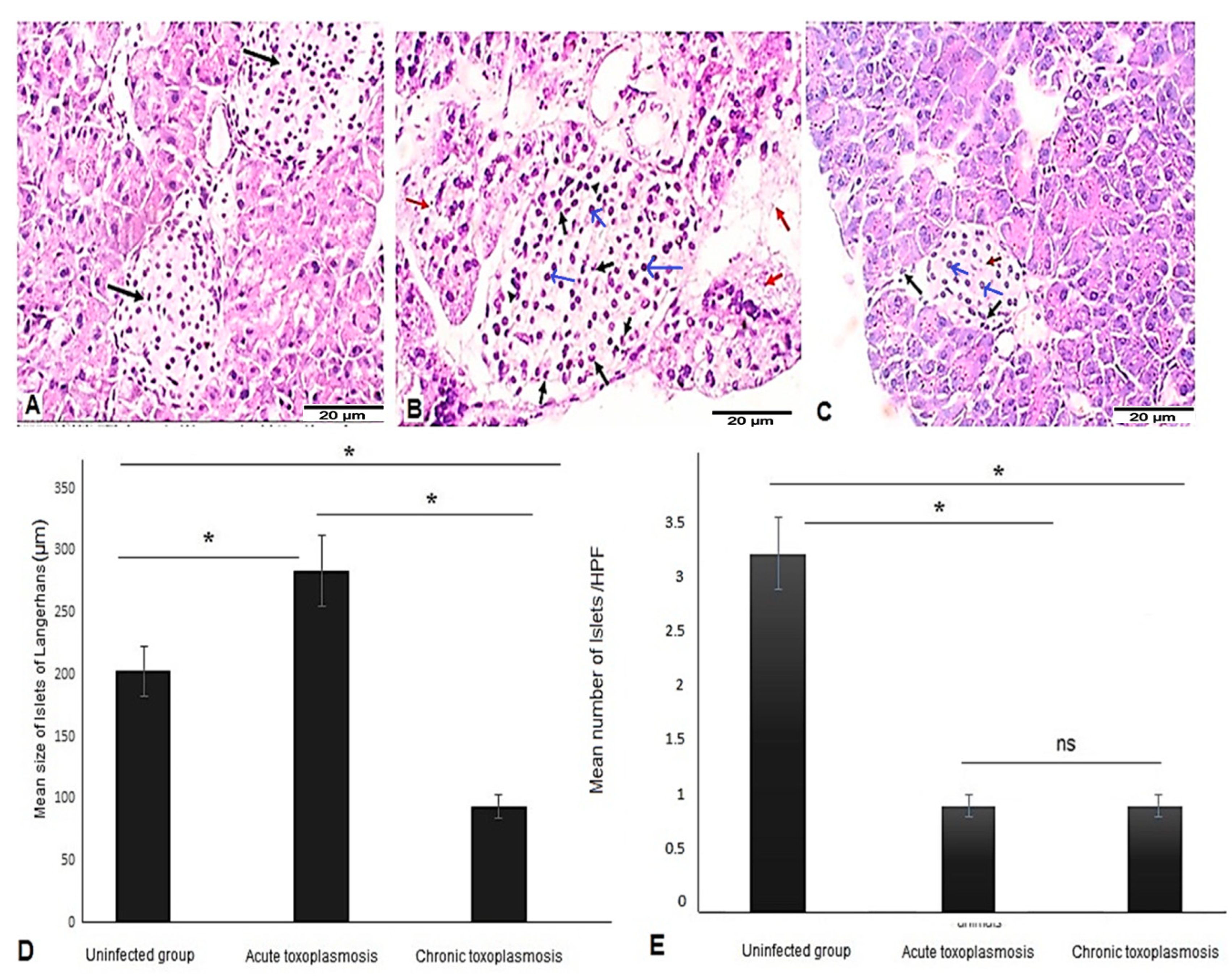
Figure 1. Acute and chronic toxoplasmosis are associated with pancreatic pathological changes and reduced the numbers of islets of Langerhans. Pancreatic tissue sections of mice from different groups (n = 10/group) were stained with H & E stain and imaged at 400×. (A) Representative image of pancreatic tissue sections of uninfected mice showing uniform rounded islets (black arrows) within pancreatic acini, with no inflammation, edema or necrosis. (B) Representative image of pancreatic tissue sections from the acute toxoplasmosis group showing enlarged islets of Langerhans with β cells (blue arrows), acute inflammatory infiltrate (black arrows), edema (arrow heads), and areas of necrosis (red arrows). (C) Representative image of pancreatic tissue sections from the chronic toxoplasmosis group showing significant reduction in size of the islets of Langerhans with β cells (blue arrows), mild infiltration by chronic inflammatory cells (black arrows) and areas showing fibrous-like material (red arrow). (D) Size of islets of Langerhans in uninfected, acute, and chronic toxoplasmosis mice groups. (E) Number of islets of Langerhans in uninfected, acute, and chronic toxoplasmosis mice groups/HPF. Data are expressed as mean ± SD (n = 10). Asterisks (*) indicate a statistically significant difference; p < 0.05 and “ns” indicates insignificant difference.
Biomedicines 11 00018 g001
Figure 2. CD8+ T cells Infiltrated pancreatic islets of Langerhans of mice with chronic toxoplasmosis. Immunohistochemistry representative images of pancreatic tissue sections stained for CD8 showing absence of CD8+ T cell infiltration into islets of Langerhans of uninfected (A) and acute toxoplasmosis mice (B) but marked infiltration of CD8+ T cells into islets of Langerhans of chronic toxoplasmosis mice group (C).
Biomedicines 11 00018 g002
Figure 3. Acute and chronic toxoplasmosis are associated with significantly lower number of insulin-producing-β cells of islets of Langerhans. Pancreatic tissue sections of mice from different groups (n = 10/group) were stained with anti-insulin antibody. (A) Representative image of pancreatic tissue sections of uninfected mice with strong insulin staining. (B) Representative image of pancreatic tissue sections of the acute toxoplasmosis group with smaller stained area of islets of Langerhans (fewer β cells) compared to uninfected group. (C) Representative image of pancreatic tissue sections of the chronic toxoplasmosis group with smaller stained area of islets of Langerhans (fewer β cells) compared to the acute toxoplasmosis group. (D) Number of β cells (insulin producing cells)/HPF in islets of Langerhans of different mice groups. Data are expressed as mean ± SD (n = 10). Asterisks (*) indicate statistically significant difference; p < 0.05.
Biomedicines 11 00018 g003
Figure 4. Acute and chronic toxoplasmosis induced apoptotic cell death in pancreatic islets of Langerhans. Pancreatic tissue sections of mice from different groups (n = 10/group) were stained with anti-caspase-3 antibody. (A) Representative image of pancreatic tissue sections of uninfected mice showing weak caspase 3 signal. (B) Representative image of pancreatic tissue sections of the acute toxoplasmosis group showing stronger caspase-3 staining and higher number of caspase-3-postive cells (apoptotic cells). (C) Representative image of pancreatic tissue sections of the chronic toxoplasmosis group with high number of caspase-3-postive cells but less than the acute toxoplasmosis group. (D) Number of caspase-3 positive cells /HPF in islets of Langerhans of different mice groups. Data are expressed as mean ± SD (n = 10). Asterisks (*) indicate statistically significant difference; p < 0.05.
Biomedicines 11 00018 g004
Disclaimer/Publisher’s Note: The statements, opinions and data contained in all publications are solely those of the individual author(s) and contributor(s) and not of MDPI and/or the editor(s). MDPI and/or the editor(s) disclaim responsibility for any injury to people or property resulting from any ideas, methods, instructions or products referred to in the content.