Article

TERT and TET2 Genetic Variants Affect Leukocyte Telomere Length and Clinical Outcome in Coronary Artery Disease Patients—A Possible Link to Clonal Hematopoiesis

by
Trine B. Opstad
1,2,*,
Svein Solheim
1,
Alf-Åge R. Pettersen
1,
Are A. Kalstad
1,
Harald Arnesen
1,2 and
Ingebjørg Seljeflot
1,2
1
Center for Clinical Heart Research, Department of Cardiology, Oslo University Hospital Ullevål, Kirkeveien 166, Pb 4950 Nydalen, N-0240 Oslo, Norway
2
Faculty of Medicine, University of Oslo, 0315 Oslo, Norway
*
Author to whom correspondence should be addressed.
Biomedicines 2022, 10(8), 2027; https://doi.org/10.3390/biomedicines10082027
Submission received: 5 July 2022 / Revised: 16 August 2022 / Accepted: 16 August 2022 / Published: 19 August 2022
(This article belongs to the Special Issue Atherosclerosis: Pathophysiology and Associations with Other Diseases)

Abstract

:
Inherited and acquired mutations in hematopoietic stem cells can cause clonal expansion with increased risk of cardiovascular disease (CVD), a condition known for the clonal hematopoiesis of indeterminate potential (CHIP). Inherited genetic variants in two CHIP-associated genome loci, the telomerase gene telomerase enzyme reverse transcriptase (TERT) (rs7705526) and the epigenetic regulator ten–eleven translocation 2 (TET2) (rs2454206), were investigated in 1001 patients with stable coronary artery disease (CAD) (mean age 62 years, 22% women), with regards to cardiovascular outcome, comorbidities, and leukocyte telomere length. Over 2 years, mutated TERT increased the risk two-fold for major clinical events (MACEs) in all patients (p = 0.004), acute myocardial infarction (AMI) in male patients (p = 0.011), and stroke in female patients (p < 0.001). Mutated TET2 correlated with type 2 diabetes (p < 0.001), the metabolic syndrome (p = 0.002), as well as fasting glucose, HbA1c, and shorter telomeres (p = 0.032, p = 0.003, and p = 0.016, respectively). In conclusion, our results from stable CAD patients highlight TERTs’ role in CVD, and underline TET2s’ role in the epigenetic regulation of lifestyle-related diseases.

1. Introduction

Clonal hematopoiesis (CH) commonly arises due to the age-related acquisition of somatic mutations in hematopoietic stem cells (HSCs) of the bone marrow [1]. The condition increases the risk of hematological malignancies [1], but also cardiovascular disease (CVD) [2]. The phenomenon is known as the clonal hematopoiesis of indeterminate potential (CHIP) [3]. Inherited genetic causes may also lead to CH, by abrogating stem cell function. Single-variant genetic association analyses of CHIP recently identified multiple genome-wide-significant loci with CH association, including the telomerase enzyme reverse transcriptase (TERT) locus at chromosome 5 and the near ten-eleven translocation 2 (TET2) locus at chromosome 4. Germline, i.e., inherited genetic variants in both TERT and TET2 genes have been associated with an increasing risk of CHIP development and malignant hematopoiesis, respectively [4,5].
The telomerase enzyme, known to be active in germ cells, pluripotent embryonic stem cells and hematopoietic progenitor cells, seems also to be active in differentiated, non-dividing, somatic cells in the cardiovascular system, sustaining the role of telomere biology in CVD [6]. Telomeres protect the ends of chromosomes, consisting of repeated DNA sequences (TTAGGG) and associated proteins [7]. Telomerase synthesizes telomeric DNA to compensate for and counteract the loss of sequences upon cell division; however, its overexpression may lead to cancer [8]. As telomere shortening in general is related to organismal and cardiovascular ageing, a delicate balance in telomere stability may thus be required to achieve healthy ageing. We recently reported shorter leukocyte telomere length (LTL) in young and elderly healthy subjects with hereditary coronary heart disease [9]. Short telomeres in blood cells may lead to somatic mutations, and potentially CH, and vice versa, manifested CHIP may shorten LTL [10]. Furthermore, genetically predicted longer telomere lengths were associated with altered clonal somatic copy number in peripheral leukocytes [5], potentially also predisposing CH; thus, a bidirectional causality between telomere lengths and CHIP has been suggested [10]. Several TERT germline mutations predisposed to CH have been identified, including the single-point rs7705526 mutation, previously associated with increased leukocyte telomere length (LTL), myeloproliferative neoplasms, and increased risk of developing CHIP [4,11,12].
TET2 is expressed in hematopoietic cells and is important for normal hematopoiesis. The enzyme regulates gene expression via the DNA demethylation of cytosine on DNA targets, and oxidizes 5-methyl cytosine to 5-hydroxymethylcytosine, thus epigenetically modulating the genome [13]. The TET family of proteins are thought to be involved in the pathogenesis of complex metabolic-related diseases [14] and TET2 is involved in telomere maintenance and chromosomal stability. Most TET2 somatic and germline mutations are loss-of-function mutations, located in the enzymes’ catalytic domain in the C-terminal part, consequently leading to hyper-methylated DNA [13]. Somatic mutations inducing TET2 deficiencies may, in addition to malignancies, also cause atherosclerosis and adverse CVD outcomes, mediated by macrophage pro-inflammatory activation and interleukin upregulation [15,16]. Several germline TET2 mutations have been investigated according to myelodysplastic malignancies, including the Ile1762Val variant in exon 11 [13,17]. So far, no germline TET2 mutation has been reported with regards to CVD.
We aimed to investigate two genetic variants, reported as germline mutations [4,5], that could potentially induce somatic mutations in HSCs ensuing CHIP development: the TERT intron 5 rs7705526 variant and the TET2 p.Ile1762Val rs2454206 missense mutation in patients with stable coronary artery disease (CAD). Our hypothesis was that these variants could affect LTLs, cardiometabolic status, and clinical outcome, with potential causality related to CHIP or its development.

2. Materials and Methods

2.1. Study Population

This study is a post-hoc analysis of 1001 angiographically verified CAD patients enrolled in the Aspirin Non-Responsiveness and Clopidogrel Endpoint Trial (ASCET), with a mean age of 62 years (22% were females and 97% were of western European descent) [18]. Patients were followed for a minimum of 2 years, and the primary clinical endpoint major adverse events (MACEs) included the first event of either a nonfatal acute myocardial infarction (AMI), unstable angina pectoris (UAP), stroke, and all-cause mortality. An endpoint committee evaluated the endpoints without access to laboratory data. No patients were lost to follow-up, and if they were unable to attend the final visit, clinical endpoints were recorded on request.
At inclusion, clinical subgroups were defined as follows: previous MI, as recorded by patients medical files; hypertension (HT), i.e., individuals previously diagnosed or treated HT; diabetes, i.e., individuals with treated type 2 diabetes and/or fasting glucose > 7.0 mmol/L; and metabolic syndrome (Mets), according to modified NCEP ATP III criteria [19]. These was a threshold value of at least three of the following: (1) waist circumferences ≥ 102 cm in men and ≥88 cm in women; (2) triglycerides ≥ 1.69 mmol/L; (3) HDL-cholesterol < 1.04 mmol/L in males and <1.29 mmol/L in females; (4) blood pressure ≥ 130/85 mm Hg or diagnosed or treated HT; and (5) fasting glucose ≥ 6.1 mmol/L.
The Regional Committee of Medical Research Ethics South-Eastern Norway approved the ASCET study. All research in the present study was performed in accordance with relevant guidelines and regulations. The participants conformed to the Declaration of Helsinki and written informed consent was obtained from all patients. The ASCET study was registered at clinical trial.gov, http://www.clinicaltrial.com (accessed on 22 September 2005), with the identification number NCT00222261.

2.2. Laboratory Methods

Blood samples were collected at baseline between 8.00 and 10.00 a.m. in fasting conditions. Serum was prepared by centrifugation within 1 h at 2500× g in 10 min for routine analyses. EDTA whole-blood samples were kept frozen at −80 °C until DNA extraction, performed with the MagNA Pure LC DNA Isolation Kit on the MagNA Pure LC Instrument (Roche Diagnostics, GmbH, Mannheim, Germany). Extracted DNA was tested for purity and quantity using a NanoDrop 1000 spectrophotometer (Thermo Scientific, Wilmington, DE, USA) and stored at −80 °C. LTL analysis was performed in a subgroup of the first included patients (n = 262).

2.3. Genotype Analysis

The allelic discrimination of TERT C/A (rs7705526) and TET2 p.Ile1762Val A/G (rs2454206) variants was performed with real-time PCR on the ViiA7 instrument (Applied Biosystems, Thermo Fisher Scientific), using the TaqMan single-nucleotide polymorphism (SNP) assays ID C_189441058_10 and ID C_11566753_20, respectively (Life Technologies dba Invitrogen, Pleasanton, CA, USA). The TaqPath ProAmp Master Mix was used for the TERT assay and TaqMan Universal PCR Master Mix for the TET2 assay. Non-template samples were included in each run to exclude contamination of samples, and 5% of the samples were re-ran as quality control, with 99% concordance.

2.4. Leukocyte Telomere Length (LTL) Determination

An equal amount of extracted DNA per experiment (2 ng/μL) was used to measure LTLs using a single-plex quantitative RT- PCR [20]. PCR amplification was performed on the ViiA7 instrument (Applied Biosystems by Life Technologies, Foster City, CA, USA), using telomere-specific primers (Invitrogen by Thermo Fisher Scientific, Waltham, MA, USA) (Supplementary Table S1) and GoTaq PCR Master Mix (Promega, Madison, WI, USA). LTLs were relatively quantified (RQ) to the single-copy gene (SCG) SB34 (Invitrogen by Thermo Fisher Scientific) with specific SCG primers (Supplementary Table S1) and an internal reference sample. The primers for both targets were diluted to a final concentration of 4 pmol/µL. PCR conditions for both targets were as follows: an initial step at 95 °C for 10 min followed by 40 cycles of 95 °C for 15 s and 60 °C for 1 min. A template negative control was included in each run. Each sample was run in triplicates and individual amplification curves for all samples of both assays were carefully validated. Technical triplicates with a SD exceeding 0.5 Ct were excluded from the analysis, with two remaining valuable parallels.

2.5. Statistical Analysis

Data were presented as mean (+/−SD), median (25th, 75th percentile), or proportions (%), as appropriate. The unpaired two-sampled Student t-test and one-way ANOVA were used for the analysis of normal distributed continuous data, and the Mann–Whitney U test and Kruskal–Wallis test were used for skewed continuous data, when appropriate, for comparisons between two or more groups, respectively. Chi-squared tests were used to compare the proportions. A binary logistic regression model was used in association between the TERT variant and clinical endpoints, adjusting for age and sex (included in the model by convention), and previous MI and stroke (more frequent present at inclusion in subjects suffering from new clinical events). The TERT and TET2 variants were tested with the Hardy–Weinberg equilibrium (x2 > 0.05, both). The level of statistical significance was set to p < 0.05. SPSS version 26 (SPSS Inc., Chicago, IL, USA) was used for all statistical analyses.

3. Results

In the investigated population, 106 MACEs were recorded after 2 years (AMI (n = 36), stroke (n = 28), UAP (n = 33) and deaths (n = 9)). Baseline characteristics according to the presence of endpoints or not are presented in Table 1, with previous MI and stroke being more frequent in patients suffering from a new clinical event. Independently of MACEs, the mean age in the population was 62 years, and 22% were women. The presence of comorbidities was as follows: 20% had type 2 diabetes, 24% had Mets, 56% had hypertension, and 20% were current smokers. 98% of patients were on statin treatment, and many used anti-hypertensive medications.

3.1. Frequencies of the TERT and TET2 Mutations

DNA was available in 995 samples and the investigated variants were successfully analyzed in all, except for one TERT measurement. Genotypes and variant allele frequencies (VAFs) in the CAD subjects, stratified by sex, are presented in Table 2. VAFs for the TERT C/A and TET2 p.Ile1762Val A/G variants were 0.324 and 0.346, respectively. A higher frequency of the TERT variant was observed in men vs. women (p = 0.033).

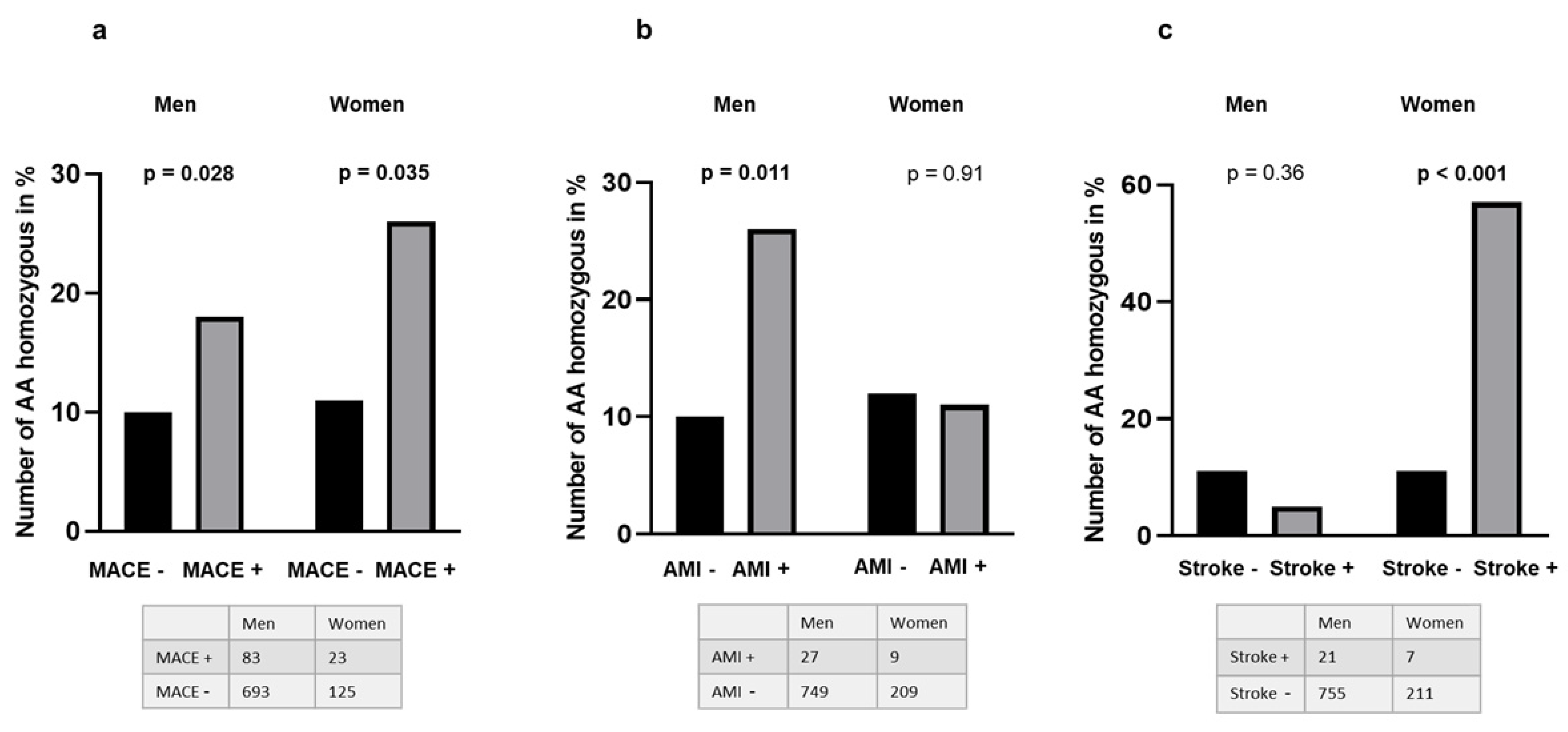
3.2. Presence of the TERT and TET2 Mutations as Related to Clinical Outcome and Comorbidity

The variants’ influence on clinical outcome is presented in Table 3. An increasing number of TERT variant alleles (A) correlated with an increased risk of MACEs (p = 0.010). In terms of categorization, the AA genotype vs. the CC and CA genotypes increased the risk further (p = 0.003), with an OR of 2.2 (95 % confidence interval [CI] of 1.3, 3.7), which was still significant when adjusting for age, sex, previous MI, and stroke (OR = 2.2, 95% CI [1.3, 3.7], p = 0.004). As the TERT mutation was more frequent in men, we analyzed the data separately. The results are illustrated in Figure 1, showing an increased risk of MACE in both genders: in men with an OR = 2.0 (95% CI (1.1, 3.6), p = 0.028) and in women with an OR = 2.9 (95% CI (1.0, 8.), p = 0.035) (Figure 1a). With the mutation, an increased risk of new-onset AMI was found in men only (OR = 3.0, 95% CI [1.2, 7.3], p = 0.011, n = 27) (Figure 1b), whereas an increased risk of new onset stroke was observed in women only (OR = 10.9, 95% CI [2.3, 51.8], p < 0.001, n = 7) (Figure 1c). The TERT mutation was not associated with previous MI and stroke, or other comorbidities (Table 3).
The TET2 mutation was not associated with MACE (Table 3), but was significantly and more frequently present in patients with type 2 diabetes (p < 0.001) and with Mets (p = 0.002). In line with this, significant associations were observed with increasing fasting glucose levels across AA-AG-GG genotypes: mean (SD) 6.0 (0.1)-6.0 (1.0)-6.5 (0.2) mmol/L (p = 0.032) and increasing HbA1c levels 5.9 (0.04)-6.0 (0.04)-6.2 (0.11)% (p = 0.003).

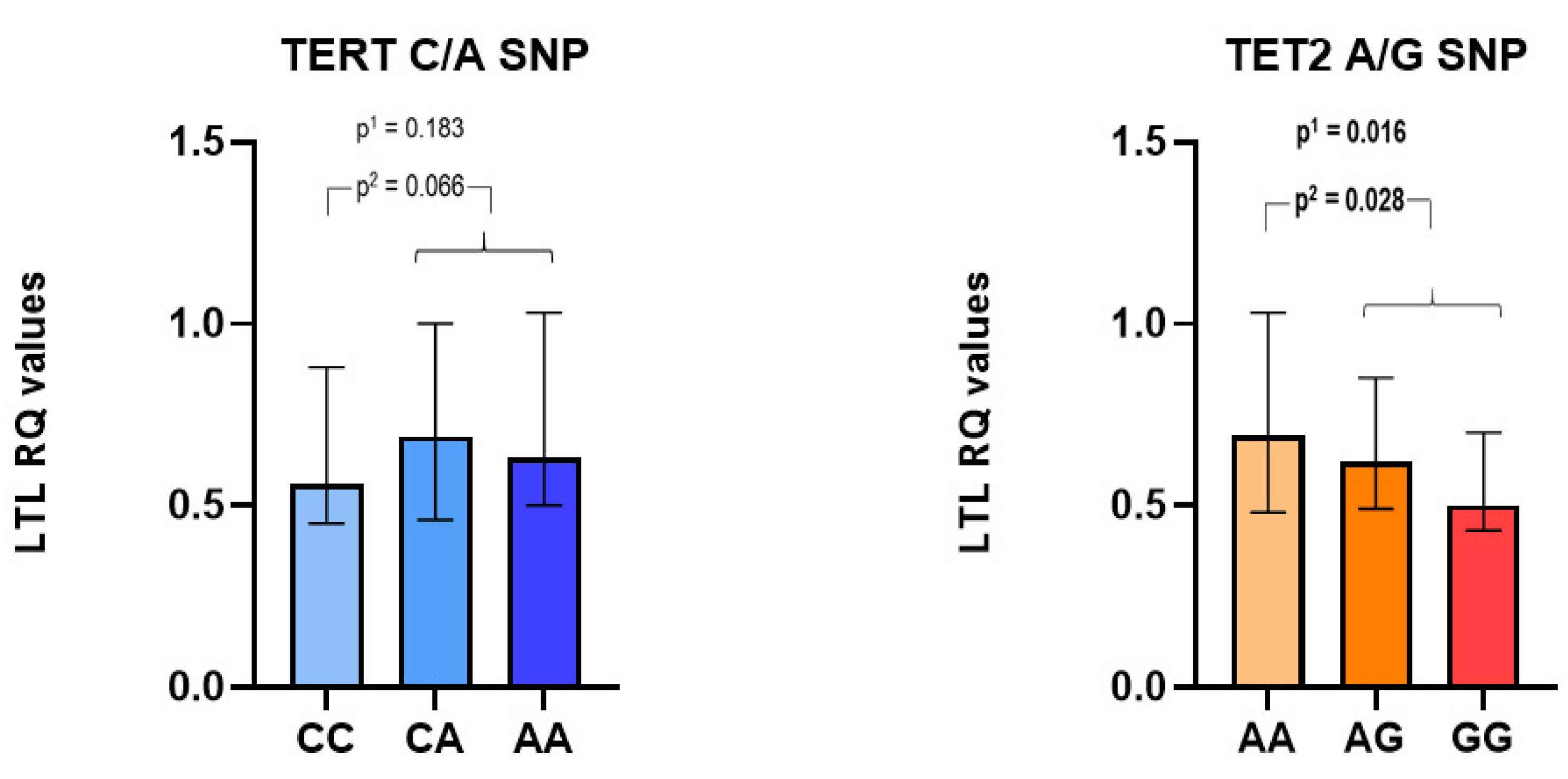
3.3. TERT and TET2 Mutations as Related to Leukocyte Telomere Lengths (LTLs)

In Figure 2, we illustrated the relationship between TERT and TET2 mutations, respectively, and LTLs (RQ levels) measured in the subset of CAD patients (n = 262). The TERT A allele correlated with borderline significant longer LTLs (p = 0.066), as compared to the CC wild type, with an overall non-significant difference in LTLs between genotypes (p = 0.183). LTLs were significantly shorter with an increasing number of TET2 variant G-alleles (p = 0.016).

4. Discussion

The main finding in our study is that the selected germline intronic TERT C/A variant (rs7705526) correlated with composite clinical endpoints during two-year follow-up in patients with stable CAD, with an increased risk of AMI exclusively in men and an increased risk of stroke exclusively in women. The investigated TET2 genetic variant (rs2454206), with an amino acid substitution of isoleucine to valine at position 1762, correlated with shorter telomeres and the presence of type 2 diabetes and Mets, and accordingly with fasting glucose and HbA1c levels.
By simultaneously analyzing germline and somatic mutations with blood-derived whole-genome sequencing (WGS), recent comprehensive studies have demonstrated that germline mutations influence the acquisition of somatic mutations in blood cells [11,12,21]. Inherited causes of CHIP include multiple genetic variants at the TERT and TET2 locus [4]. The investigated TERT genetic variant (rs7705526) in intron 5 is in strong linkage disequilibrium (r2 = 0.55) with the TERT genetic variant rs34002450 in intron 3, both identified as lead variants in the TERT locus, and as germline genetic determinants of CHIP [4,21], the latter with a 1.3-fold increased risk of CHIP development (TOPMed project) [4]. The rs34002450 presented with a 1.37-fold increased risk of developing CH in the Iceland deCODE genetics project [21]. Thus, the TERT-CHIP association may indicate the role of telomerase activity in CH. We observed borderline significantly longer LTLs in TERT rs7705526 A-allele carriers, as previously reported by others [5,11]. We also observed that LTLs, independently of the investigate gene variants, were not linked to MACE, and were measured in a subpopulation, indicating underpowered analysis and/or the fact that the already-manifested CAD status in our population may have affected the results. Telomerase overexpression has been reported to transform cultured cells into cancerous cells [22], and failure of the TERT gene may deteriorate genome integrity, enabling the acquisition of somatic mutations in hematopoietic cells and the further development of CHIP. As TERT is also active in the cells of the cardiovascular system, any interference with TERT activity may contribute to CVDs [6]. The TERT rs7705526 variant has been shown to lead to the CHIP-related somatic mutation JAK2 p.V617F [23], supporting the possibility of the causational TERT A-allele-CHIP development in our CAD population.
Although numbers are low, the observed sex dimorphism in the frequency of the TERT rs77055526 variant and in the type of MACE may indicate different underlying pathophysiological mechanisms in men and women with regards to the involvement of TERT in CVD development, in need of further investigation. Indeed, the use of hormone replacement therapy or merely a postmenopausal state might have influenced the association found between the onset of strokes in women and the TERT mutation. Unfortunately, we do not have data to explore any influence.
TET2 is one of the most common somatically mutated genes in CH and CHIP [1,21,24] and multiple germline TET2 mutations have been investigated according to myeloid malignancies [13,17,25]. The TET2 p.Ile1762Val genetic variant is located in the catalytic domain of the TET2 gene; thus, an eventual loss of TET2 function may lead to DNA hypermethylation and subsequent altered gene expression in blood cells. We observed that the variant was present at a significantly higher frequencies in both type 2 diabetes and Mets subjects, with altered glucose and HbA1c levels. Inherited TET2 mutations ensuing life-long epigenetic changes may have altered pathways in glucose regulation. TET2 has been suggested to facilitate the transcriptional activity of peroxisome proliferator-activated receptor gamma (PPARƴ), involved in insulin sensitivity; thus, a TET2 loss-of-function may have promoted insulin resistance [26]. The TET2 p.Ile1762Val variant has also previously been reported to correlate with liver PPARƴ coactivator 1 alpha (PGC1A)-methylation levels and non-alcoholic fatty liver disease [27]. TET2 has also been proposed to regulate PPARƴ transcription in adipocytes [26], which can partly explain the observed Mets and type 2 diabetes association in the present study. The TET2 rs2454206 genetic variant has also been reported to correlate with diabetes [14], and mice experiments suggest that TET2 loss-of-function-driven clonal hematopoiesis can contribute to insulin resistance and type 2 diabetes [28].
Both the TERT mutation (rs7705526) and the TET2 (rs2454206) are reported to significantly correlate with levels of blood pressure in the Common Metabolic Diseases Knowledge Portal (hugeamp.org). The lack of these associations in our study might be due to medication status and/or the population itself consisting exclusively of patients with stable CAD.
The TET2 variant correlated with shorter telomeres, to our knowledge not previously reported in humans. As TET enzymes are important for telomere stability, the investigated TET2 variant may potentially have induced TET2 deficiency and subsequently telomere loss [29]. Experiments with TET2-depleted mice resulted in shorter telomeres, explained by an upregulation of DNA methyltransferase, which decreases 5-hydroxymetylcytosine levels and increases the methylation status at sub-telomeres, the region between telomeric caps and chromatin [29]. Aberrant methylation on sub-telomeric DNA may have certain effects on telomere lengths, correlating with age-related diseases [30], underpinning the connection between diabetes and Mets. The detected shorter telomeres with the TET2 variant in our population may have hypothetically induced somatic mutations, CH and CHIP-related CAD, which indirectly may have accelerated telomere attrition, although any causality cannot be drawn.
Our study has several limitations. We investigated candidate SNPs with only two variants; thus, any influence of other known TERT and TET2 genetic variants is not explored. The population consisted exclusively of medically treated patients with stable CAD. With the low numbers of LTL analyses, these results should be regarded as explorative and hypothesis-generating, and must be taken with caution. As others have reported an association between the TERT rs7705526 mutation and longer telomeres, our lack of a statistical significant association ensues the possibility of a type 1 statistical error. Its effect on cardiovascular outcome may however underpin the idea that longer telomeres may induce CHIP development, and herein the observed increased risk of MACE, despite other mechanisms of the observed associations, cannot be excluded. The low number in studied subgroups may have also influenced results with regards to clinical endpoints.

5. Conclusions

The TERT rs7705526 mutation correlated with an increased risk of clinical adverse events in our CAD population, potentially related to CHIP development. The TET2 p.Ile1762Val missense mutation rs2454206 correlated with type 2 diabetes and Mets, along with dysregulated glucose metabolism, illustrating epigenetic regulation as a bridge between inherited and environmental causes in the development of disease. The associated shorter telomeres may reflect their manifested CAD.

Supplementary Materials

The following supporting information can be downloaded at: https://www.mdpi.com/article/10.3390/biomedicines10082027/s1. Supplementary Table S1: Nucleotide sequence for the telomere and single-copy gene analyses.

Author Contributions

Conceptualization, T.B.O., S.S., H.A. and I.S.; methodology, T.B.O. and I.S., software, T.B.O. and A.-Å.R.P.; validation, T.B.O. and I.S.; formal analysis, A.-Å.R.P. and T.B.O.; investigation, T.B.O., S.S., A.-Å.R.P., H.A. and I.S.; resources, H.A. and I.S.; data curation, A.-Å.R.P. and T.B.O.; writing—original draft preparation, T.B.O.; writing—review and editing, S.S., A.-Å.R.P., A.A.K., H.A. and I.S.: visualization, T.B.O.; supervision, I.S.; project administration, T.B.O., S.S., A.-Å.R.P., H.A. and I.S.: funding acquisition, T.B.O., A.-Å.R.P., H.A. and I.S. All authors have read and agreed to the published version of the manuscript.

Funding

This research was funded by the Norwegian Council for Cardiovascular Diseases and Stein Erik Hagen Foundation for Clinical Heart Research, Oslo Norway.

Institutional Review Board Statement

The Regional Committee of Medical Research Ethics South-Eastern Norway approved the ASCET study.

Informed Consent Statement

Informed consent was obtained from all subjects involved in the study.

Data Availability Statement

The data presented in this study are available on request from the corresponding author. The data are not publicly available due to the privacy of the patients.

Acknowledgments

Vibeke Bratseth and Sissel Åkra are acknowledged for their expert technical assistance.

Conflicts of Interest

The authors declare no conflict of interest.

References

  1. Jaiswal, S.; Fontanillas, P.; Flannick, J.; Manning, A.; Grauman, P.V.; Mar, B.G.; Lindsley, R.C.; Mermel, C.H.; Burtt, N.; Chavez, A.; et al. Age-related clonal hematopoiesis associated with adverse outcomes. N. Engl. J. Med. 2014, 371, 2488–2498. [Google Scholar] [CrossRef]
  2. Jaiswal, S.; Natarajan, P.; Silver, A.J.; Gibson, C.J.; Bick, A.G.; Shvartz, E.; McConkey, M.; Gupta, N.; Gabriel, S.; Ardissino, D.; et al. Clonal Hematopoiesis and Risk of Atherosclerotic Cardiovascular Disease. N. Engl. J. Med. 2017, 377, 111–121. [Google Scholar] [CrossRef]
  3. Steensma, D.P.; Bejar, R.; Jaiswal, S.; Lindsley, R.C.; Sekeres, M.A.; Hasserjian, R.P.; Ebert, B.L. Clonal hematopoiesis of indeterminate potential and its distinction from myelodysplastic syndromes. Blood 2015, 126, 9–16. [Google Scholar] [CrossRef]
  4. Bick, A.G.; Weinstock, J.S.; Nandakumar, S.K.; Fulco, C.P.; Bao, E.L.; Zekavat, S.M.; Szeto, M.D.; Liao, X.; Leventhal, M.J.; Nasser, J.; et al. Inherited causes of clonal haematopoiesis in 97,691 whole genomes. Nature 2020, 586, 763–768. [Google Scholar] [CrossRef]
  5. Brown, D.W.; Lin, S.H.; Loh, P.R.; Chanock, S.J.; Savage, S.A.; Machiela, M.J. Genetically predicted telomere length is associated with clonal somatic copy number alterations in peripheral leukocytes. PLoS Genet. 2020, 16, e1009078. [Google Scholar] [CrossRef]
  6. Zurek, M.; Altschmied, J.; Kohlgrüber, S.; Ale-Agha, N.; Haendeler, J. Role of Telomerase in the Cardiovascular System. Genes 2016, 7, 29. [Google Scholar] [CrossRef]
  7. Blackburn, E.H. Structure and function of telomeres. Nature 1991, 350, 569–573. [Google Scholar] [CrossRef]
  8. Hannen, R.; Bartsch, J.W. Essential roles of telomerase reverse transcriptase hTERT in cancer stemness and metastasis. FEBS Lett. 2018, 592, 2023–2031. [Google Scholar] [CrossRef]
  9. Opstad, T.B.; Kalstad, A.A.; Holte, K.B.; Berg, T.J.; Solheim, S.; Arnesen, H.; Seljeflot, I. Shorter Leukocyte Telomere Lengths in Healthy Relatives of Patients with Coronary Heart Disease. Rejuv. Res. 2020, 23, 324–332. [Google Scholar] [CrossRef]
  10. Nakao, T.; Bick, A.G.; Taub, M.A.; Zekavat, S.M.; Uddin, M.M.; Niroula, A.; Carty, C.L.; Lane, J.; Honigberg, M.C.; Weinstock, J.S.; et al. Bidirectional Mendelian randomization supports bidirectional causality between telomere length and clonal hematopoiesis of intermediate potential. medRxiv 2021. [Google Scholar] [CrossRef]
  11. Bojesen, S.E.; Pooley, K.A.; Johnatty, S.E.; Beesley, J.; Michailidou, K.; Tyrer, J.P.; Edwards, S.L.; Pickett, H.A.; Shen, H.C.; Smart, C.E.; et al. Multiple independent variants at the TERT locus are associated with telomere length and risks of breast and ovarian cancer. Nat. Genet. 2013, 45, 371–384. [Google Scholar] [CrossRef]
  12. Bao, E.L.; Nandakumar, S.K.; Liao, X.; Bick, A.G.; Karjalainen, J.; Tabaka, M.; Gan, O.I.; Havulinna, A.S.; Kiiskinen, T.T.J.; Lareau, C.A.; et al. Inherited myeloproliferative neoplasm risk affects haematopoietic stem cells. Nature 2020, 586, 769–775. [Google Scholar] [CrossRef]
  13. Bussaglia, E.; Anton, R.; Nomdedeu, J.F.; Fuentes-Prior, P. TET2 missense variants in human neoplasia. A proposal of structural and functional classification. Mol. Genet. Genom. Med. 2019, 7, e00772. [Google Scholar] [CrossRef]
  14. Pirola, C.J.; Scian, R.; Gianotti, T.F.; Dopazo, H.; Rohr, C.; Martino, J.S.; Castaño, G.O.; Sookoian, S. Epigenetic Modifications in the Biology of Nonalcoholic Fatty Liver Disease: The Role of DNA Hydroxymethylation and TET Proteins. Medicine 2015, 94, e1480. [Google Scholar] [CrossRef]
  15. Sano, S.; Oshima, K.; Wang, Y.; MacLauchlan, S.; Katanasaka, Y.; Sano, M.; Zuriaga, M.A.; Yoshiyama, M.; Goukassian, D.; Cooper, M.A.; et al. Tet2-Mediated Clonal Hematopoiesis Accelerates Heart Failure Through a Mechanism Involving the IL-1ꞵNLRP3 Inflammasome. J. Am. Coll. Cardiol. 2018, 71, 875–886. [Google Scholar] [CrossRef]
  16. Fuster, J.J.; MacLauchlan, S.; Zuriaga, M.A.; Polackal, M.N.; Ostriker, A.C.; Chakraborty, R.; Wu, C.L.; Sano, S.; Muralidharan, S.; Rius, C.; et al. Clonal hematopoiesis associated with TET2 deficiency accelerates atherosclerosis development in mice. Science 2017, 355, 842–847. [Google Scholar] [CrossRef]
  17. Hu, J.; Xu, J.; Tian, T.; Xie, J.; Fan, L.; Zhu, G.; Xia, T.; Chen, X.; Tan, Y.; Chen, X.; et al. TET2 rs2454206, TET2 rs12498609 and ASXL1 rs3746609 single nucleotide polymorphisms in patients with myelodysplastic syndromes. Blood Cells Mol. Dis. 2019, 74, 44–50. [Google Scholar] [CrossRef]
  18. Pettersen, A.A.; Seljeflot, I.; Abdelnoor, M.; Arnesen, H. High On-Aspirin Platelet Reactivity and Clinical Outcome in Patients with Stable Coronary Artery Disease: Results From ASCET (Aspirin Nonresponsiveness and Clopidogrel Endpoint Trial). J. Am. Heart Assoc. 2012, 1, e000703. [Google Scholar] [CrossRef]
  19. Grundy, S.M.; Cleeman, J.I.; Daniels, S.R.; Donato, K.A.; Eckel, R.H.; Franklin, B.A.; Gordon, D.J.; Krauss, R.M.; Savage, P.J.; Smith, S.C., Jr.; et al. Diagnosis and management of the metabolic syndrome: An American Heart Association/National Heart, Lung, and Blood Institute Scientific Statement. Circulation 2005, 112, 2735–2752. [Google Scholar] [CrossRef]
  20. Cawthon, R.M. Telomere measurement by quantitative PCR. Nucleic Acids Res. 2002, 30, e47. [Google Scholar] [CrossRef]
  21. Zink, F.; Stacey, S.N.; Norddahl, G.L.; Frigge, M.L.; Magnusson, O.T.; Jonsdottir, I.; Thorgeirsson, T.E.; Sigurdsson, A.; Gudjonsson, S.A.; Gudmundsson, J.; et al. Clonal hematopoiesis, with and without candidate driver mutations, is common in the elderly. Blood 2017, 130, 742–752. [Google Scholar] [CrossRef]
  22. Mitchell, J.R.; Collins, K. Human telomerase activation requires two independent interactions between telomerase RNA and telomerase reverse transcriptase. Mol. Cell 2000, 6, 361–371. [Google Scholar] [CrossRef]
  23. Hinds, D.A.; Barnholt, K.E.; Mesa, R.A.; Kiefer, A.K.; Do, C.B.; Eriksson, N.; Mountain, J.L.; Francke, U.; Tung, J.Y.; Nguyen, H.M.; et al. Germ line variants predispose to both JAK2 V617F clonal hematopoiesis and myeloproliferative neoplasms. Blood 2016, 128, 1121–1128. [Google Scholar] [CrossRef]
  24. Buscarlet, M.; Provost, S.; Zada, Y.F.; Barhdadi, A.; Bourgoin, V.; Lépine, G.; Mollica, L.; Szuber, N.; Dubé, M.-P.; Busque, L. DNMT3A and TET2 dominate clonal hematopoiesis and demonstrate benign phenotypes and different genetic predispositions. Blood 2017, 130, 753–762. [Google Scholar] [CrossRef]
  25. Hamed, N.A.; Elhalawani, N.A.; Kassem, H.S.; Ayad, M.W.; Dammag, E.A. The Prognostic Significance of TET2 Single Nucleotide Polymorphism in Egyptian Chronic Myeloid Leukemia. Mediterr. J. Hematol. Infect. Dis. 2020, 12, e2020004. [Google Scholar]
  26. Villivalam, S.D.; Kim, J.; Kang, S. DNMT3a and TET2 in adipocyte insulin sensitivity. Oncotarget 2018, 9, 35289–35290. [Google Scholar] [CrossRef]
  27. Sookoian, S.; Rosselli, M.S.; Gemma, C.; Burgueño, A.L.; Fernández Gianotti, T.; Castaño, G.O.; Pirola, C.J. Epigenetic regulation of insulin resistance in nonalcoholic fatty liver disease: Impact of liver methylation of the peroxisome proliferator-activated receptor γ coactivator 1α promoter. Hepatology 2010, 52, 1992–2000. [Google Scholar] [CrossRef]
  28. Fuster, J.J.; Zuriaga, M.A.; Zorita, V.; MacLauchlan, S.; Polackal, M.N.; Viana-Huete, V.; Ferrer-Pérez, A.; Matesanz, N.; Herrero-Cervera, A.; Sano, S.; et al. TET2-Loss-of-Function-Driven Clonal Hematopoiesis Exacerbates Experimental Insulin Resistance in Aging and Obesity. Cell Rep. 2020, 33, 108326. [Google Scholar] [CrossRef]
  29. Yang, J.; Guo, R.; Wang, H.; Ye, X.; Zhou, Z.; Dan, J.; Wang, H.; Gong, P.; Deng, W.; Yin, Y.; et al. Tet Enzymes Regulate Telomere Maintenance and Chromosomal Stability of Mouse ESCs. Cell Rep. 2016, 15, 1809–1821. [Google Scholar] [CrossRef]
  30. Hu, H.; Li, B.; Duan, S. The Alteration of Subtelomeric DNA Methylation in Aging-Related Diseases. Front. Genet. 2018, 9, 697. [Google Scholar] [CrossRef]
Figure 1. Association of the TERT rs7705526 mutation with MACE in certain subcategories. Tables underneath each figure denote actual numbers in different groups. (a) Frequency of TERT rs7705526 (C/A) homozygous subjects (AA) according to major clinical events (MACEs), separated by gender. Black and gray columns represent the % of AA homozygous without and with MACE, respectively. (b) Frequency of TERT rs7705526 (C/A) homozygous subjects (AA) according to acute myocardial infarction (AMI), separated by gender. Black columns represent the % of AA homozygous without AMI in both gender. The gray columns represent the % of AA homozygous suffering from AMI in men and women, respectively. (c) Frequency of TERT rs7705526 (C/A) homozygous subjects (AA) according to stroke, separated by gender. Black columns represent the % of AA homozygous patients without stroke. The gray columns represent the % of AA homozygous suffering from strokes in women and men, respectively.
Biomedicines 10 02027 g001
Figure 2. Relatively quantified (RQ) leukocyte telomere length (LTL), related to TERT rs7705526 (C/A) and TET2 rs2454206 (A/G) genotypes. p1 values refer to the difference in LTLs between genotypes, whereas p2 values refer to differences in LTLs between the presence of the variant allele compared to the wild type. In this subset, LTLs were not associated with MACE (n = 34), independent of TERT and TET2 genotypes (median RQ level (25, 75 percentiles): 0.67 (0.51, 0.91) as compared to without MACE (n = 228): 0.61 (0.46, 0.96)).
Biomedicines 10 02027 g002
Table 1. Baseline characteristics according to presence of clinical composite endpoints after 2 year in the CAD population.
With Endpoints (n = 106)Without Endpoint (n = 895)p
Age (years, mean (range))63 (41–80)62 (36–81)0.499
Men/women n (%)83/23 (78/22)700/195 (78/22)0.983
Type 2 diabetes Mellitus n (%)24 (23)176 (20)0.469
Previous myocardial infarction n (%)57 (54)380 (43)0.026
Metabolic syndrome (%)25 (24)219 (25)0.836
Previous stroke n (%)6 (6)21 (2.3)0.047
Hypertension n (%)63 (59)493 (55)0.394
SBP mm/Hg140 (125, 150)140 (125, 150)0.831
DBP mm/Hg80 (75, 90)80 (75, 90)0.616
Current smokers n (%)23 (22)180 (20)0.666
BMI (kg/m2) a27.4 (4.0)27.7 (9.4)0.742
Total cholesterol (mmol/L)4.5 (1.0)4.6 (1.0)0.877
HDL cholesterol (mmol/L)1.3 (0.4)1.3 (0.4)0.898
LDL cholesterol (mmol/L)2.5 (0.8)2.5 (0.8)0.758
Triglycerides (mmol/L) a1.5 (0.9)1.6 (1.1)0.887
Fasting glucose (mmol/L)6.1 (1.7)6.0 (1.9)0.914
HbA1c (%)6.05 (0.87)5.97 (0.91)0.42
Medication (%)
Statins98990.524
β-lockers74760.867
Nitrates27210.145
ACE inhibitors31260.32
ARB26240.711
CCB27250.656
Diuretics26220.417
Values are mean (SD) or numbers (%) if not otherwise stated, a median levels (25, 75. percentile). SD: standard deviation, SBP: systolic blood pressure, DBP: diastolic blood pressure, BMI: body mass index, HDL: high-density lipoprotein, LDL: low-density lipoprotein, ACE: angiotensin-converting enzyme, ARB: angiotensin receptor blocker, CCB: calcium channel blocker. p-values are chi-square test for categorical variables and t-test or Mann–Whitney test for continuous variables, referring to differences between patients with and without clinical endpoint. Bold text represents significant p-values (p < 0.05).
Table 2. Frequencies of the TERT rs7705526 and TET2 rs2454206 genetic variants in CAD patients, stratified by gender.
TERT Genotypes TET2 p.Ile1762Val
CCCAAAVAFpAAAGGGVAFp
CAD patients4614211120.324 4344341270.346
Men (n = 777)346345850.3330.033342339960.3420.74
Women
(n = 218)
11576270.299 9295310.360
VAF; variant allele frequency p-values refer to difference in VAF between sex. Bold text represents a significant p-value (p < 0.05).
Table 3. The presence of the TERT rs7705526 and TET2 rs2454206 genetic variants in the CAD population, as related to clinical outcome after 2 years, and comorbidity at baseline.
Clinical Statusn aTERT Genotypes TET2 Genotypes
CCCAAAVAFpAAAGGGVAFp
Composite endpointYes1064144210.4060.0104946110.3210.70
No889420377910.315 3853881160.348
Diabetes type 2Yes1989385200.3150.8519874820.417<0.001
No797368336920.327 360352850.328
Metabolic syndromeYes242114105230.3120.609799470.3970.002
No751347315890.329 337334800.330
Previous MIYes433209173510.3180.40190193510.3410.69
No561252248610.330 244241760.350
Previous StrokeYes2616730.2500.2581260.4620.196
No9674454131090.327 4254221210.343
HypertensionYes554242245670.3420.153226249800.3690.063
No440219176450.302 208185470.317
a actual numbers in different subgroups. VAF; variant allele frequency, MI; myocardial infarction, p-values refer to difference in genotype frequencies between actual subgroups, using the chi-square test. Bold text represents significant p-values (p < 0.05).
Publisher’s Note: MDPI stays neutral with regard to jurisdictional claims in published maps and institutional affiliations.

