
In Vivo and Ex Vivo Gene Electrotransfer in Ophthalmological Disorders

 ,
,
Abstract
:1. Introduction
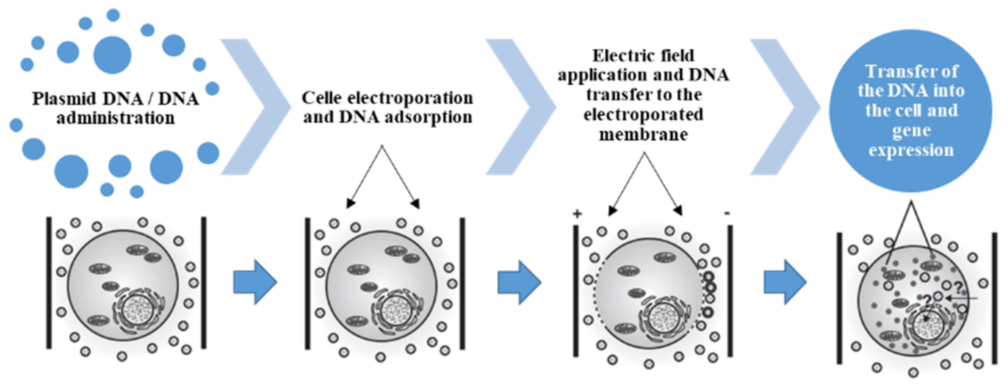
2. Methods
3. Results
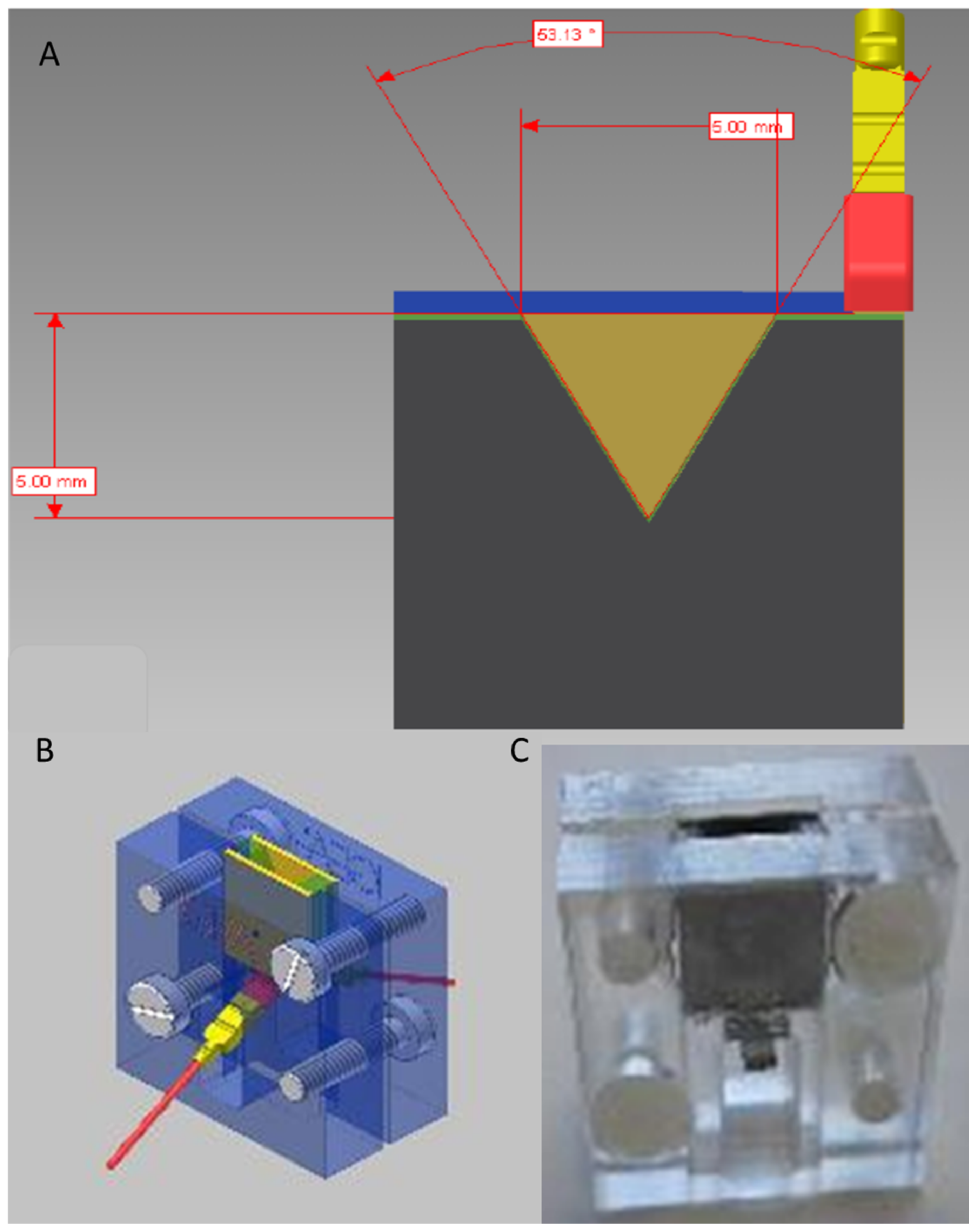
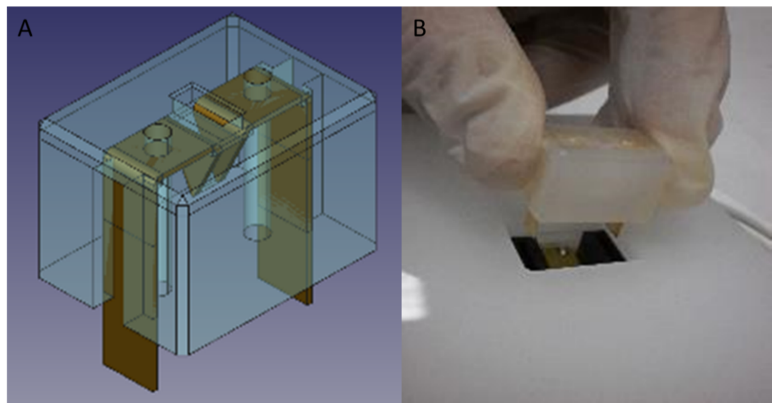
4. IGEA Experience in the Project “Transposon-Based, Targeted Ex Vivo Gene Therapy to Treat Age-Related Macular Degeneration (TargetAMD)”
5. Discussion
6. Conclusions
Author Contributions
Funding
Institutional Review Board Statement
Informed Consent Statement
Data Availability Statement
Conflicts of Interest
References
- Wirth, T.; Parker, N.; Ylä-Herttuala, S. History of gene therapy. Gene 2013, 1525, 162–169. [Google Scholar] [CrossRef] [PubMed]
- Bulcha, J.T.; Wang, Y.; Ma, H.; Tai, P.W.L.; Gao, G. Viral vector platforms within the gene therapy landscape. Sig. Transduct. Target. 2021, 6, 53. [Google Scholar] [CrossRef] [PubMed]
- Tang, R.; Xu, Z. Gene therapy: A double-edged sword with great powers. Mol. Cell Biochem. 2020, 474, 73–81. [Google Scholar] [CrossRef] [PubMed]
- Uthra, S.; Kumaramanickavel, G. Gene therapy in ophthalmology. Oman J. Ophthalmol. 2009, 2, 108–110. [Google Scholar] [CrossRef]
- Neumann, E.; Schaefer-Ridder, M.; Wang, Y.; Hofschneider, P.H. Gene transfer into mouse lyoma cells by electroporation in high electric fields. EMBO 1982, 1, 841–845. [Google Scholar] [CrossRef]
- Fechheimer, M.; Boylan, J.F.; Parker, S.; Sisken, J.E.; Patel, G.L.; Zimmer, S.G. Transfection of mammalian cells with plasmid DNA by scrape loading and sonication loading. Proc. Natl. Acad. Sci. USA 1987, 84, 8463–8467. [Google Scholar] [CrossRef]
- Fraley, R.; Subramani, S.; Berg, P.; Papahadjopoulos, D. Introduction of liposome-encapsulated SV40 DNA into cells. J. Biol. Chem. 1980, 255, 10431–10435. [Google Scholar] [CrossRef]
- Gao, X.; Huang, L. Potentiation of cationic liposome-mediated gene delivery by polycations. Biochemistry 1996, 35, 1027–1036. [Google Scholar] [CrossRef]
- Conley, S.M.; Cai, X.; Naash, M.I. Nonviral ocular gene therapy: Assessment and future directions. Curr. Opin. Mol. Ther. 2008, 10, 456–463. [Google Scholar]
- Trigueros, S.; Domenech, E.B.; Toulis, V.; Marfany, G. In vitro gene delivery in retinal pigment epitheliumcells by plasmid dna-wrapped gold nanoparticles. Genes 2019, 10, 289. [Google Scholar] [CrossRef]
- Dezawa, M.; Takano, M.; Negishi, H.; Mo, X.; Oshitari, T.; Sawada, H. Gene transfer into retinal ganglion cells by in vivo electroporation: A new approach. Micron 2002, 33, 1–6. [Google Scholar] [CrossRef]
- Matsuda, T.; Cepko, C.L. Electroporation and RNA interference in the rodent retina in vivo and in vitro. Proc. Natl. Acad. Sci. USA 2004, 101, 16–22. [Google Scholar] [CrossRef] [PubMed]
- Athanasopoulos, T.; Munye, M.M.; Yanez-Munoz, R.J. Non integrating gene therapy vectors. Hematol. Oncol. Clin. N. Am. 2017, 31, 753–770. [Google Scholar] [CrossRef] [PubMed]
- Peeters, L.; Sanders, N.N.; Braeckmans, K.; Boussery, K.; Van de Voorde, J.; De Smedt, S.C.; Demeester, J. Vitreous: A barrier to nonviral ocular gene therapy. Invest. Ophthalmol. Vis. Sci. 2005, 46, 3553–3561. [Google Scholar] [CrossRef] [PubMed]
- Moore, N.A.; Bracha, P.; Hussain, R.M.; Morral, N.; Ciulla, T.A. Gene therapy for age-related macular degeneration. Expert Opin. Biol. 2017, 17, 1235–1244. [Google Scholar] [CrossRef]
- Chatterjee, P.R.; Dutta, H.; Sarkar, K. Prospects of gene therapy for ocular diseases. J. Indian Med. Assoc. 2000, 98, 746–747. [Google Scholar]
- Anand, V.; Duffy, B.; Yang, Z.; Dejneka, N.S.; Maguire, A.M.; Bennett, J. A deviant immune response to viral proteins and transgene product is generated on subretinal administration of adenovirus and adeno-associated virus. Mol. Ther. 2002, 5, 125–132. [Google Scholar] [CrossRef]
- Willett, K.L.; Bennett, J. Immunology of AAV-mediated gene transferin the eye. Front. Immunol. 2013, 4, 261. [Google Scholar] [CrossRef]
- Pichi, F.; Morara, M.; Veronese, C.; Nucci, P.; Ciardella, A.P. Multimodal imaging in hereditary retinal diseases. J. Ophthalmol. 2013, 634351. [Google Scholar] [CrossRef]
- Amato, A.; Arrigo, A.; Aragona, E.; Manitto, M.P.; Saladino, A.; Bandello, F.; Battaglia Parodi, M. Gene Therapy in Inherited Retinal Diseases: An Update on Current State of the Art. Front. Med. 2021, 8, 750586. [Google Scholar] [CrossRef]
- Dhurandhar, D.; Sahoo, N.K.; Mariappan, I.; Narayanan, R. Gene therapy in retinal diseases: A review. Indian J. Ophthalmol. 2021, 69, 2257–2265. [Google Scholar] [CrossRef] [PubMed]
- Pietrzyk, J.J. Gene therapy-hopes and fears. Folia Med. Cracov. 1998, 39, 131–138. [Google Scholar] [PubMed]
- Zelenin, A.V. Gene therapy: Ethical aspects and problems of genetic safety. Genetika 1999, 35, 1605–1612. [Google Scholar] [PubMed]
- Grodeland, G.; Bogen, B. Efficient vaccine against pandemic influenza: Combining DNA vaccination and targeted delivery to MHC class II molecules. Expert Rev. Vaccines 2015, 14, 805–814. [Google Scholar] [CrossRef]
- Ogunremi, O.; Pasick, J.; Kobinger, G.P.; Hannaman, D.; Berhane, Y.; Clavijo, A.; van Drunen Littel-van den Hurk, S. A single electroporation delivery of a DNA vaccine containing the hemagglutinin gene of Asian H5N1 avian influenza virus generated a protective antibody response in chickens against a North American virus strain. Clin. Vaccine Immunol. 2013, 20, 491–500. [Google Scholar] [CrossRef]
- Sullenger, B.A.; Nair, S. From the RNA world to the clinic. Science 2016, 352, 1417–1420. [Google Scholar] [CrossRef]
- Henshaw, J.W.; Yuan, F. Field distribution and DNA transport in solid tumors during electric field-mediated gene delivery. J. Pharm. Sci. 2008, 97, 691–711. [Google Scholar] [CrossRef]
- Ramamoorth, M.; Narvekar, A. Non viral vectors in gene therapy- an overview. J. Clin. Diagn. Res. JCDR 2015, 9, GE01–GE06. [Google Scholar] [CrossRef]
- Al-Dosari, M.S.; Gao, X. Nonviral gene delivery: Principle, limitations, and recent progress. AAPS J. 2009, 11, 671–681. [Google Scholar] [CrossRef]
- Cervia, L.D.; Yuan, F. Current Progress in Electrotransfection as a Nonviral Method for Gene Delivery. Mol. Pharm. 2018, 15, 3617–3624. [Google Scholar] [CrossRef]
- Available online: https://www.targetamd.eu/ (accessed on 7 June 2022).
- Oshima, Y.; Sakamoto, T.; Yamanaka, I.; Nishi, T.; Ishibashi, I.; Inomata, H. Targeted gene transfer to corneal endothelium in vivo by electric pulse. Gene 1998, 5, 1347–1354. [Google Scholar] [CrossRef] [PubMed]
- Oshima, Y.; Sakamoto, T.; Kawano, Y.; Hata, Y.; Yoshikawa, H.; Sonoda, K.H.; Ishibashi, T.; Inomata, H. Synergistic effect of electric pulses and bleomycin on cultured rabbit subconjunctival fibroblasts. Graefes Arch. Clin. Exp. Ophthalmol. 1998, 236, 52–60. [Google Scholar] [CrossRef] [PubMed]
- Oshima, Y.; Sakamoto, T.; Nakamura, T.; Tahara, Y.; Goto, Y.; Ishibashi, T.; Inomata, H. The comparative benefits of glaucoma filtering surgery with an electric-pulse targeted drug delivery system demonstrated in an animal model. Ophthalmology 1999, 106, 1140–1146. [Google Scholar] [CrossRef]
- Sakamoto, T.; Oshima, Y.; Nakagawa, K.; Ishibashi, T.; Inomata, H.; Sueishi, K. Target gene transfer of tissue plasminogen activator to cornea by electric pulse inhibits intracameral fibrin formation and corneal cloudiness. Hum. Gene Ther. 1999, 10, 2551–2557. [Google Scholar] [CrossRef]
- Oshima, Y.; Sakamoto, T.; Hisatomi, T.; Tsutsumi, C.; Sassa, Y.; Ishibashi, T.; Inomata, H. Targeted gene transfer to corneal stroma in vivo by electric pulses. Exp. Eye Res. 2002, 74, 191–198. [Google Scholar] [CrossRef] [PubMed]
- Blair-Parks, K.; Weston, B.C.; Dean, D.A. High-level gene transfer to the cornea using electroporation. J. Gene Med. 2002, 4, 92–100. [Google Scholar] [CrossRef] [PubMed]
- Yu, W.Z.; Li, X.X.; She, H.C.; He, P.Y.; Dong, J.Q.; Rui, M.; Ma, D.L. Gene transfer of kringle 5 of plasminogen by electroporation inhibits corneal neovascularization. Ophthalmic Res. 2003, 35, 239–246. [Google Scholar] [CrossRef]
- Mamiya, K.; Ohguro, H.; Ohguro, I.; Metoki, T.; Ishikawa, F.; Yamazaki, H.; Takano, Y.; Ito, T.; Nakazawa, M. Effects of matrix metalloproteinase-3 gene transfer by electroporation in glaucoma filter surgery. Exp. Eye Res. 2004, 79, 405–410. [Google Scholar] [CrossRef]
- Chalberg, T.W.; Genise, H.L.; Vollrath, D.; Calos, M.P. phiC31 integrase confers genomic integration and long-term transgene expression in rat retina. Investig. Ophthalmol. Vis. Sci. 2005, 46, 2140–2146. [Google Scholar] [CrossRef]
- He, Z.; Pipparelli, A.; Manissolle, C.; Acquart, S.; Garraud, O.; Gain, P.; Thuret, G. Ex vivo gene electrotransfer to the endothelium of organ cultured human corneas. Ophthalmic Res. 2010, 43, 43–55. [Google Scholar] [CrossRef]
- Touchard, E.; Kowalczuk, L.; Bloquel, C.; Naud, M.C.; Bigey, P.; Behar-Cohen, F. The ciliary smooth muscle electrotransfer: Basic principles and potential for sustained intraocular production of therapeutic proteins. J. Gene Med. 2010, 12, 904–919. [Google Scholar] [CrossRef] [PubMed]
- Kowalczuk, L.; Touchard, E.; Omri, S.; Jonet, L.; Klein, C.; Valamanes, F.; Berdugo, M.; Bigey, P.; Massin, P.; Jeanny, J.C.; et al. Placental growth factor contributes to micro-vascular abnormalization and blood-retinal barrier breakdown in diabetic retinopathy. PLoS ONE 2011, 6, e17462. [Google Scholar] [CrossRef]
- Touchard, E.; Berdugo, M.; Bigey, P.; El Sanharawi, M.; Savoldelli, M.; Naud, M.C.; Jeanny, J.C.; Behar-Cohen, F. Suprachoroidal electrotransfer: A nonviral gene delivery method to transfect the choroid and the retina without detaching the retina. Mol. Ther. 2012, 20, 1559–1570. [Google Scholar] [CrossRef] [PubMed]
- Touchard, E.; Benard, R.; Bigot, K.; Laffitte, J.D.; Buggage, R.; Bordet, T.; Behar-Cohen, F. Non-viral ocular gene therapy, pEYS606, for the treatment of non-infectious uveitis: Preclinical evaluation of the medicinal product. J. Control. Release 2018, 285, 244–251. [Google Scholar] [CrossRef]
- Fleckenstein, M.; Keenan, T.D.L.; Guymer, R.H.; Chakravarthy, U.; Schmitz-Valckenberg, S.; Klaver, C.C.; Wong, W.T.; Chew, E.Y. Age-related macular degeneration. Nat. Rev. Dis. Primers. 2021, 6, 31. [Google Scholar] [CrossRef]
- Campochiaro, P.A.; Nguyen, Q.D.; Shah, S.M.; Klein, M.L.; Holz, E.; Frank, R.N.; Saperstein, D.A.; Gupta, A.; Stout, J.T.; Macko, J.; et al. Adenoviral vector-delivered pigment epithelium-derived factor for neovascular age-related maculardegeneration: Results of a phase I clinical trial. Hum Gene 2006, 17, 167–176. [Google Scholar] [CrossRef]
- Kropp, M.; Harmening, N.; Bautzová, T.; Asrih, M.; Sealy, G.; Johnen, S.; Izsvák, Z.; Marie, C.; Scherman, D.; van den Berg, J.; et al. Development of GMP-compliant production of freshly isolated and transfected iris pigment epithelial (IPE) cells to treat age-related macular degeneration (AMD). Hum. Gene Ther. 2017, 28, A126. [Google Scholar]
- Stolba, U.; Ansari-Shahrezaei, S.; Hagen, S.; Stattin, M.; Schmid, S.; Kropp, M.; Harmening, N.; Thumann, G.; TargetAMD Group. Neovascular age-related macular degeneration in Austria—Expert review and introduction to the TargetAMD approach. Spektrum Der. Augenheilkd. 2017, 31, 206. [Google Scholar] [CrossRef]
- Available online: https://cordis.europa.eu/project/id/305134/reporting/it (accessed on 7 June 2022).
- Kachi, S.; Oshima, Y.; Esumi, N.; Kachi, M.; Rogers, B.; Zack, D.J.; Campochiaro, P.A. Nonviral ocular gene transfer. Gene Ther. 2005, 12, 843–851. [Google Scholar] [CrossRef]
- Kalia, Y.N.; Naik, A.; Garrison, J.; Guy, R.H. Iontophoretic drug delivery. Adv. Drug Deliv. Rev. 2004, 56, 619–658. [Google Scholar] [CrossRef]
- Katahira, T.; Nakamura, H. Gene silencing in chick embryos with a vector-based small interfering RNA system. Dev. Growth Differ. 2003, 45, 361–367. [Google Scholar] [CrossRef] [PubMed]
- Klenchin, V.A.; Sukharev, S.I.; Serov, S.M.; Chernomordik, L.V.; YuA, C. Electrically induced DNA uptake by cells is a fast process involving DNA electrophoresis. Biophys. J. 1991, 60, 804–811. [Google Scholar] [CrossRef]
- Dujardin, N.; Préat, V. Delivery of DNA to skin by electroporation. Methods Mol. Biol. 2004, 245, 215–226. [Google Scholar] [CrossRef] [PubMed]
- Titomirov, A.V.; Sukharev, S.; Kistanova, E. In vivo electroporation and stable transformation of skin cells of newborn mice by plasmid DNA. Biochim. Biophys. Acta 1991, 1088, 131–134. [Google Scholar] [CrossRef]
- Zhang, L.; Li, L.; Hoffmann, G.A.; Hoffman, R.M. Depth-targeted efficient gene delivery and expression in the skin by pulsed electric fields: An approach to gene therapy of skin aging and other diseases. Biochem. Biophys. Res. Commun. 1996, 220, 633–636. [Google Scholar] [CrossRef]
- Suzuki, T.; Shin, B.C.; Fujikura, K.; Matsuzaki, T.; Takata, K. Direct gene transfer into rat liver cells by in vivo electroporation. FEBS Lett. 1998, 425, 436–440. [Google Scholar] [CrossRef]
- Heller, R.; Jaroszeski, M.; Atkin, A.; Moradpour, D.; Gilbert, R.; Wands, J.; Nicolau, C. In vivo gene electroinjection and expression in rat liver. FEBS Lett. 1996, 389, 225–258. [Google Scholar] [CrossRef]
- Aihara, H.; Miyazaki, J. Gene transfer into muscle by electroporation in vivo. Nat. Biotechnol. 1998, 16, 867–870. [Google Scholar] [CrossRef]
- Mir, L.M.; Bureau, M.F.; Rangara, R.; Schwartz, B.; Scherman, D. Long-term, high level in vivo gene expression after electric pulse-mediated gene transfer into skeletal muscle. C R Acad. Sci. III 1998, 321, 893–899. [Google Scholar] [CrossRef]
- McMahon, J.M.; Wells, D.J. Electroporation for gene transfer to skeletal muscles: Current status. Bio. Drugs 2004, 18, 155–165. [Google Scholar] [CrossRef]
- Bloquel, C.; Bejjani, R.A.; Bigey, P.; Bedioui, F.; Doat, M.; BenEzra, D.; Scherman, D.; Behar-Cohen, F. Plasmid electrotransfer of eye ciliary muscle: Principle and uveitis therapeutic efficacy using hTNF- soluble receptor. FASEB J. 2006, 20, 389–391. [Google Scholar] [CrossRef] [PubMed]
- McMahon, J.M.; Signori, E.; Wells, K.E.; Fazio, V.M.; Wells, D.J. Optimisation of electrotransfer of plasmid into skeletal muscle by pretreatment with hyaluronidase—increased expression with reduced muscle damage. Gene Ther. 2001, 8, 1264–1270. [Google Scholar] [CrossRef] [PubMed]
- Rabussay, D.; Dev, N.B.; Fewell, J.; Smith, L.; Widera, G.; Zhang, L. Enhancement of therapeutic drug and DNA delivery into cells by electroporation. J. Phys. D Appl. Phys. 2003, 36, 348–363. [Google Scholar] [CrossRef]
- Young, J.L.; Dean, D.A. Electroporation-mediated gene delivery. Adv. Genet. 2015, 89, 49–88. [Google Scholar] [CrossRef] [PubMed]
- Duvall, C.L.; Prokop, A.; Charles, A.; Jeffrey, G.; Davidson, M. Chapter-35. Gene delivery into cells and tissues. In Principles of Tissue Engineering, 4th ed.; Lanza, R.P., Langer, R.S., Vacanti, J., Eds.; Elsevier/AP: Amsterdam, The Netherlands, 2014; pp. 687–723. [Google Scholar] [CrossRef]


| Study Design | ||||||||
|---|---|---|---|---|---|---|---|---|
| Manuscript | Study Information | Shape and Material of the Electrode(s) | Pulse Number and Duration | PAUSE Duration/Pulse Frequency | Applied Voltage/Current Electric Field | Results | ||
| Oshima et al. [32,33,34] | rat in vivo | corneal endothelium | ring-shaped (distance of 0.5 mm) | stainless steel | 8 pulses 50 ms | Not reported | from 5 to 40 V/cm | β-Galactosidase strongest expression at 20 V. Expression limited to corneal endothelial cells. No cell damage or inflammation detected. |
| Sakamoto et al. [35] | rat in vivo | corneal endothelium | circular (Ø of 0.5 mm) | stainless steel | 8 pulses 50 ms | 80 ms | Not reported | Presence of plasminogen activator for 4 days. Significant decrease in fibrin formation. No histological damage detected. |
| Oshima et al. [36] | Rat in vivo | corneal stroma | bipolar linear (gap of 10 mm) | stainless steel | 8 pulses 50 ms | 75 ms | from 10 to 30 V/cm | Effective and selective gene transfer using eight electric pulses of 20 V for 50 ms. No cell damage or inflammation detected with voltages lower than 30 V. |
| Blair-Parks et al. [37] | mouse in vivo | corneal endothelium and stroma | 2 golden-plated (3 mm apart) | gold | 8 pulses 10 ms | Not reported | 0, 100, 200, 400 V/cm | Nanogram levels of gene product expression. No trauma, edema or inflammation detected with field strength up to 200 V/cm. |
| Yu et al. [38] | rat in vivo | cornea | 2 golden-plated (5 mm apart) | gold | 8 pulses 10 ms | Not reported | 200 V/cm | K5 gene effectively transfected. Effective inhibition of corneal neovascularization. |
| Mamiya et al. [39] | rabbit in vivo | conjunctiva | cup-shaped (not reported) | Not reported | 2 × 5 pulses 50 ms | Not reported | 5 V/cm | MMP-3 plasmid correctly expressed. Combination with surgery caused longer survival of the filtering bleb and decreased levels of IOP. |
| Chalberg et al. [40] | rat in vivo | retinal pigment epithelium | 2 circular (Ø of 7 mm, 14 mm apart) | Not reported | 5 pulses 100 ms | 950 ms | 100 V/cm | Strongest transgene expression after 48 h. 85-fold higher long-term expression achieved with co-injection of integrase. |
| He et al. [41] | human ex vivo | corneal endothelium | 2 convex-shaped (Ø of 13 mm, 2.5 mm apart, 8.4 mm curvature radius) | stainless steel | 8 pulses 100 ms | 1 Hz | 125 mA | eGFP expression detected in all transfected corneas. |
| Touchard et al. [42] | rat/rabbit in vivo | ciliary muscle | semi-annular sheet anode wire cathode (Ø of 0.25/0.5 mm) | platinum/ iridium | 8 pulses 20 ms | 5 Hz | 15/20 V | Plasmid effectively transfected in ciliary muscle fibers. Long-lasting gene transfer (>5 months). |
| Kowalczuk et al. [43] | rat in vivo | ciliary muscle | semi-annular sheet anode wire cathode (Ø of 0.25 mm) | platinum/ iridium | 8 pulses 20 ms | 5 Hz | 200 V/cm | In diabetic retinas, over-expression of rPGF-1 induced glial activation and proliferation. |
| Touchard et al. [44] | rat in vivo | suprachoroidal space | semi-annular sheet anode semi-annular wire cathode | platinum/ iridium | 8 pulses 20 ms | 5 Hz | 40 V/cm | Strongest transfection efficacy at 60 V/cm (8 pulses, 20 ms, 5 Hz). No ocular complications recorded. |
| curved sheet anode semi-annular sheet cathode | platinum/ iridium | 8 pulses 20 ms | 5 Hz | 14, 30, 60, 120 V/cm | ||||
| Touchard et al. [45] | rat in vivo | suprachoroidal space | semi-annular sheet anode wire cathode | platinum/ iridium | 8 pulses 10 ms | 5 Hz | 200 V/cm | Significant reduction of ocular inflammation observed. In EAU, significant protection of photoreceptors observed after pEYS606 treatment. |
Publisher’s Note: MDPI stays neutral with regard to jurisdictional claims in published maps and institutional affiliations. |
© 2022 by the authors. Licensee MDPI, Basel, Switzerland. This article is an open access article distributed under the terms and conditions of the Creative Commons Attribution (CC BY) license (https://creativecommons.org/licenses/by/4.0/).
Share and Cite
Fusco, R.; Perazzolo Gallo, G.; Di Bernardo, E.; D’Alessio, V.; Ronchetti, M.; Cadossi, M.; Cadossi, R. In Vivo and Ex Vivo Gene Electrotransfer in Ophthalmological Disorders. Biomedicines 2022, 10, 1889. https://doi.org/10.3390/biomedicines10081889
Fusco R, Perazzolo Gallo G, Di Bernardo E, D’Alessio V, Ronchetti M, Cadossi M, Cadossi R. In Vivo and Ex Vivo Gene Electrotransfer in Ophthalmological Disorders. Biomedicines. 2022; 10(8):1889. https://doi.org/10.3390/biomedicines10081889
Chicago/Turabian StyleFusco, Roberta, Giacomo Perazzolo Gallo, Elio Di Bernardo, Valeria D’Alessio, Mattia Ronchetti, Matteo Cadossi, and Ruggero Cadossi. 2022. "In Vivo and Ex Vivo Gene Electrotransfer in Ophthalmological Disorders" Biomedicines 10, no. 8: 1889. https://doi.org/10.3390/biomedicines10081889
APA StyleFusco, R., Perazzolo Gallo, G., Di Bernardo, E., D’Alessio, V., Ronchetti, M., Cadossi, M., & Cadossi, R. (2022). In Vivo and Ex Vivo Gene Electrotransfer in Ophthalmological Disorders. Biomedicines, 10(8), 1889. https://doi.org/10.3390/biomedicines10081889