
Carbon Graphitization: Towards Greener Alternatives to Develop Nanomaterials for Targeted Drug Delivery


Abstract
:1. Introduction
2. Carbon Nanomaterials (CNMs)
3. The Graphitization Process
4. Greener Alternatives to Traditional Graphitization for CNM Production
4.1. CNMs from Waste
4.2. CNMs from Sustainable Natural Sources
4.3. Atmospheric Particulate as Carbon Source for CNMs
4.4. Greener Routes to Prepare CNMs from Graphite- or Coal-Based Products
4.5. CO2 as Carbon Source to Produce CNMs
5. Applications of CNMs for Targeted Drug Delivery
5.1. Fullerenes
5.2. CNOs
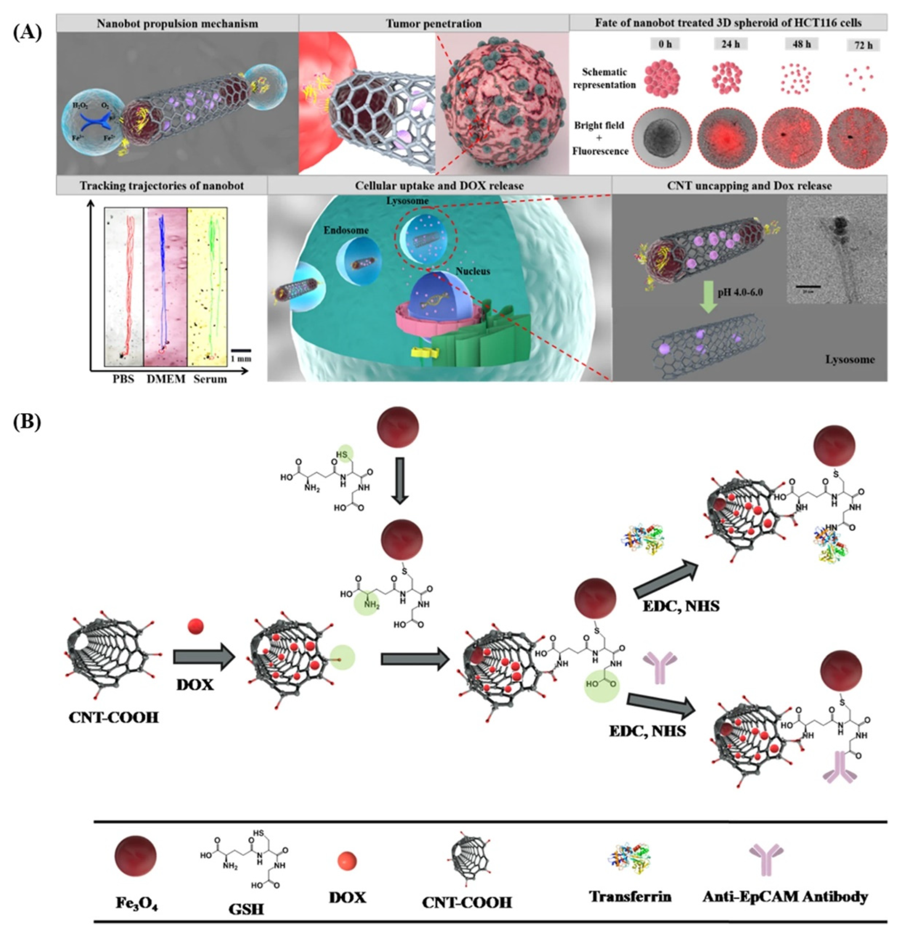
5.3. CNTs
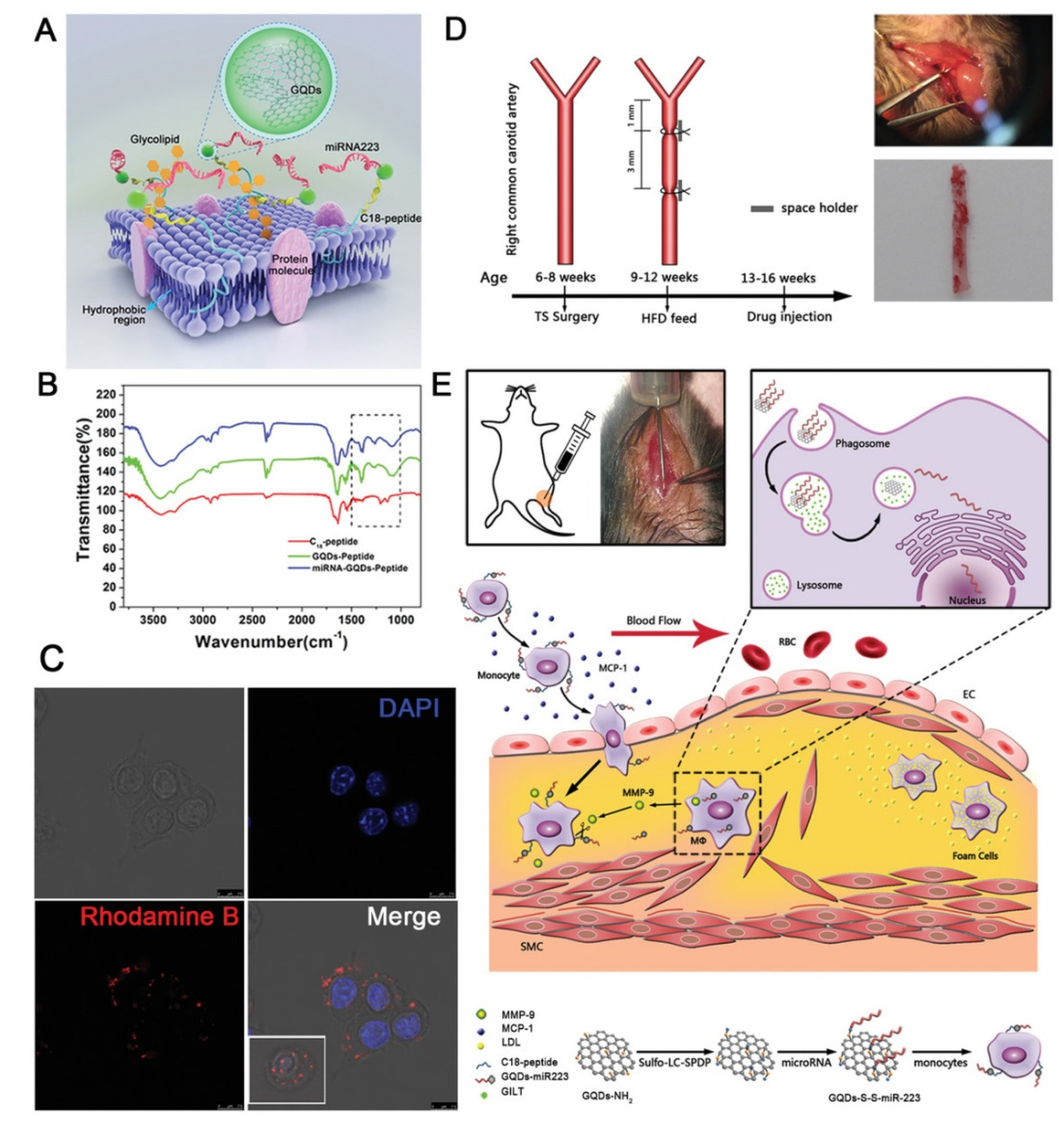
5.4. GQDs
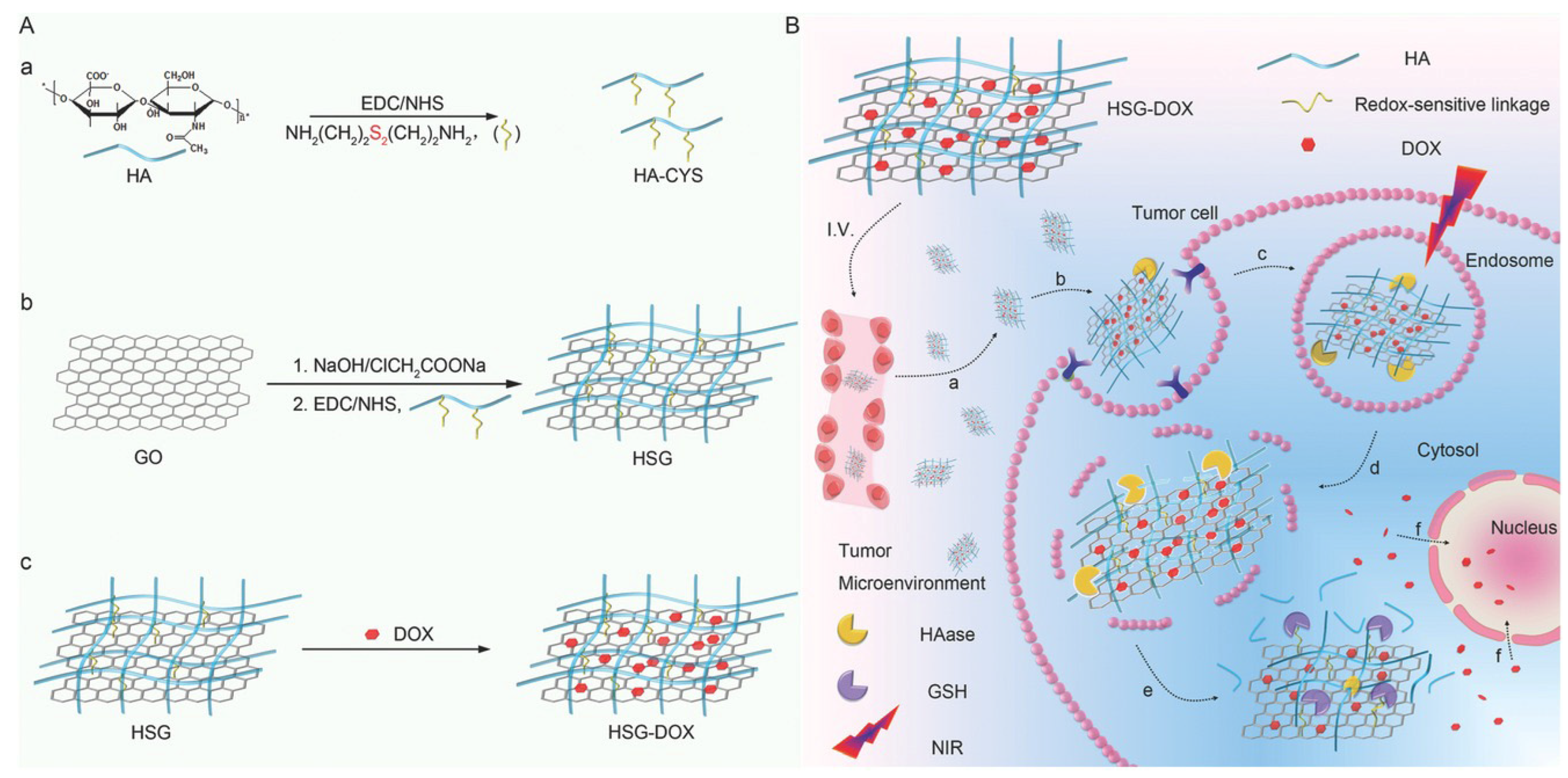
5.5. Graphene-Based 2D Materials
6. Conclusions
Author Contributions
Funding
Institutional Review Board Statement
Informed Consent Statement
Data Availability Statement
Acknowledgments
Conflicts of Interest
References
- Mateo, J.; Steuten, L.; Aftimos, P.; André, F.; Davies, M.; Garralda, E.; Geissler, J.; Husereau, D.; Martinez-Lopez, I.; Normanno, N.; et al. Delivering precision oncology to patients with cancer. Nat. Med. 2022, 28, 658–665. [Google Scholar] [CrossRef]
- Marchesan, S.; Prato, M. Nanomaterials for (nano)medicine. ACS Med. Chem. Lett. 2013, 4, 147–149. [Google Scholar] [CrossRef] [Green Version]
- Rosenkrans, Z.T.; Ferreira, C.A.; Ni, D.; Cai, W. Internally responsive nanomaterials for activatable multimodal imaging of cancer. Adv. Healthc. Mater. 2021, 10, e2000690. [Google Scholar] [CrossRef] [PubMed]
- Paramasivam, G.; Palem, V.V.; Sundaram, T.; Sundaram, V.; Kishore, S.C.; Bellucci, S. Nanomaterials: Synthesis and applications in theranostics. Nanomaterials 2021, 11, 3228. [Google Scholar] [CrossRef] [PubMed]
- Mitchell, M.J.; Billingsley, M.M.; Haley, R.M.; Wechsler, M.E.; Peppas, N.A.; Langer, R. Engineering precision nanoparticles for drug delivery. Nat. Rev. Drug Discov. 2021, 20, 101–124. [Google Scholar] [CrossRef]
- Marchesan, S.; Melchionna, M.; Prato, M. Carbon nanostructures for nanomedicine: Opportunities and challenges. Full. Nanotub. Carbon Nanostruct. 2014, 22, 190–195. [Google Scholar] [CrossRef]
- Erol, O.; Uyan, I.; Hatip, M.; Yilmaz, C.; Tekinay, A.B.; Guler, M.O. Recent advances in bioactive 1D and 2D carbon nanomaterials for biomedical applications. Nanomedicine 2018, 14, 2433–2454. [Google Scholar] [CrossRef] [PubMed] [Green Version]
- Bazaka, K.; Jacob, M.V.; Ostrikov, K. Sustainable life cycles of natural-precursor-derived nanocarbons. Chem. Rev. 2016, 116, 163–214. [Google Scholar] [CrossRef] [PubMed] [Green Version]
- Sun, W.; Lee, J.; Zhang, S.; Benyshek, C.; Dokmeci, M.R.; Khademhosseini, A. Engineering precision medicine. Adv. Sci. 2019, 6, 1801039. [Google Scholar] [CrossRef] [Green Version]
- Speranza, G. Carbon nanomaterials: Synthesis, functionalization and sensing applications. Nanomaterials 2021, 11, 967. [Google Scholar] [CrossRef]
- Jolly, A.; Miao, D.; Daigle, M.; Morin, J.-F. Emerging bottom-up strategies for the synthesis of graphene nanoribbons and related structures. Angew. Chem. Int. Ed. 2020, 59, 4624–4633. [Google Scholar] [CrossRef]
- Bruno, F.; Sciortino, A.; Buscarino, G.; Soriano, M.L.; Ríos, Á.; Cannas, M.; Gelardi, F.; Messina, F.; Agnello, S. A comparative study of top-down and bottom-up carbon nanodots and their interaction with mercury ions. Nanomaterials 2021, 11, 1265. [Google Scholar] [CrossRef] [PubMed]
- Ventrella, A.; Camisasca, A.; Fontana, A.; Giordani, S. Synthesis of green fluorescent carbon dots from carbon nano-onions and graphene oxide. RSC Adv. 2020, 10, 36404–36412. [Google Scholar] [CrossRef]
- Georgakilas, V.; Perman, J.A.; Tucek, J.; Zboril, R. Broad family of carbon nanoallotropes: Classification, chemistry, and applications of fullerenes, carbon dots, nanotubes, graphene, nanodiamonds, and combined superstructures. Chem. Rev. 2015, 115, 4744–4822. [Google Scholar] [CrossRef] [PubMed]
- Basso, L.; Cazzanelli, M.; Orlandi, M.; Miotello, A. Nanodiamonds: Synthesis and application in sensing, catalysis, and the possible connection with some processes occurring in space. Appl. Sci. 2020, 10, 4094. [Google Scholar] [CrossRef]
- Bottari, G.; Herranz, M.Á.; Wibmer, L.; Volland, M.; Rodríguez-Pérez, L.; Guldi, D.M.; Hirsch, A.; Martín, N.; D’Souza, F.; Torres, T. Chemical functionalization and characterization of graphene-based materials. Chem. Sov. Rev. 2017, 46, 4464–4500. [Google Scholar] [CrossRef] [Green Version]
- Calvaresi, M.; Quintana, M.; Rudolf, P.; Zerbetto, F.; Prato, M. Rolling up a graphene sheet. ChemPhysChem 2013, 14, 3447–3453. [Google Scholar] [CrossRef] [PubMed]
- Mchedlov-Petrossyan, N.O. Fullerenes in liquid media: An unsettling intrusion into the solution chemistry. Chem. Rev. 2013, 113, 5149–5193. [Google Scholar] [CrossRef] [PubMed]
- Giordani, S.; Camisasca, A.; Maffeis, V. Carbon nano-onions: A valuable class of carbon nanomaterials in biomedicine. Curr. Med. Chem. 2019, 26, 6915–6929. [Google Scholar] [CrossRef]
- Yang, F.; Wang, M.; Zhang, D.; Yang, J.; Zheng, M.; Li, Y. Chirality pure carbon nanotubes: Growth, sorting, and characterization. Chem. Rev. 2020, 120, 2693–2758. [Google Scholar] [CrossRef] [PubMed]
- Eatemadi, A.; Daraee, H.; Karimkhanloo, H.; Kouhi, M.; Zarghami, N.; Akbarzadeh, A.; Abasi, M.; Hanifehpour, Y.; Joo, S.W. Carbon nanotubes: Properties, synthesis, purification, and medical applications. Nanoscale Res. Lett. 2014, 9, 393. [Google Scholar] [CrossRef] [Green Version]
- Karousis, N.; Suarez-Martinez, I.; Ewels, C.P.; Tagmatarchis, N. Structure, properties, functionalization, and applications of carbon nanohorns. Chem. Rev. 2016, 116, 4850–4883. [Google Scholar] [CrossRef]
- Liu, J.; Li, R.; Yang, B. Carbon dots: A new type of carbon-based nanomaterial with wide applications. ACS Cent. Sci. 2020, 6, 2179–2195. [Google Scholar] [CrossRef]
- Luo, H.; Yu, G. Preparation, bandgap engineering, and performance control of graphene nanoribbons. Chem. Mater. 2022, 34, 3588–3615. [Google Scholar] [CrossRef]
- Adorinni, S.; Cringoli, M.C.; Perathoner, S.; Fornasiero, P.; Marchesan, S. Green approaches to carbon nanostructure-based biomaterials. Appl. Sci. 2021, 11, 2490. [Google Scholar] [CrossRef]
- Ugarte, D. Onion-like graphitic particles. Carbon 1995, 33, 989–993. [Google Scholar] [CrossRef]
- Vicentini, N.; Gatti, T.; Salerno, M.; Gomez, Y.S.H.; Bellon, M.; Gallio, S.; Marega, C.; Filippini, F.; Menna, E. Effect of different functionalized carbon nanostructures as fillers on the physical properties of biocompatible poly(L-lactic acid) composites. Mater. Chem. Phys. 2018, 214, 265–276. [Google Scholar] [CrossRef]
- Piovesana, S.; Iglesias, D.; Melle-Franco, M.; Kralj, S.; Cavaliere, C.; Melchionna, M.; Laganà, A.; Capriotti, A.L.; Marchesan, S. Carbon nanostructure morphology templates nanocomposites for phosphoproteomics. Nano Res. 2020, 13, 380–388. [Google Scholar] [CrossRef]
- Tonellato, M.; Piccione, M.; Gasparotto, M.; Bellet, P.; Tibaudo, L.; Vicentini, N.; Bergantino, E.; Menna, E.; Vitiello, L.; Di Liddo, R. Commitment of autologous human multipotent stem cells on biomimetic poly-l-lactic acid-based scaffolds is strongly influenced by structure and concentration of carbon nanomaterial. Nanomaterials 2020, 10, 415. [Google Scholar] [CrossRef] [PubMed] [Green Version]
- Iglesias, D.; Melle-Franco, M.; Kurbasic, M.; Melchionna, M.; Abrami, M.; Grassi, M.; Prato, M.; Marchesan, S. Oxidized nanocarbons-tripeptide supramolecular hydrogels: Shape matters! ACS Nano 2018, 12, 5530–5538. [Google Scholar] [CrossRef]
- Marchesan, S.; Prato, M. Under the lens: Carbon nanotube and protein interaction at the nanoscale. Chem. Commun. 2015, 51, 4347–4359. [Google Scholar] [CrossRef]
- Sun, H.; Ren, J.; Qu, X. Carbon nanomaterials and DNA: From molecular recognition to applications. Acc. Chem. Res. 2016, 49, 461–470. [Google Scholar] [CrossRef] [PubMed]
- Pinals, R.L.; Yang, D.; Lui, A.; Cao, W.; Landry, M.P. Corona exchange dynamics on carbon nanotubes by multiplexed fluorescence monitoring. J. Am. Chem. Soc. 2019, 142, 1254–1264. [Google Scholar] [CrossRef]
- Palmieri, V.; Perini, G.; De Spirito, M.; Papi, M. Graphene oxide touches blood: In vivo interactions of bio-coronated 2D materials. Nanoscale Horiz. 2019, 4, 273–290. [Google Scholar] [CrossRef]
- Cai, K.; Wang, A.Z.; Yin, L.; Cheng, J. Bio-nano interface: The impact of biological environment on nanomaterials and their delivery properties. J. Control. Release 2017, 263, 211–222. [Google Scholar] [CrossRef] [PubMed]
- Marchesan, S.; Kostarelos, K.; Bianco, A.; Prato, M. The winding road for carbon nanotubes in nanomedicine. Mater. Today 2015, 18, 12–19. [Google Scholar] [CrossRef]
- Gazzi, A.; Fusco, L.; Orecchioni, M.; Ferrari, S.; Franzoni, G.; Yan, J.S.; Rieckher, M.; Peng, G.; Lucherelli, M.A.; Vacchi, I.A.; et al. Graphene, other carbon nanomaterials and the immune system: Toward nanoimmunity-by-design. J. Phys. Mater. 2020, 3, 034009. [Google Scholar] [CrossRef]
- Chen, M.; Qin, X.; Zeng, G. Biodegradation of carbon nanotubes, graphene, and their derivatives. Trends Biotechnol. 2017, 35, 836–846. [Google Scholar] [CrossRef]
- Rozhin, P.; Abdel Monem Gamal, J.; Giordani, S.; Marchesan, S. Carbon nanomaterials (CNMs) and enzymes: From nanozymes to cnm-enzyme conjugates and biodegradation. Materials 2022, 15, 1037. [Google Scholar] [CrossRef]
- Marchesan, S.; Melchionna, M.; Prato, M. Wire up on carbon nanostructures! How to play a winning game. ACS Nano 2015, 9, 9441–9450. [Google Scholar] [CrossRef] [Green Version]
- Loh, K.P.; Ho, D.; Chiu, G.N.C.; Leong, D.T.; Pastorin, G.; Chow, E.K.H. Clinical applications of carbon nanomaterials in diagnostics and therapy. Adv. Mater. 2018, 30, 1802368. [Google Scholar] [CrossRef] [PubMed]
- Plonska-Brzezinska, M.E. Carbon nanomaterials: Perspective of their applications in biomedicine. Curr. Med. Chem. 2019, 26, 6832–6833. [Google Scholar] [CrossRef] [PubMed]
- Teradal, N.L.; Jelinek, R. Carbon nanomaterials in biological studies and biomedicine. Adv. Healthc. Mater. 2017, 6, 1700574. [Google Scholar] [CrossRef] [PubMed]
- Mehra, N.K.; Jain, A.K.; Nahar, M. Carbon nanomaterials in oncology: An expanding horizon. Drug Discov. Today 2018, 23, 1016–1025. [Google Scholar] [CrossRef]
- Augustine, S.; Singh, J.; Srivastava, M.; Sharma, M.; Das, A.; Malhotra, B.D. Recent advances in carbon based nanosystems for cancer theranostics. Biomater. Sci. 2017, 5, 901–952. [Google Scholar] [CrossRef]
- Bartelmess, J.; Quinn, S.; Giordani, S. Carbon nanomaterials: Multi-functional agents for biomedical fluorescence and raman imaging. Chem. Soc. Rev. 2015, 44, 4672–4698. [Google Scholar] [CrossRef]
- Rezaee, M.; Behnam, B.; Banach, M.; Sahebkar, A. The yin and yang of carbon nanomaterials in atherosclerosis. Biotechnol. Adv. 2018, 36, 2232–2247. [Google Scholar] [CrossRef]
- Xin, Q.; Shah, H.; Nawaz, A.; Xie, W.; Akram, M.Z.; Batool, A.; Tian, L.; Jan, S.U.; Boddula, R.; Guo, B. Antibacterial carbon-based nanomaterials. Adv. Mater. 2019, 31, 1804838. [Google Scholar] [CrossRef]
- Unal, M.A.; Bayrakdar, F.; Nazir, H.; Besbinar, O.; Gurcan, C.; Lozano, N.; Arellano, L.M.; Yalcin, S.; Panatli, O.; Celik, D.; et al. Graphene oxide nanosheets interact and interfere with SARS-CoV-2 surface proteins and cell receptors to inhibit infectivity. Small 2021, 17, 2101483. [Google Scholar] [CrossRef]
- Ashrafizadeh, M.; Mohammadinejad, R.; Kailasa, S.K.; Ahmadi, Z.; Afshar, E.G.; Pardakhty, A. Carbon dots as versatile nanoarchitectures for the treatment of neurological disorders and their theranostic applications: A review. Adv. Coll. Interface Sci. 2020, 278, 102123. [Google Scholar] [CrossRef]
- Gupta, N.; Rai, D.B.; Jangid, A.K.; Kulhari, H. A review of theranostics applications and toxicities of carbon nanomaterials. Curr. Drug Metab. 2019, 20, 506–532. [Google Scholar] [CrossRef] [PubMed]
- Yuan, X.; Zhang, X.; Sun, L.; Wei, Y.; Wei, X. Cellular toxicity and immunological effects of carbon-based nanomaterials. Particle Fibre Toxicol. 2019, 16, 18. [Google Scholar] [CrossRef]
- Fadeel, B.; Kostarelos, K. Grouping all carbon nanotubes into a single substance category is scientifically unjustified. Nat. Nanotechnol. 2020, 15, 164. [Google Scholar] [CrossRef] [Green Version]
- Graphene Standards. Available online: https://www.thegraphenecouncil.org/page/GrapheneStandards (accessed on 21 May 2022).
- Xiarchos, I.; Morozinis, A.K.; Kavouras, P.; Charitidis, C.A. Nanocharacterization, materials modeling, and research integrity as enablers of sound risk assessment: Designing responsible nanotechnology. Small 2020, 16, 2001590. [Google Scholar] [CrossRef] [PubMed]
- Zhu, Y.; Qu, B.; Andreeva, D.V.; Ye, C.; Novoselov, K.S. Graphene standardization: The lesson from the East. Mater. Today 2021, 47, 9–15. [Google Scholar] [CrossRef]
- Kotzabasaki, M.; Sotiropoulos, I.; Charitidis, C.; Sarimveis, H. Machine learning methods for multi-walled carbon nanotubes (mwcnt) genotoxicity prediction. Nanoscale Adv. 2021, 3, 3167–3176. [Google Scholar] [CrossRef]
- Romanos, N.; Kalogerini, M.; Koumoulos, E.P.; Morozinis, A.; Sebastiani, M.; Charitidis, C. Innovative data management in advanced characterization: Implications for materials design. Mater. Today Commun. 2019, 20, 100541. [Google Scholar] [CrossRef]
- Rao, R.; Pint, C.L.; Islam, A.E.; Weatherup, R.S.; Hofmann, S.; Meshot, E.R.; Wu, F.; Zhou, C.; Dee, N.; Amama, P.B.; et al. Carbon nanotubes and related nanomaterials: Critical advances and challenges for synthesis toward mainstream commercial applications. ACS Nano 2018, 12, 11756–11784. [Google Scholar] [CrossRef] [PubMed] [Green Version]
- Li, H.; Zhang, H.; Li, K.; Zhang, J.; Sun, M.; Su, B. Catalytic graphitization of coke carbon by iron: Understanding the evolution of carbon structure, morphology and lattice fringes. Fuel 2020, 279, 118531. [Google Scholar] [CrossRef]
- Flygare, M.; Svensson, K. Quantifying crystallinity in carbon nanotubes and its influence on mechanical behaviour. Mater. Today Commun. 2019, 18, 39–45. [Google Scholar] [CrossRef]
- Mochida, I.; Egashira, M.; Korai, Y.; Yokogawa, K. Structural changes of fullerene by heat-treatment up to graphitization temperature. Carbon 1997, 35, 1707–1712. [Google Scholar] [CrossRef]
- Fadeel, B.; Bussy, C.; Merino, S.; Vázquez, E.; Flahaut, E.; Mouchet, F.; Evariste, L.; Gauthier, L.; Koivisto, A.J.; Vogel, U.; et al. Safety assessment of graphene-based materials: Focus on human health and the environment. ACS Nano 2018, 12, 10582–10620. [Google Scholar] [CrossRef] [PubMed]
- Tasis, D.; Tagmatarchis, N.; Bianco, A.; Prato, M. Chemistry of carbon nanotubes. Chem. Rev. 2006, 106, 1105–1136. [Google Scholar] [CrossRef]
- Andrews, R.; Jacques, D.; Qian, D.; Dickey, E.C. Purification and structural annealing of multiwalled carbon nanotubes at graphitization temperatures. Carbon 2001, 39, 1681–1687. [Google Scholar] [CrossRef]
- Zhu, S.; Xu, G. Single-walled carbon nanohorns and their applications. Nanoscale 2010, 2, 2538–2549. [Google Scholar] [CrossRef] [PubMed]
- Bartolome, J.P.; Fragoso, A. Preparation and characterization of carbon nano-onions by nanodiamond annealing and functionalization by radio-frequency Ar/O2 plasma. Fuller. Nanotub. Carbon Nanostruct. 2017, 25, 327–334. [Google Scholar] [CrossRef]
- Li, L.; Wu, G.; Yang, G.; Peng, J.; Zhao, J.; Zhu, J.-J. Focusing on luminescent graphene quantum dots: Current status and future perspectives. Nanoscale 2013, 5, 4015–4039. [Google Scholar] [CrossRef] [Green Version]
- Yogesh, G.K.; Shukla, S.; Sastikumar, D.; Koinkar, P. Progress in pulsed laser ablation in liquid (plal) technique for the synthesis of carbon nanomaterials: A review. Appl. Phys. A 2021, 127, 810. [Google Scholar] [CrossRef]
- Rathinavel, S.; Priyadharshini, K.; Panda, D. A review on carbon nanotube: An overview of synthesis, properties, functionalization, characterization, and the application. Mater. Sci. Eng. B 2021, 268, 115095. [Google Scholar] [CrossRef]
- Tripathi, K.M.; Tran, T.S.; Kim, Y.J.; Kim, T. Green fluorescent onion-like carbon nanoparticles from flaxseed oil for visible light induced photocatalytic applications and label-free detection of Al(III) ions. ACS Sustain. Chem. Eng. 2017, 5, 3982–3992. [Google Scholar] [CrossRef]
- Jung, S.; Myung, Y.; Das, G.S.; Bhatnagar, A.; Park, J.-W.; Tripathi, K.M.; Kim, T. Carbon nano-onions from waste oil for application in energy storage devices. New J. Chem. 2020, 44, 7369–7375. [Google Scholar] [CrossRef]
- Xin, Y.; Odachi, K.; Shirai, T. Fabrication of ultra-bright carbon nano-onions via a one-step microwave pyrolysis of fish scale waste in seconds. Green Chem. 2022, 24, 3969–3976. [Google Scholar] [CrossRef]
- Gunture; Dalal, C.; Kaushik, J.; Garg, A.K.; Sonkar, S.K. Pollutant-soot-based nontoxic water-soluble onion-like nanocarbons for cell imaging and selective sensing of toxic cr(VI). ACS Appl. Bio Mater. 2020, 3, 3906–3913. [Google Scholar] [CrossRef]
- Chowdhury, S.N.; Tung, T.T.; Ta, Q.T.H.; Gunture; Castro, M.; Feller, J.F.; Sonkar, S.K.; Tripathi, K.M. Upgrading of diesel engine exhaust waste into onion-like carbon nanoparticles for integrated degradation sensing in nano-biocomposites. New J. Chem. 2021, 45, 3675–3682. [Google Scholar] [CrossRef]
- Gunture; Kaushik, J.; Garg, A.K.; Saini, D.; Khare, P.; Sonkar, S.K. Pollutant diesel soot derived onion-like nanocarbons for the adsorption of organic dyes and environmental assessment of treated wastewater. Ind. Eng. Chem. Res. 2020, 59, 12065–12074. [Google Scholar] [CrossRef]
- Liu, X.; Ren, J.; Licht, G.; Wang, X.; Licht, S. Carbon nano-onions made directly from CO2 by molten electrolysis for greenhouse gas mitigation. Adv. Sustain. Syst. 2019, 3, 1900056. [Google Scholar] [CrossRef]
- Liu, X.; Licht, G.; Wang, X.; Licht, S. Controlled growth of unusual nanocarbon allotropes by molten electrolysis of CO2. Catalysts 2022, 12, 125. [Google Scholar] [CrossRef]
- Sun, W.; Zhang, X.; Jia, H.-R.; Zhu, Y.-X.; Guo, Y.; Gao, G.; Li, Y.-H.; Wu, F.-G. Water-dispersible candle soot–derived carbon nano-onion clusters for imaging-guided photothermal cancer therapy. Small 2019, 15, 1804575. [Google Scholar] [CrossRef]
- Zhou, M.; Li, Q.; Zhong, S.; Chen, J.; Lin, H.; Wu, X.-L. Facile large scale fabrication of magnetic carbon nano-onions for efficient removal of bisphenol a. Mater. Chem. Phys. 2017, 198, 186–192. [Google Scholar] [CrossRef]
- Sang, S.; Yang, S.; Guo, A.; Gao, X.; Wang, Y.; Zhang, C.; Cui, F.; Yang, X. Hydrothermal synthesis of carbon nano-onions from citric acid. Asian J. Chem. 2020, 15, 3428–3431. [Google Scholar] [CrossRef]
- Jin, H.; Wu, S.; Li, T.; Bai, Y.; Wang, X.; Zhang, H.; Xu, H.; Kong, C.; Wang, H. Synthesis of porous carbon nano-onions derived from rice husk for high-performance supercapacitors. Appl. Surf. Sci. 2019, 488, 593–599. [Google Scholar] [CrossRef]
- Patiño-Carachure, C.; Martínez-Vargas, S.; Flores-Chan, J.E.; Rosas, G. Synthesis of carbon nanostructures by graphite deformation during mechanical milling in air. Fuller. Nanotub. Carbon Nanostruct. 2020, 28, 869–876. [Google Scholar] [CrossRef]
- Elessawy, N.A.; El-Sayed, E.M.; Ali, S.; Elkady, M.F.; Elnouby, M.; Hamad, H.A. One-pot green synthesis of magnetic fullerene nanocomposite for adsorption characteristics. J. Water Process. Eng. 2020, 34, 101047. [Google Scholar] [CrossRef]
- Douglas, A.; Carter, R.; Li, M.; Pint, C.L. Toward small-diameter carbon nanotubes synthesized from captured carbon dioxide: Critical role of catalyst coarsening. ACS Appl. Mater. Interfaces 2018, 10, 19010–19018. [Google Scholar] [CrossRef]
- Liu, X.; Licht, G.; Licht, S. The green synthesis of exceptional braided, helical carbon nanotubes and nanospiral platelets made directly from CO2. Mater. Today Chem. 2021, 22, 100529. [Google Scholar] [CrossRef]
- Chen, Y.; Wang, M.; Lu, S.; Tu, J.; Jiao, S. Electrochemical graphitization conversion of CO2 through soluble navo3 homogeneous catalyst in carbonate molten salt. Electrochim. Acta 2020, 331, 135461. [Google Scholar] [CrossRef]
- Hu, L.; Song, Y.; Ge, J.; Zhu, J.; Han, Z.; Jiao, S. Electrochemical deposition of carbon nanotubes from CO2 in CaCl2–NaCl-based melts. J. Mater. Chem. A 2017, 5, 6219–6225. [Google Scholar] [CrossRef]
- Arcaro, S.; Berutti, F.A.; Alves, A.K.; Bergmann, C.P. Mwcnts produced by electrolysis of molten carbonate: Characteristics of the cathodic products grown on galvanized steel and nickel chrome electrodes. Appl. Surf. Sci. 2019, 466, 367–374. [Google Scholar] [CrossRef]
- Hu, L.; Yang, W.; Yang, Z.; Xu, J. Fabrication of graphite via electrochemical conversion of CO2 in a CaCl2 based molten salt at a relatively low temperature. RSC Adv. 2019, 9, 8585–8593. [Google Scholar] [CrossRef] [Green Version]
- Wang, X.; Liu, X.; Licht, G.; Licht, S. Calcium metaborate induced thin walled carbon nanotube syntheses from CO2 by molten carbonate electrolysis. Sci. Rep. 2020, 10, 15146. [Google Scholar] [CrossRef]
- Wang, X.; Sharif, F.; Liu, X.; Licht, G.; Lefler, M.; Licht, S. Magnetic carbon nanotubes: Carbide nucleated electrochemical growth of ferromagnetic cnts from CO2. J. CO2 Util. 2020, 40, 101218. [Google Scholar] [CrossRef]
- Wang, X.; Licht, G.; Liu, X.; Licht, S. One pot facile transformation of CO2 to an unusual 3-D nano-scaffold morphology of carbon. Sci. Rep. 2020, 10, 21518. [Google Scholar] [CrossRef]
- Ren, J.; Johnson, M.; Singhal, R.; Licht, S. Transformation of the greenhouse gas CO2 by molten electrolysis into a wide controlled selection of carbon nanotubes. J. CO2 Util. 2017, 18, 335–344. [Google Scholar] [CrossRef] [Green Version]
- Li, Z.; Yuan, D.; Wu, H.; Li, W.; Gu, D. A novel route to synthesize carbon spheres and carbon nanotubes from carbon dioxide in a molten carbonate electrolyzer. Inorg. Chem. Front. 2018, 5, 208–216. [Google Scholar] [CrossRef]
- Johnson, M.; Ren, J.; Lefler, M.; Licht, G.; Vicini, J.; Liu, X.; Licht, S. Carbon nanotube wools made directly from CO2 by molten electrolysis: Value driven pathways to carbon dioxide greenhouse gas mitigation. Mater. Today Energy 2017, 5, 230–236. [Google Scholar] [CrossRef]
- Wang, X.; Licht, G.; Liu, X.; Licht, S. CO2 utilization by electrolytic splitting to carbon nanotubes in non-lithiated, cost-effective, molten carbonate electrolytes. Adv. Sustain. Syst. 2022, 6, 2100481. [Google Scholar] [CrossRef]
- Chan, K.F.; Ranade, A.K.; Desai, P.; Hazan, M.A.; Azman, N.F.I.; Shaharifin, S.; Kalita, G.; Mamat, M.S.; Tanemura, M.; Yaakob, Y. Upcycling the barbeque grease into carbon nanomaterials. Carbon Trends 2022, 6, 100143. [Google Scholar] [CrossRef]
- Tripathi, N.; Pavelyev, V.; Islam, S.S. Synthesis of carbon nanotubes using green plant extract as catalyst: Unconventional concept and its realization. Appl. Nanosci. 2017, 7, 557–566. [Google Scholar] [CrossRef] [Green Version]
- Salah, N.; Abdel-wahab, M.S.; Alshahrie, A.; Alharbi, N.D.; Khan, Z.H. Carbon nanotubes of oil fly ash as lubricant additives for different base oils and their tribology performance. RSC Adv. 2017, 7, 40295–40302. [Google Scholar] [CrossRef] [Green Version]
- Yao, D.; Wang, C.-H. Pyrolysis and in-line catalytic decomposition of polypropylene to carbon nanomaterials and hydrogen over fe- and ni-based catalysts. Appl. Energy 2020, 265, 114819. [Google Scholar] [CrossRef]
- Aboul-Enein, A.A.; Awadallah, A.E. Production of nanostructure carbon materials via non-oxidative thermal degradation of real polypropylene waste plastic using La2O3 supported Ni and Ni–Cu catalysts. Polym. Degrad. Stab. 2019, 167, 157–169. [Google Scholar] [CrossRef]
- Aboul-Enein, A.A.; Awadallah, A.E. Production of nanostructured carbon materials using Fe–Mo/MgO catalysts via mild catalytic pyrolysis of polyethylene waste. Chem. Eng. J. 2018, 354, 802–816. [Google Scholar] [CrossRef]
- Tripathi, P.K.; Durbach, S.; Coville, N.J. Synthesis of multi-walled carbon nanotubes from plastic waste using a stainless-steel cvd reactor as catalyst. Nanomaterials 2017, 7, 284. [Google Scholar] [CrossRef] [PubMed]
- Aboul-Enein, A.A.; Adel-Rahman, H.; Haggar, A.M.; Awadallah, A.E. Simple method for synthesis of carbon nanotubes over Ni-Mo/Al2O3 catalyst via pyrolysis of polyethylene waste using a two-stage process. Fuller. Nanotub. Carbon Nanostruct. 2017, 25, 211–222. [Google Scholar] [CrossRef]
- Aboul-Enein, A.A.; Awadallah, A.E.; Abdel-Rahman, A.A.H.; Haggar, A.M. Synthesis of multi-walled carbon nanotubes via pyrolysis of plastic waste using a two-stage process. Fuller. Nanotub. Carbon Nanostruct. 2018, 26, 443–450. [Google Scholar] [CrossRef]
- Bajad, G.; Vijayakumar, R.P.; Rakhunde, P.; Hete, A.; Bhade, M. Processing of mixed-plastic waste to fuel oil, carbon nanotubes and hydrogen using multi–core reactor. Chem. Eng. Process. 2017, 121, 205–214. [Google Scholar] [CrossRef]
- Jia, J.; Veksha, A.; Lim, T.-T.; Lisak, G. In situ grown metallic nickel from x–Ni (x = La, Mg, Sr) oxides for converting plastics into carbon nanotubes: Influence of metal–support interaction. J. Clean. Prod. 2020, 258, 120633. [Google Scholar] [CrossRef]
- Liu, X.; Sun, H.; Wu, C.; Patel, D.; Huang, J. Thermal chemical conversion of high-density polyethylene for the production of valuable carbon nanotubes using Ni/AAO membrane catalyst. Energy Fuels 2018, 32, 4511–4520. [Google Scholar] [CrossRef]
- Liu, X.; Shen, B.; Wu, Z.; Parlett, C.M.A.; Han, Z.; George, A.; Yuan, P.; Patel, D.; Wu, C. Producing carbon nanotubes from thermochemical conversion of waste plastics using ni/ceramic based catalyst. Chem. Eng. Sci. 2018, 192, 882–891. [Google Scholar] [CrossRef] [Green Version]
- Panahi, A.; Wei, Z.; Song, G.; Levendis, Y.A. Influence of stainless-steel catalyst substrate type and pretreatment on growing carbon nanotubes from waste postconsumer plastics. Ind. Eng. Chem. Res. 2019, 58, 3009–3023. [Google Scholar] [CrossRef]
- Yao, D.; Wu, C.; Yang, H.; Zhang, Y.; Nahil, M.A.; Chen, Y.; Williams, P.T.; Chen, H. Co-production of hydrogen and carbon nanotubes from catalytic pyrolysis of waste plastics on ni-fe bimetallic catalyst. Energy Convers. Manag. 2017, 148, 692–700. [Google Scholar] [CrossRef]
- Yao, D.; Yang, H.; Hu, Q.; Chen, Y.; Chen, H.; Williams, P.T. Carbon nanotubes from post-consumer waste plastics: Investigations into catalyst metal and support material characteristics. Appl. Catal. B 2021, 280, 119413. [Google Scholar] [CrossRef]
- Zhao, N.; Wu, Q.; Zhang, X.; Yang, T.; Li, D.; Zhang, X.; Ma, C.; Liu, R.; Xin, L.; He, M. Chemical vapor deposition growth of single-walled carbon nanotubes from plastic polymers. Carbon 2022, 187, 29–34. [Google Scholar] [CrossRef]
- Haggar, A.M.; Awadallah, A.E.; Aboul-Enein, A.A.; Sayed, G.H. Non-oxidative conversion of real low density polyethylene waste into hydrogen and carbon nanomaterials over mgo supported bimetallic co-mo catalysts with different total co-mo contents. Chem. Eng. Sci. 2022, 247, 117092. [Google Scholar] [CrossRef]
- Yao, D.; Li, H.; Mohan, B.C.; Prabhakar, A.K.; Dai, Y.; Wang, C.-H. Conversion of waste plastic packings to carbon nanomaterials: Investigation into catalyst material, waste type, and product applications. ACS Sustain. Chem. Eng. 2022, 10, 1125–1136. [Google Scholar] [CrossRef]
- Moo, J.G.S.; Veksha, A.; Oh, W.-D.; Giannis, A.; Udayanga, W.D.C.; Lin, S.-X.; Ge, L.; Lisak, G. Plastic derived carbon nanotubes for electrocatalytic oxygen reduction reaction: Effects of plastic feedstock and synthesis temperature. Electrochem. Commun. 2019, 101, 11–18. [Google Scholar] [CrossRef]
- Hamid, Z.A.; Azim, A.A.; Mouez, F.A.; Rehim, S.S.A. Challenges on synthesis of carbon nanotubes from environmentally friendly green oil using pyrolysis technique. J. Anal. Appl. Pyrolysis 2017, 126, 218–229. [Google Scholar] [CrossRef]
- Sarno, M.; Senatore, A.; Scarpa, D.; Cirillo, C. “Green” synthesis of nanocarbons for reduced friction and wear. Lubricants 2020, 8, 13. [Google Scholar] [CrossRef] [Green Version]
- De Menezes, F.D.; dos Reis, S.R.R.; Pinto, S.R.; Portilho, F.L.; do Vale Chaves e Mello, F.; Helal-Neto, E.; da Silva de Barros, A.O.; Alencar, L.M.R.; de Menezes, A.S.; dos Santos, C.C.; et al. Graphene quantum dots unraveling: Green synthesis, characterization, radiolabeling with 99mTc, in vivo behavior and mutagenicity. Mater. Sci. Eng. C 2019, 102, 405–414. [Google Scholar] [CrossRef] [PubMed]
- Nirala, N.R.; Khandelwal, G.; Kumar, B.; Vinita; Prakash, R.; Kumar, V. One step electro-oxidative preparation of graphene quantum dots from wood charcoal as a peroxidase mimetic. Talanta 2017, 173, 36–43. [Google Scholar] [CrossRef] [PubMed]
- Jovanović, S.P.; Syrgiannis, Z.; Budimir, M.D.; Milivojević, D.D.; Jovanovic, D.J.; Pavlović, V.B.; Papan, J.M.; Bartenwerfer, M.; Mojsin, M.M.; Stevanović, M.J.; et al. Graphene quantum dots as singlet oxygen producer or radical quencher—The matter of functionalization with urea/thiourea. Mater. Sci. Eng. C 2020, 109, 110539. [Google Scholar] [CrossRef] [PubMed]
- Wang, Z.; Chen, D.; Gu, B.; Gao, B.; Wang, T.; Guo, Q.; Wang, G. Biomass-derived nitrogen doped graphene quantum dots with color-tunable emission for sensing, fluorescence ink and multicolor cell imaging. Spectrochim. Acta A Mol. Biomol. Spectrosc. 2020, 227, 117671. [Google Scholar] [CrossRef]
- Chen, W.; Shen, J.; Lv, G.; Li, D.; Hu, Y.; Zhou, C.; Liu, X.; Dai, Z. Green synthesis of graphene quantum dots from cotton cellulose. ChemistrySelect 2019, 4, 2898–2902. [Google Scholar] [CrossRef]
- Chen, W.; Li, D.; Tian, L.; Xiang, W.; Wang, T.; Hu, W.; Hu, Y.; Chen, S.; Chen, J.; Dai, Z. Synthesis of graphene quantum dots from natural polymer starch for cell imaging. Green Chem. 2018, 20, 4438–4442. [Google Scholar] [CrossRef]
- Hoan, B.T.; Tam, P.D.; Pham, V.-H. Green synthesis of highly luminescent carbon quantum dots from lemon juice. J. Nanotechnol. 2019, 2019, 2852816. [Google Scholar] [CrossRef] [Green Version]
- Qu, C.; Zhang, D.; Yang, R.; Hu, J.; Qu, L. Nitrogen and sulfur co-doped graphene quantum dots for the highly sensitive and selective detection of mercury ion in living cells. Spectrochim. Acta A Mol. Biomol. Spectrosc. 2019, 206, 588–596. [Google Scholar] [CrossRef] [PubMed]
- Xu, S.; Liu, Y.; Yang, H.; Zhao, K.; Li, J.; Deng, A. Fluorescent nitrogen and sulfur co-doped carbon dots from casein and their applications for sensitive detection of Hg2+ and biothiols and cellular imaging. Anal. Chim. Acta 2017, 964, 150–160. [Google Scholar] [CrossRef]
- Liu, Q.; Zhang, J.; He, H.; Huang, G.; Xing, B.; Jia, J.; Zhang, C. Green preparation of high yield fluorescent graphene quantum dots from coal-tar-pitch by mild oxidation. Nanomaterials 2018, 8, 844. [Google Scholar] [CrossRef] [Green Version]
- Zhu, J.; Tang, Y.; Wang, G.; Mao, J.; Liu, Z.; Sun, T.; Wang, M.; Chen, D.; Yang, Y.; Li, J.; et al. Green, rapid, and universal preparation approach of graphene quantum dots under ultraviolet irradiation. ACS Appl. Mater. Interfaces 2017, 9, 14470–14477. [Google Scholar] [CrossRef]
- Kumawat, M.K.; Thakur, M.; Gurung, R.B.; Srivastava, R. Graphene quantum dots for cell proliferation, nucleus imaging, and photoluminescent sensing applications. Sci. Rep. 2017, 7, 15858. [Google Scholar] [CrossRef]
- Singh, A.; Khare, P.; Verma, S.; Bhati, A.; Sonker, A.K.; Tripathi, K.M.; Sonkar, S.K. Pollutant soot for pollutant dye degradation: Soluble graphene nanosheets for visible light induced photodegradation of methylene blue. ACS Sustain. Chem. Eng. 2017, 5, 8860–8869. [Google Scholar] [CrossRef]
- Khare, P.; Singh, A.; Verma, S.; Bhati, A.; Sonker, A.K.; Tripathi, K.M.; Sonkar, S.K. Sunlight-induced selective photocatalytic degradation of methylene blue in bacterial culture by pollutant soot derived nontoxic graphene nanosheets. ACS Sustain. Chem. Eng. 2018, 6, 579–589. [Google Scholar] [CrossRef]
- Yoon, J.-C.; Dai, X.; Kang, K.-N.; Hwang, J.; Kwak, M.-J.; Ding, F.; Jang, J.-H. Graphitization with suppressed carbon loss for high-quality reduced graphene oxide. ACS Nano 2021, 15, 11655–11666. [Google Scholar] [CrossRef]
- Mahmoud, A.E.D.; Hosny, M.; El-Maghrabi, N.; Fawzy, M. Facile synthesis of reduced graphene oxide by tecoma stans extracts for efficient removal of ni (II) from water: Batch experiments and response surface methodology. Sustain. Environ. Res. 2022, 32, 22. [Google Scholar] [CrossRef]
- El Essawy, N.A.; Ali, S.M.; Farag, H.A.; Konsowa, A.H.; Elnouby, M.; Hamad, H.A. Green synthesis of graphene from recycled pet bottle wastes for use in the adsorption of dyes in aqueous solution. Ecotoxicol. Environ. Saf. 2017, 145, 57–68. [Google Scholar] [CrossRef]
- Ezzat, M.N.; Ali, Z.T.A. Green approach for fabrication of graphene from polyethylene terephthalate (pet) bottle waste as reactive material for dyes removal from aqueous solution: Batch and continuous study. Sustain. Mater. Technol. 2022, 32, e00404. [Google Scholar] [CrossRef]
- Pandey, S.; Karakoti, M.; Dhali, S.; Karki, N.; SanthiBhushan, B.; Tewari, C.; Rana, S.; Srivastava, A.; Melkani, A.B.; Sahoo, N.G. Bulk synthesis of graphene nanosheets from plastic waste: An invincible method of solid waste management for better tomorrow. Waste Manag. 2019, 88, 48–55. [Google Scholar] [CrossRef]
- Cui, L.; Wang, X.; Chen, N.; Ji, B.; Qu, L. Trash to treasure: Converting plastic waste into a useful graphene foil. Nanoscale 2017, 9, 9089–9094. [Google Scholar] [CrossRef]
- Gong, P.; Tang, C.; Wang, B.; Xiao, T.; Zhu, H.; Li, Q.; Sun, Z. Precise CO2 reduction for bilayer graphene. ACS Cent. Sci. 2022, 8, 394–401. [Google Scholar] [CrossRef] [PubMed]
- Liu, X.; Wang, X.; Licht, G.; Licht, S. Transformation of the greenhouse gas carbon dioxide to graphene. J. CO2 Util. 2020, 36, 288–294. [Google Scholar] [CrossRef]
- Ahamed, A.; Veksha, A.; Yin, K.; Weerachanchai, P.; Giannis, A.; Lisak, G. Environmental impact assessment of converting flexible packaging plastic waste to pyrolysis oil and multi-walled carbon nanotubes. J. Hazard. Mater. 2020, 390, 121449. [Google Scholar] [CrossRef] [PubMed]
- El Essawy, N.A.; Konsowa, A.H.; Elnouby, M.; Farag, H.A. A novel one-step synthesis for carbon-based nanomaterials from polyethylene terephthalate (PET) bottles waste. J. Air. Waste. Manag. Assoc. 2017, 67, 358–370. [Google Scholar] [CrossRef] [PubMed] [Green Version]
- Mulay, M.R.; Chauhan, A.; Patel, S.; Balakrishnan, V.; Halder, A.; Vaish, R. Candle soot: Journey from a pollutant to a functional material. Carbon 2019, 144, 684–712. [Google Scholar] [CrossRef]
- Li, F.; Zhou, C.; Yang, P.; Wang, B.; Hu, J.; Wei, J.; Yu, Q. Direct synthesis of carbon nanotubes on fly ash particles to produce carbon nanotubes/fly ash composites. Front. Struct. Civ. Eng. 2019, 13, 1405–1414. [Google Scholar] [CrossRef]
- Liang, C.; Chen, Y.; Wu, M.; Wang, K.; Zhang, W.; Gan, Y.; Huang, H.; Chen, J.; Xia, Y.; Zhang, J.; et al. Green synthesis of graphite from CO2 without graphitization process of amorphous carbon. Nat. Commun. 2021, 12, 119. [Google Scholar] [CrossRef] [PubMed]
- Motiei, M.; Rosenfeld Hacohen, Y.; Calderon-Moreno, J.; Gedanken, A. Preparing carbon nanotubes and nested fullerenes from supercritical CO2 by a chemical reaction. J. Am. Chem. Soc. 2001, 123, 8624–8625. [Google Scholar] [CrossRef] [PubMed]
- Allaedini, G.; Tasirin, S.M.; Aminayi, P. Synthesis of CNTs via chemical vapor deposition of carbon dioxide as a carbon source in the presence of nimgo. J. Alloys Compd. 2015, 647, 809–814. [Google Scholar] [CrossRef]
- Strudwick, A.J.; Weber, N.E.; Schwab, M.G.; Kettner, M.; Weitz, R.T.; Wünsch, J.R.; Müllen, K.; Sachdev, H. Chemical vapor deposition of high quality graphene films from carbon dioxide atmospheres. ACS Nano 2015, 9, 31–42. [Google Scholar] [CrossRef] [Green Version]
- Sato, T.; Sugime, H.; Noda, S. CO2-assisted growth of millimeter-tall single-wall carbon nanotube arrays and its advantage against h2o for large-scale and uniform synthesis. Carbon 2018, 136, 143–149. [Google Scholar] [CrossRef]
- Ren, J.; Li, F.-F.; Lau, J.; González-Urbina, L.; Licht, S. One-pot synthesis of carbon nanofibers from CO2. Nano Lett. 2015, 15, 6142–6148. [Google Scholar] [CrossRef]
- Ren, J.; Licht, S. Tracking airborne CO2 mitigation and low cost transformation into valuable carbon nanotubes. Sci. Rep. 2016, 6, 27760. [Google Scholar] [CrossRef] [PubMed] [Green Version]
- Perini, G.; Palmieri, V.; Ciasca, G.; De Spirito, M.; Papi, M. Unravelling the potential of graphene quantum dots in biomedicine and neuroscience. Int. J. Mol. Sci. 2020, 21, 3712. [Google Scholar] [CrossRef]
- Tupone, M.G.; Panella, G.; d’Angelo, M.; Castelli, V.; Caioni, G.; Catanesi, M.; Benedetti, E.; Cimini, A. An update on graphene-based nanomaterials for neural growth and central nervous system regeneration. Int. J. Mol. Sci. 2021, 22, 13047. [Google Scholar] [CrossRef]
- Fang, J.; Islam, W.; Maeda, H. Exploiting the dynamics of the epr effect and strategies to improve the therapeutic effects of nanomedicines by using epr effect enhancers. Adv. Drug Deliv. Rev. 2020, 157, 142–160. [Google Scholar] [CrossRef]
- Bort, G.; Lux, F.; Dufort, S.; Crémillieux, Y.; Verry, C.; Tillement, O. EPR-mediated tumor targeting using ultrasmall-hybrid nanoparticles: From animal to human with theranostic aguix nanoparticles. Theranostics 2020, 10, 1319–1331. [Google Scholar] [CrossRef] [PubMed]
- Bagheri, A.R.; Aramesh, N.; Bilal, M.; Xiao, J.; Kim, H.W.; Yan, B. Carbon nanomaterials as emerging nanotherapeutic platforms to tackle the rising tide of cancer—A review. Bioorg. Med. Chem. 2021, 51, 116493. [Google Scholar] [CrossRef] [PubMed]
- Clara, J.A.; Monge, C.; Yang, Y.; Takebe, N. Targeting signalling pathways and the immune microenvironment of cancer stem cells—A clinical update. Nat. Rev. Clin. Oncol. 2020, 17, 204–232. [Google Scholar] [CrossRef] [PubMed]
- Du, Z.; Gao, N.; Wang, X.; Ren, J.; Qu, X. Near-infrared switchable fullerene-based synergy therapy for alzheimer’s disease. Small 2018, 14, 1801852. [Google Scholar] [CrossRef]
- Xiao, L.; Huang, R.; Zhang, Y.; Li, T.; Dai, J.; Nannapuneni, N.; Chastanet, T.R.; Chen, M.; Shen, F.H.; Jin, L.; et al. A new formyl peptide receptor-1 antagonist conjugated fullerene nanoparticle for targeted treatment of degenerative disc diseases. ACS Appl. Mater. Interfaces 2019, 11, 38405–38416. [Google Scholar] [CrossRef] [PubMed]
- Chen, K.; Wang, Y.; Liang, H.; Xia, S.; Liang, W.; Kong, J.; Liang, Y.; Chen, X.; Mao, M.; Chen, Z.; et al. Intrinsic biotaxi solution based on blood cell membrane cloaking enables fullerenol thrombolysis in vivo. ACS Appl. Mater. Interfaces 2020, 12, 14958–14970. [Google Scholar] [CrossRef]
- Wang, L.; Wang, Y.; Hao, J.; Dong, S. Magnetic fullerene-DNA/hyaluronic acid nanovehicles with magnetism/reduction dual-responsive triggered release. Biomacromolecules 2017, 18, 1029–1038. [Google Scholar] [CrossRef] [PubMed]
- Wang, H.; Liang, Y.; Yin, Y.; Zhang, J.; Su, W.; White, A.M.; Bin, J.; Xu, J.; Zhang, Y.; Stewart, S.; et al. Carbon nano-onion-mediated dual targeting of p-selectin and p-glycoprotein to overcome cancer drug resistance. Nat. Commun. 2021, 12, 312. [Google Scholar] [CrossRef]
- Wang, D.; Ren, Y.; Shao, Y.; Yu, D.; Meng, L. Facile preparation of doxorubicin-loaded and folic acid-conjugated carbon nanotubes@poly(N-vinyl pyrrole) for targeted synergistic chemo–photothermal cancer treatment. Bioconjug. Chem. 2017, 28, 2815–2822. [Google Scholar] [CrossRef] [PubMed]
- Wang, D.; Meng, L.; Fei, Z.; Hou, C.; Long, J.; Zeng, L.; Dyson, P.J.; Huang, P. Multi-layered tumor-targeting photothermal-doxorubicin releasing nanotubes eradicate tumors in vivo with negligible systemic toxicity. Nanoscale 2018, 10, 8536–8546. [Google Scholar] [CrossRef] [PubMed]
- Gangrade, A.; Mandal, B.B. Injectable carbon nanotube impregnated silk based multifunctional hydrogel for localized targeted and on-demand anticancer drug delivery. ACS Biomater. Sci. Eng. 2019, 5, 2365–2381. [Google Scholar] [CrossRef]
- Singh, R.P.; Sharma, G.; Sonali; Singh, S.; Bharti, S.; Pandey, B.L.; Koch, B.; Muthu, M.S. Chitosan-folate decorated carbon nanotubes for site specific lung cancer delivery. Mater. Sci. Eng. C 2017, 77, 446–458. [Google Scholar] [CrossRef]
- Milosavljevic, V.; Krejcova, L.; Guran, R.; Buchtelova, H.; Wawrzak, D.; Richtera, L.; Heger, Z.; Kopel, P.; Adam, V. Exceptional release kinetics and cytotoxic selectivity of oxidised mwcnts double-functionalised with doxorubicin and prostate-homing peptide. Colloids Surf. B 2017, 156, 123–132. [Google Scholar] [CrossRef]
- Cao, Y.; Huang, H.-Y.; Chen, L.-Q.; Du, H.-H.; Cui, J.-H.; Zhang, L.W.; Lee, B.-J.; Cao, Q.-R. Enhanced lysosomal escape of ph-responsive polyethylenimine–betaine functionalized carbon nanotube for the codelivery of survivin small interfering rna and doxorubicin. ACS Appl. Mater. Interfaces 2019, 11, 9763–9776. [Google Scholar] [CrossRef] [PubMed]
- Lin, Q.-J.; Xie, Z.-B.; Gao, Y.; Zhang, Y.-F.; Yao, L.; Fu, D.-L. Lyp-1-fMWNTs enhanced targeted delivery of MBD1 siRNA to pancreatic cancer cells. J. Cell. Mol. Med. 2020, 24, 2891–2900. [Google Scholar] [CrossRef] [PubMed]
- Chen, W.; Yang, S.; Wei, X.; Yang, Z.; Liu, D.; Pu, X.; He, S.; Zhang, Y. Construction of aptamer-sirna chimera/PEI/5-FU/carbon nanotube/collagen membranes for the treatment of peritoneal dissemination of drug-resistant gastric cancer. Adv. Healthc. Mater. 2020, 9, 2001153. [Google Scholar] [CrossRef]
- Hu, Y.; Niemeyer, C.M. Designer DNA–silica/carbon nanotube nanocomposites for traceable and targeted drug delivery. J. Mater. Chem. B 2020, 8, 2250–2255. [Google Scholar] [CrossRef] [Green Version]
- Ding, X.; Su, Y.; Wang, C.; Zhang, F.; Chen, K.; Wang, Y.; Li, M.; Wang, W. Synergistic suppression of tumor angiogenesis by the co-delivering of vascular endothelial growth factor targeted sirna and candesartan mediated by functionalized carbon nanovectors. ACS Appl. Mater. Interfaces 2017, 9, 23353–23369. [Google Scholar] [CrossRef] [PubMed]
- Andhari, S.S.; Wavhale, R.D.; Dhobale, K.D.; Tawade, B.V.; Chate, G.P.; Patil, Y.N.; Khandare, J.J.; Banerjee, S.S. Self-propelling targeted magneto-nanobots for deep tumor penetration and pH-responsive intracellular drug delivery. Sci. Rep. 2020, 10, 4703. [Google Scholar] [CrossRef] [Green Version]
- Khodadadei, F.; Safarian, S.; Ghanbari, N. Methotrexate-loaded nitrogen-doped graphene quantum dots nanocarriers as an efficient anticancer drug delivery system. Mater. Sci. Eng. C 2017, 79, 280–285. [Google Scholar] [CrossRef]
- Li, S.; Zhou, S.; Li, Y.; Li, X.; Zhu, J.; Fan, L.; Yang, S. Exceptionally high payload of the IR780 iodide on folic acid-functionalized graphene quantum dots for targeted photothermal therapy. ACS Appl. Mater. Interfaces 2017, 9, 22332–22341. [Google Scholar] [CrossRef]
- Shi, S.-W.; Li, Y.-H.; Zhang, Q.-L.; Yang, S.-P.; Liu, J.-G. Targeted and NIR light-controlled delivery of nitric oxide combined with a platinum(IV) prodrug for enhanced anticancer therapy. J. Mater. Chem. B 2019, 7, 1867–1874. [Google Scholar] [CrossRef] [PubMed]
- Liu, J.; Li, R.S.; He, M.; Xu, Z.; Xu, L.Q.; Kang, Y.; Xue, P. Multifunctional sGQDs-corm@HA nanosheets for bacterial eradication through cascade-activated “nanoknife” effect and photodynamic/CO gas therapy. Biomaterials 2021, 277, 121084. [Google Scholar] [CrossRef]
- Nasrollahi, F.; Koh, Y.R.; Chen, P.; Varshosaz, J.; Khodadadi, A.A.; Lim, S. Targeting graphene quantum dots to epidermal growth factor receptor for delivery of cisplatin and cellular imaging. Mater. Sci. Eng. C 2019, 94, 247–257. [Google Scholar] [CrossRef] [PubMed]
- Yu, C.; Long, Z.; Qiu, Q.; Liu, F.; Xu, Y.; Zhang, T.; Guo, R.; Zhong, W.; Huang, S.; Chen, S. Graphene quantum dots-based targeted nanoprobes detecting drug delivery, imaging, and enhanced chemotherapy of nasopharyngeal carcinoma. Bioeng. Transl. Med. 2021, 7, e10270. [Google Scholar] [CrossRef]
- Liu, F.; Ding, N.; Huo, D.; Yang, G.; Wei, K.; Guan, G.; Li, Y.; Yang, J.; Wang, T.; Wang, Y.; et al. Surface-engineered monocyte inhibits atherosclerotic plaque destabilization via graphene quantum dot-mediated microrna delivery. Adv. Healthc. Mater. 2019, 8, 1900386. [Google Scholar] [CrossRef]
- Yin, T.; Liu, J.; Zhao, Z.; Zhao, Y.; Dong, L.; Yang, M.; Zhou, J.; Huo, M. Redox sensitive hyaluronic acid-decorated graphene oxide for photothermally controlled tumor-cytoplasm-selective rapid drug delivery. Adv. Funct. Mater. 2017, 27, 1604620. [Google Scholar] [CrossRef]
- Shao, L.; Zhang, R.; Lu, J.; Zhao, C.; Deng, X.; Wu, Y. Mesoporous silica coated polydopamine functionalized reduced graphene oxide for synergistic targeted chemo-photothermal therapy. ACS Appl. Mater. Interfaces 2017, 9, 1226–1236. [Google Scholar] [CrossRef] [PubMed]
- De Sousa, M.; Visani de Luna, L.A.; Fonseca, L.C.; Giorgio, S.; Alves, O.L. Folic-acid-functionalized graphene oxide nanocarrier: Synthetic approaches, characterization, drug delivery study, and antitumor screening. ACS Appl. Nano Mater. 2018, 1, 922–932. [Google Scholar] [CrossRef]
- Huang, C.; Wu, J.; Jiang, W.; Liu, R.; Li, Z.; Luan, Y. Amphiphilic prodrug-decorated graphene oxide as a multi-functional drug delivery system for efficient cancer therapy. Mater. Sci. Eng. C 2018, 89, 15–24. [Google Scholar] [CrossRef]
- Oz, Y.; Barras, A.; Sanyal, R.; Boukherroub, R.; Szunerits, S.; Sanyal, A. Functionalization of reduced graphene oxide via thiol–maleimide “click” chemistry: Facile fabrication of targeted drug delivery vehicles. ACS Appl. Mater. Interfaces 2017, 9, 34194–34203. [Google Scholar] [CrossRef]
- Du, P.; Yan, J.; Long, S.; Xiong, H.; Wen, N.; Cai, S.; Wang, Y.; Peng, D.; Liu, Z.; Liu, Y. Tumor microenvironment and nir laser dual-responsive release of berberine 9-o-pyrazole alkyl derivative loaded in graphene oxide nanosheets for chemo-photothermal synergetic cancer therapy. J. Mater. Chem. B 2020, 8, 4046–4055. [Google Scholar] [CrossRef]
- Lu, Y.-J.; Lin, P.-Y.; Huang, P.-H.; Kuo, C.-Y.; Shalumon, K.T.; Chen, M.-Y.; Chen, J.-P. Magnetic graphene oxide for dual targeted delivery of doxorubicin and photothermal therapy. Nanomaterials 2018, 8, 193. [Google Scholar] [CrossRef] [PubMed] [Green Version]
- Song, M.-M.; Xu, H.-L.; Liang, J.-X.; Xiang, H.-H.; Liu, R.; Shen, Y.-X. Lactoferrin modified graphene oxide iron oxide nanocomposite for glioma-targeted drug delivery. Mater. Sci. Eng. C 2017, 77, 904–911. [Google Scholar] [CrossRef]
- Xiong, S.; Luo, J.; Wang, Q.; Li, Z.; Li, J.; Liu, Q.; Gao, L.; Fang, S.; Li, Y.; Pan, H.; et al. Targeted graphene oxide for drug delivery as a therapeutic nanoplatform against Parkinson’s disease. Biomater. Sci. 2021, 9, 1705–1715. [Google Scholar] [CrossRef] [PubMed]
- Yin, F.; Hu, K.; Chen, Y.; Yu, M.; Wang, D.; Wang, Q.; Yong, K.-T.; Lu, F.; Liang, Y.; Li, Z. SiRNA delivery with PEGylated graphene oxide nanosheets for combined photothermal and genetherapy for pancreatic cancer. Theranostics 2017, 7, 1133–1148. [Google Scholar] [CrossRef] [PubMed] [Green Version]
- Đurašević, S.; Nikolić, G.; Todorović, A.; Drakulić, D.; Pejić, S.; Martinović, V.; Mitić-Ćulafić, D.; Milić, D.; Kop, T.J.; Jasnić, N.; et al. Effects of fullerene C(60) supplementation on gut microbiota and glucose and lipid homeostasis in rats. Food Chem. Toxicol. 2020, 140, 111302. [Google Scholar] [CrossRef] [PubMed]
- Dalal, C.; Saini, D.; Garg, A.K.; Sonkar, S.K. Fluorescent carbon nano-onion as bioimaging probe. ACS Appl. Bio Mater. 2021, 4, 252–266. [Google Scholar] [CrossRef] [PubMed]
- Marchesan, S.; Ballerini, L.; Prato, M. Nanomaterials for stimulating nerve growth. Science 2017, 356, 1010–1011. [Google Scholar] [CrossRef] [Green Version]
- Li, X.; Yang, W.; Xie, H.; Wang, J.; Zhang, L.; Wang, Z.; Wang, L. Cnt/sericin conductive nerve guidance conduit promotes functional recovery of transected peripheral nerve injury in a rat model. ACS Appl. Mater. Interfaces 2020, 12, 36860–36872. [Google Scholar] [CrossRef]
- Kunisaki, A.; Kodama, A.; Ishikawa, M.; Ueda, T.; Lima, M.D.; Kondo, T.; Adachi, N. Carbon-nanotube yarns induce axonal regeneration in peripheral nerve defect. Sci. Rep. 2021, 11, 19562. [Google Scholar] [CrossRef] [PubMed]
- Marchesan, S.; Bosi, S.; Alshatwi, A.; Prato, M. Carbon nanotubes for organ regeneration: An electrifying performance. Nano Today 2016, 11, 398–401. [Google Scholar] [CrossRef]
- Amin, D.R.; Sink, E.; Narayan, S.P.; Abdel-Hafiz, M.; Mestroni, L.; Peña, B. Nanomaterials for cardiac tissue engineering. Molecules 2020, 25, 5889. [Google Scholar] [CrossRef] [PubMed]
- Barrejón, M.; Marchesan, S.; Alegret, N.; Prato, M. Carbon nanotubes for cardiac tissue regeneration: State of the art and perspectives. Carbon 2021, 184, 641–650. [Google Scholar] [CrossRef]
- Chernova, T.; Murphy, F.A.; Galavotti, S.; Sun, X.M.; Powley, I.R.; Grosso, S.; Schinwald, A.; Zacarias-Cabeza, J.; Dudek, K.M.; Dinsdale, D.; et al. Long-fiber carbon nanotubes replicate asbestos-induced mesothelioma with disruption of the tumor suppressor gene CDKN2A (INK4A/ARF). Curr. Biol. 2017, 27, 3302–3314.e3306. [Google Scholar] [CrossRef] [Green Version]
- Hansen, S.F.; Lennquist, A. Carbon nanotubes added to the sin list as a nanomaterial of very high concern. Nat. Nanotechnol. 2020, 15, 3–4. [Google Scholar] [CrossRef]
- Iglesias, D.; Bosi, S.; Melchionna, M.; Da Ros, T.; Marchesan, S. The glitter of carbon nanostructures in hybrid/composite hydrogels for medicinal use. Curr. Top. Med. Chem. 2016, 16, 1976–1989. [Google Scholar] [CrossRef] [PubMed] [Green Version]
- Rezapour, M.R.; Myung, C.W.; Yun, J.; Ghassami, A.; Li, N.; Yu, S.U.; Hajibabaei, A.; Park, Y.; Kim, K.S. Graphene and graphene analogs toward optical, electronic, spintronic, green-chemical, energy-material, sensing, and medical applications. ACS Appl. Mater. Interfaces 2017, 9, 24393–24406. [Google Scholar] [CrossRef] [PubMed]


| Source | Tile of Standard | Publication Organization | Standard Number | Classification | Status |
|---|---|---|---|---|---|
| International | Nanotechnologies—Vocabulary—Part 13: Graphene and related two-dimensional (2D) materials | International Organization for Standardization (ISO) | ISO/TS 80004-13:2017 | Terminology | Accept payment https://www.iso.org/standard/64741.html accessed on 21 May 2022 |
| Nanotechnologies—Matrix of properties and measurement techniques for graphene and related two-dimensional (2D) materials | ISO/TR 19733:2019 | Measurement/ Method | Accept payment https://www.iso.org/standard/66188.html accessed on 21 May 2022 | ||
| Nanomanufacturing—Key control characteristics—Part 6–4: Graphene—Surface conductance measurement using resonant cavity | International Electrotechnical Commission (IEC) | IEC TS 62607-6-4:2016 | Measurement/ Method | Accept payment https://webstore.iec.ch/publication/25950 accessed on 21 May 2022 | |
| China/ national | Nanotechnologies—Vocabulary—Part 13: Graphene and related two-dimensional (2D) materials | National Technical Committee 279 on Nanotechnology of Standardization Administration of China | GB/T 30544.13-2018 | Terminology | Accept payment https://www.chinesestandard.net/China/Chinese.aspx/GBT30544.13-2018 accessed on 21 May 2022 |
| Nanotechnologies—Determination of specific surface area of graphene materials—Methylene blue adsorption method | GB/Z 38062-2019 | Measurement/ Method | Open http://www.cssn.net.cn/cssn/front/110526250.html accessed on 21 May 2022 | ||
| Nanotechnologies—Quantitative analysis of the surface oxygen functional groups on graphene materials—Chemical titration method | GB/T 38114-2019 | Measurement/ Method | Open http://www.gb688.cn/bzgk/gb/newGbInfo?hcno=05223E5FA0DF26920BA548B964F0928E accessed on 21 May 2022 | ||
| Graphene zinc coatings | Ministry of Industry and Information Technology, China | HG/T 5573-2019 | Application | Open http://std.samr.gov.cn/hb/search/stdHBDetailed?id=9F25957A194447DAE05397BE0A0A0983 accessed on 21 May 2022 | |
| China/ associations | Graphene-enhanced extreme pressure lithium grease for construction machinery | Zhongguancun Standardization Association, Beijing, China | T/ZSA 74-2019 | Application | Accept payment http://www.ttbz.org.cn/StandardManage/BuyDetail/32676/ accessed on 21 May 2022 |
| Graphene-modified rigid electric heating pad | T/ZSA 73-2019 | Application | Accept payment http://www.ttbz.org.cn/StandardManage/BuyDetail/32675/ accessed on 21 May 2022 | ||
| Graphene-modified flexible electric heating film | T/ZSA 9001.01-2017 | Application | Private http://www.ttbz.org.cn/StandardManage/Detail/21754/ accessed on 21 May 2022 | ||
| Epoxy graphene zinc primer | China Coating Industry Association | T/CNCIA 01003-2017 | Application | Open http://www.ttbz.org.cn/StandardManage/Detail/22146/ accessed on 21 May 2022 | |
| Waterborne graphene electromagnetic shielding coating for architecture | T/CNCIA 01004-2017 | Application | Open http://www.ttbz.org.cn/StandardManage/Detail/22147/ accessed on 21 May 2022 | ||
| Graphene heating tiles | Guangdong Enterprise Innovation and Development Association, Guangdong Province, China | T/GDID 1012-2019 | Application | Private http://www.ttbz.org.cn/StandardManage/Detail/32376/ accessed on 21 May 2022 | |
| Graphene hollow yarn fabric with antibacterial and deodorant | Nantong textile industry association, Jiangsu Province, China | T/NTTIC 022-2019 | Application | Private http://www.ttbz.org.cn/StandardManage/Detail/32223/ accessed on 21 May 2022 | |
| Graphene materials terminology and designation | Zhongguancun Huaqing Innovation Alliance of the Graphene Industry, Beijing, China | T/CGIA 001-2018 | Terminology | Accept payment http://www.ttbz.org.cn/StandardManage/BuyDetail/23102/ accessed on 21 May 2022 | |
| Determination of silicon content in graphene materials—Molybdenum blue spectrophotometry | T/CGIA 013-2019 | Measurement/ Method | Accept payment http://www.ttbz.org.cn/StandardManage/BuyDetail/30289/ accessed on 21 May 2022 | ||
| Determination of metallic elements in graphene materials—Inductively coupled plasma emission spectrometry | T/CGIA 012-2019 | Measurement/ Method | Accept payment http://www.ttbz.org.cn/StandardManage/BuyDetail/30288/ accessed on 21 May 2022 | ||
| Test method of iodine adsorption number for graphene materials | T/CGIA 011-2019 | Measurement/ Method | Accept payment http://www.ttbz.org.cn/StandardManage/Detail/29810/ accessed on 21 May 2022 | ||
| Guidance on naming of products containing graphene materials | T/CGIA 002-2018 | Measurement/ Method | Accept payment http://www.ttbz.org.cn/StandardManage/BuyDetail/23101/ accessed on 21 May 2022 | ||
| Graphene-enhanced extreme pressure lithium grease for construction machinery | T/CGIA 31-2019 | Application | Accept payment http://www.ttbz.org.cn/StandardManage/BuyDetail/30287/ accessed on 21 May 2022 | ||
| Graphene materials conductive suspension for use in lithium-ion battery application | T/CGIA 032-2019 | Application | Private standard@c-gia.org | ||
| Electric infrared radiant heating film made by printing ink-based graphene materials | T/CGIA 030-2017 | Application | Private http://www.ttbz.org.cn/StandardManage/Detail/4045/ accessed on 21 May 2022 | ||
| Test method for identification of graphene materials in fibers—Transmission electron microscope (TEM) method | China National Textile and Apparel Council | T/CNTAC 21-2018 | Measurement/ Method | Private http://www.ttbz.org.cn/StandardManage/Detail/25301/ accessed on 21 May 2022 |
| Carbon Nanostructure | Green Process | Carbon Source | Applications | Ref. |
|---|---|---|---|---|
| CNOs | Pyrolysis | Flaxseed oil | Photocatalysis Al(III) detection | [71] |
| Pyrolysis | Waste frying oil | Capacitors | [72] | |
| Microwave pyrolysis | Fish scale | LED | [73] | |
| Soxhlet purification | Pollutant soot | Cell imaging Cr(VI) detection Strain sensing Dyes removal | [74,75,76] | |
| Molten salt electrolysis | CO2 | n.a. | [77,78] | |
| Candle burning | Candle soot | Cancer therapy Bioimaging Bisphenol A removal | [79,80] | |
| Hydrothermal | Citric acid | n.a. | [81] | |
| Catalyzed carbonization | Rice husk | Capacitors | [82] | |
| Ball milling | Graphite | n.a. | [83] | |
| Fullerenes | Catalytic thermal decomposition | Plastic waste | Dyes removal | [84] |
| CNTs | Molten salt electrolysis | CO2 | n.a. | [77,78,85,86,87,88,89,90,91,92,93,94,95,96,97] |
| CVD | Barbeque grease | n.a. | [98] | |
| CVD | Plant extract | n.a. | [99] | |
| CVD | Flying ash | Lubricants | [100] | |
| CVD | Plastic waste | n.a. | [101,102,103,104,105,106,107,108,109,110,111,112,113,114,115] | |
| CVD | Plastic waste | Adsorption | [116] | |
| CVD | Plastic waste | Oxygen reduction | [117] | |
| Spray pyrolysis | Coconut and olive oil | n.a. | [118] | |
| Catalyzed pyrolysis | Plastic waste | Lubricants | [119] | |
| GQDs | Electrochemistry | Graphite | Radioimaging | [120] |
| Electrochemistry | Wood charcoal | Peroxidase mimic | [121] | |
| Gamma irradiation | Graphite | Photodynamic therapy | [122] | |
| Hydrothermal | Fruit | Bioimaging Ag(I) sensing | [123] | |
| Hydrothermal | Cotton | Bioimaging | [124] | |
| Hydrothermal | Starch | Bioimaging | [125] | |
| Hydrothermal | Lemon juice | n.a. | [126] | |
| Pyrolysis | Citric acid | Hg(II) sensing | [127] | |
| Pyrolysis | Casein | Hg(II) and thiols sensing Bioimaging | [128] | |
| Mild oxidation | Coal tar pitch | n.a. | [129] | |
| UV irradiation | Salicylic acid | Bioimaging | [130] | |
| Microwave | Grape seed | Bioimaging | [131] | |
| Graphene-based 2D materials | Soxhlet purification | Pollutant soot | Dyes degradation | [132,133] |
| Catalytic reduction | Graphene oxide | n.a. | [134] | |
| Reduction with plant extract | Graphene oxide | Ni(II) removal | [135] | |
| Chemical graphitization and reduction with ascorbic acid | Charcoal | Lubricants | [119] | |
| Thermal decomposition | Plastic waste | Dyes removal | [136,137] | |
| Pyrolysis and ball milling | Plastic waste | Energy storage | [138] | |
| CVD | Plastic waste | Electrodes | [139] | |
| CVD | CO2 | n.a. | [140] | |
| Molten salt electrolysis | CO2 | n.a. | [141] |
| Plastic Polymer | Pyrolysis Temperature (°C) | CVD Temperature (°C) | Catalyst | Condensation Step | ID/IG | Ref. |
|---|---|---|---|---|---|---|
| PP | 500 | 800 | Fe-Ni | No | 0.82 | [101] |
| PP | 500 | 700 | Cu-Ni/La2O3 | Yes | 0.69 | [102] |
| LDPE 1 | 400 | 750 | Fe-Mo/MgO | Yes | 0.51 | [103] |
| PP | 500 | 900 | Stainless-steel | No | 0.48 | [104] |
| LDPE 1 | 600 | 700 | Ni-Mo/Al2O3 | Yes | 0.93 | [105] |
| LDPE 1 | 700 | 650 | Ni-Mo/Al2O3 | Yes | 1.26 | [106] |
| PP | 700 | 650 | Ni-Mo/Al2O3 | Yes | 1.31 | [106] |
| MP 2 | 700 | 900 | Ni-Mo/MgO | Yes | 0.71 | [107] |
| LDPE 1 | 700 | 800 | Ni/La | Yes | 0.47 | [108] |
| PP | 700 | 800 | Ni/La | Yes | 0.42 | [108] |
| HDPE 3 | 500 | 700 | Ni/AAO | No | n.a. | [109] |
| HDPE 3 | 500 | 700 | Ni/ceramic | No | n.a. | [110] |
| PE | 800 | 800 | Stainless-steel | No | 0.36 | [111] |
| MP 2 | 500 | 800 | Ni-Fe | No | 0.52 | [112,113] |
| LDPE 1, PP | 450 | 800 | Co/MgO | No | Low | [114] |
| LDPE 1 | 500 | 700 | Co-Mo/MgO | Yes | 0.70 | [115] |
| LDPE 1, PP, MP 2 | 600 | 500 | NiO/CaCO3 | No | ≈1.5 | [117] |
| LDPE 1, PP, MP 2 | 600 | 800 | NiO/CaCO3 | No | ≈0.4 | [117] |
| CNM Type | Therapeutic Agent | Targeting Agent | Disease | Ref. |
|---|---|---|---|---|
| Fullerene | Fullerene | KLVFF peptide | Alzheimer | [159] |
| Fullerene | FIFIFK peptide | Disc diseases | [160] | |
| Fullerenol | Blood-cell membrane | Thrombotic disease | [161] | |
| Doxorubicin | Hyaluronic acid (HA) | Cancer | [162] | |
| CNOs | Doxorubicin HM30181A | Fucoidan | Cancer | [163] |
| CNTs | Doxorubicin | Folic acid | Cancer | [164,165,166] |
| Docetaxel Coumarin-6 | Folic acid | Lung cancer | [167] | |
| Doxorubicin | Prostate-homing peptide | Prostate cancer | [168] | |
| Doxorubicin Survivin siRNA | BR2 peptide | Cancer | [169] | |
| MBD1 siRNA | LyP-1 peptide | Pancreatic cancer | [170] | |
| AS1411 aptamer 5-fluorouracil p38 MAPK siRNA | AS1411 aptamer | Peritoneal dissemination of gastric cancer | [171] | |
| Doxorubicin | DNA | Cancer | [172] | |
| VEGF siRNA Candesartan | Candesartan | Cancer | [173] | |
| Doxorubicin | Transferrin anti-EpCAM mAb | Cancer | [174] | |
| GQDs | Methotrexate | Methotrexate | Cancer | [175] |
| IR780 iodide | Folic acid | Cancer | [176] | |
| Pt(IV) prodrug NO | Folic acid | Cancer | [177] | |
| CO | Hyaluronic acid (HA) | Bacterial infections | [178] | |
| Cisplatin | scFvB10 | Breast cancer | [179] | |
| Doxorubicin Cisplatin | GE11 peptide | Nasopharyngeal carcinoma | [180] | |
| miRNA223 | Monocyte | Atherosclerosis | [181] | |
| Graphene-based 2D materials | Doxorubicin | Hyaluronic acid (HA) | Cancer | [182,183] |
| Camptothecin | Folic acid | Cancer | [184] | |
| Doxorubicin | Folic acid | Cancer | [185] | |
| Doxorubicin | RGDfC peptide | Cancer | [186] | |
| Berberine AS1411 aptamer | AS1411 aptamer | Nucleolin-positive cancer | [187] | |
| Doxorubicin | Cetuximab | Colon carcinoma | [188] | |
| Doxorubicin | Lactoferrin | Glioma | [189] | |
| Puerarin | Lactoferrin | Parkinson | [190] | |
| HDAC 1 siRNA K-Ras siRNA | Folic acid | Pancreatic cancer | [191] |
Publisher’s Note: MDPI stays neutral with regard to jurisdictional claims in published maps and institutional affiliations. |
© 2022 by the authors. Licensee MDPI, Basel, Switzerland. This article is an open access article distributed under the terms and conditions of the Creative Commons Attribution (CC BY) license (https://creativecommons.org/licenses/by/4.0/).
Share and Cite
Marin, D.; Marchesan, S. Carbon Graphitization: Towards Greener Alternatives to Develop Nanomaterials for Targeted Drug Delivery. Biomedicines 2022, 10, 1320. https://doi.org/10.3390/biomedicines10061320
Marin D, Marchesan S. Carbon Graphitization: Towards Greener Alternatives to Develop Nanomaterials for Targeted Drug Delivery. Biomedicines. 2022; 10(6):1320. https://doi.org/10.3390/biomedicines10061320
Chicago/Turabian StyleMarin, Davide, and Silvia Marchesan. 2022. "Carbon Graphitization: Towards Greener Alternatives to Develop Nanomaterials for Targeted Drug Delivery" Biomedicines 10, no. 6: 1320. https://doi.org/10.3390/biomedicines10061320
APA StyleMarin, D., & Marchesan, S. (2022). Carbon Graphitization: Towards Greener Alternatives to Develop Nanomaterials for Targeted Drug Delivery. Biomedicines, 10(6), 1320. https://doi.org/10.3390/biomedicines10061320