Review

Contribution of Nanomaterials to the Development of Electrochemical Aptasensors for the Detection of Antimicrobial Residues in Food Products

Laboratory of Fougeres, European Union Reference Laboratory (EU-RL) for Antimicrobial and Dye Residue Control in Food-Producing Animals, Bâtiment Bioagropolis-La Haute Marche-Javené, 35302 Fougères, France
Chemosensors 2021, 9(4), 69; https://doi.org/10.3390/chemosensors9040069
Submission received: 26 February 2021 / Revised: 25 March 2021 / Accepted: 26 March 2021 / Published: 30 March 2021

Abstract

:
The detection of antimicrobial residues in food products of animal origin is of utmost importance. Indeed antimicrobial residues could be present in animal derived food products because of animal treatments for curative purposes or from illegal use. The usual screening methods to detect antimicrobial residues in food are microbiological, immunological or physico-chemical methods. The development of biosensors to propose sensitive, cheap and quick alternatives to classical methods is constantly increasing. Aptasensors are one of the major trends proposed in the literature, in parallel with the development of immunosensors based on antibodies. The characteristics of electrochemical sensors (i.e., low cost, miniaturization, and portable instrumentation) make them very good candidates to develop screening methods for antimicrobial residues in food products. This review will focus on the recent advances in the development of electrochemical aptasensors for the detection of antimicrobial residues in food products. The contribution of nanomaterials to improve the performance characteristics of electrochemical aptasensors (e.g., Sensitivity, easiness, stability) in the last ten years, as well as signal amplification techniques will be highlighted.

1. Introduction

Antibiotic residues could be present in animal derived-food products because of veterinary treatments for curative or preventive purposes. The persistence of antimicrobial residues in food products may present certain risks for the consumer (e.g., allergies, toxicological issues) and may be problematic for the food industry (i.e., Milk transformation into cheese or yoghurts). Furthermore, it could lead to the development of antimicrobial resistance in zoonotic bacteria that could have an impact on human health by transfer of antimicrobial resistance to human pathogenic bacteria [1]. In the European Union (EU), there are two kinds of veterinary medicines: authorized substances for which Maximum Residue Limits (MRL) were set [2,3] and banned substances with reference points for action (RPA) [4,5]. Monitoring and surveillance plans are in place in the EU to control the absence of banned substances and to measure the levels of residues of authorized substances [6]. The first step of the control is based on screening methods. Screening methods have to be sensitive, simple, quick, specific or selective, inexpensive and with high throughput of samples [4]. Classical screening methods used to monitor antimicrobial residues in food products are: microbiological (e.g., Tube tests, plate tests), immunological (e.g., Enzyme linked immunosorbent assay (ELISA), dipstick tests), and physico-chemical methods (e.g., Liquid chromatography coupled to mass spectrometry in tandem (LC-MS/MS)). Biosensors represent a promising development path to improve the current performance of screening methods. Indeed, microbiological methods are inexpensive, with a wide spectrum of detection and simple, but they often lack sensitivity for some families of antimicrobials. Immunological methods are useful especially for banned substances because they are very specific in general for the detection of one compound or a family of compound. Some dipsticks tests are available for a multiplex detection of two to more families of antimicrobials. They are quick and simple methods; however their cost increases as the number of families detected increases. LC-MS/MS methods are increasingly used at the screening step because of their high sensitivity and their ability to identify and quantify the target analytes, even at the screening step. However these methods are costly. We therefore need to develop innovative methods with high sensitivity, and at minimal cost.
The first developed biosensors for the detection of antimicrobial residues were immunosensors [7]. The detection was based on polyclonal or monoclonal antibodies. ELISA tests for the detection of antimicrobial residues were developed previously and so antibodies were already available. Moreover antibodies present a strong affinity for the target analytes and broad specificity; the production of monoclonal antibodies is reproducible and in unlimited quantity [8]. However they are only active and stable under physiological conditions, there is a lot of batch to batch variation (i.e., polyclonal) and monoclonal antibodies are expensive.
The major limiting factor in the development of a biosensor is the bioreceptor. Therefore the development of innovative bioreceptors such as aptamers is a promising path to develop biosensors with better performance characteristics. Aptamers are oligonucleotide sequences (single-stranded DNA or RNA molecules). They are synthesized by a multi-step iterative process of in vitro selection and amplification cycles that is also called SELEX (systematic evolution of ligands by exponential enrichment). Aptamers are gaining use in the development of biosensors because of numerous advantages [9]. First, the target is not limited to immunological molecules. Moreover aptamers have enhanced affinity to the target, while being more stable (i.e., Molecule itself, immobilized surfaces) and reusable. They can be easily modified to enable their immobilization by combinatorial chemistry [10]. Their production is simple, reproducible, and can be automated.
Piro et al. compared the performances of immunosensors and aptasensors [11]. This review showed that the sensitivity of immunosensors is often better than sensitivity of aptasensors. However the progresses in transduction techniques and signal amplification strategies have led to the development of very sensitive aptasensors.
Amaya-Gonzalez et al. (2013) highlighted the innovative aspects of aptasensors and their potential benefits for the control of food pathogens to improve consumer protection (Figure 1) [12]. Similar benefits can be expected for the development of innovative screening methods for monitoring antimicrobial residues in food products. Aptasensors are increasingly exploited for the detection of antibiotic residues. According to a Scopus search with the keywords “aptasensors” and “antibiotic,” the first article published on this topic dated back in 2007. However the majority of articles were published from 2015 until now (90% of the publications).
Different types of aptasensors for the detection of antimicrobials in food products were developed based on three main transduction principles: optical, electrochemical, and mass-sensitive. This review is focused on electrochemical aptasensors because of their high potential (i.e., Simple, low cost, quick, sensitive, and portable). The contribution of nanotechnologies to the development of biosensors and in particular to electrochemical aptasensors has been tremendous in recent years. This review will highlight the main types of nanomaterials used for the development of electrochemical aptasensors for the detection of antimicrobial residues. The role of nanomaterials in the improvement of electrochemical aptasensors performance will be discussed, as well as amplification strategies.

2. Electrochemical Aptasensors for the Detection of Antimicrobial Residues

The first aptamers for antimicrobial residues were published at the end of 1990’s and included those with a specific binding to aminoglycosides [13], chloramphenicol [14], and tetracycline [15]. Nowadays, aptamers for the specific detection of chloramphenicol (CAP), kanamycin (KAN), streptomycin (STR), tetracycline (TTC), and oxytetracycline (OTC) are commonly described. The antimicrobial families for which aptamers have been developed were beta-lactams (e.g., Penicillin, ampicillin), aminoglycosides (e.g., Kanamycin, streptomycin, tobramycin, neomycin), tetracyclines (tetracycline, oxytetracycline), fluoroquinolones (e.g., Ciprofloxacin, danofloxacin, enrofloxacin and ofloxacin), lincomycin, chloramphenicol and sulphonamides (e.g., Sulfadimethoxine) [16]. The 3-dimensional structure of some aptamer-antibiotic complexes by crystallographic analysis is available on a public database named Protein Data Bank (https://www.rcsb.org/ (25 March 2021)). Some aptamers were specific for one antimicrobial (i.e., Chloramphenicol (CAP)) while others were selective for the detection of several antimicrobials of the same family. For instance, an aptamer anti-penicillin could also bind to other beta-lactams such as ampicillin and amoxicillin. Kwon et al. developed a colorimetric aptasensor for the detection of the tetracyclines’ family [17]. The same aptamer was able to detect tetracycline, oxytetracycline, doxycycline and chlortetracycline. This characteristic was of utmost importance to develop selective aptasensors, with a wide spectrum of detection.
The first aptasensors developed in the second half of 1990s were optical aptasensors. However, the development of electrochemical aptasensors gradually increased in early 2000 [18]. The first articles dealing with electrochemical aptasensors developed for the detection of antimicrobial residues were published from 2010 [19,20]. The performance characteristics common to all electrochemical sensors are—simplicity, rapidity in detection, cost-effectiveness, and the ease of miniaturization, which are necessary for point of care (POC) applications of biosensors. Different types of electrochemical biosensors can be distinguished—amperometric, potentiometric, voltametric (e.g., Differential pulse voltammetry (DPV), cyclic voltammetry (CV)), conductimetric and impedimetric (i.e., Electrochemical impedance spectroscopy (EIS)). Gaudin reviewed the principles of each technique, their advantages and drawbacks [8]. The current review will highlight that voltametric techniques (i.e., Differential Pulse Voltammetry (DPV), Square Wave Voltammetry (SWV)) are most commonly used, especially due to higher sensitivities than potentiometric and amperometric ones. Moreover electrochemical derived techniques (photoelectrochemical (PEC), electrochemiluminescence (ECL)) could be applied to the detection of antimicrobial residues. PEC and ECL techniques are a combination of optical and electrochemical methods. So they have the advantages of both methods. In the PEC technique, the light is employed as the excitation source and electricity is produced and detected. A photoactive material is deposited onto the electrode surface and its excitation under light irradiation induced electron transfer that produced current. Conversely in the ECL technique, a potential (i.e., the excitation source) is applied to an ECL luminophore (e.g., Luminol, ruthenium(II) tris-bipyridine (Ru(bpy)32+))-modified electrode and the luminescence is emitted and measured.

2.1. Different Strategies of Development of Aptasensors

Due to the aptamer intrinsic properties, two main different strategies could be used to develop electrochemical aptasensors [21]. The electrochemical detection of aptasensors could be based on two principles: target induced conformation change (i.e., direct conformational change of aptamers in the presence of the target) and target-induced displacement format.

2.1.1. Change of Aptamer Conformation

Aptamers are characterized by a specific 3D conformation. Different natural aptamer conformations can be observed.
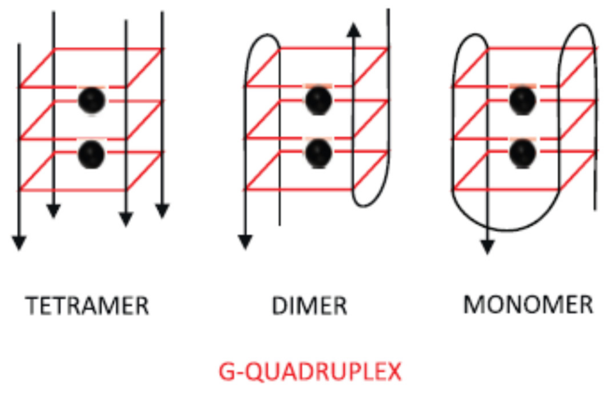
One well-known example is the antiparallel-G-quadruplex aptamer-target complex structure [22,23]. Guanine rich nucleic acids can self-assemble into four-stranded guanine (G)-quadruplex structures (Figure 2) [24]. This structure is based on base pairings of the Hoogsteen type forming a plateau of four guanine residues, also called a “quartet”. The parallel and uninterrupted stacking of at least two quartets, intercalated by a monovalent cation (sodium or potassium) stabilizing the structure, constitutes the G-quadruplex structure. Aptamers could recognize their target by means of three-dimensional folding of their chain.
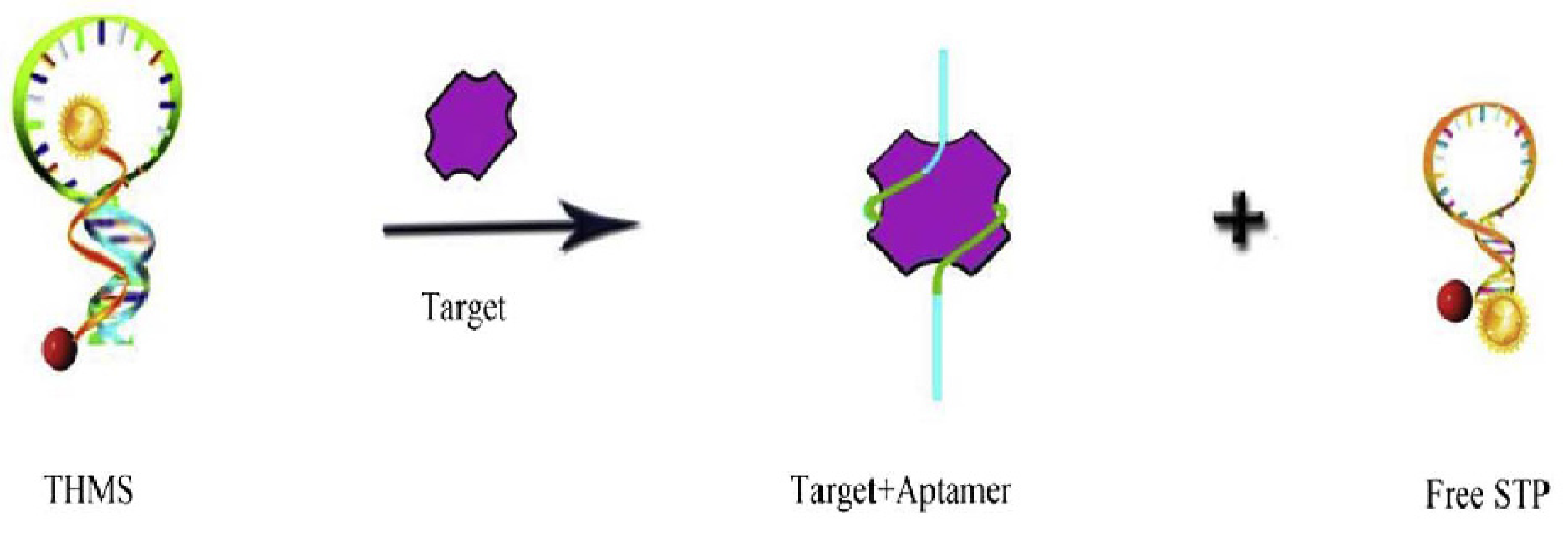
The Triple-helix molecular switch (THMS) that relies on the autonomous formation of DNA triplex can also be used for the development of aptasensors [25]. THMS is usually constituted of an aptamer sequence with two arm fragments or a DNA sequence that forms a hairpin structure in the presence of other parts of THMS, a sequence that is trapped between two arm fragments of aptamer sequence or DNA sequence, sometimes called signal transduction probe (STP) (Figure 3) [26]. The binding of the target to the aptamer modified the THMS structure. In this case, the conformation change was detected because the fluorophore and the quencher were close in the STP structure, while they were far from each other in the THMS structure. The fluorescence signal was quenched and therefore the signal decreased (“signal off” sensor). This THMS structure was less employed in the development of electrochemical sensors for the detection of antimicrobial residues than the G-Quadruplex structure. This structure was very much used to develop biosensors based on fluorescence. It lent itself better to this mode of detection because of the quenching effect.
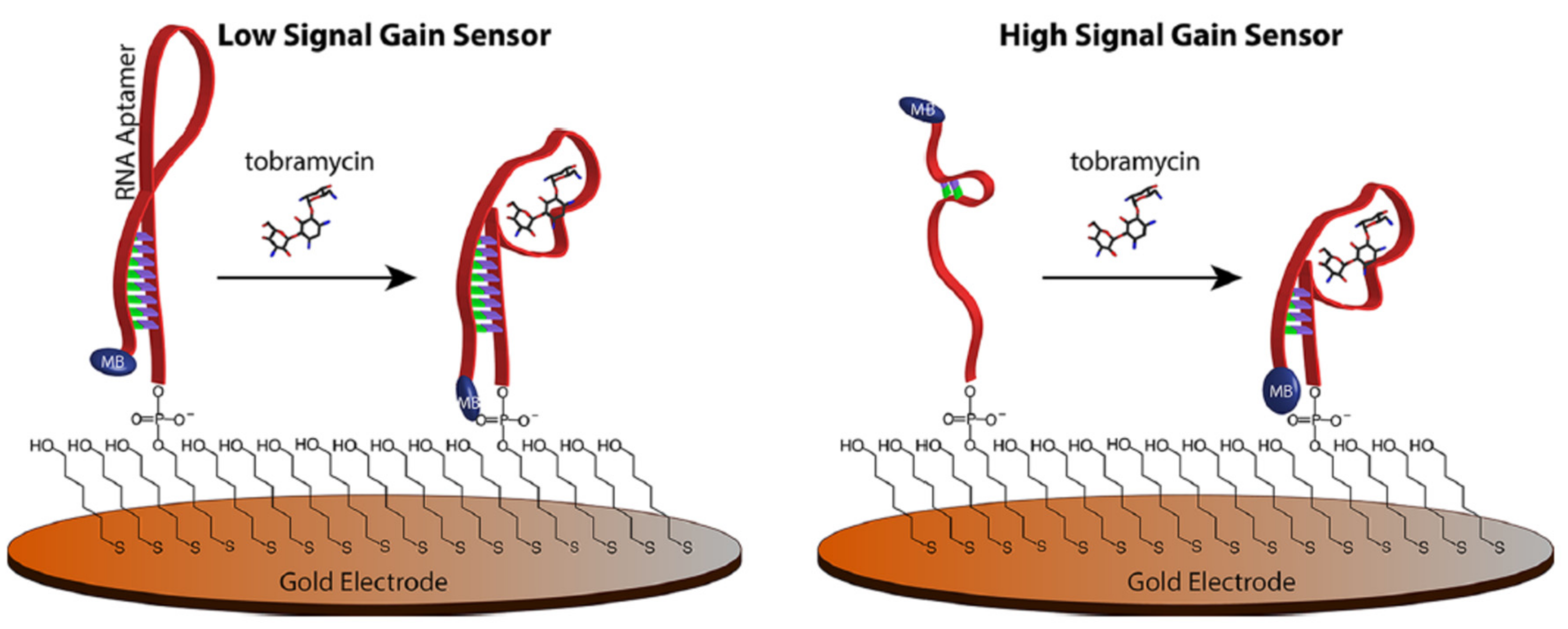
The initial conformation of the aptamer will influence the sensitivity of the developed aptasensor. Indeed, when one extremity of the aptamer is functionalized with a redox probe, the initial proximity (or the distance of the redox probe from the surface of the electrode) is determined by the aptamer conformation. Thus when the aptamer binds to the target, the distance gain of the redox probe with the electrode will be greater or less depending on the initial distance. Schoukroun-Barnes et al. developed strategies to better exploit the target binding-induced changes in aptamer conformation and/or flexibility (Figure 4) [27]. They rationally designed and tested three mutant (modified) aptamer sequences to support larger conformation changes. The structure of the parent aptamer sequence is a stem-loop structure, even in the absence of target. Therefore, the aptamer conformation change when binding to the target is relatively small. This observation may explain the lack of sensitivity of the aptasensors developed first with the parent RNA sequence. Therefore the authors modified the RNA sequence in order to destabilize their structure. Doing that they planned to increase the conformation change and therefore to improve the sensitivity of the developed aptasensors. The three modified aptamers were designed following two criteria: firstly bases involved in binding with the target were conserved; secondly the modified sequence should exhibit minimal secondary structure. The performance of the electrochemical aptasensors developed using the modified aptamers were compared with those obtained with the parent RNA aptamer sequence. With one of the modified aptamers (D2), the sensitivity was increased about 10 times.
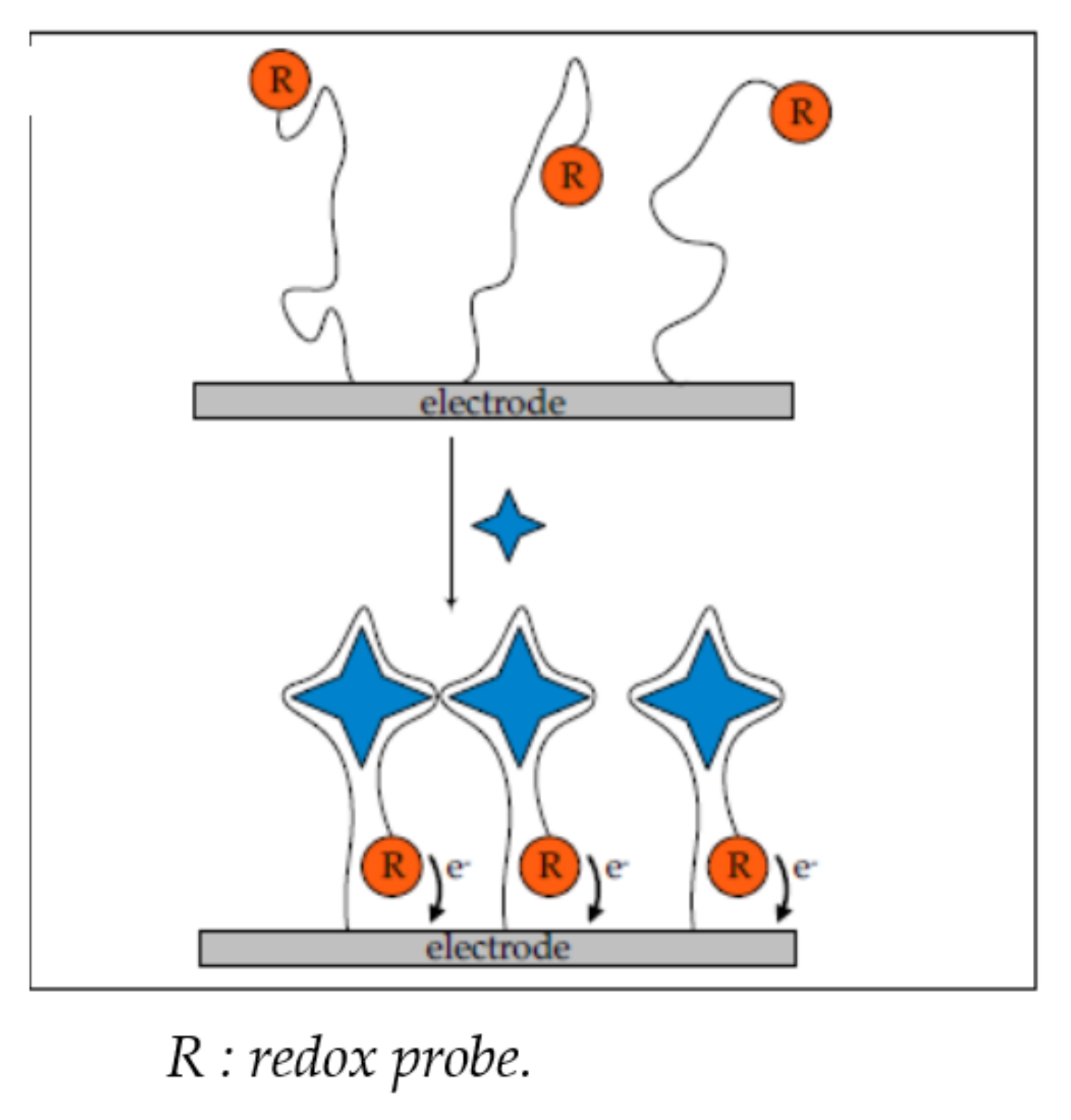
The conformation of a given aptamer is heavily affected by various factors, such as temperature, pH, type, and concentration of cations [21]. The change of structure or conformation (e.g., hairpin, pseudo-node) of the aptamer when it interacts with the target analyte can be electrochemically detected. The electrochemical detection is possible when the 3′ and 5′ extremities of the aptamer are labelled with electrochemical tags (e.g., methylene blue (MB), ferrocene)) [21]. This is a labelled technique that required the binding of redox probe to one aptamer’s extremity (Figure 5).
Another possibility is to introduce charge-bearing particles into solution like hexamine-ruthenium(III) ([Ru(NH3)6]3+) (RuHex) that is a redox cation which is attracted by the negatively charged phosphate backbone of aptamers [28].
The different aptasensors can also be categorized as signal on or signal off sensors. When the target analyte binds to the aptamer (grafted onto the electrode surface), the conformational change of the aptamer induces a decrease of the distance between the electroactive compounds and the electrode surface. Therefore the current is increased in the presence of the target analyte (“signal on” sensor) (Figure 5). For instance, Yu et al. developed an electrochemical aptasensor for the detection of penicillin G in milk [29] using MB as the electrochemical tag. Two different aptamers functionalized with MB were immobilized onto a gold electrode. The change of conformation of the aptamer in the presence of penicillin G induced an increase of the current measured by Square Wave Voltammetry (SWV) because the electrochemical tag was brought closer to the electrode surface by the conformation change (signal on). On the contrary, the change of aptamer conformation could increase the distance between the redox probe and the electrode surface. Therefore the signal is decreased, it is a “signal off” method [30].
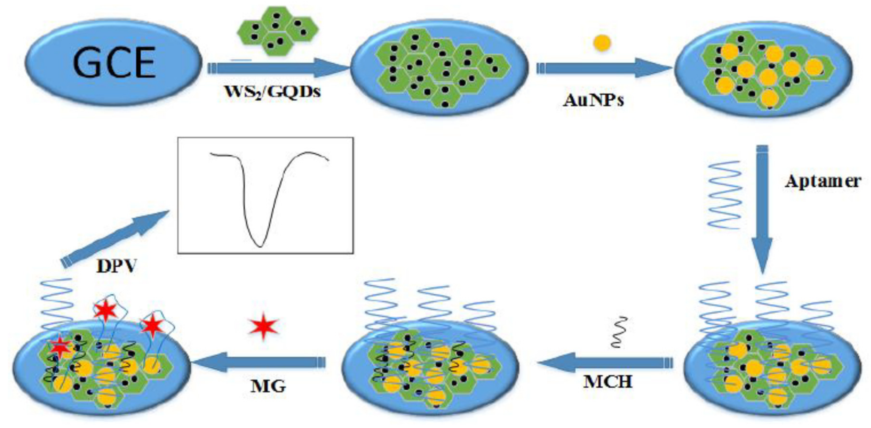
Techniques without labelling are also available because aptamers are polyanionic molecules. Aptamers possess intrinsic electrochemical properties that could be used for the detection. When the aptamer binds to its target analyte, the produced electrochemical signal is modified and is different from the signal of the free aptamer. The development of electrochemical aptasensors without labelling is of interest because there is no need to modify the aptamer sequence. Wang et al. developed a label free electrochemical aptasensors for the detection of malachite green [31] (Figure 6).
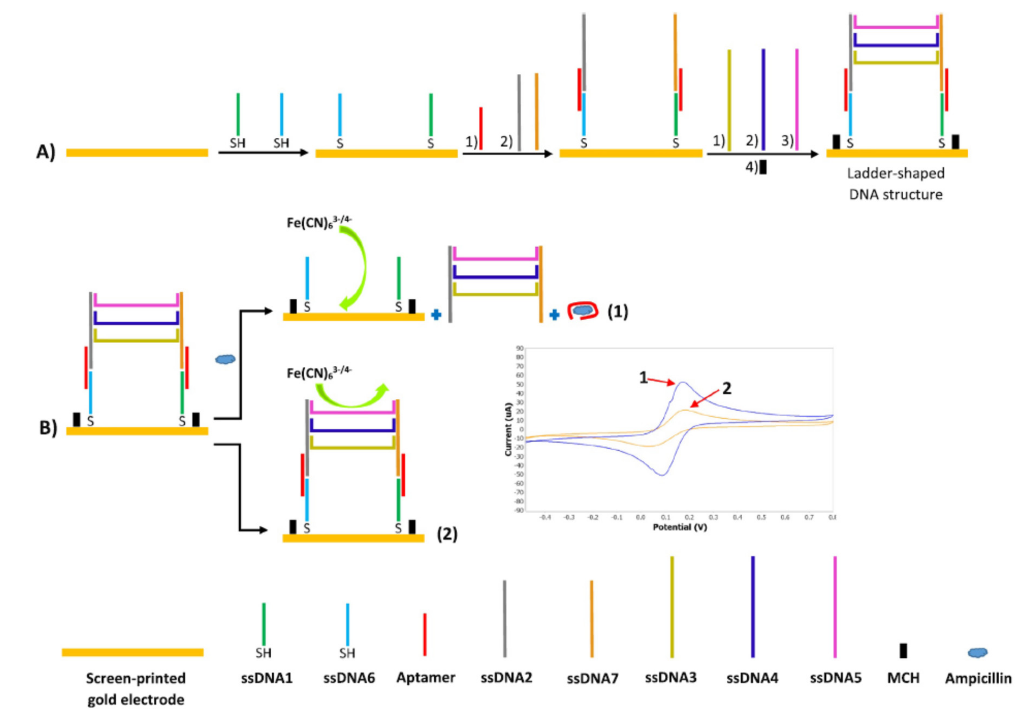
Taghdisi et al. developed a label-free electrochemical aptasensor based on ladder-shaped DNA structure for the detection of ampicillin in milk [32] (Figure 7). A ladder-shaped DNA structure composed of 6 ssDNA is formed onto the surface of a screen printed gold electrode (SPGE). One extremity of two of them is complementary with one part of the aptamer sequence. In the absence of the target, the ladder-shaped DNA structure remains intact because it is stabilized with the aptamer. The redox probe [Fe(CN6)]3−/4− is at distance of the electrode surface because of steric hindrance and electrostatic repulsion coming from the DNA structure. The electrochemical signal is therefore weak. In the presence of the target, the aptamer is released from the ladder-shaped DNA structure, which itself detaches from the electrode. Then [Fe(CN6)]3−/4− can come into contact with the electrode and the signal increases.
Chiorcea-Paquim and Oliveira-Brett reviewed the strategies developed for the fabrication of G-quadruplex-based biosensors with electrochemical detection [24]. Two strategies are presented: G-quadruplex aptasensors and hemin/G-quadruplex peroxidase-mimicking DNAzymes biosensors.
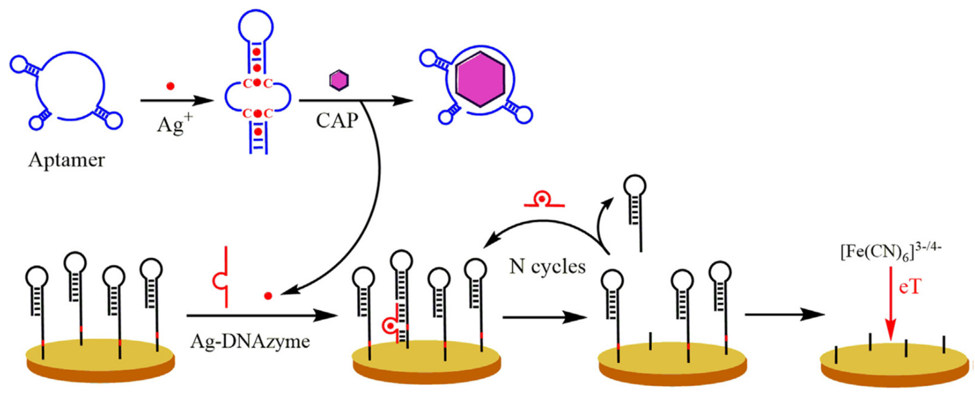
Hemin/G-quadruplex peroxidase-mimicking DNAzymes are constituted of DNA oligonucleotide that first adopted a G-quadruplex structure and then formed a complex with hemin. Hemin (ferric chloride heme) is an iron-containing porphyrin with chlorine. DNAzymes are a class of metalloenzymes that use DNA molecules exclusively for catalysis, and aptamers for binding the target. They are widely used for the development of aptasensors for various targets, especially because they are more stable than natural enzymes and because of their excellent catalytic activity [33]. DNAzymes were widely used to develop aptasensors for the detection of antimicrobials [34,35,36]. For instance Huang et al. developed an aptasensor for chloramphenicol (CAP), based on an Ag-DNAzyme [37] (Figure 8). Silver ions (Ag+) are bound to the C-C mismatches of the aptamer, inducing a change of conformation. In the presence of CAP, the conformation of the aptamer is modified and the Ag+ ions are released (the Ag-DNAzyme). The DNAzyme is activated in the presence of Ag+ ions and so the Ag-DNAzyme cleaved the substrate DNA immobilized on the electrode surface. Therefore the electrochemical response of [Fe(CN)6]3−/4− is increased. The signal increased proportionally to the CAP concentration. Furthermore the signal is amplified because the DNAzyme released after cleavage of one DNA is available to hybridize another DNA on the electrode and cleave it again.

2.1.2. Target-Induced Displacement

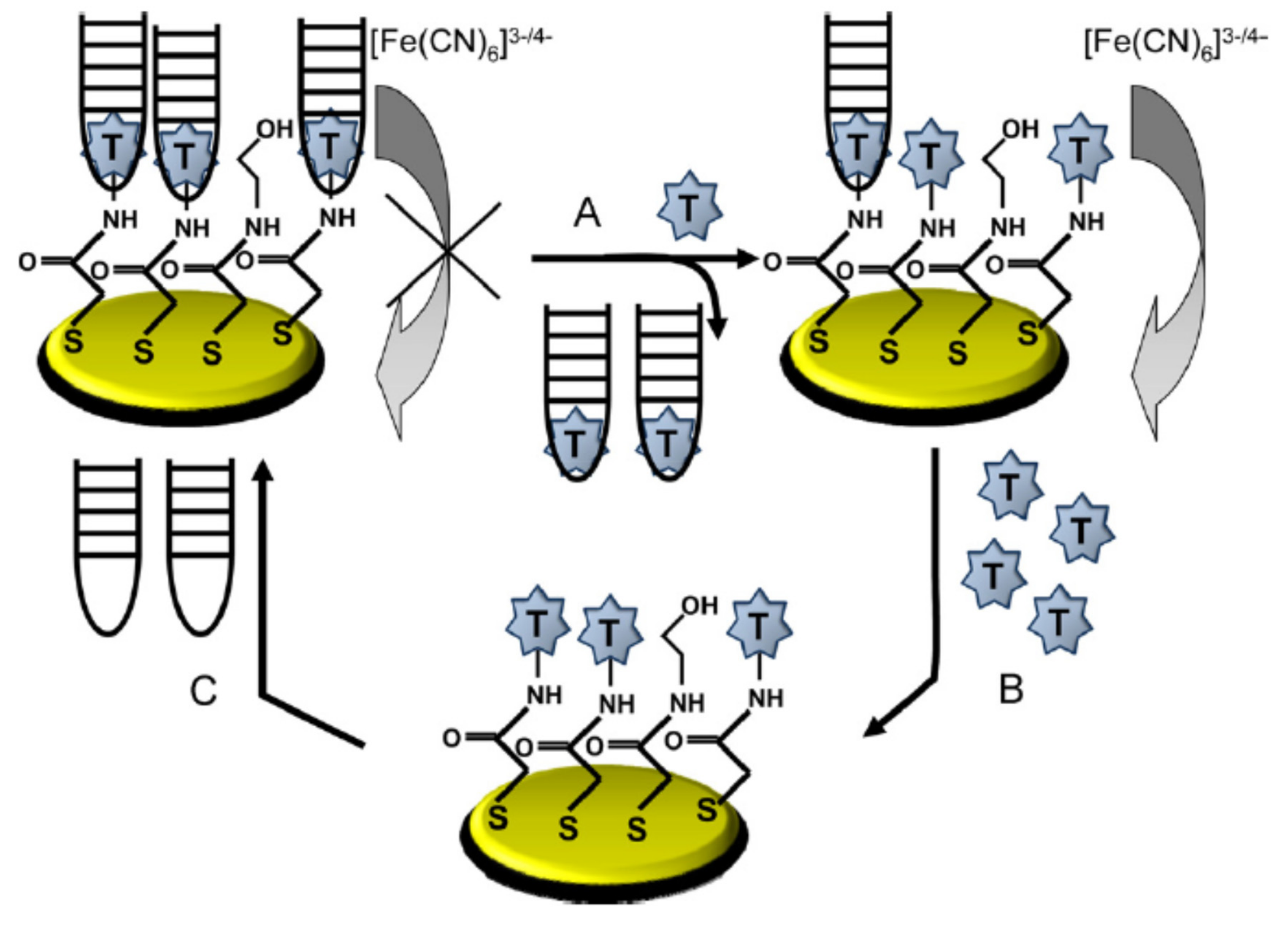
This method is independent of the aptamer change of conformation. An aptasensor based on direct target induced displacement was developed for the detection of tobramycin (Figure 9) [19]. The aptamer first binds to the target immobilized onto the electrode and then is displaced by the target present in the analyzed sample. Therefore less aptamers are binding to the electrode and therefore the overall signal tends to decrease (signal off). In this particular case, the aptamer is not functionalized with redox probe, so it is a non-labelled technique.
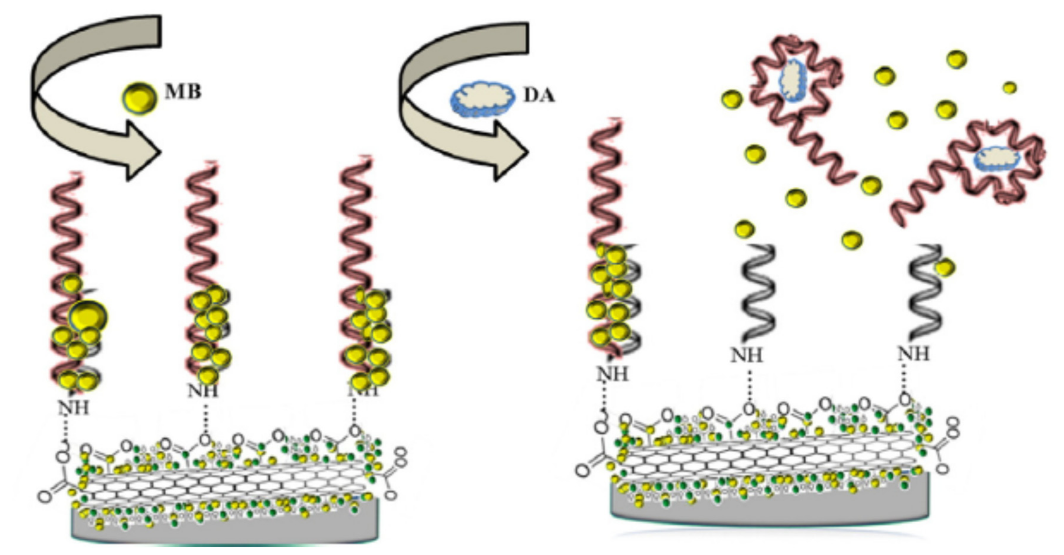
A different approach was based on a complementary DNA (cDNA) that was initially linked to the aptamer [30]. In this case, the target-induced displacement format was based on the displacement of the cDNA by the target. When the target was present, the cDNA was released because the affinity of the target to the aptamer was higher. Therefore the conformation of the aptamer was modified, the distance between the redox probe and the electrode was decreased and the current increased, it is a “signal on” method. This review illustrated an mode of amplification of the signal [30]. Indeed electroactive components (redox probe) were intercalated into the aptamer sequence bound to the cDNA. Therefore the current was amplified due to the presence of multiple redox probes. When the target is present, cDNA and redox probes were released. Consequently, the distance increased and the current decreased (signal off). The signal amplification is of utmost importance to improve the sensitivity of aptasensors for the detection of antimicrobial residues and to reach very low levels of analytes (i.e., µg/kg). Figure 10 illustrated the principle of target-induced aptamer displacement and the signal amplification by accumulation of the redox probe [38]. A gold electrode was modified with a nanocomposite containing gold nanoparticles, polyethylenimine and acid-oxidized carbon nanotubes (AuNPs/PEI/CNTs-COOH). An ssDNA was immobilized onto this nanocomposite modified electrode. Then the aptamer was added that hybridized with the cDNA. Then methylene blue (MB) was accumulated onto the electrode surface. When the target was added, the formed complex aptamer-target was released from the electrode surface also removing the accumulated MB. Therefore the signal decreased when the concentration of target increased.
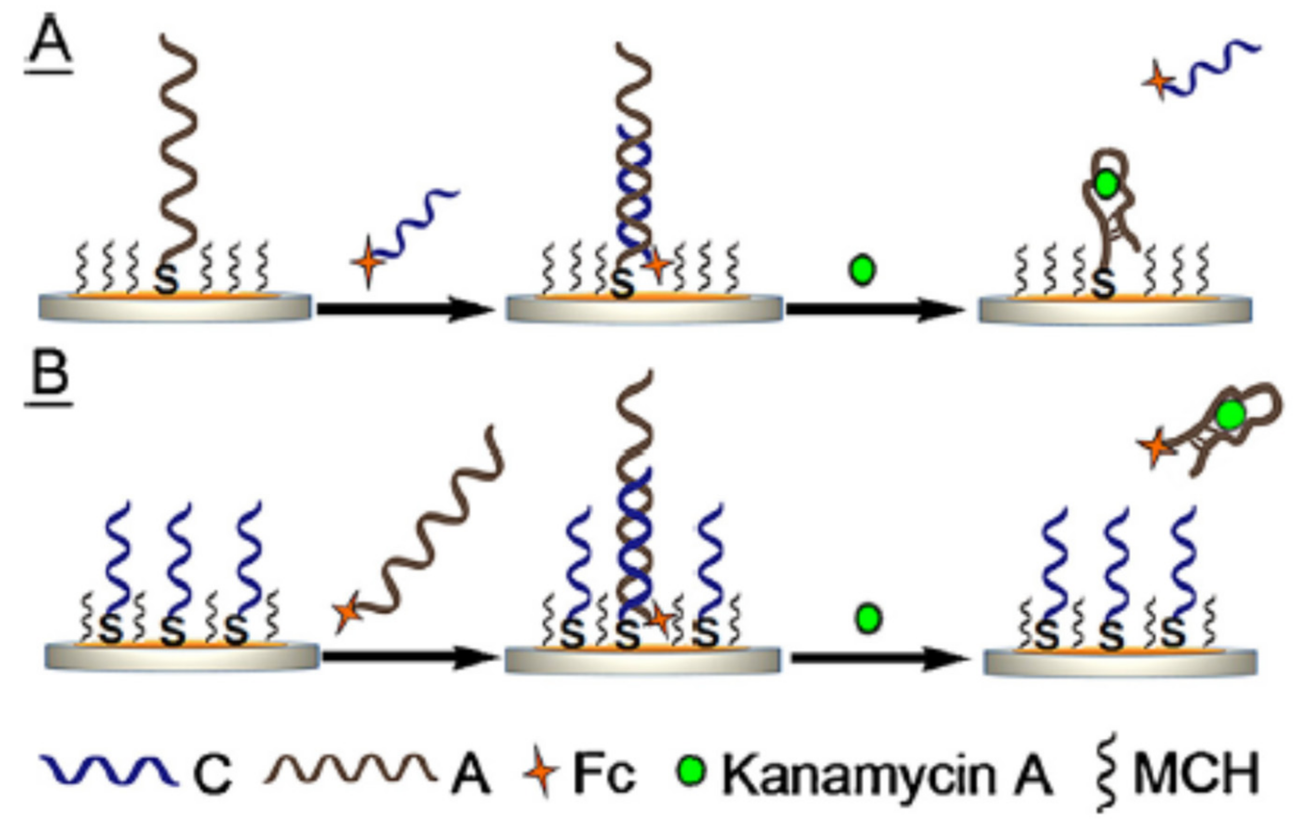
An electrochemical aptasensor was developed for the detection of kanamycin based on target-induced signaling probe shifting mechanism [39]. The complex capture probe (i.e. the aptamer) with the assistant probe was bound onto the surface of a gold electrode. The signaling probe was a cDNA modified at one extremity with a redox probe (Methylene blue (MB)) (labelled method). When the signaling probe was injected onto the electrode surface, the signal was weak (background signal) because the redox probe was far from the electrode. On the contrary when the target was added, the target bound to the capture probe that was released from the assistant probe. The assistant probe was then free to bind to the signaling probe that was also complementary with the assistant probe. The redox probe was then closer to the electrode surface and the signal increased (signal on method). A different strategy was implemented by Liu et al. Their electrochemical aptasensor for the detection of kanamycin A was based on signaling-probe displacement using two approaches (Figure 11) [40]. In the first approach, a thiolated aptamer was immobilized onto a gold electrode. Then a cDNA functionalized with ferrocene (Fc) as redox probe was added and bound to the aptamer. Fc was close to the surface and so the current signal was high. When the target was present in the sample, it bound to the aptamer and the Fc tagged cDNA was released. Therefore the signal decreased when the target was present (signal off method). The second approach was based on a thiolated cDNA immobilized onto the electrode. The aptamer functionalized with Fc was added and binds to the cDNA. When the target was present, it bound to the Fc tagged aptamer and therefore the complex aptamer-target was released. Again, the signal decreased in the presence of the target (signal off method). The sensitivity obtained with the first approach (aptamer acted as capture probe onto the electrode) was better than with the second one. The authors observed that the DNA probe density was higher with sensor B (second approach). Furthermore non-specific adsorption of kanamycin (positively charged in the buffer) on DNA probes (negatively charged) may be higher. Therefore the sensitivity decreased.
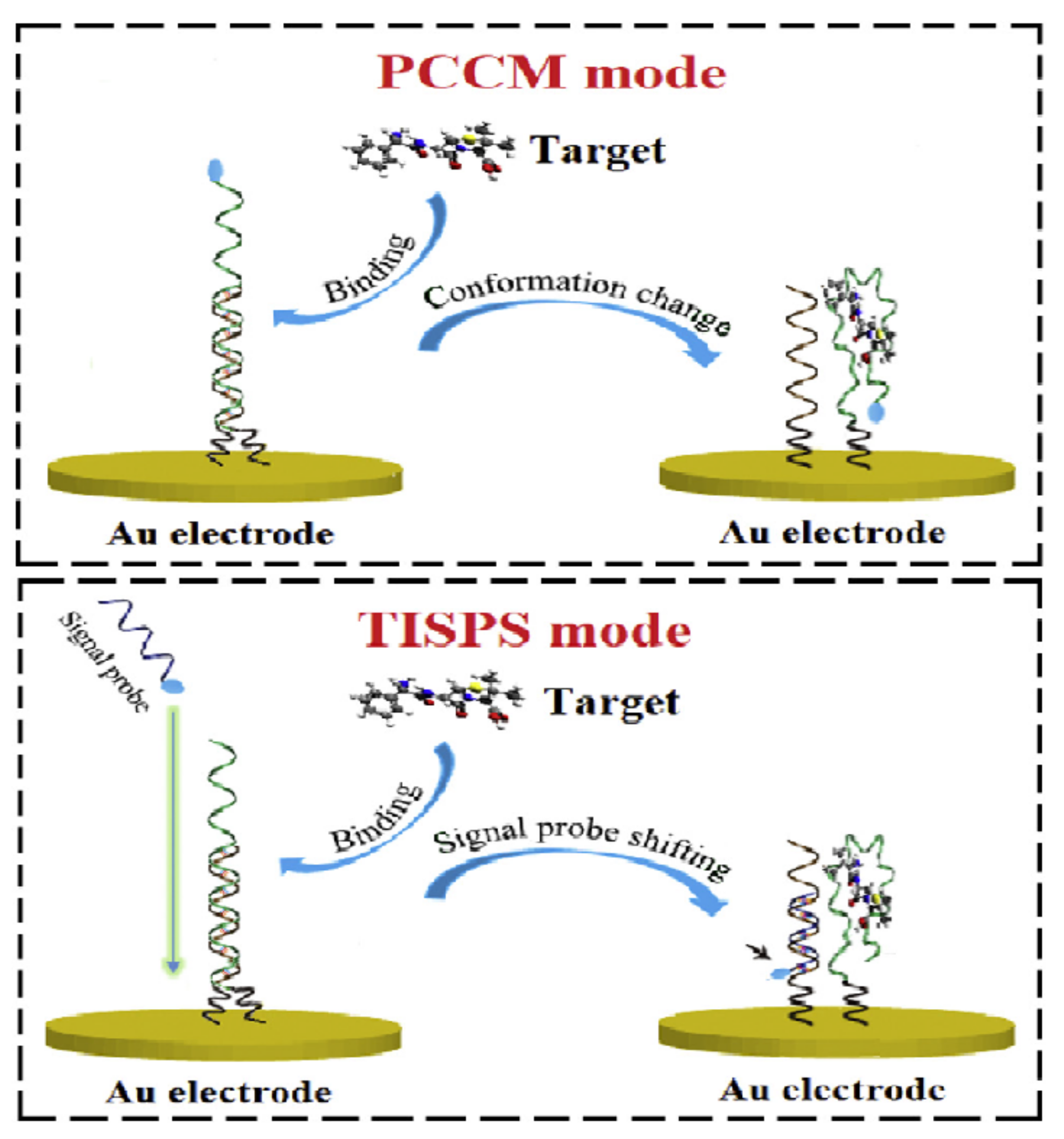
These two different signal transduction mechanisms (i.e., Conformation change and target-induced displacement) were employed to develop two electrochemical aptasensors for the detection of kanamycin (Figure 12) [41]. The first one was based on the conformation change of the aptamer-MB when the complex aptamer-target was formed (probe conformation changing mode (PCCM)). The aptamer was functionalized with MB as an electrochemical tag. Therefore this conformation change made the MB closer to the electrode surface and the signal was increased (signal on method). The second one was based on the target-induced signal probe shifting (TISPS). A signaling probe functionalized with MB was free in solution. When the target bound to the aptamer, the complex aptamer-cDNA immobilized onto the gold electrode was broken. The cDNA was free to bind to the signaling probe. Finally the redox probe MB was close to the electrode surface and the signal was increased (signal on method). The authors concluded that the sensitivity of the TISPS based sensor was better because the background current was weak. The electron transfer (ET) was lower with this sensor in the absence of the target because the signaling probe (MB) was in solution and far from the electrode surface. However when analyzing matrices (tap water and diluted milk), this sensor was slower and the anti-fouling properties ware reduced.

2.2. Different Immobilisation Techniques of Aptamers

In some cases, the first step of the aptasensor development consists in the immobilization of the aptamer onto the electrode surface (or other surfaces (e.g., Nanomaterials like gold NPs)). The sensitivity of the developed aptasensors will be influenced by the probe density on the electrode surface that means that an attention has to be paid on the concentration of aptamer used to fabricate sensing monolayers [27]. Therefore the immobilization of the aptamer is a crucial step. Aptamer sequences have to be modified to covalently immobilize them onto electrode surfaces. The aptamer is usually modified at the 5′-end for electrode grafting (e.g., thiolated) and at the 3′- end, with a redox probe (e.g., MB, Fc).
Oberhaus et al. recently published a review of immobilization techniques for aptamers on gold electrodes (2017–2019) [10]. The first approach consists of the direct immobilization of thiolated aptamers by covalent linkage. Thiolation involves in the introduction of the sulfhydryl (SH) group. There is a strong interaction between SH and gold that is used for the immobilization via Au–S bonds (covalent binding) and non-specific absorption. Thiol-containing molecules such as mercaptoethanol could be used in combination with the thiolated aptamers to remove non-specific binding of aptamers onto the electrode. Consequently MCH ensures the control of aptamer orientation in the upright position, due to electrostatic interactions, and so the accessibility of the immobilized aptamers [42].
Amino and thiolated aptamers are the most commonly used aptamers for the development of electrochemical aptasensors [43]. Comparative assays allowed concluding that the initial modification density of amino aptamers was higher. The produced electrochemical signal was higher and the sensitivity was better. However the stability of the amino modified aptamers was lower than the one of thiolated aptamers and these latter can be reused. Therefore thiolated aptamers were better candidates for the construction of electrochemical aptasensors.
One other possibility is to insert short linkers (e.g., alkyl chain, 3-mercaptopropionic acid (MPA) or poly(thymine) linker) between the aptamer and the surface. Furthermore, the streptavidin/biotin interaction can be used for the immobilization. This procedure usually required an initial functionalization of DNA (i.e., Biotinylated aptamer or cDNA) and of the electrode surface (or nanomaterials (e.g., AuNPs [44]) with streptavidin. The binding streptavidin/biotin is very strong and stable (one of the strongest non covalent bond known), because the affinity of biotin towards streptavidin is high. Finally, nanomaterials (e.g., reduced graphene oxide) may be used as immobilization platforms.
Aptamers can also be immobilized onto electrodes via a diazonium coupling mechanism based on electrografting [45]. Screen printed carbon electrodes (SPCE) were previously functionalized with 4-carboxyphenyl (4-CP) diazonium salt (4-COOH-Ar-N2+) that is the diazonium derivative of 4-aminobenzoic acid (4-ABA). Then the carboxyl group of 4-CP reacted with N-(3-dimethylaminopropyl)-N′-ethylcarbodiimide hydrochloride (EDC) and N-hydroxysuccinimide (NHS). NHS is a chemical reagent used to convert carboxyl groups to amine reactive NHS esters. EDC is utilized as a carboxyl-activating agent for amide bonding with primary amines. Finally the 3′ NH2 modified aptamer was bound covalently to 4-CP. The immobilization by diazotization is easy to perform, stable, and produced uniform surface modification. Furthermore the surfaces produced by this mechanism allowed to develop sensitive aptasensors [45]. The diazonium coupling mechanism can be used to immobilize aptamers on different materials (e.g., gold, carbon, carbon nanotubes).
Fouling substances consisted in interfering compounds that can be matrix compounds (i.e., Proteins) that could interfere with the electrochemical signal detection. Antifouling strategies for optical biosensors are commonly used, even based on high molecular weight molecule such as polyethylene glycol (PEG). Few antifouling strategies compatible with electrochemical measurements are available to reduce non-specific protein adsorption on the electrode surface. Oberhaus et al. have reviewed the such antifouling strategies compatible with electrochemical sensors [10]. Self-assembled monolayers of aromatic thiols (i.e., p-aminothiophenol (p-ATP) and p-mercaptobenzoic acid (p-MBA)) that improved electron transfer could be used to immobilize aptamers on the electrode surface. The immobilization with a high surface density allows lower detection limits. Furthermore this scheme of immobilization gives a very low non-specific binding and a lower background signal.

2.3. Nanomaterials

Nowadays, nanotechnologies and nanomaterials (NMs) in particular are essential to the improvement of the performance of biosensors and especially in the case of electrochemical biosensors [46]. Electrochemical biosensors could be categorized according to the different nature of the nanoparticles used.
The classification of nanomaterials could be based on various criteria. Nanomaterials could be classified as per the size and dimensions. There are four types of nanomaterials such as zero dimension (e.g., Gold (Au) and silver (Ag) nanoparticles (NPs), Quantum Dots (QDs)) (1–50 nm), one dimension (e.g., metals (Au, Ag), metal oxides (ZnO, TiO2), QDs) (1–100 nm), two dimensions (2D) (e.g., thin-films, thin-film multilayers, nanosheets), and three dimensions (3D) [47].
They could be also classified as per composition: carbon nanomaterials (e.g., carbon nanotubes (CNT), graphene, graphene oxide (GO)), metallic (e.g., gold and silver NPs) and non-metallic nanoparticles (e.g., SiO2 NMs), metal compound nanomaterials (e.g., QDs).
Nanomaterials involved in the development of aptasensors could have different functions: carrier, enhancer, reporter, catalyst (nanoenzyme like), quencher, separator (Table 1) [48,49]. Carrier and enhancer NMs could be used in electrochemical aptasensors to amplify the signal because of large surface area, high surface-to-volume ratio, favorable electronic properties, and electrocatalytic activity. The immobilization of NMs onto the electrode surface could amplify the signal by increasing the surface of interaction. Another possibility to amplify the signal is to use NMs immobilized with signaling materials (e.g., ferrocene). NMs act as catalyst when they exhibit enzyme like activities, for instance Horseradish peroxidase (HRP) like activity (e.g., noble metal NPs, metallic oxide NPs and composite NPs). This characteristic could be used to develop sensitive electrochemical aptasensors because these NMs can catalyze the degradation of hydrogen peroxide. An electrochemical reporter NM is used as a signal molecule (signal probe) (e.g., Metal NPs, metallic oxide NPs and QDs). Quencher NMs (e.g., AuNPs) can be used in aptasensors based on electrogenerated chemiluminescence (ECL) quenching. The main NMS acting as separator are magnetic nanoparticles (MNPs). They are used to extract a target analyte from a complex matrix that is the case for food products. These NMs are used to remove or at least decrease matrix effects due to the presence of many constituents in the matrix. As it is shown in Table 1, each type of NMs can have several properties that can be used simultaneously in the development of a biosensor (e.g., Carrier and enhancer, carrier and catalyst).
Different types of nanomaterials can be employed for the development of analytical methods for biological and chemical contaminants in food products, especially Carbon nanotubes (CNTs) based and graphene based electrochemical sensors [50]. Single wall CNT (SWCNT), multi-wall CNTs (MWCNT) or graphene functionalized with an aptamer and deposited on an electrode enhance the electrochemical changes in the presence of a specific target analyte. Nanocomposites (e.g., AuNPs composited with polymers) can also be used to increase electrochemical efficiency [51].
The characteristics and the performance of electrochemical and electrochemically derived aptasensors for the detection of antimicrobial residues in food products were presented in Table 2 and Table 3. This review of applications of electrochemical aptasensors for the detection of antimicrobial residues in animal derived food products was presented in relation to the different classes of NMs. As noticed in Table 2 and Table 3 the great majority of aptasensors are now developed based on nanomaterials.

2.3.1. Carbon Nanomaterials (e.g., Carbon Nanotubes (CNT), Graphene, Graphene Oxide (GO))

Carbon nanotubes (CNTs) are sheets of graphene rolled into a cylinder. Graphene is a one-atom thick layer that is arranged into a honeycomb lattice structure. CNTs are divided into Single Wall carbon nanotubes (SWCNTs) made from one layer of carbon atoms and Multi-walled carbon nanotubes (MWCNTs) consisting of several layers. MWCNTs are formed from the folding of graphene layers into carbon cylinders [46]. Graphene oxide (GO) is a derivative of graphene that is produced by chemical oxidation of graphite. Reduced graphene oxide (rGO) is obtained from GO through elimination of some oxygen containing groups.
Carbon nanomaterials properties allow to improve the performance of electrochemical aptasensors [132]. CNTs are characterized by good electrical conductivity, large surface area and multiple functionalization possibilities. The large surface area is interesting to immobilize a large amount of aptamers, that influenced the sensitivity of the aptasensor. The good electrical conductivity is exploited to increase the signal (i.e., current). This is also favorable to reach low limits of detection. These characteristics allowed to develop sensitive, easy to prepare, cost-effective and stable electrochemical aptasensors.
A SWCNT based aptasensor was developed for the detection of oxytetracycline in water samples [97]. A gold electrode surface was pre-grafted with SWCNTs and then the probe DNA (complementary to part of the aptamer sequence) was immobilized onto SWCNTs surface. A competition is performed by mixing samples with the aptamer that are injected onto the electrode surface. When no oxytetracycline is present in the sample, the aptamers are free to bind to the DNA-probe on the surface. Therefore the production of current is high. On the contrary when oxytetracycline is present in the sample, less aptamers are free to bind to the DNA-probes surface and so the current signal is decreased (signal off). The authors highlighted the main advantage of this sensor compared to other SWCNTs assembled sensors that is the controlled and uniform assembly of the SWCNTs at the desired locations. The electrode is more stable and can be regenerated by removing the aptamers and reused (over 20 times).
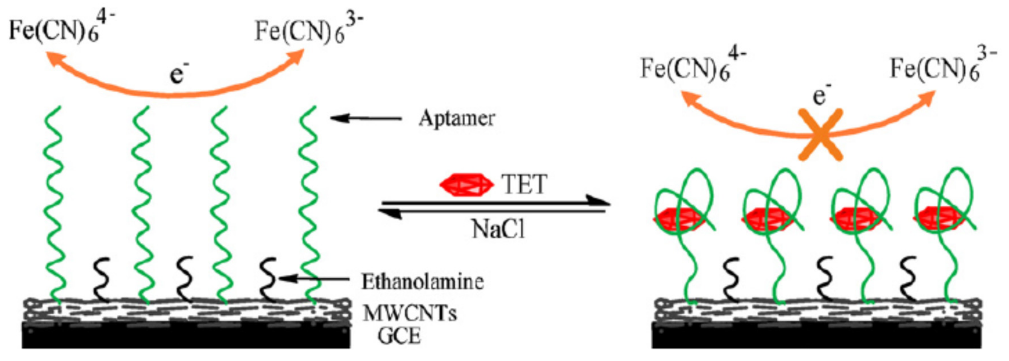
Zhou et al. developed an electrochemical aptasensor for the detection of tetracycline in milk (Figure 13) based on MWCNTs-modified electrodes [133]. A glassy carbon electrode (GCE) was first functionalized with MWCNTs. Non-specific binding was removed by immobilising ethanolamine onto the electrode surface. The aptamer is immobilized onto the MWCNTs electrode surface. When there is no tetracycline in the sample, the electron transfer occurred due to the presence of the redox probe Fe(CN)63−/Fe(CN)64−. The signal measured by DPV is high. When the sample contained tetracycline, its binding to the aptamer induced a change of conformation of the aptamer. Therefore the electron transfer is prevented due to the conformation change (label free signal-off method). The homogeneous immobilization of MWCNTs allowed us to increase the electrode conductivity and the loading amount of aptamers, therefore producing higher currents and to improve the sensor sensitivity. Furthermore the immobilization of aptamers onto this surface is so stable that the electrode can be reused several times.
Among the carbon NMs, we have observed in the literature review that electrochemical aptasensors were more currently developed using MWCNTs than SWCNTs for the detection of antimicrobials (Table 2 and Table 3). Furthermore graphene oxide (GO) were also commonly used for the modification of electrodes [65,67,127,134]. An electrochemical aptasensor was developed for the detection of sulfadimethoxine in milk [73]. A signal probe functionalized with AuNPs decorated C60-rGOTb (C60-rGO-Tb-AuNPs) is used as the signal tracer. Toluidine blue (Tb) is the redox probe A gold electrode was immobilized with the aptamer and a capture probe (cDNA). When sulfadimethoxine was present in a sample, the aptamer was released from the electrode surface. Then the capture probe was free to bind with the signaling probe functionalized with C60-rGO-Tb-AuNPs. The current increased proportionally to the sulfadimethoxine concentration. The signal was amplified due to addition of an enzyme (RecJf exonuclease) that digested the aptamer when bound to the target. Therefore the target is again free to interact with aptamers on the surface of the electrodes and a new cycle started.

2.3.2. Metallic Nanomaterials (e.g., Gold NPs, Silver NPs)

Metallic nanomaterials are essentially gold (Au) and silver (Ag) nanoparticles (NPs) that are often used as tracers (enhancer, reporter effects) in electrochemical aptasensors. Furthermore, metallic nanoparticles (MNPs) could be used as separators due to their magnetic properties (magnetics beads (MBs)). Magnetic nanoparticles (MNPs) or Magnetic Beads (MBs) are metal compound nanomaterials that could be used as separators when the binding element such as an aptamer is immobilized onto the surface of the MBs [135]. Ferrous (Fe3O4 NPs) and ferric oxide (Fe2O3 NPs) are the most common and widely used magnetic nanoparticles (MNPs).
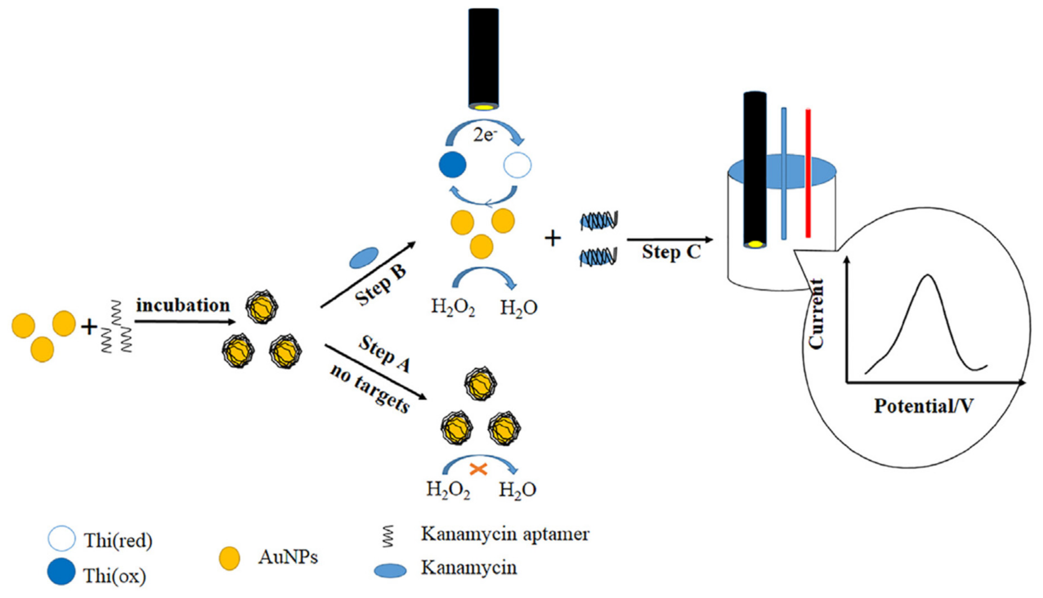
AuNPs and Platinum (Pt)NPs could serve as catalysts, respectively due to their mimicking peroxidase activity and catechol oxidase-like activity. The peroxidase-like activity of gold nanoparticles (AuNPs) was used in the development of an electrochemical aptasensor for the detection of kanamycin in honey (Figure 14) [136]. AuNPs are characterized by large surface area to volume ratio, good electrical properties, high surface reaction activity, small particle size and good surface properties [46]. Furthermore, AuNPs have a mimetic peroxidase activity. The aptamer anti-kanamycin was adsorbed onto AuNPs by electrostatic interactions that blocked the catalytic sites. Therefore in the absence of kanamycin, the peroxidase-like activity of AuNPs was inhibited. When kanamycin was present in a sample, the aptamer bound to the target and was released from the AuNPs. Therefore the peroxidase-like activity of AuNPs was recovered. The reduction of H2O2 by AuNPs generated an electron transfer that produced oxidized thionine (Thi). A current was produced that could be measured by LSV. This aptasensor was based on the target-induced displacement of the aptamer.
An electrochemical aptasensor for the detection of chloramphenicol (CAP) in milk was developed using silver nanoparticles (AgNPs) [53]. A GCE was modified with a combination of rGO and AgNPs (GCE@Gr/AgNPs). Then aptamers were immobilized onto this modified surface, with 6-mercapto-1-hexanol (MCH) as blocking reagent to prevent non-specific binding. In the presence of CAP, the aptamer captured its target at the electrode surface. CAP was reduced by the nanocomposite rGO-AgNPs and thus an electrochemical signal measured by linear sweep voltammetry (LSV) was produced (signal on).

2.3.3. Metal Compound Nanomaterials (e.g., QDs, UCNPs, MOFs)

Quantum dots (QDs) are semiconductor nanocrystals that are constituted of a metal (e.g., Pb, Cd, Zn, Cu) and sulfur S (Na2S). They act as signal tags by exploiting the oxidation potentials of the associated metal ions ion (e.g., Pb2+, Cd2+, Zn2+,Cu2+) in the construction of electrochemical aptasensors. Various QDs can be synthetized with different chemical compositions and surface functionalization possibilities.
Three different Quantum dots (QDs) were used to develop an electrochemical aptasensor for the multiplex detection of streptomycin, chloramphenicol and tetracycline in milk [90]. Three capture DNAs (capt-DNAs), each one complementary of one part of three cDNA1s, were immobilized onto the surface of a gold electrode. Then the three aptamers (Ap-DNAs) and the three cDNA1s, each one complementary of one part of one Ap-DNA were mixed and injected onto the electrode surface. The three cDNA1 were also complementary of part of the capt-DNA. When one of the target analytes is present in a sample, the corresponding aptamer bound to the target, thus the cDNA1 was free to hybridize with the corresponding capt-DNA. Three cDNA2 labelled each with one QDs (i.e. PbS, CdS, and ZnS) complementary with a third part of the cDNA1 (different part compared to capt-DNAs) were added. Thus in the presence of one of the target, the quantity of QDs present onto the surface was high and the signal measured by SWASV (square wave anodic stripping voltammetry) was increased (signal on). The dissolution of QDs tags with nitric acid (HNO3) that released Pb2+, Cd2+ and Zn2+ induced the electrochemical signal. The three QDs were different, made of PbS, CdS, and ZnS that gave signals at different potentials (respectively −0.6V, −0.8 and −1.1 V). Therefore the aptasensor could identify which target was present. When none of the targets was present, no QDs could be immobilized onto the electrode surface, so that the signal was low. The multiplex potential of this electrochemical aptasensor was highly interesting. Moreover the detection limits reported were low: 10 nM (5.8 ng/mL), 5 nM (1.6 ng/mL) and 20 nM (8.8 ng/mL) respectively for streptomycin, CAP and tetracycline. It has to be noticed that the LOD of CAP is too high compared to the EU regulatory limit (0.3 ng/mL).
Upconversion nanoparticles (UCNP) can be produced with different surface ligands as well as variable emission range. UCNPs are able to convert near infrared radiations with lower energy into visible radiations with higher energy via a nonlinear optical process. The surface of upconverting nanoparticles could be modified (e.g., amine functionalized) to immobilize antibodies or aptamers. No applications were found of using UCNPs to develop electrochemical aptasensor for the detection of antimicrobials in food products.
Most of the works published on the development of electrochemical aptasensors for the detection of antimicrobials in food products are based on Metal Organic frameworks (MOFs). MOFs are consisting of metal nodes coordinated to organic ligands to form one-, two-, or three-dimensional structures. MOFs are especially attractive for use as catalysts. An impedimetric aptasensor based on a Ce-MOF@MCA nanocomposite was developed for the detection of oxytetracycline [96]. MCA was formed by the reaction of cyanuric acid with melamine that formed a porous organic framework nanomaterial. Then a Ce-based metal organic framework (Ce-MOF)@MCA was produced. A gold electrode (AuE) was pre-grafted with this Ce-MOF@MCA nanocomposite onto which the aptamer was immobilized. In the presence of the target, the complex aptamer-target was formed inducing a conformation change of the aptamer. This conformation change prevented the redox probes to come close to the electrode surface so the signal measured by electrochemical impedance spectroscopy decreased (signal off).
A multiplex electrochemical aptasensor based on two different MOFs (PbS, CdS) was developed for the detection of kanamycin and chloramphenicol in milk [86]. The two aptamers were immobilized onto magnetic beads (MBs). Then two complementary DNAs (cDNAs) tagged with MOFs were hybridized, one with the aptamer anti-kanamycin and the other one with the aptamer anti-CAP (one PbS, the other one CdS). When one of the target was present, the aptamer-target complex was formed onto the MB and the cDNA-tagged MOF was released in the supernatant. The supernatant was deposited onto a glassy carbon electrode (GCE). Therefore when one of the target was present, the quantity of NMOFs released in the supernatant and the electrochemical signal of the corresponding ion (Pb2+ or Cd2+) was high (signal on). On the contrary when no targets were present, the signals were weak because no MOF was released in the supernatant. Each MOF was specific for the detection of one of the two targets. Therefore the identification of the analyte was possible.
He et al. developed an impedimetric aptasensor for the detection of penicillin in milk [95]. Two different Ag(I) metal−organic frameworks (Ag-MOFs) were prepared based on two counter anions (SiF62− and CH3SO3). A gold electrode was coated with the aptamer and with Ag-MOFs. In the presence of the target, the complex aptamer-target is formed. When the concentration of penicillin increased, the impedance of electron transfer of [Fe(CN)6]3−/4− was enhanced. The Ag2SiF6-MOF-based aptasensor was found to be more sensitive (LOD 0.849 pg/mL) and selective than the Ag2CH3SO3-MOF one.
Different kinds of metal oxide nanomaterials, such as TiO2, Fe2O3, Al2O3, ZnO, andSiO2 are used for the development of electrochemical aptasensors. A photoelectrochemical (PEC) aptasensor was developed for the detection of ofloxacin in milk based on the combination of TiO2 nanotube array (NTA) with Ag2S nanoparticles [118]. The synergistic effect of both NMs produced a better photoactive material characterized by a wider absorption range of visible light. The polydopamine (PDA) film reinforced the co-sensitization of TiO2 composites. The PDA film was able to immobilize more aptamers onto the surface. The aptamer was then immobilized onto the modified surface with 6-mercapto-1-hexanol (MCH) to remove non-specific binding. The PEC signal enhancement of this aptasensor was caused by the combination of an increase of the quantity of immobilized aptamers and an enhanced visible-light PEC activity. The PEC signal decreased in the presence of ofloxacin in a sample (signal-off).

2.3.4. Conductive Polymers

Polymers are organic macromolecules, composed by repeated entities, called mer. Conductive polymers are organic polymers that conduct electricity (e.g., Polyaniline, polyacetylene, polypyrrole). The conductivity of a conductive polymer is increased when the concentration of the charged dopant increased. Polyaniline is one of the most promising conductive polymers.
A label-free multiplex electrochemical aptasensor was developed for the detection of ampicillin and kanamycin based on poly(3,4-ethylenedioxythiophene (PEDOT) as the conductive polymer [137]. The aptamer was covalently immobilized to the polymer film. PEDOT is stable in phosphate buffers, and highly conductive. The developed aptasensor was able to detect ampicillin in milk below its regulatory limit (4 ng/mL). Zhao et al. developed an electrochemical aptasensor for the detection of penicillin based on a PEDOT-AuNPs composite [79]. The detection limit was low (0.057 ng/mL).
A Carbon Black (CB)-oligolactide composite was used to develop an impedimetric aptasensor for the detection of kanamycin in milk [94]. A GCE was first co-immobilized with carbon black and chitosan. Chitosan is a natural biocompatible polyoxide that could be combined with other conductive polymers like polypyrrole or with other NMs. In this case, CB was added into the chitosan matrix to improve the aptasensor performance because of its high surface-to-volume ratio and electrochemical properties. Then the covalent immobilization of the aptamer using EDC/NHS was reliable due to this porous CS-CB layer. In the presence of streptomycin, the charge transfer resistance decreased (signal off). The limit of detection was 0.3 nM.

2.3.5. Combination of Several Nanomaterials

Increasing number of electrochemical aptasensors for the detection of antimicrobial residues in animal derived food products use a combination of several nanomaterials to potentiate their effects.
A nanoporous PtTi/graphene oxide-Fe3O4/MWCNT-Fe3O4 nanocomposite was used to develop a label-free aptasensor for the detection and quantification of penicillin (Figure 15) [65]. This aptasensor was based on a combination of several carbon and metal-based nanomaterials to produce a synergistic effect of all the materials. This combination enhanced the signal intensity due to the combined NMs properties and increased the immobilization rate of aptamers. The authors assumed that this synergistic effect would increase the sensitivity of the sensor compared to a sensor using a single NM, which was demonstrated in this study (detection limit 25.3 pg/mL). A concentration as low as 0.05 ng/mL of penicillin was detected in a milk sample, while the regulatory limit (Maximum Residue Limit (MRL)) was set at 4 ng/mL. The first step of the fabrication of the aptasensor consisted in the immobilization of a nanocomposite material consisted of graphene and iron oxide (magnetite) (GR-Fe3O4) onto the GCE surface. Then a second layer of multi-walled carbon nanotubes and iron oxide (MWCNTs-Fe3O4) was grafted. A third layer of nanoporous platinum and titanium (NP-PtTi) alloy was deposited. The aptamer anti-penicillin was immobilized onto the modified GCE. Finally Bovine serum albumin (BAS) was added to the surface to remove non-specific binding effects. In the presence of penicillin in a sample injected onto the electrode surface, the target bound to the aptamer. The conformation change was responsible for a decrease of the current signal that was measured by DPV.
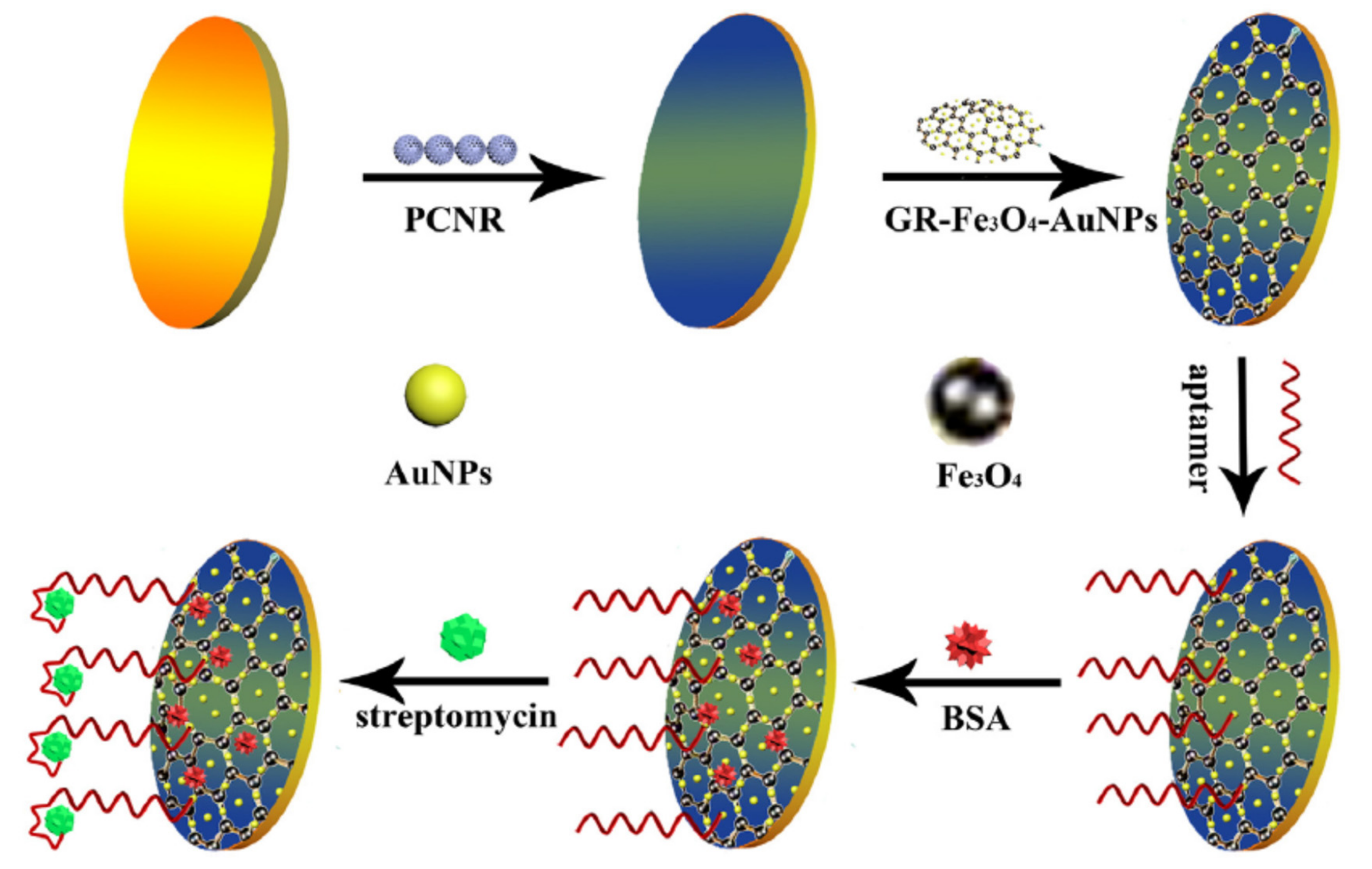
An electrochemical aptasensor was fabricated based on a combination of nanomaterials for signal amplification, porous carbon nanorods (PCNR) and multifunctional graphene nanocomposites (GR–Fe3O4–AuNPs) (Figure 16) [77]. The first step was the immobilization of a PCNR layer, followed by an assembly of graphene nanocomposites and gold nanoparticles. The role of the first layer was to increase the surface area for aptamer immobilization. The second layer function was to improve the current signal due to the electrocatalytic characteristics of graphene nanocomposites (GR–Fe3O4) and AuNPs. The high electron transfer capability of both nanomaterials was potentiated in the construction of this aptasensor. The aptamer anti-streptomycin was immobilized onto these layers, in combination with BSA that reduced the non-specific binding. The aptamer hindered electron transfer between the redox probe [Fe(CN)6]3−/4− in solution and the electrode. In the absence of streptomycin, the aptamer remained free and the current signal was high. When streptomycin was present in a milk sample, it bound to the aptamer that changed of conformation. The electron transfer was much more hindered than without streptomycin. The current signal decreased when streptomycin concentration increased.
A photoelectrochemical (PEC) aptasensor based on a ternary composite structure was developed for the detection of kanamycin in milk [122]. The heterostructure was formed with 3D flower-like TiO2, MoS2 and gold nanoparticles. The authors reported similar improvements of the NMS composite to the aptasensor performance characteristics: acceleration of the electron transfer, increase of the amount of immobilized aptamers and improvement of the visible light excitation. The detection limit was 0.05 nM of kanamycin.
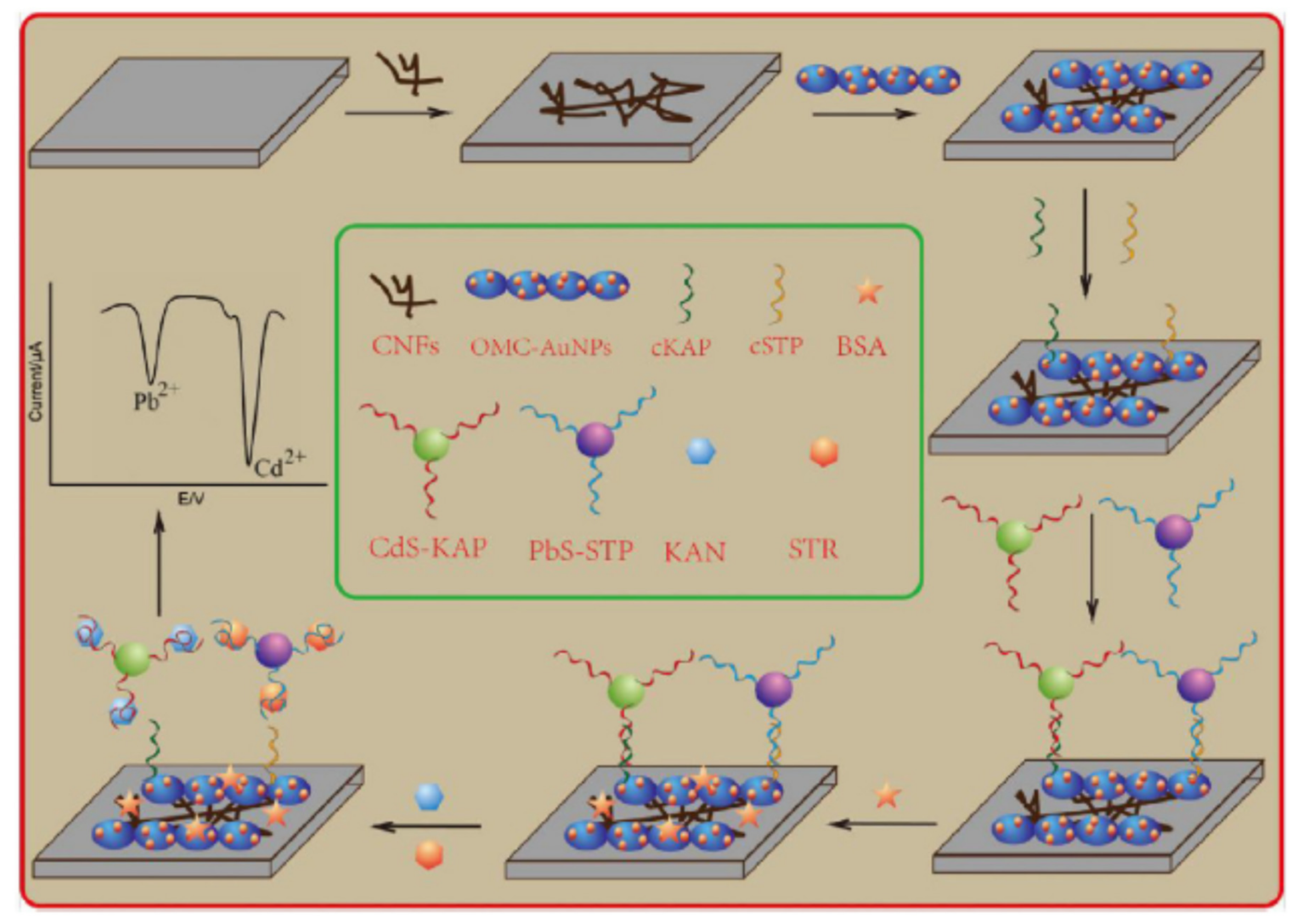
Carbon nanofibers (CNFs) and mesoporous carbon-gold nanoparticles (OMC-AuNPs) were used to promote the electron transfer in the development of an electrochemical aptasensor for the multiplex detection of kanamycin (KAN) and streptomycin (STR) in milk (Figure 17) [138]. CNFs and OMC-AuNPs were successively grafted onto a SPCE. Two different complementary sDNA probes were immobilized onto the modified SPCE. Then the two aptamers tagged with QDs (CdS Apt-KAN and PbS Apt-STR) hybridized with the cDNA probes on the electrode surface. When one of the target was present, the complex aptamer-target was released far from the electrode and the cDNA remained immobilized. The Cd and Pb stripping peak intensities were correlated to the concentration of kanamycin and streptomycin respectively.
A pencil graphite electrode (PGE) was modified with reduced graphene oxide (rGO) and AuNPs to develop an electrochemical aptasensor for the detection of sulfadimethoxine in fish [99]. The aptamer was immobilized onto the modified electrode. When the target was present, the complex aptamer-target increased the value of charge transfer resistance (Rct). The limit of detection was 3.7 × 10⁻¹⁶M. After fish sample preparation, the lowest detected concentration in the matrix was 1.10−10 M.

2.4. Different Amplification Techniques

For the detection of residual levels as it is the case for antimicrobials residues in food products, signal amplification is often necessary. Nanomaterials as it was explained before greatly contributed to signal amplification. But there are other techniques available for signal amplification.
Aptamers are flexible for designing different biosensors because they can be replicated or cleaved with various DNA enzymes for signal amplification (e.g., Exonucleases, polymerases). Some amplification techniques are based on enzymes able to digest DNA that are called nucleases or exonucleases. Different enzymes could be used to improve the sensitivity of electrochemical aptasensors.
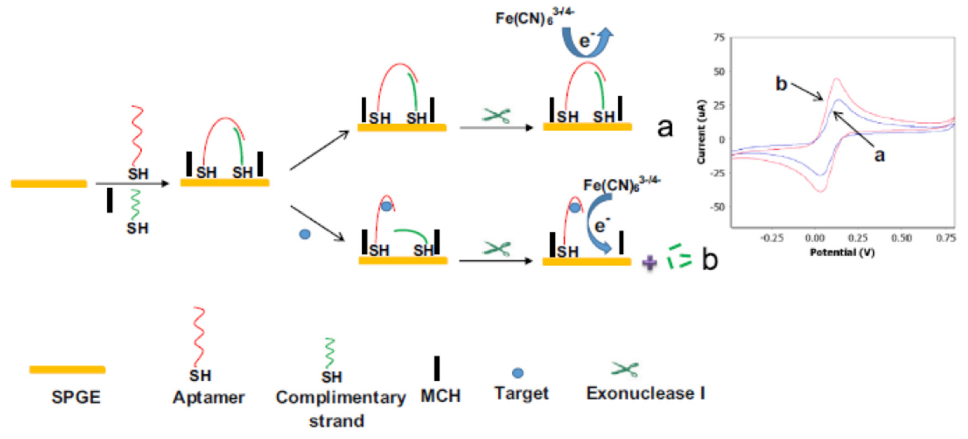
Exonuclease I (Exo I) was used to develop an electrochemical aptasensor for the detection of streptomycin in milk (Figure 18) [80]. Exo I selectively digests the 3′-end of ssDNA. The aptamer and a complementary strand cDNA were immobilized onto a screen printed gold electrode (SPGE). Their hybridization formed a bridge that prevented the redox probe [Fe(CN)6]3−/4− to reach the electrode surface. The signal is low. When the target was present, it bound to the aptamer. Therefore the bridge was broken and the enzyme Exo I digested the cDNA on the electrode. So the redox probe could come closer to the electrode surface and the produced current was high (signal on).
The triggered cleavage activity of nuclease P1 by aptamer-target complex was used to develop an electrochemical aptasensor for sulfadimethoxine detection [70]. The capacity of nuclease P1 to digest only ssDNA was used. The aptamer hybridized to a cDNA that was immobilized onto the surface of a gold electrode. Upon addition of nuclease P1 and antibody anti-dsDNA, the complex aptamer-cDNA (dsDNA) could not be digested by nuclease P1 and the antibody bound to the ds DNA. So the redox probe [Fe(CN6)]3−/4− could not reach the surface of the electrode and the electrochemical signal was weak. When the target of the aptamer was present, the aptamer-target complex was formed because of high affinity constants. Nuclease P1 therefore digested the single strand cDNA on the electrode and less antibodies were able to bind to ds-DNA. The electrode surface was more accessible to the redox probe and finally the signal was increased (signal on method).
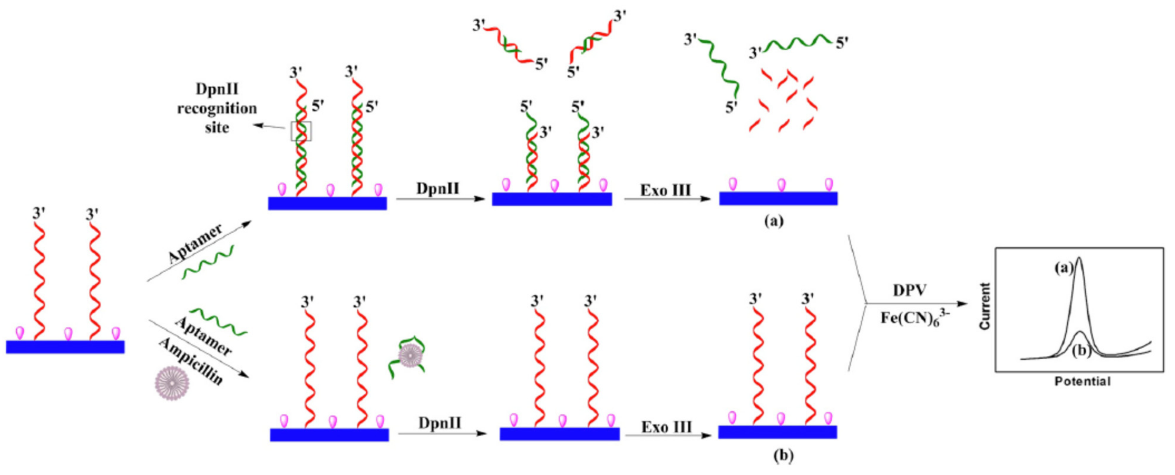
An electrochemical aptasensor based on the combined action of two enzymes, the restriction endonuclease DpnII and the exonuclease Exo III, was developed for the detection of ampicillin in milk (Figure 19) [69]. A cDNA capture probe was immobilized onto the surface of glassy carbon electrode (GCE). The aptamer hybridized with this probe, forming the active binding site for the DpnII. This enzyme cut part of the dsDNA that was released in solution. Thus the remaining dsDNA on the electrode was digested the by Exo III, as well as the released part of the cDNA. By addition of the redox probe [Fe(CN)6]3−/4−, the DPV signal was high. When the target (ampicillin) was present, the aptamer bound to its target. Therefore the two enzymes could not make their catalytic actions because no ds DNA was formed onto the electrode surface. The signal was weak (signal off).
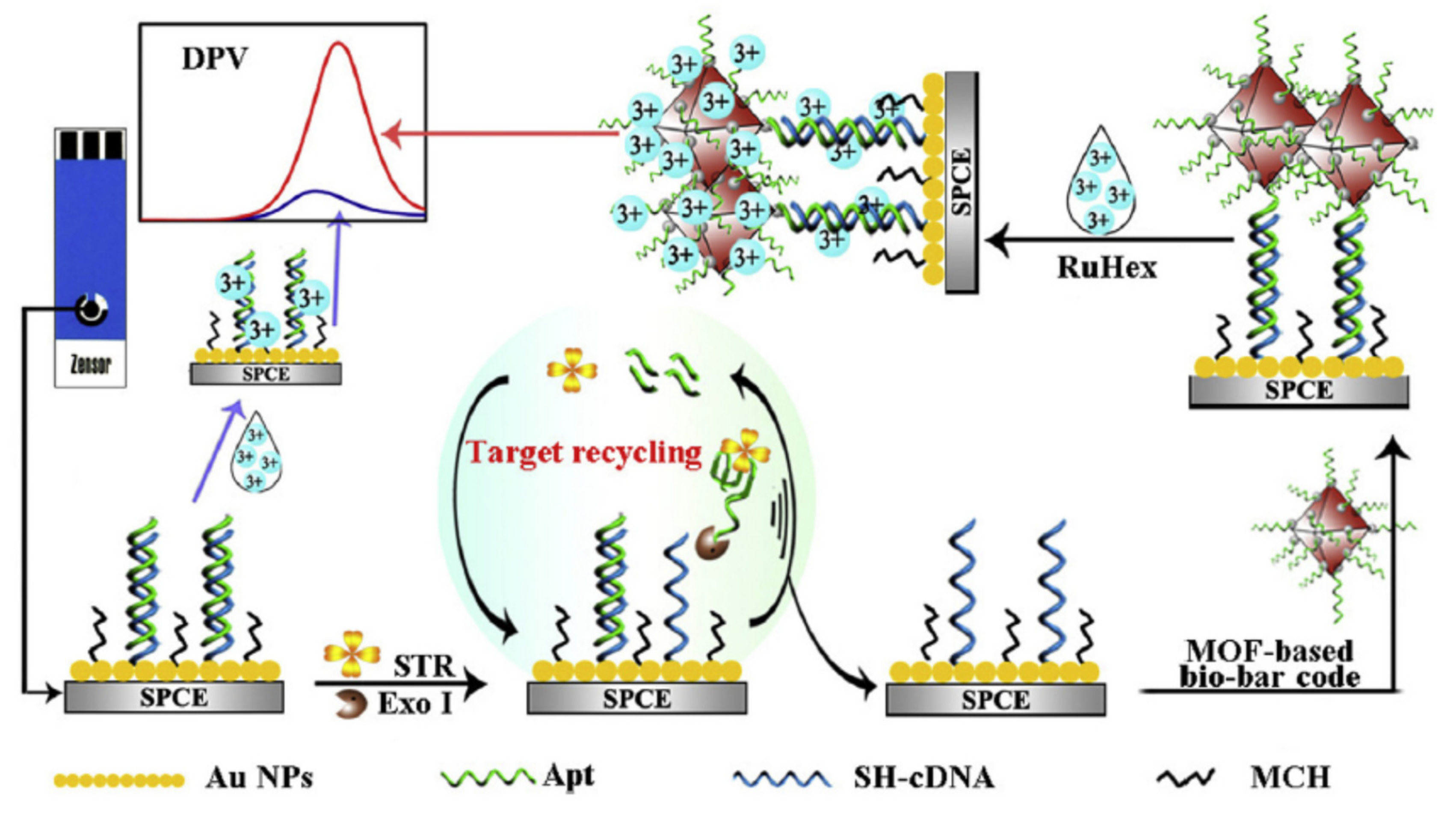
The enzyme assisted-target recycling amplification (TRCA) is another amplification strategy. It consists in the digestion of the aptamer by an exonuclease. So the target is released and free to do a new cycle of binding with other aptamer sequence. This technique was employed to develop an electrochemical aptasensor for the detection of streptomycin using Ru Hex as the redox probe in solution (Figure 20) [28]. Screen-printed carbon electrodes (SPCE) were pre-grafted with AuNPs and then co-immobilized with the aptamer anti-streptomycin and a complementary cDNA. When streptomycin was present in the sample, the aptamer was released after forming a complex with streptomycin. The enzyme Exo I digested the aptamer from its 3′-end. Therefore the target was free to start new cycles of complexation with other aptamers, so more aptamers were released from the electrode surface and more cDNA are free on the electrode surface. Then metal-organic frameworks (MOFs) immobilized with aptamers anti-streptomycin were injected onto the modified SPCE, so that the MOF-aptamers bound to the cDNA remaining free onto the electrode surface. Finally the high number of aptamers bound onto the MOFs attracted RuHex added in solution. A high current was produced (signal on) and measured by DPV. A dual amplification of the signal was performed due to target recycling amplification (TRCA) and use of MOFs-bio bar codes (dsDNA formed by the hybridization of the aptamer with a cDNA).
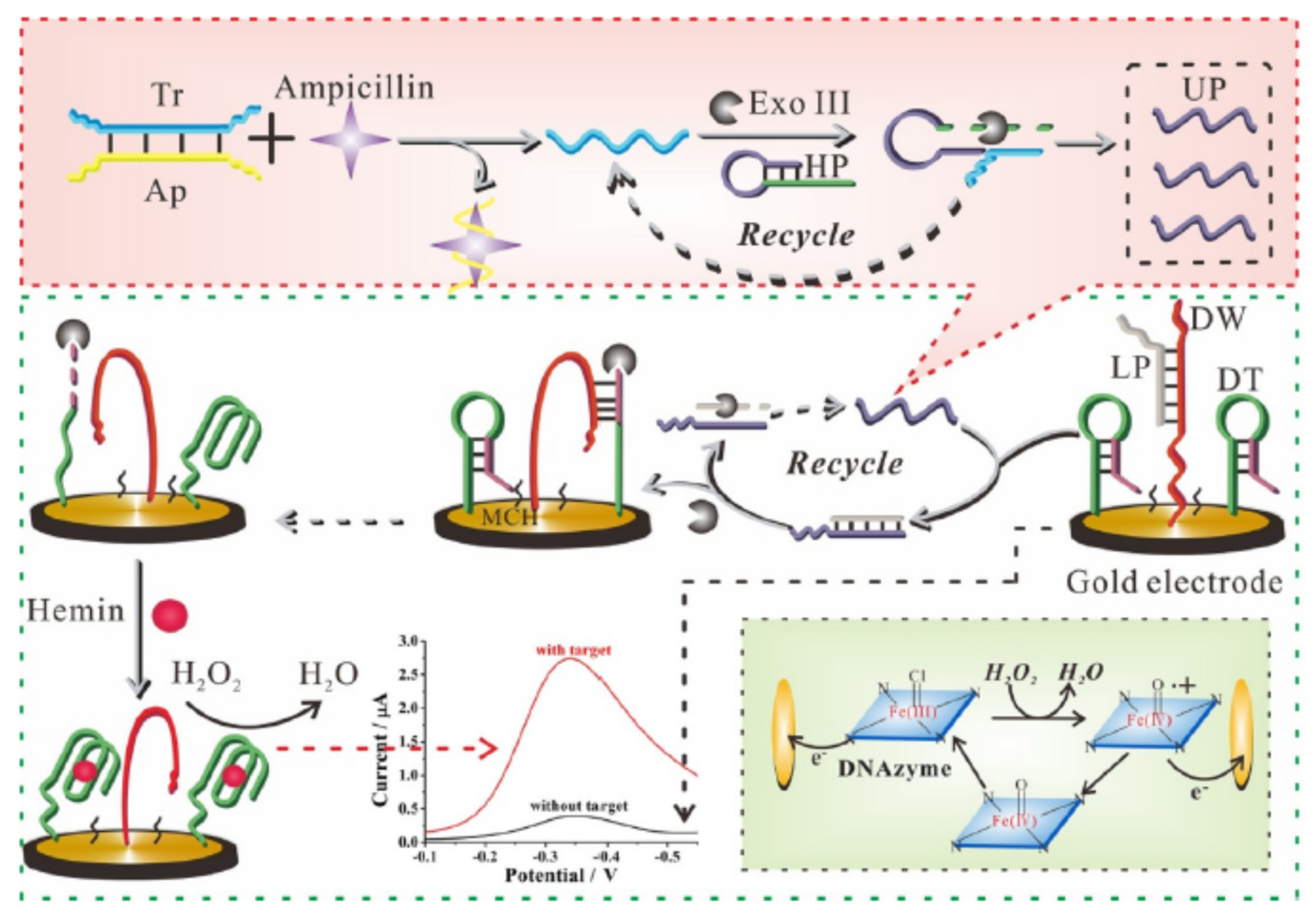
Zhang et al. developed an electrochemical aptasensor for the detection of ampicillin in milk (Figure 21) [35]. The first stage of amplification was a target-triggered enzymatic cyclic amplification. The aptamer was hybridized with trigger ssDNA in solution. In the presence of the target, a complex aptamer-target was formed and the trigger was released. After addition of hairpin probe (HP), the trigger bound to HP and the Exonuclease III (Exo III) could digest part of the HP that was complementary to a short sequence of the trigger. Then the trigger was released and was ready to perform a new cycle of amplification. Unlocking probe (UP) was formed at each cycle and so was amplified by the repetition of cycles. The more ampicillin there was in the sample, the more UP production increased. If the target was absent, the above solution dropped on electrode surfaces did not contain UP, but only aptamer-trigger complexes. So the signal was weak (background signal). The second stage of amplification was linked to the presence of a high amount of UP. DNA-track (DT) that contained hemin aptamer and a cDNA complementary to DW and a complex DNA walker (DW)-Locking probe (LP) were grafted onto a gold electrode. The complex DW-LP was resistant to the Exo III-catalyzed degradation. When the deposited solution was concentrated in UP, UP hybridized with LP and DW was released to hybridize with DT. In the complex UP-LP, the LP sequence was digested by Exo III, UP was released and a new cycle could start. Moreover the Exonuclease digested part of the dsDNA formed by DT and DW. The remaining part of DT was the aptamer sequence. By combination of this sequence with hemin, the aptamer adopted a c Hemin-G quadruplex conformation that mimicked peroxidase activity. Finally this mimicking enzyme procured the reduction of H2O2 and the signal was increased. The more UP there is in the solution, the more the amount of mimicking enzyme formed increased and therefore the more the signal increased. The amplification came from the use of Exonuclease III which occurred at two stages of this protocol.
There are mainly two main nucleic acid amplification techniques used for the development of aptasensors: hybridization chain reaction (HCR) and rolling circle amplification (RCA).
The hybridization chain reaction (HCR) technique is an isothermal enzyme-free amplification. The HCR amplification is based on a chain reaction of hybridization events between alternating H1 and H2 hairpins that form a double helix structure (Figure 22). This structure grows until the there is no more hairpin available in solution.
This technique was used to develop an electrochemical aptasensor based on triple helix aptamer probe with catalyzed hairpin assembly (CHA) signal amplification for the detection of tetracycline [140] The THMS structure was formed by the assembly of the aptamer loop with two arms and a trigger probe. This structure was broken in the presence of the target by the formation of a complex aptamer-target. In this example the single-stranded DNA initiator of the chain reaction was the released trigger probe. The released trigger probe hybridized first with a complementary DNA with hairpin structure (H1) tagged with ferrocene. The other hairpin structure-probe cDNA (H2) tagged with ferrocene also was added and hybridized with H1, so that the released probe was free and started a new cycle. Exo III digested the double helix structure formed by the hybridization of H1-H2 (dsDNA) and ferrocene was released in solution near the surface of the gold electrode. So the signal was high in the presence of the target. When no target was present, the THMS structure was not broken, so the probe was not released and the signal was low.
A dual amplification system was used to develop an electrochemical aptasensor for the detection of kanamycin in milk [76]. HCR was one of the amplification technique, based on two hybridized DNA probes S2-S3, S2 containing the aptamer sequence; the other one was target recycling amplification. In the presence of kanamycin, the complex kanamycin-aptamer (S2) could hybridize with a third DNA probe S3. Thus kanamycin was released for the next cycle. A low detection limit of 16 fM was obtained for the detection of kanamycin. Liu et al. developed an aptasensor for the detection of streptomycin combining two modes of detection: electrochemical and PEC (dual-ratiometric aptasensor) [106]. The amplification technique used was HCR based on two DNA probes HP1 and HP2. The chain reaction started when the target bound to the aptamer and so one part of the sDNA became free to hybridize with HP1 and HP2. The bifunctional redox probe MB was intercalated into the formed dsDNA and so produced amplified electrochemical and PEC signals. The detection limit was low (10−11 M).
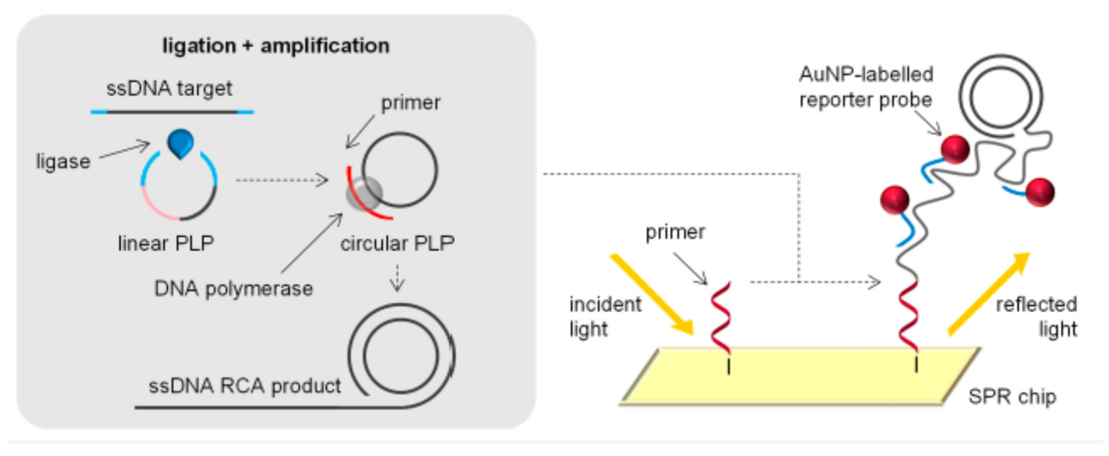
Rolling circle amplification (RCA) is an isothermal DNA amplification technique. The basic principle is the replication of specific DNA sequences [10]. The template used to initiate the RCA is a ssDNA minicircle (padlock probe) that hybridizes to the target sequence (Figure 23). A long single-stranded DNA concatemer of the original DNA target is generated. For instance an aptamer sequence could be replicated several times by RCA. Aptamers tagged with AuNPs could be electrochemically detected. The amplification of the aptamer will also increase the number of tagged AuNPs and therefore the signal will increase. This technique will improve the sensitivity of the aptasensor.
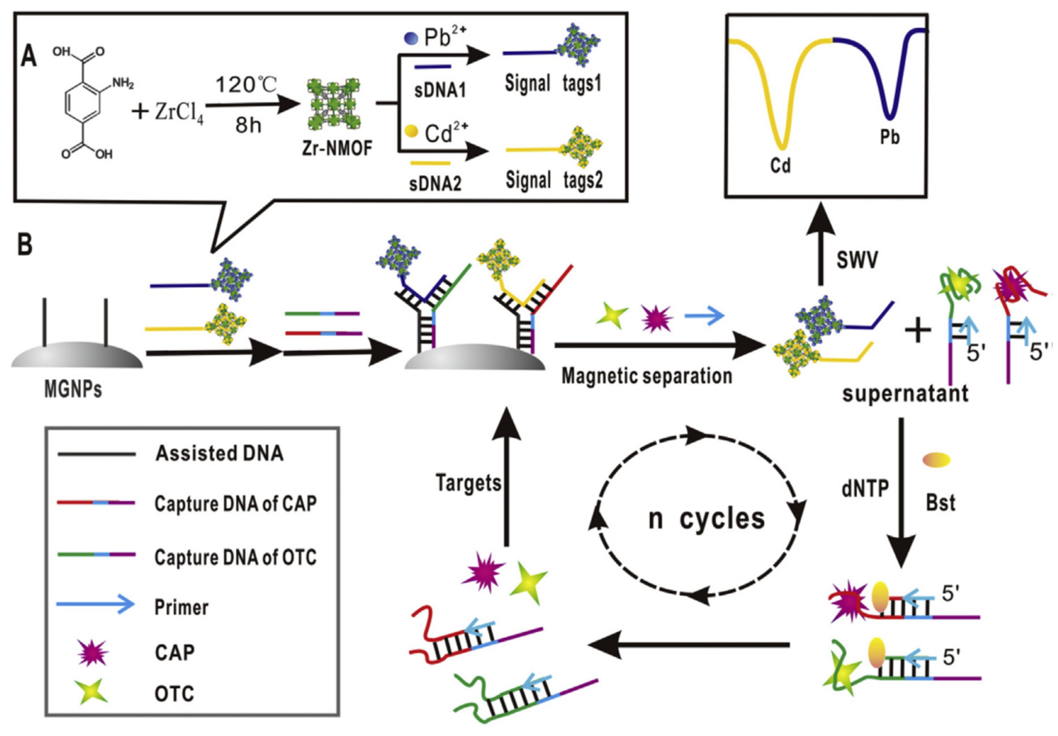
Strand displacement reaction (SDR) technique could be used as a signal amplification strategy. A multiplex electrochemical aptasensor milk based on circular strand-replacement DNA polymerization (CSRP) was developed for the detection of chloramphenicol (CAP) and oxytetracycline (OTC) in milk (Figure 24) [87]. Magnetic beads (MB) were immobilized with assisted ssDNA (or capture cDNA). Signal probes tagged with QDs (PbS for OTC and CdS for CAP) and the two aptamers anti-CAP and anti-OTC were hybridized with the assisted DNAs. Two different structure were formed, one for each aptamer, by the hybridization of assisted DNA, signal tag and aptamer. When one of the target was present, the aptamer-complex was formed and released from the MBs in the supernatant. The corresponding signal tag was also released from the MBs. The signals produced by the signal tags could be electrochemically measured (Cd, Pb). The amplification was performed by adding a primer, a polymerase (Bst DNA polymerase) and deoxynucleotide triphosphates (dNTPs) that produced a ssDNA complementary to the aptamer sequence. So the complex aptamer-target was broken by this elongation and the target was free again to interact with a new aptamer. The amplification cycles could continue. When there was no target analyte, the heterostructure formed by assisted DNA, aptamer and signal probe remained immobilized onto the MBs. No signal tags were released in the supernatant. So the signal was weak for both tags. Therefore the measured current increased the target concentration (signal on).

3. Conclusions and Perspectives

During the past decade, many works have been performed on the development of electrochemical aptasensors for the detection of antimicrobial residues in animal derived food products. Most of the electrochemical aptasensors developed for the detection of antimicrobial residues in food products were based on voltametric techniques (e.g., DPV, SWV), but also the PEC technique amongst the electrochemical-derived techniques was mainly employed. These aptasensors published were essentially developed for the detection of a relatively limited spectrum of antibiotics including kanamycin, chloramphenicol, tetracycline, oxytetracycline, ampicillin and penicillin. Therefore there is still a lot of work to achieve and to develop aptasensors for a wider variety of antimicrobial residues. A key limitation for the development of such aptasensors for antimicrobial residues is fundamentally the lack of availability of aptamers for many antimicrobial residues. Currently immunoassays are predominant on the market because of the availability of specific antibodies, even though their specificity and affinity are not optimal for the purpose. New aptamers have to be developed in the short term to increase the impact and enhance the usage of aptasensors in the agri-food industry.
The specificity of aptamers for banned substances such as for CAP has drawn a great interest for the obvious reason that the screening method should avoid false positive results (positive result while the sample did not contain the banned compound). The selectivity of some aptamers able to bind to several molecules from the same family has allowed new exciting developments whereby one single aptasensor can be designed for a whole family of antimicrobial residues. The interest is still high for the development of selective aptamers for other authorized families of antimicrobials (e.g., Sulphonamides, quinolones, macrolides, etc.).
Ideally, electrochemical aptasensors for the detection of antimicrobial residues can take advantage of multiplex sensors over conventional screening methods (i.e. Microbiological, physico-chemical). Some applications cited here demonstrated that multiplexing is possible by using several aptamers in parallel. The different targets could be discriminated for instance by using different signal tags for the multiple targets (e.g., QDs: PbS, CdS).
Screening methods need to be very sensitive to achieve the very low concentrations, sometimes below 1 ng/mL (or ng/g). The development of POC testing for field applications would be of great interest to develop a cost-effective, rapid and sensitive self-control. We have demonstrated in this review that nanomaterials are increasingly used in the development of selective and sensitive electrochemical aptasensors because of their outstanding characteristics: electrochemical properties (e.g., High electron transfer), high capacity of aptamer immobilization (i.e. High surface to volume ratio), ease of NMs functionalization, magnetic properties (i.e., Magnetic beads for target separation), enzyme-mimicking activities (e.g., AuNPs). Attention has to be paid to the toxicity of some nanomaterials. The tendency is also to combine different nanomaterials to obtain synergistic effects. The development of more performant aptasensors is strongly related to the use of nanomaterials and nucleic acid signal amplification techniques.
This review has also highlighted that all the developments we have described are only at the stage of proofs-of-concepts. Even though when some prototype aptasensor tests were carried for the detection of antimicrobial residues in real samples, real validation assays are urgently needed. Moreover, this review clearly emphasized that most of the real samples tested were milk samples because it is a liquid matrix, the easiest food matrix to handle with. The performance of electrochemical aptasensors for the detection of antimicrobial residues in food products should be tested in real samples (e.g., milk, meat, eggs, honey) because the sensitivity of the method is highly decreased in complex matrices compared to assays in buffer solutions.

Funding

This research received no external funding.

Acknowledgments

This work was financially supported by the European Union Reference Laboratory for Antibiotic Veterinary Medicinal Product and Dye Residues in Food at the French Agency for Food, Environmental and Occupational Health & Safety (ANSES). The author would like to thank the director of ANSES Fougères, Tahar Ait Ali, for his careful proofreading of the manuscript and his valuable advice.

Conflicts of Interest

The author declares no conflict of interest.

References

  1. van Duijkeren, E.; Schwarz, C.; Bouchard, D.; Catry, B.; Pomba, C.; Baptiste, K.E.; Moreno, M.A.; Rantala, M.; Ružauskas, M.; Sanders, P.; et al. The use of aminoglycosides in animals within the EU: Development of resistance in animals and possible impact on human and animal health: A review. J. Antimicrob. Chemother. 2019, 74, 2480–2496. [Google Scholar] [CrossRef]
  2. European Commission. Commission Regulation (EC) N° 470/2009 laying down Community procedures for the establishment of residue limits of pharmacologically active substances in foodstuffs of animal origin. Off. J. EU 2009, L152, 11–22.
  3. European Commission. Council Regulation (EU) (2010) N 37/2010 of 22 December 2009 on pharmacologically active substances and their classification regarding maximum residue limits in foodstuffs of animal origin. Off. J. EU 2009, L15, 1–72. [Google Scholar]
  4. European Commission. Commission Decision (EC) N° 2002/657 of 12 August 2002 implementing Council Directive 96/23/EC concerning the performance of analytical methods and interpretation of results. Off. J. EU 2002, L221, 8–36. [Google Scholar]
  5. European Commission. Commission Regulation (EU) 2019/1871 of 7 November 2019 on reference points for action for non-allowed pharmacologically active substances present in food of animal origin and repealing Decision 2005/34/EC (Text with EEA relevance). Off. J. EU 2019, L289, 41–46. [Google Scholar]
  6. European Commission. Regulation (EU) 2017/625 of the European Parliament and of the Council of 15 March 2017 on official controls and other official activities performed to ensure the application of food and feed law, rules on animal health and welfare, plant health and plant protection products. Off. J. EU 2017, L95, 1–142. [Google Scholar]
  7. Marco, M.-P.; Gee, S.; Hammock, B.D. Immunochemical techniques for environmental analysis I. Immunosensors. TrAC Trends Anal. Chem. 1995, 14, 341–350. [Google Scholar] [CrossRef]
  8. Gaudin, V. Advances in biosensor development for the screening of antibiotic residues in food products of animal origin—A comprehensive review. Biosens. Bioelectron. 2017, 90, 363–377. [Google Scholar] [CrossRef]
  9. Gaudin, V. The Growing Interest in Development of Innovative Optical Aptasensors for the Detection of Antimicrobial Residues in Food Products. Biosensors 2020, 10, 21. [Google Scholar] [CrossRef] [Green Version]
  10. Oberhaus, F.V.; Frense, D.; Beckmann, D. Immobilization Techniques for Aptamers on Gold Electrodes for the Electrochemical Detection of Proteins: A Review. Biosensors 2020, 10, 45. [Google Scholar] [CrossRef]
  11. Piro, B.; Shi, S.; Reisberg, S.; Noël, V.; Anquetin, G. Comparison of Electrochemical Immunosensors and Aptasensors for Detection of Small Organic Molecules in Environment, Food Safety, Clinical and Public Security. Biosensors 2016, 6, 7. [Google Scholar] [CrossRef] [PubMed] [Green Version]
  12. Amaya-González, S.; De-los-Santos-Álvarez, N.; Miranda-Ordieres, A.J.; Lobo-Castañón, M.J. Aptamer-Based Analysis: A Promising Alternative for Food Safety Control. Sensors 2013, 13, 16292–16311. [Google Scholar] [CrossRef] [PubMed] [Green Version]
  13. Wang, Y.; Killian, J.; Hamasaki, K.; Rando, R.R. RNA Molecules That Specifically and Stoichiometrically Bind Aminoglycoside Antibiotics with High Affinities. Biochemistry 1996, 35, 12338–12346. [Google Scholar] [CrossRef] [PubMed]
  14. Burke, D.H.; Hoffman, D.C.; Brown, A.; Hansen, M.; Pardi, A.; Gold, L. RNA aptamers to the peptidyl transferase inhibitor chloramphenicol. Chem. Biol. 1997, 4, 833–843. [Google Scholar] [CrossRef] [Green Version]
  15. Berens, C.; Thain, A.; Schroeder, R. A tetracycline-binding RNA aptamer. Bioorg. Med. Chem. 2001, 9, 2549–2556. [Google Scholar] [CrossRef]
  16. Mehlhorn, A.; Rahimi, P.; Joseph, Y. Aptamer-Based Biosensors for Antibiotic Detection: A Review. Biosensors 2018, 8, 54. [Google Scholar] [CrossRef] [Green Version]
  17. Kwon, Y.S.; Ahmad Raston, N.H.; Gu, M.B. An ultra-sensitive colorimetric detection of tetracyclines using the shortest aptamer with highly enhanced affinity. Chem. Commun. 2014, 50, 40–42. [Google Scholar] [CrossRef]
  18. Hianik, T.; Wang, J. Electrochemical Aptasensors—Recent Achievements and Perspectives. Electroanalysis 2009, 21, 1223–1235. [Google Scholar] [CrossRef]
  19. González-Fernández, E.; de-los-Santos-Álvarez, N.; Lobo-Castañón, M.J.; Miranda-Ordieres, A.J.; Tuñón-Blanco, P. Impedimetric aptasensor for tobramycin detection in human serum. Biosens. Bioelectron. 2011, 26, 2354–2360. [Google Scholar] [CrossRef]
  20. Kim, Y.-J.; Kim, Y.S.; Niazi, J.H.; Gu, M.B. Electrochemical aptasensor for tetracycline detection. Bioprocess Biosyst. Eng. 2010, 33, 31. [Google Scholar] [CrossRef]
  21. Nguyen, T.; Hilton, J.P.; Lin, Q. Emerging applications of aptamers to micro- and nanoscale biosensing. Microfluid. Nanofluidics 2009, 6, 347. [Google Scholar] [CrossRef]
  22. Sharma, A.; Chandra Singh, A.; Bacher, G.; Bhand, S. Recent Advances in Aptamer- Based Biosensors for Detection of Antibiotic Residues. Aptamers Synth. Antibodies 2016, 2, 43–54. [Google Scholar]
  23. Viglasky, V.; Hianik, T. Potential uses of G-quadruplex-forming aptamers. Gen. Physiol. Biophys. 2013, 32, 149–172. [Google Scholar] [CrossRef] [PubMed]
  24. Chiorcea-Paquim, A.-M.; Oliveira-Brett, A.M. Redox behaviour of G-quadruplexes. Electrochim. Acta 2014, 126, 162–170. [Google Scholar] [CrossRef]
  25. Bagheri, E.; Abnous, K.; Alibolandi, M.; Ramezani, M.; Taghdisi, S.M. Triple-helix molecular switch-based aptasensors and DNA sensors. Biosens. Bioelectron. 2018, 111, 1–9. [Google Scholar] [CrossRef] [PubMed]
  26. Jalalian, S.H.; Karimabadi, N.; Ramezani, M.; Abnous, K.; Taghdisi, S.M. Electrochemical and optical aptamer-based sensors for detection of tetracyclines. Trends Food Sci. Technol. 2018, 73, 45–57. [Google Scholar] [CrossRef]
  27. Schoukroun-Barnes, L.R.; Wagan, S.; White, R.J. Enhancing the Analytical Performance of Electrochemical RNA Aptamer-Based Sensors for Sensitive Detection of Aminoglycoside Antibiotics. Anal. Chem. 2014, 86, 1131–1137. [Google Scholar] [CrossRef]
  28. Meng, X.; Gu, H.; Yi, H.; He, Y.; Chen, Y.; Sun, W. Sensitive detection of streptomycin in milk using a hybrid signal enhancement strategy of MOF-based bio-bar code and target recycling. Anal. Chim. Acta 2020, 1125, 1–7. [Google Scholar] [CrossRef] [PubMed]
  29. Yu, Z.; Cui, P.; Xiang, Y.; Li, B.; Han, X.; Shi, W.; Yan, H.; Zhang, G. Developing a fast electrochemical aptasensor method for the quantitative detection of penicillin G residue in milk with high sensitivity and good anti-fouling ability. Microchem. J. 2020, 157, 105077. [Google Scholar] [CrossRef]
  30. Kim, Y.S.; Gu, M.B. Advances in Aptamer Screening and Small Molecule Aptasensors. In Biosensors Based on Aptamers and Enzymes; Gu, M.B., Kim, H.-S., Eds.; Springer: Berlin/Heidelberg, Germany, 2014; pp. 29–67. [Google Scholar] [CrossRef]
  31. Wang, Q.; Qin, X.; Geng, L.; Wang, Y. Label-Free Electrochemical Aptasensor for Sensitive Detection of Malachite Green Based on Au Nanoparticle/Graphene Quantum Dots/Tungsten Disulfide Nanocomposites. Nanomaterials 2019, 9, 229. [Google Scholar] [CrossRef] [Green Version]
  32. Taghdisi, S.M.; Danesh, N.M.; Nameghi, M.A.; Ramezani, M.; Alibolandi, M.; Abnous, K. An electrochemical sensing platform based on ladder-shaped DNA structure and label-free aptamer for ultrasensitive detection of ampicillin. Biosens. Bioelectron. 2019. [Google Scholar] [CrossRef]
  33. Alizadeh, N.; Salimi, A.; Hallaj, R. Hemin/G-Quadruplex Horseradish Peroxidase-Mimicking DNAzyme: Principle and Biosensing Application. Adv. Biochem. Eng. Biotechnol. 2017, 170, 85–106. [Google Scholar] [CrossRef]
  34. Zhang, R.; Li, S.; Wang, J.; Qu, X.; Zhao, Y.; Liu, S.; Wang, Y.; Huang, J.; Yu, J. Entropy-driven spliced DNA walking machine for label-free electrochemical detection of antibiotics. Sens. Actuators B 2020, 320, 128385. [Google Scholar] [CrossRef]
  35. Zhang, R.; Wang, Y.; Qu, X.; Li, S.; Zhao, Y.; Liu, S.; Huang, J. Exonuclease III-powered DNA Walking Machine for Label-free and Ultrasensitive Electrochemical Sensing of Antibiotic. Sens. Actuators B 2019, 297, 126771. [Google Scholar] [CrossRef]
  36. Han, C.; Li, R.; Li, H.; Liu, S.; Xu, C.; Wang, J.; Wang, Y.; Huang, J. Ultrasensitive voltammetric determination of kanamycin using a target-triggered cascade enzymatic recycling couple along with DNAzyme amplification. Microchim. Acta 2017, 184, 2941–2948. [Google Scholar] [CrossRef]
  37. Huang, Y.; Zheng, J.; Wang, L.; Duan, X.; Wang, Y.; Xiang, Y.; Li, G. Sensitive Detection of Chloramphenicol Based on Ag-DNAzyme-Mediated Signal Amplification Modulated by DNA/Metal Ion Interaction. Biosens. Bioelectron. 2018. [Google Scholar] [CrossRef] [PubMed]
  38. Azadbakht, A.; Roushani, M.; Abbasi, A.R.; Menati, S.; Derikvand, Z. A label-free aptasensor based on polyethyleneimine wrapped carbon nanotubes in situ formed gold nanoparticles as signal probe for highly sensitive detection of dopamine. Mater. Sci. Eng. C 2016, 68, 585–593. [Google Scholar] [CrossRef] [PubMed]
  39. Li, F.; Yu, Z.; Han, X.; Shi, W.; Liu, Y.; Yan, H.; Zhang, G. A signal-on electrochemical aptasensor for highly sensitive and specific detection of kanamycin based on target-induced signaling probe shifting mechanism. Sens. Actuators B 2018, 273, 480–487. [Google Scholar] [CrossRef]
  40. Liu, R.; Yang, Z.; Guo, Q.; Zhao, J.; Ma, J.; Kang, Q.; Tang, Y.; Xue, Y.; Lou, X.; He, M. Signaling-Probe Displacement Electrochemical Aptamer-based Sensor (SD-EAB) for Detection of Nanomolar Kanamycin A. Electrochim. Acta 2015, 182, 516–523. [Google Scholar] [CrossRef]
  41. Han, X.; Yu, Z.; Li, F.; Shi, W.; Fu, C.; Yan, H.; Zhang, G. Two kanamycin electrochemical aptamer-based sensors using different signal transduction mechanisms: A comparison of electrochemical behavior and sensing performance. Bioelectrochemistry 2019, 129, 270–277. [Google Scholar] [CrossRef]
  42. Herne, T.M.; Tarlov, M.J. Characterization of DNA Probes Immobilized on Gold Surfaces. J. Am. Chem. Soc. 1997, 119, 8916–8920. [Google Scholar] [CrossRef]
  43. Nan, M.-N.; Bi, Y.; Xue, H.-L.; Long, H.-T.; Xue, S.-L.; Pu, L.-M.; Prusky, D. Modification performance and electrochemical characteristics of different groups of modified aptamers applied for label-free electrochemical impedimetric sensors. Food Chem. 2021, 337, 127761. [Google Scholar] [CrossRef]
  44. Li, F.; Wang, X.; Sun, X.; Guo, Y.; Zhao, W. A dual-signal amplification strategy for kanamycin based on ordered mesoporous carbon-chitosan/gold nanoparticles-streptavidin and ferrocene labelled DNA. Anal. Chim. Acta 2018, 1033, 185–192. [Google Scholar] [CrossRef]
  45. Sharma, A.; Istamboulie, G.; Hayat, A.; Catanante, G.; Bhand, S.; Marty, J.L. Disposable and portable aptamer functionalized impedimetric sensor for detection of kanamycin residue in milk sample. Sens. Actuators B 2017, 245, 507–515. [Google Scholar] [CrossRef]
  46. Tanimu, A.; Lawal, M.A. electrochemical-sensors-using-nanomaterials—A-mini-review. Res. Rev. J. Chem. 2017, 6, 38–48. [Google Scholar]
  47. Malhotra, B.D.; Ali, M.A. Chapter 1—Nanomaterials in Biosensors: Fundamentals and Applications. In Nanomaterials for Biosensors; Malhotra, B.D., Ali, M.A., Eds.; William Andrew Publishing: Amsterdam, The Netherlands, 2018; pp. 1–74. [Google Scholar] [CrossRef]
  48. Yang, T.; Huang, H.; Zhu, F.; Lin, Q.; Zhang, L.; Liu, J. Recent Progresses in Nanobiosensing for Food Safety Analysis. Sensors 2016, 16, 1118. [Google Scholar] [CrossRef] [Green Version]
  49. Abi, A.; Mohammadpour, Z.; Zuo, X.; Safavi, A. Nucleic acid-based electrochemical nanobiosensors. Biosens. Bioelectron. 2018, 102, 479–489. [Google Scholar] [CrossRef] [PubMed]
  50. Lim, M.C.; Kim, Y.R. Analytical Applications of Nanomaterials in Monitoring Biological and Chemical Contaminants in Food. J Microbiol. Biotechnol. 2016, 26, 1505–1516. [Google Scholar] [CrossRef] [Green Version]
  51. Sharma, R.; Ragavan, K.V.; Thakur, M.S.; Raghavarao, K.S.M.S. Recent advances in nanoparticle based aptasensors for food contaminants. Biosens. Bioelectron. 2015, 74, 612–627. [Google Scholar] [CrossRef]
  52. Hu, M.; Wang, Y.; Yang, J.; Sun, Y.; Xing, G.; Deng, R.; Hu, X.; Zhang, G. Competitive electrochemical immunosensor for maduramicin detection by multiple signal amplification strategy via hemin@Fe-MIL-88NH2/AuPt. Biosens. Bioelectron. 2019, 111554. [Google Scholar] [CrossRef]
  53. Liu, S.; Lai, G.; Zhang, H.; Yu, A. Amperometric aptasensing of chloramphenicol at a glassy carbon electrode modified with a nanocomposite consisting of graphene and silver nanoparticles. Microchim. Acta 2017, 184, 1445–1451. [Google Scholar] [CrossRef]
  54. Huang, S.; Gan, N.; Zhang, X.; Wu, Y.; Shao, Y.; Jiang, Z.; Wang, Q. Portable fluoride-selective electrode as signal transducer for sensitive and selective detection of trace antibiotics in complex samples. Biosens. Bioelectron. 2019, 128, 113–121. [Google Scholar] [CrossRef]
  55. Yao, Y.; Jiang, C.; Ping, J. Flexible Freestanding Graphene Paper-based Potentiometric Enzymatic Aptasensor for Ultrasensitive Wireless Detection of Kanamycin. Biosens. Bioelectron. 2018. [Google Scholar] [CrossRef]
  56. Ebrahimi Vafaye, S.; Rahman, A.; Safaeian, S.; Adabi, M. An electrochemical aptasensor based on electrospun carbon nanofiber mat and gold nanoparticles for the sensitive detection of Penicillin in milk. J. Food Meas. Charact. 2021, 15, 876–882. [Google Scholar] [CrossRef]
  57. Alawad, A.; Istamboulié, G.; Calas-Blanchard, C.; Noguer, T. A reagentless aptasensor based on intrinsic aptamer redox activity for the detection of tetracycline in water. Sens. Actuators B 2019, 288, 141–146. [Google Scholar] [CrossRef]
  58. Shi, Z.; Hou, W.; Jiao, Y.; Guo, Y.; Sun, X.; Zhao, J.; Wang, X. Ultra-sensitive aptasensor based on IL and Fe3O4 nanoparticles for tetracycline detection. Int. J. Electrochem. Sci. 2017, 12, 7426–7434. [Google Scholar] [CrossRef]
  59. Guo, Y.; Zhang, Q.; Xu, Q.; Ma, N.; Sun, X.; Wang, X. Fabrication of aptasensors modified by MWCNTs-CS/Fe3O4-CS based on SPEs. Int. J. Electrochem. Sci. 2016, 11, 1691–1698. [Google Scholar]
  60. Taghdisi Heidarian, S.M.; Tavanaee Sani, A.; Danesh, N.M.; Ramezani, M.; Alibolandi, M.; Gerayelou, G.; Abnous, K.; Taghdisi, S.M. A novel electrochemical approach for the ultrasensitive detection of fluoroquinolones based on a double-labelled aptamer to surpass complementary strands of aptamer lying flat. Sens. Actuators B 2021, 334, 129632. [Google Scholar] [CrossRef]
  61. Huang, W.; Zhou, Y.; Zhan, D.; Lai, G. Homogeneous biorecognition reaction-induced assembly of DNA nanostructures for ultrasensitive electrochemical detection of kanamycin antibiotic. Anal. Chim. Acta 2021, 1154, 338317. [Google Scholar] [CrossRef]
  62. Lu, M.; Cao, C.; Wang, F.; Liu, G. A polyethyleneimine reduced graphene oxide/gold nanocubes based electrochemical aptasensor for chloramphenicol detection using single-stranded DNA-binding protein. Mater. Des. 2021, 199, 109409. [Google Scholar] [CrossRef]
  63. Roushani, M.; Rahmati, Z.; Farokhi, S.; Hoseini, S.J.; Fath, R.H. The development of an electrochemical nanoaptasensor to sensing chloramphenicol using a nanocomposite consisting of graphene oxide functionalized with (3-Aminopropyl) triethoxysilane and silver nanoparticles. Mater. Sci. Eng. C 2020, 108, 110388. [Google Scholar] [CrossRef]
  64. Bi, H.; Wu, Y.; Wang, Y.; Liu, G.; Ning, G.; Xu, Z. A molecularly imprinted polymer combined with dual functional Au@Fe3O4 nanocomposites for sensitive detection of kanamycin. J. Electroanal. Chem. 2020, 870, 114216. [Google Scholar] [CrossRef]
  65. Guo, W.; Umar, A.; Alsaiari, M.A.; Wang, L.; Pei, M. Ultrasensitive and selective label-free aptasensor for the detection of penicillin based on nanoporous PtTi/graphene oxide-Fe3O4/MWCNT-Fe3O4 nanocomposite. Microchem. J. 2020, 158, 105270. [Google Scholar] [CrossRef]
  66. Li, F. Magnetic bead-based electrochemical aptasensor doped with multi-wall carbon nanotubes for the detection of ampicillin in milk. Int. J. Electrochem. Sci. 2020, 7520–7530. [Google Scholar] [CrossRef]
  67. Sebastian, N.; Yu, W.-C.; Balram, D. Electrochemical detection of an antibiotic drug chloramphenicol based on a graphene oxide/hierarchical zinc oxide nanocomposite. Inorg. Chem. Front. 2019, 6, 82–93. [Google Scholar] [CrossRef]
  68. Zhang, R.; Wang, Y.; Qu, X.; Li, S.; Zhao, Y.; Liu, S.; Zhang, F.; Huang, J.; Yu, J. A Label-free Electrochemical Platform for Antibiotics Detection Based on Cascade Enzymatic Amplification Coupled with Split G-quadruplex DNAzyme. Analyst 2019, 144. [Google Scholar] [CrossRef]
  69. Wang, T.; Yin, H.; Zhang, Y.; Wang, L.; Du, Y.; Zhuge, Y.; Ai, S. Electrochemical aptasensor for ampicillin detection based on the protective effect of aptamer-antibiotic conjugate towards DpnII and Exo III digestion. Talanta 2019, 197, 42–48. [Google Scholar] [CrossRef] [PubMed]
  70. Bai, Z.; Chen, Y.; Li, F.; Zhou, Y.; Yin, H.; Ai, S. Electrochemical aptasensor for sulfadimethoxine detection based on the triggered cleavage activity of nuclease P1 by aptamer-target complex. Talanta 2019, 204, 409–414. [Google Scholar] [CrossRef]
  71. Li, L.; Liu, X.; Yang, L.; Zhang, S.; Zheng, H.; Tang, Y.; Wong, D.K.Y. Amplified oxygen reduction signal at a Pt-Sn-modified TiO2 nanocomposite on an electrochemical aptasensor. Biosens. Bioelectron. 2019, 142, 111525. [Google Scholar] [CrossRef]
  72. He, B.; Wang, L.; Dong, X.; Yan, X.; Li, M.; Yan, S.; Yan, D. Aptamer-based thin film gold electrode modified with gold nanoparticles and carboxylated multi-walled carbon nanotubes for detecting oxytetracycline in chicken samples. Food Chem. 2019, 300, 125179. [Google Scholar] [CrossRef]
  73. You, H.; Bai, L.; Yuan, Y.; Zhou, J.; Bai, Y.; Mu, Z. An amperometric aptasensor for ultrasensitive detection of sulfadimethoxine based on exonuclease-assisted target recycling and new signal tracer for amplification. Biosens. Bioelectron. 2018, 117, 706–712. [Google Scholar] [CrossRef] [PubMed]
  74. Li, F.; Guo, Y.; Wang, X.; Sun, X. Multiplexed aptasensor based on metal ions labels for simultaneous detection of multiple antibiotic residues in milk. Biosens. Bioelectron. 2018, 115, 7–13. [Google Scholar] [CrossRef]
  75. Chen, Z.; Lai, G.; Liu, S.; Yu, A. Ultrasensitive electrochemical aptasensing of kanamycin antibiotic by enzymatic signal amplification with a horseradish peroxidase-functionalized gold nanoprobe. Sens. Actuators B 2018, 273, 1762–1767. [Google Scholar] [CrossRef]
  76. Hong, F.; Chen, X.; Cao, Y.; Dong, Y.; Wu, D.; Hu, F.; Gan, N. Enzyme- and label-free electrochemical aptasensor for kanamycin detection based on double stir bar-assisted toehold-mediated strand displacement reaction for dual-signal amplification. Biosens. Bioelectron. 2018, 112, 202–208. [Google Scholar] [CrossRef] [PubMed]
  77. Yin, J.; Guo, W.; Qin, X.; Zhao, J.; Pei, M.; Ding, F. A sensitive electrochemical aptasensor for highly specific detection of streptomycin based on the porous carbon nanorods and multifunctional graphene nanocomposites for signal amplification. Sens. Actuators B 2017, 241, 151–159. [Google Scholar] [CrossRef]
  78. Taghdisi, S.M.; Danesh, N.M.; Ramezani, M.; Abnous, K. A novel M-shape electrochemical aptasensor for ultrasensitive detection of tetracyclines. Biosens. Bioelectron. 2016, 85, 509–514. [Google Scholar] [CrossRef] [PubMed]
  79. Zhao, J.; Guo, W.; Pei, M.; Ding, F. GR–Fe3O4NPs and PEDOT–AuNPs composite based electrochemical aptasensor for the sensitive detection of penicillin. Anal. Methods 2016, 8, 4391–4397. [Google Scholar] [CrossRef]
  80. Mohammad Danesh, N.; Ramezani, M.; Sarreshtehdar Emrani, A.; Abnous, K.; Taghdisi, S.M. A novel electrochemical aptasensor based on arch-shape structure of aptamer-complimentary strand conjugate and exonuclease I for sensitive detection of streptomycin. Biosens. Bioelectron. 2016, 75, 123–128. [Google Scholar] [CrossRef] [PubMed]
  81. Yin, J.; Guo, W.; Qin, X.; Pei, M.; Wang, L.; Ding, F. A regular “signal attenuation” electrochemical aptasensor for highly sensitive detection of streptomycin. New J. Chem. 2016, 40, 9711–9718. [Google Scholar] [CrossRef]
  82. Hamidi-Asl, E.; Dardenne, F.; Blust, R.; De Wael, K. An Improved Electrochemical Aptasensor for Chloramphenicol Detection Based on Aptamer Incorporated Gelatine. Sensors 2015, 15, 7605–7618. [Google Scholar] [CrossRef] [Green Version]
  83. Yan, L.; Luo, C.; Cheng, W.; Mao, W.; Zhang, D.; Ding, S. A simple and sensitive electrochemical aptasensor for determination of Chloramphenicol in honey based on target-induced strand release. J. Electroanal. Chem. 2012, 687, 89–94. [Google Scholar] [CrossRef]
  84. Shen, Z.; He, L.; Cao, Y.; Hong, F.; Zhang, K.; Hu, F.; Lin, J.; Wu, D.; Gan, N. Multiplexed electrochemical aptasensor for antibiotics detection using metallic-encoded apoferritin probes and double stirring bars-assisted target recycling for signal amplification. Talanta 2019, 197, 491–499. [Google Scholar] [CrossRef] [PubMed]
  85. Huang, S.; Gan, N.; Li, T.; Zhou, Y.; Cao, Y.; Dong, Y. Electrochemical aptasensor for multi-antibiotics detection based on endonuclease and exonuclease assisted dual recycling amplification strategy. Talanta 2018, 179, 28–36. [Google Scholar] [CrossRef]
  86. Chen, M.; Gan, N.; Zhou, Y.; Li, T.; Xu, Q.; Cao, Y.; Chen, Y. A novel aptamer- metal ions- nanoscale MOF based electrochemical biocodes for multiple antibiotics detection and signal amplification. Sens. Actuators B 2017, 242, 1201–1209. [Google Scholar] [CrossRef]
  87. Chen, M.; Gan, N.; Li, T.; Wang, Y.; Xu, Q.; Chen, Y. An electrochemical aptasensor for multiplex antibiotics detection using Y-shaped DNA-based metal ions encoded probes with NMOF substrate and CSRP target-triggered amplification strategy. Anal. Chim. Acta 2017, 968, 30–39. [Google Scholar] [CrossRef]
  88. Yan, Z.; Gan, N.; Li, T.; Cao, Y.; Chen, Y. A sensitive electrochemical aptasensor for multiplex antibiotics detection based on high-capacity magnetic hollow porous nanotracers coupling exonuclease-assisted cascade target recycling. Biosens. Bioelectron. 2016, 78, 51–57. [Google Scholar] [CrossRef]
  89. Pilehvar, S.; Mehta, J.; Dardenne, F.; Robbens, J.; Blust, R.; De Wael, K. Aptasensing of Chloramphenicol in the Presence of Its Analogues: Reaching the Maximum Residue Limit. Anal. Chem. 2012, 84, 6753–6758. [Google Scholar] [CrossRef]
  90. Xue, J.; Liu, J.; Wang, C.; Tian, Y.; Zhou, N. Simultaneous electrochemical detection of multiple antibiotic residues in milk based on aptamers and quantum dots. Anal. Methods 2016, 8, 1981–1988. [Google Scholar] [CrossRef]
  91. Yuan, R.; Yan, Z.; Shaga, A.; He, H. Design and fabrication of an electrochemical sensing platform based on a porous organic polymer for ultrasensitive ampicillin detection. Sens. Actuators B 2021, 327, 128949. [Google Scholar] [CrossRef]
  92. He, X.; Han, H.; Shi, W.; Dong, J.; Lu, X.; Yang, W.; Lu, X. A label-free electrochemical DNA biosensor for kanamycin detection based on diblock DNA with poly-cytosine as a high affinity anchor on graphene oxide. Anal. Methods 2020, 12, 3462–3469. [Google Scholar] [CrossRef]
  93. Mohammad-Razdari, A.; Ghasemi-Varnamkhasti, M.; Rostami, S.; Izadi, Z.; Ensafi, A.A.; Siadat, M. Development of an electrochemical biosensor for impedimetric detection of tetracycline in milk. J. Food Sci. Technol. 2020. [Google Scholar] [CrossRef]
  94. Kulikova, T.; Gorbatchuk, V.; Stoikov, I.; Rogov, A.; Evtugyn, G.; Hianik, T. Impedimetric Determination of Kanamycin in Milk with Aptasensor Based on Carbon Black-Oligolactide Composite. Sensors 2020, 20, 4738. [Google Scholar] [CrossRef]
  95. He, H.; Wang, S.-Q.; Han, Z.-Y.; Tian, X.-H.; Zhang, W.-W.; Li, C.-P.; Du, M. Construction of electrochemical aptasensors with Ag(I) metal−organic frameworks toward high-efficient detection of ultra-trace penicillin. Appl. Surf. Sci. 2020, 531, 147342. [Google Scholar] [CrossRef]
  96. Zhou, N.; Ma, Y.; Hu, B.; He, L.; Wang, S.; Zhang, Z.; Lu, S. Construction of Ce-MOF@COF hybrid nanostructure: Label-free aptasensor for the ultrasensitive detection of oxytetracycline residues in aqueous solution environments. Biosens. Bioelectron. 2019, 127, 92–100. [Google Scholar] [CrossRef]
  97. Yildirim-Tirgil, N.; Lee, J.; Cho, H.; Lee, H.; Somu, S.; Busnaina, A.; Gu, A.Z. A SWCNT based aptasensor system for antibiotic oxytetracycline detection in water samples. Anal. Methods 2019, 11, 2692–2699. [Google Scholar] [CrossRef]
  98. Mohammad-Razdari, A.; Ghasemi-Varnamkhasti, M.; Izadi, Z.; Ensafi, A.A.; Rostami, S.; Siadat, M. An impedimetric aptasensor for ultrasensitive detection of Penicillin G based on the use of reduced graphene oxide and gold nanoparticles. Microchim. Acta 2019, 186, 372. [Google Scholar] [CrossRef]
  99. Mohammad-Razdari, A.; Ghasemi-Varnamkhasti, M.; Izadi, Z.; Rostami, S.; Ensafi, A.A.; Siadat, M.; Losson, E. Detection of sulfadimethoxine in meat samples using a novel electrochemical biosensor as a rapid analysis method. J. Food Compos. Anal. 2019, 82, 103252. [Google Scholar] [CrossRef]
  100. Hu, X.; Goud, K.Y.; Kumar, V.S.; Catanante, G.; Li, Z.; Zhu, Z.; Marty, J.L. Disposable electrochemical aptasensor based on carbon nanotubes- V2O5-chitosan nanocomposite for detection of ciprofloxacin. Sens. Actuators B 2018, 268, 278–286. [Google Scholar] [CrossRef]
  101. Liu, X.; Hu, M.; Wang, M.; Song, Y.; Zhou, N.; He, L.; Zhang, Z. Novel Nanoarchitecture of Co-MOF-on-TPN-COF Hybrid: Ultralowly Sensitive Bioplatform of Electrochemical Aptasensor toward Ampicillin. Biosens. Bioelectron. 2018. [Google Scholar] [CrossRef] [PubMed]
  102. Paniel, N.; Istamboulié, G.; Triki, A.; Lozano, C.; Barthelmebs, L.; Noguer, T. Selection of DNA aptamers against penicillin G using Capture-SELEX for the development of an impedimetric sensor. Talanta 2017, 162, 232–240. [Google Scholar] [CrossRef]
  103. Xu, Q.-C.; Zhang, Q.-Q.; Sun, X.; Guo, Y.-M.; Wang, X.-Y. Aptasensors modified by antimony tin oxide nanoparticle-chitosan based on interdigitated array microelectrodes for tetracycline detection. RSC Adv. 2016, 6, 17328–17335. [Google Scholar] [CrossRef]
  104. Pilehvar, S.; Dierckx, T.; Blust, R.; Breugelmans, T.; De Wael, K. An Electrochemical Impedimetric Aptasensing Platform for Sensitive and Selective Detection of Small Molecules Such as Chloramphenicol. Sensors 2014, 14, 12059–12069. [Google Scholar] [CrossRef]
  105. Peng, J.; Yang, J.; Chen, B.; Zeng, S.; Zheng, D.; Chen, Y.; Gao, W. Design of ultrathin nanosheet subunits ZnIn2S4 hollow nanocages with enhanced photoelectric conversion for ultrasensitive photoelectrochemical sensing. Biosens. Bioelectron. 2021, 175, 112873. [Google Scholar] [CrossRef]
  106. Liu, D.; Meng, S.; Shen, X.; Li, Y.; Yan, X.; You, T. Dual-ratiometric aptasensor for streptomycin detection based on the in-situ coupling of photoelectrochemical and electrochemical assay with a bifunctional probe of methylene blue. Sens. Actuators B 2021, 332, 129529. [Google Scholar] [CrossRef]
  107. Liu, X.; Jiang, Y.; Luo, J.; Guo, X.; Ying, Y.; Wen, Y.; Yang, H.; Wu, Y. A SnO2/Bi2S3-based photoelectrochemical aptasensor for sensitive detection of tobramycin in milk. Food Chem. 2021, 344, 128716. [Google Scholar] [CrossRef] [PubMed]
  108. Yan, T.; Zhang, X.; Ren, X.; Lu, Y.; Li, J.; Sun, M.; Yan, L.; Wei, Q.; Ju, H. Fabrication of N-GQDs and AgBiS2 dual-sensitized ZIFs-derived hollow ZnxCo3-xO4 dodecahedron for sensitive photoelectrochemical aptasensing of ampicillin. Sens. Actuators B 2020, 320, 128387. [Google Scholar] [CrossRef]
  109. Chen, Y.; Wang, Y.; Yan, P.; Ouyang, Q.; Dong, J.; Qian, J.; Chen, J.; Xu, L.; Li, H. Co3O4 nanoparticles/graphitic carbon nitride heterojunction for photoelectrochemical aptasensor of oxytetracycline. Anal. Chim. Acta 2020, 1125, 299–307. [Google Scholar] [CrossRef]
  110. Dong, J.; Li, H.; Yan, P.; Xu, L.; Zhang, J.; Qian, J.; Chen, J.; Li, H. A label-free photoelectrochemical aptasensor for tetracycline based on Au/BiOI composites. Inorg. Chem. Commun. 2019, 109, 107557. [Google Scholar] [CrossRef]
  111. Zhu, Y.; Yan, K.; Xu, Z.; Wu, J.; Zhang, J. Cathodic “signal-on” photoelectrochemical aptasensor for chloramphenicol detection using hierarchical porous flower-like Bi-BiOI@C composite. Biosens. Bioelectron. 2019. [Google Scholar] [CrossRef] [PubMed]
  112. Zhu, X.; Gao, L.; Tang, L.; Peng, B.; Huang, H.; Wang, J.; Yu, J.; Ouyang, X.; Tan, J. Ultrathin PtNi nanozyme based self-powered photoelectrochemical aptasensor for ultrasensitive chloramphenicol detection. Biosens. Bioelectron. 2019, 111756. [Google Scholar] [CrossRef]
  113. Zhang, X.; Yan, T.; Wu, T.; Feng, Y.; Sun, M.; Yan, L.; Du, B.; Wei, Q. Fabrication of hierarchical MIL-68(In)-NH2/MWCNT/CdS composites for constructing label-free photoelectrochemical tetracycline aptasensor platform. Biosens. Bioelectron. 2019. [Google Scholar] [CrossRef]
  114. You, F.; Zhu, M.; Ding, L.; Xu, Y.; Wang, K. Design and construction of Z-scheme Bi2S3/nitrogen-doped graphene quantum dots: Boosted photoelectric conversion efficiency for high-performance photoelectrochemical aptasensing of sulfadimethoxine. Biosens. Bioelectron. 2019, 130, 230–235. [Google Scholar] [CrossRef] [PubMed]
  115. Ge, L.; Xu, Y.; Ding, L.; You, F.; Liu, Q.; Wang, K. Perovskite-type BiFeO3/ultrathin graphite-like carbon nitride nanosheets p-n heterojunction: Boosted visible-light-driven photoelectrochemical activity for fabricating ampicillin aptasensor. Biosens. Bioelectron. 2019, 124–125, 33–39. [Google Scholar] [CrossRef]
  116. Ge, L.; Liu, Q.; Jiang, D.; Ding, L.; Wen, Z.; Guo, Y.; Ding, C.; Wang, K. Oxygen vacancy enhanced photoelectrochemical performance of Bi2MoO6/B, N co-doped graphene for fabricating lincomycin aptasensor. Biosens. Bioelectron. 2019, 135, 145–152. [Google Scholar] [CrossRef]
  117. Qin, X.; Wang, Q.; Geng, L.; Shu, X.; Wang, Y. A “signal-on” photoelectrochemical aptasensor based on graphene quantum dots-sensitized TiO2 nanotube arrays for sensitive detection of chloramphenicol. Talanta 2019, 197, 28–35. [Google Scholar] [CrossRef] [PubMed]
  118. Qin, X.; Geng, L.; Wang, Q.; Wang, Y. Photoelectrochemical aptasensing of ofloxacin based on the use of a TiO2 nanotube array co-sensitized with a nanocomposite prepared from polydopamine and Ag2S nanoparticles. Microchim. Acta 2019, 186, 430. [Google Scholar] [CrossRef]
  119. Wang, Y.; Bian, F.; Qin, X.; Wang, Q. Visible light photoelectrochemical aptasensor for chloramphenicol by using a TiO2 nanorod array sensitized with Eu(III)-doped CdS quantum dots. Microchim. Acta 2018, 185, 161. [Google Scholar] [CrossRef] [PubMed]
  120. Zhou, Y.; Sui, C.; Yin, H.; Wang, Y.; Wang, M.; Ai, S. Tungsten disulfide (WS2) nanosheet-based photoelectrochemical aptasensing of chloramphenicol. Microchim. Acta 2018, 185, 453. [Google Scholar] [CrossRef] [PubMed]
  121. Ge, L.; Li, H.; Du, X.; Zhu, M.; Chen, W.; Shi, T.; Hao, N.; Liu, Q.; Wang, K. Facile one-pot synthesis of visible light-responsive BiPO4/nitrogen doped graphene hydrogel for fabricating label-free photoelectrochemical tetracycline aptasensor. Biosens. Bioelectron. 2018, 111, 131–137. [Google Scholar] [CrossRef]
  122. Liu, X.; Liu, P.; Tang, Y.; Yang, L.; Li, L.; Qi, Z.; Li, D.; Wong, D.K.Y. A photoelectrochemical aptasensor based on a 3D flower-like TiO2-MoS2-gold nanoparticle heterostructure for detection of kanamycin. Biosens. Bioelectron. 2018, 112, 193–201. [Google Scholar] [CrossRef]
  123. Xu, X.; Liu, D.; Luo, L.; Li, L.; Wang, K.; You, T. Photoelectrochemical aptasensor based on CdTe quantum dots-single walled carbon nanohorns for the sensitive detection of streptomycin. Sens. Actuators B 2017, 251, 564–571. [Google Scholar] [CrossRef]
  124. Li, Y.; Tian, J.; Yuan, T.; Wang, P.; Lu, J. A sensitive photoelectrochemical aptasensor for oxytetracycline based on a signal “switch off-on” strategy. Sens. Actuators B 2017, 240, 785–792. [Google Scholar] [CrossRef]
  125. Okoth, O.K.; Yan, K.; Liu, Y.; Zhang, J. Graphene-doped Bi2S3 nanorods as visible-light photoelectrochemical aptasensing platform for sulfadimethoxine detection. Biosens. Bioelectron. 2016, 86, 636–642. [Google Scholar] [CrossRef]
  126. Cheng, S.; Liu, H.; Zhang, H.; Chu, G.; Guo, Y.; Sun, X. Ultrasensitive electrochemiluminescence aptasensor for kanamycin detection based on silver nanoparticle-catalyzed chemiluminescent reaction between luminol and hydrogen peroxide. Sens. Actuators B 2020, 304, 127367. [Google Scholar] [CrossRef]
  127. Li, S.; Liu, C.; Yin, G.; Zhang, Q.; Luo, J.; Wu, N. Aptamer-molecularly imprinted sensor base on electrogenerated chemiluminescence energy transfer for detection of lincomycin. Biosens. Bioelectron. 2017, 91, 687–691. [Google Scholar] [CrossRef] [PubMed]
  128. Miao, Y.-B.; Ren, H.-X.; Gan, N.; Zhou, Y.; Cao, Y.; Li, T.; Chen, Y. A triple-amplification SPR electrochemiluminescence assay for chloramphenicol based on polymer enzyme-linked nanotracers and exonuclease-assisted target recycling. Biosens. Bioelectron. 2016, 86, 477–483. [Google Scholar] [CrossRef]
  129. Feng, X.; Gan, N.; Zhang, H.; Yan, Q.; Li, T.; Cao, Y.; Hu, F.; Yu, H.; Jiang, Q. A novel “dual-potential” electrochemiluminescence aptasensor array using CdS quantum dots and luminol-gold nanoparticles as labels for simultaneous detection of malachite green and chloramphenicol. Biosens. Bioelectron. 2015, 74, 587–593. [Google Scholar] [CrossRef]
  130. Feng, X.; Gan, N.; Lin, S.; Li, T.; Cao, Y.; Hu, F.; Jiang, Q.; Chen, Y. Ratiometric electrochemiluminescent aptasensor array for antibiotic based on internal standard method and spatial-resolved technique. Sens. Actuators B 2016, 226, 305–311. [Google Scholar] [CrossRef]
  131. Gai, P.; Gu, C.; Hou, T.; Li, F. Ultrasensitive Self-Powered Aptasensor Based on Enzyme Biofuel Cell and DNA Bioconjugate: A Facile and Powerful Tool for Antibiotic Residue Detection. Anal. Chem. 2017, 89, 2163–2169. [Google Scholar] [CrossRef]
  132. Vasilescu, A.; Hayat, A.; Gáspár, S.; Marty, J.-L. Advantages of Carbon Nanomaterials in Electrochemical Aptasensors for Food Analysis. Electroanalysis 2018, 30, 2–19. [Google Scholar] [CrossRef]
  133. Zhou, L.; Li, D.-J.; Gai, L.; Wang, J.-P.; Li, Y.-B. Electrochemical aptasensor for the detection of tetracycline with multi-walled carbon nanotubes amplification. Sens. Actuators B 2012, 162, 201–208. [Google Scholar] [CrossRef]
  134. Golkarieh, A.-M.; Nasirizadeh, N.; Jahanmardi, R. Fabrication of an electrochemical sensor with Au nanorods-graphene oxide hybrid nanocomposites for in situ measurement of cloxacillin. Mater. Sci. Eng. C 2021, 118, 111317. [Google Scholar] [CrossRef]
  135. Cao, L.; Zhang, Q.; Dai, H.; Fu, Y.; Li, Y. Separation/Concentration-signal-amplification in-One Method Based on Electrochemical Conversion of Magnetic Nanoparticles for Electrochemical Biosensing. Electroanalysis 2018, 30, 517–524. [Google Scholar] [CrossRef]
  136. Wang, C.; Liu, C.; Luo, J.; Tian, Y.; Zhou, N. Direct electrochemical detection of kanamycin based on peroxidase-like activity of gold nanoparticles. Anal. Chim. Acta 2016, 936, 75–82. [Google Scholar] [CrossRef] [PubMed]
  137. Daprà, J.; Lauridsen, L.H.; Nielsen, A.T.; Rozlosnik, N. Comparative study on aptamers as recognition elements for antibiotics in a label-free all-polymer biosensor. Biosens. Bioelectron. 2013, 43, 315–320. [Google Scholar] [CrossRef] [PubMed]
  138. Li, F.; Wang, X.; Sun, X.; Guo, Y. Multiplex electrochemical aptasensor for detecting multiple antibiotics residues based on carbon fiber and mesoporous carbon-gold nanoparticles. Sens. Actuators B 2018, 265, 217–226. [Google Scholar] [CrossRef]
  139. Qian, M.; Hu, W.; Wang, L.; Wang, Y.; Dong, Y. A Non-Enzyme and Non-Label Sensitive Fluorescent Aptasensor Based on Simulation-Assisted and Target-Triggered Hairpin Probe Self-Assembly for Ochratoxin a Detection. Toxins 2020, 12, 376. [Google Scholar] [CrossRef]
  140. Wang, Y.; Yao, L.; Ning, G.; Wu, Y.; Wu, S.; Mao, S.; Liu, G.-Q. An electrochemical strategy for tetracycline detection coupled triple helix aptamer probe with catalyzed hairpin assembly signal amplification. Biosens. Bioelectron. 2019, 143, 111613. [Google Scholar] [CrossRef]
  141. Leonardo, S.; Toldrà, A.; Campàs, M. Biosensors Based on Isothermal DNA Amplification for Bacterial Detection in Food Safety and Environmental Monitoring. Sensors 2021, 21, 602. [Google Scholar] [CrossRef] [PubMed]
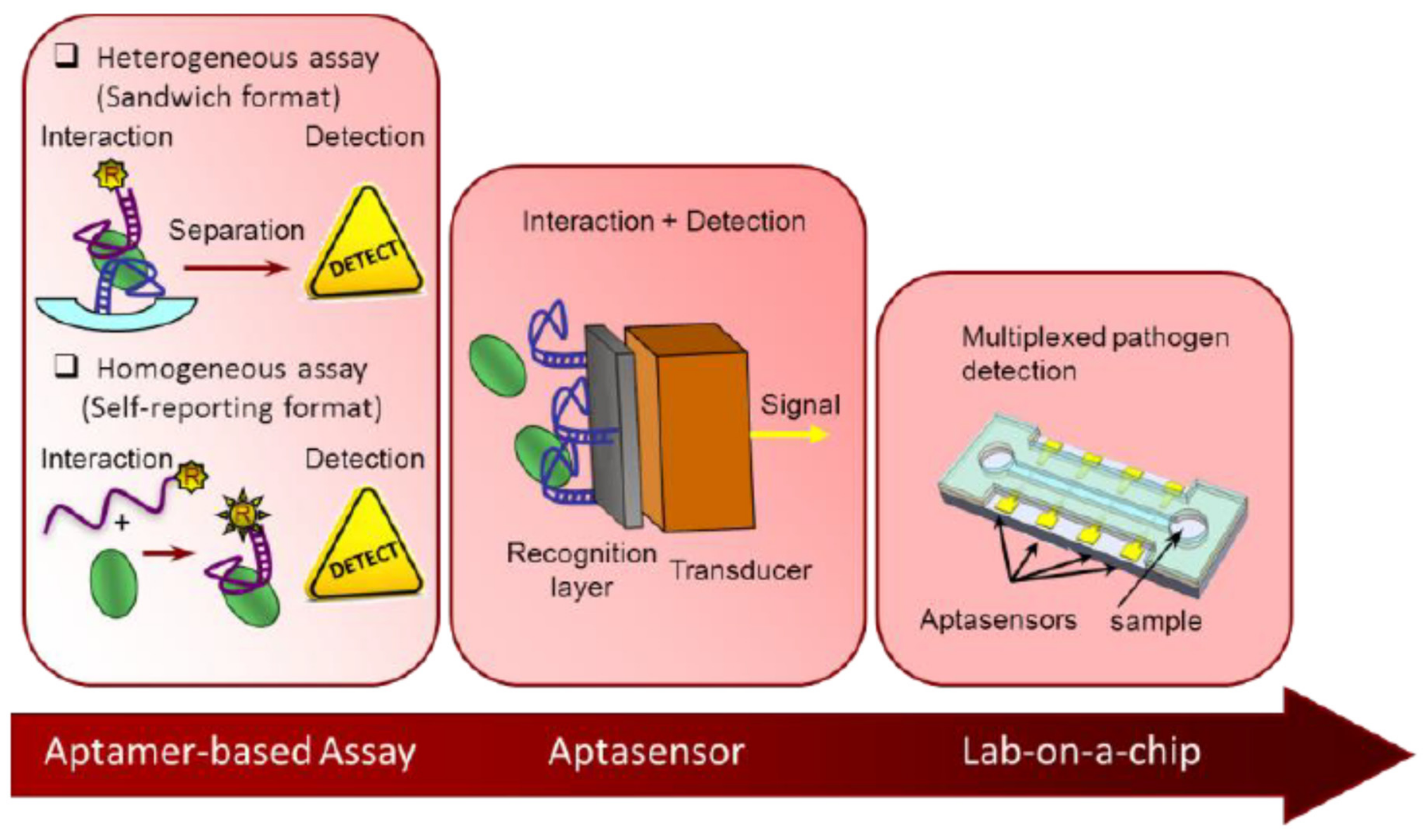
Figure 1. Aptamer-Based Analysis: A Promising Alternative for Food Safety Control. Reprinted from reference [12]. This article is an open access article distributed under the terms and conditions of the Creative Commons Attribution license (http://creativecommons.org/licenses/by/4.0/ (25 March 2021)).
Chemosensors 09 00069 g001
Figure 2. Schematic representation of tetramer, dimer and monomer G-quadruplex structures. The black balls represent the cations that stabilize the structure. Reprinted and adapted from reference [24] with permission from Elsevier.
Chemosensors 09 00069 g002
Figure 3. Schematic illustration of the conformation change of the Triple-helix molecular switch (THMS) structure in the presence of the target Reprinted from reference [26] with permission from Elsevier.
Chemosensors 09 00069 g003
Figure 4. Schematic illustration of two electrochemical aptasensor architectures: a low signal gain sensor and a high signal gain sensor. The latter illustration shows a larger change in conformation upon target binding thus leading to a high signal change. Reprinted with permission from Schoukroun-Barnes LR, Wagan S, White RJ. 2014. Enhancing the Analytical Performance of Electrochemical RNA Aptamer-Based Sensors for Sensitive Detection of Aminoglycoside Antibiotics. Anal. Chem. 86:1131–1137. Copyright (2013) American Chemical Society.
Chemosensors 09 00069 g004
Figure 5. Working principle of a labelled electrochemical aptasensor based on target induced conformation change. The aptamer was previously labelled with a redox probe on its free extremity. The aptamer conformation was modified in the presence of the target analyte (a) the distance between the redox probe to the electrode surface was decreased (“signal on”). Reprinted from reference [16]. This article is an open access article distributed under the terms and conditions of the Creative Commons Attribution (CC BY) license (http://creativecommons.org/licenses/by/4.0/ 25 March 2021).
Chemosensors 09 00069 g005
Figure 6. Working principle of a label free electrochemical aptasensor for the detection of malachite green. Reprinted from reference [31]. This article is an open access article distributed under the terms and conditions of the Creative Commons Attribution (CC BY) license (http://creativecommons.org/licenses/by/4.0/ 25 March 2021).
Chemosensors 09 00069 g006
Figure 7. Schematic illustration of a label-free electrochemical aptasensor based on ladder-shaped DNA structure for the detection of ampicillin in milk. Reprinted from reference [32] with permission from Elsevier.
Chemosensors 09 00069 g007
Figure 8. Principle of the aptasensors based on an Ag-DNAzyme for the detection of chloramphenicol (CAP). Reprinted from [37] with permission from Elsevier.
Chemosensors 09 00069 g008
Figure 9. Working principle of an aptasensor based on aptamer displacement assay. (A) Incubation of tobramycin with the aptamer-modified electrode. (B) Sensing phase regeneration. (C) Saturation of the sensing phase with the aptamer. Reprinted from reference [19] with permission from Elsevier.
Chemosensors 09 00069 g009
Figure 10. Schematic illustration of the target-induced aptamer displacement format and signal amplification by accumulation of redox probes. Reprinted and extracted from reference [38] with permission from Elsevier.
Chemosensors 09 00069 g010
Figure 11. Schematic illustration of two approaches for the development of an electrochemical aptasensor for the detection of kanamycin A based on signaling-probe displacement. Reprinted from reference [40]. with permission from Elsevier.
Chemosensors 09 00069 g011
Figure 12. Two electrochemical aptasensors for the detection of kanamycin using two different signal transduction mechanisms: probe conformation changing mode (PCCM) and target-induced signal probe shifting (TISPC). Reprinted from reference [41] with permission from Elsevier.
Chemosensors 09 00069 g012
Figure 13. Fabrication of aptasensors modified with multi-walled carbon nanotubes (MWCNTs). Reprinted from reference [133]. with permission from Elsevier.
Chemosensors 09 00069 g013
Figure 14. Schematic illustration of an electrochemical aptasensor for the detection of kanamycin in honey based on peroxidase-like activity of gold nanoparticles. Reprinted from reference [136] with permission from Elsevier.
Chemosensors 09 00069 g014
Figure 15. Schematic representation of a label-free aptasensor for the detection of penicillin based on nanoporous PtTi/graphene oxide-Fe3O4/MWCNT-Fe3O4 nanocomposite. Reprinted from reference [65] with permission from Elsevier.
Chemosensors 09 00069 g015
Figure 16. Schematic representation of an electrochemical aptasensor for the detection of streptomycin based on the porous carbon nanorods and multifunctional graphene nanocomposites. Reprinted from reference [77] with permission from Elsevier.
Chemosensors 09 00069 g016
Figure 17. Multiplex electrochemical aptasensor for the multiplex detection of kanamycin and streptomycin in milk based on carbon fiber (CNFs) and mesoporous carbon-gold nanoparticles (OMC-AuNPs). Reprinted from reference [138] with permission from Elsevier.
Chemosensors 09 00069 g017
Figure 18. Schematic illustration of the signal amplification by Exonuclease I for the detection of streptomycin in milk. Reprinted from reference [80] with permission from Elsevier.
Chemosensors 09 00069 g018
Figure 19. Electrochemical aptasensor for ampicillin detection based on the protective effect of aptamer-antibiotic complex towards DpnII and Exo III digestion. Reprinted from reference [69] with permission from Elsevier.
Chemosensors 09 00069 g019
Figure 20. Schematic representation of an electrochemical aptasensor for the detection of streptomycin in milk. The signal was amplified based on target recycling amplification (TRCA). Reprinted from reference [28] with permission from Elsevier.
Chemosensors 09 00069 g020
Figure 21. Schematic illustration of a label-free electrochemical aptasensor for the detection of ampicillin based on target-triggered enzymatic cyclic amplification. Reprinted from reference [35] with permission from Elsevier.
Chemosensors 09 00069 g021
Figure 22. Schematic illustration of a fluorescent aptasensor for the detection of ochratoxin A based on hybridization chain reaction (HCR). Reprinted from reference [139]. This article is an open access article distributed under the terms and conditions of the Creative Commons Attribution (CC BY) license (http://creativecommons.org/licenses/by/4.0/ 25 March 2021).
Chemosensors 09 00069 g022
Figure 23. Schematic illustration of an optical aptasensor based on Rolling circle amplification (RCA). Reprinted from [141]. This article is an open access article distributed under the terms and conditions of the Creative Commons Attribution (CC BY) license (https://creativecommons.org/licenses/by/4.0/ 25 March 2021)).
Chemosensors 09 00069 g023
Figure 24. Schematic representation of a multiplex electrochemical aptasensor for the detection of chloramphenicol (CAP) and oxytetracycline (OTC) in milk based on circular strand-replacement DNA polymerization (CSRP). Reprinted from reference [87] with permission from Elsevier.
Chemosensors 09 00069 g024
Table 1. Summary of types and functions of nanomaterials. Adapted from reference [48]. This article is an open access article distributed under the terms and conditions of the Creative Commons Attribution (CC-BY) license (http://creativecommons.org/licenses/by/4.0/ (25 March 2021)).
Nanomaterials (NMs)
Main FunctionMetallic NMMetal Compound NMNon-Metallic NMCarbon NMsNanostructures
CarrierAuNPsQDsSiO2 NPsGraphene, CNTsDNA nanostructures
EnhancerAuNPs, AgNPsCuO NPsPolyaniline NPsCNTs
ReporterAuNPs, AgNPsQDs, UCNPs CDs
QuencherAuNPs Graphene, CNTs
CatalystPtNPsCuO NPs
ReporterMetal nanoclusters
Separator Fe3O4 NPs
Ag NPs: silver nanoparticles; AuNPs: gold nanoparticles; CDs: carbon dots; CNTs: carbon nanotubes; CuO NPs: Cupric oxide nanoparticles; Fe3O4 NPs: magnetite; QDs: quantum dots; SiO2 NPs: Silicon dioxide nanoparticles; UCNPs: upconversion nanoparticles.
Table 2. Electrochemical aptasensors for the detection of antimicrobial residues in food products.
Electrode@NMsAnalyteFood ProductDetection Technique LOD (ng/mL)References
GCE@AuNPs/MD-BSA/Ab1 + hemin@MOFs/AuPt-Ab2-HRP + HRPMaduramicinEggsAmperometric0.045 ng/mL[52]
GCE@rGO/AgNPsCAPMilkAmperometric0.65 ng/mL[53]
Au bar a@AuNPs/cDNA (S2) + Apt-MOFs (F-) + Au bar b-cDNA (S3) + FSEKanamycin, CAPMilk, fishPotentiometric0.17 ng/mL, 0.15 ng/mL[54]
GNPE@Apt + DNase IKanamycinMilkPotentiometric0.00003 ng/mL[55]
ECNF@AuNPs-AptPenicillinMilkCV0.6 ng/mL[56]
SPCE@4-CP-AptTetracyclineWaterCV0.035 ng/mL[57]
SPE@IL/Fe3O4/AptTetracyclineMilkCV0.44 ng/mL[58]
SPCE@CS-MWCNT/CS-Fe3O4/AptTetracyclineMilkCV0.44 ng/mL[59]
SPGE@cDNA1/cDNA2 + MB-Apt-MB + [Fe(CN)6]3−/4−CiprofloxacinMilkDPV0.033 ng/mL[60]
AuE@S3/S4/Apt/S1/S2/MB/AuNP/HRPKanamycinMilk, honeyDPV0.0000091 ng/mL[61]
AuE@PEI-rGO/AuNCs-AptCAPChicken muscleDPV0.67 ng/mL[62]
GCE@GO-f-/[Si-NH2]/AgNPs-Apt + [Fe(CN)6]3−/4−CAPHoney, milkDPV0.001 ng/mL[63]
GCE@CS-rGO-AuNPs-MIP + KAN + Au@Fe3O4/SH-β-CD-Fc-AptKanamycinMilkDPV0.91 ng/mL[64]
AuE@AD/TS + Apt-Primer + DWAmpicillinWaterDPV0.0003 ng/mL[34]
GCE@ GO-Fe3O4/MWCNT-Fe3O4/PtTi/AptPenicillinMilkDPV0.0253 ng/mL[65]
SPCE@Au-APtStreptomycinMilkDPV0.0026 ng/mL[28]
MBs-Ampi + GCE + biotinylated-Apt + SA-HRP + MWCNTAmpicillinMilkDPV0.00003 ng/mL[66]
GCE@GO/ZnOCAPMilk, honeyDPV3.23 ng/mL[67]
AuE@captDNA + Apt-Primer + ExoIII + Hemin + H2O2KanamycinMilkDPV0.00004 ng/mL[68]
AuE@captDNA + Apt-Primer + ExoIII + Hemin + H2O2AmpicillinMilkDPV0.027 ng/mL[35]
GCE@cDNA/Apt + DpnII + Exo III + [Fe(CN)6]3−/4−AmpicillinMilkDPV0.011 ng/mL[69]
SPGE@ssDNA1/ssDNA6/Apt/ssDNA2/ssDNA7/ssDNA3/ssDNA4/ssDNA5 + [Fe(CN)6]3−/4−AmpicillinMilkDPV0.00035 ng/mL[32]
GCE@MCH/Apt/AuNPs/GQDs-WS2/Malachite greenFishDPV1.11 ng/mL[31]
AuE@cDNA/Apt + Nuclease P1 + Ab anti-dsDNA + [Fe(CN)6]3−/4−SDMXMilkDPV0.01 ng/mL[70]
GCE@Pt–Sn@TiO2/cDNA/Apt + RecJf exonucleaseStreptomycinMilkDPV0.01 ± 0.003 ng/mL[71]
TFGE@Apt + cDNA/MWCNT/AuNPs + thionineOTCChicken meatDPV3.1 × 10−5 ng/mL[72]
GCE/OMC-CS/AuNPs-SA/Biot-cDNA1/Fc-DNA2 (Apt)KanamycinMilkDPV0.017 ng/mL[44]
AuE@Apt-cDNA + C60-rGO/Tb/AuNPs + ExoNaseSDMXMilkDPV10 × 10−6 ng/mL[73]
E@AuNPsCNFs/MWCNTs/cDNA-KAN/polyA/cDNA-STR + CdS-AptKAN + PbS/AptSTRKanamycin, streptomycinMilkDPV0.036 ng/mL
0.021 ng/mL
[74]
AuE@cDNA + biotinylated Apt + MB + AuNPs/SA/HRP + H2O2KanamycinMilkDPV0.00088 ng/mL[75]
AuE1 and AuE2/S1-S2(Apt)/S3/MBKanamycinMilkDPV7.8 × 10−6 ng/mL[76]
AuE@HP2 + Apt-primer cDNA + Exo I + HP1 + nb.BbvCI + Hemin/K++ H2O2KanamycinMilkDPV0.00024 ng/mL[36]
GCE@PCNR/GR–Fe3O4–AuNPs/AptStreptomycinmilkDPV0.028 ng/mL[77]
AuE@cDNA1/cDNA2/cDNA3/Apt +Exo I + [Fe(CN)6]3−/4−Tetracycline, doxycylineMilkDPV0.2 ng/mL[78]
GCE@PEDOT–AuNPs/GR–Fe3O4NPs/AptPenicillinMilkDPV0.057 ng/mL[79]
AuE@cDNA/Apt + Exo I + [Fe(CN)6]3−/4−StreptomycinMilkDPV8.2 ng/mL[80]
GCE@PCNR/MWCNT-CuO-AuNPs/AptStreptomycinMilk, honeyDPV0.036 ng/mL[81]
SPGE@GelB-AptCAPMilkDPV0.059 ng/mL[82]
AuE@Apt + Biotinylated detection DNA + ST-APCAPHoneyDPV0.29 ng/mL[83]
GCE + Au bar a@AuNPs/S1/S2 5AptKAN)-S’1/S’2 (AptAMP) + cDNAKAN (S4)/Apo (Pb) + cDNAAMP (S’4)/Apo (Cd) + Au bar b@AuNPs/S3Kanamycin, ampicillinMilk, fishSWV8.7 × 10−6 ng/mL, 0.00001 ng/mL[84]
AuE@HP (DNAzyme) + Apt/Ag + [Fe(CN)6]3−/4−CAPMilkSWV0.0004 ng/mL[37]
Au bar a@Apt1/cDNA1/AuNPs-EcoR1-Apt2/cDNA2/AuNPs-BamH1 + Exo I + Au bar b-dsDNA1&2/MOFs (MB, Fc)Kanamycin, CAPMilkSWV0.0000170 ng/mL, 0.0000068 ng/mL[85]
GCE@MB-Apt Probe cDNA, M-NMOF (Pb2+, Cd2+)Kanamycin, CAPMilkSWV0.0000775 ng/mL, 0.0000061 ng/mL[86]
GCE + MGNPs@aDNA/CaptDNA1&2 (=Apt + primer) + cDNA-MOFs (sDNA1 Pb, sDNA2 Cd)CAP, OTCMilkSWV0.0000107 ng/mL, 0.000022 ng/mL[87]
GCE@S3/S4 + Apt-CAP + Apt-OTC + S1-MOF (Cd) + S2-MOF (Pb) + Exo ICAP, OTCMilkSWV0.15 and 0.10 ng/mL[88]
AuE@AptCAPMilkSWV0.52 ng/mL[89]
AuE@CaptDNA + 3 Apt (STR, CAP, TTC) + 3 cDNA1s + 3 QD–cDNA2 (PbS, CdS, ZnS NCs) + HNO3Streptomycin, CAP, TTCMilkSWASV5.82 ng/mL, 1.62 ng/mL, 8.89 ng/mL[90]
AuE@POP-AptAmpicillinMilkEIS0.00000133 ng/mL[91]
GCE@MWCNT/GO/AptTetracyclineMilkImpedimetric0.0000212 ng/mL[92]
PGE@AuNPs/RGO/AptTetracyclineMilkEIS0.000000017 ng/mL[93]
GCE@CB/CS-AptKanamycinMilk, yoghurtEIS0.15 ng/mL[94]
AuE@Ag(I) MOFs/Apt + [Fe(CN)6]3−/4−PenicillinMilkEIS0.000849 ng/mL[95]
AuE@Ce-MOF/MCA/AptOTCMilkEIS17.4 × 10−6 ng/mL[96]
AuE@SWCNT-cDNA + AptOTCWaterImpedimetric1.125 ng/mL[97]
AuE@Ce-MOF/MCA-AptOTCMilkEIS0.00000037 ng/mL[96]
PGE@rGO/AuNPs-AptPenicillin GMilkEIS0.00000027 ng/mL[98]
PGE@rGO/AuNPs-AptSDMXFish, chicken, beefEIS0.000000115 ng/mL[99]
SPCE@MWCNT-V2O5-CS-Apt + [Fe(CN)6]4−/3−CiprofloxacinMilkEIS0.5 ng/mL[100]
AuE@TPN-COF@Co-MOF-AptAmpicillinMilkEIS0.217 × 10−6 ng/mL[101]
SPCE@4-CP-Apt + [Fe(CN)6]4−/3−KanamycinMilkEIS0.11 ng/mL[45]
SPCE@4-NB-Apt + [Fe(CN)6]4−/3−Penicillin GMilkEIS0.17 ng/mL[102]
IDAM@CS/TiO2 NPsTetracyclineMilkEIS3 ng/mL[103]
AuE@Apt + [Fe(CN)6]4−/3−CAP/EIS0.57 ng/mL[104]
Apo: apoferritine; Apt: aptamer; AuE: gold electrode; AuNPs gold nanoparticles; Co-MOF: Co-based metal-organic frameworks; CPE Carbon paste electrode; 4-CP: 4-carboxyphenyl; CS: chitosan; Exo-I: exonuclease; Endonuclease DpnII; endonucleases Nb.BbvCI; Fc: ferrocene; FSE: fluoride selective electrode; GR-Fe3O4: graphene and iron oxide (magnetite); AD: hairpin anchoring-DNA; CaptDNA: capture DNA; DW: hairpin DNA walker; ECNF: electrospun carbon nanofiber; EN: restriction endonuclease (EcoRI or BamHI); Fe3O4: iron oxide; GR: graphene; GR–Fe3O4NPs: magnetic graphene nanocomposite; GR–Fe3O4–AuNPs: multifunctional graphene nanocomposites; IDAMs: interdigitated array microelectrodes; MB: methylene blue; MGNPs Magnetic gold nanoparticles; MIP: molecular imprinting polymer; MOFs: metal−organic frameworks; MoS2: molybdenumdisulfide; MWCNTs: multi-walled carbo nanotubes; NP-PtTi: nanoporous platinum and titanium alloy; NCs: nanoclusters; OMC-CS: ordered mesoporous carbon-chitosan; OTC: oxytetracycline; PANI: polyaniline; PCNR: porous carbon nanorods; PEDOT: poly(3,4-ethylenedioxythiophene); PEI-rGO/AuNCs: polyethyleneimine-functionalized reduced graphene oxide and gold nanocubes composite; PGE: pencil graphite electrode; POP: porous organic polymer; Pt–Sn@TiO2: Pt and Sn nanoparticles on the surface of TiO2 nanorods; SA: streptavidin; SDMX: sulfadimethoxine; TPN-COF: terephthalonitrile-based covalent organic framework; TS: three-stranded DNA complex. Fc/β-CD-SH: β-cyclodextrin-ferrocene; APT/Fc/β-CD-SH/Au@Fe3O4.
Table 3. Electrochemical derived techniques for the aptasensing of antimicrobial residues in food products.
Electrode@NMs AnalyteFood ProductDetection TechniqueLOD (ng/g or nM)References
ITO@ZIS-HNCs/CS-AptLincomycinMilkPEC0.000034 ng/mL[105]
ITO@AuNPs/sDNA/Fc-rp/Fc-Apt + HP1-HP2 + MBStreptomycinWaterPEC&EC dual-mode10.06 ng/mL[106]
ITO@SnO2/Bi2S3-AptTobramycinMilkPEC2 ng/mL[107]
ITO@ZnxCo3-xO4/N-GQDs/AgBiS2-AptAmpicillinWaterPEC0.00009 ng/mL[108]
ITO@g-CN/Co3O4-AptOTCWaterPEC0.0016 ng/mL[109]
ITO@BiOI/Au-AptTetracycline PEC0.0002 ng/mL[110]
GCE@Bi-BiOI@C/AptCAP/PEC0.26 ng/mL[111]
FTO@BR-CN/cDNA/biotinylatedApt + SAPtNi + H2O2CAPMilkPEC0.0000084 ng/mL[112]
ITO@MIL−68(In)-NH2/MWCNT/CdS/AptTTCWaterPEC0.0067 ng/mL[113]
ITO@Bi2S3/NGQDs/CS/GA/AptSDMXMilkPEC0.009 ng/mL0[114]
ITO@BiFeO3/utg-C3N4-AptAmpicillinMilkPEC0.0000115 ng/mL[115]
ITO@Bi2MoO6/BNG-AptLincomycinMilkPEC0.0015 ng/mL[116]
TiE@ TiO2 NTs/GQDsCAPHoneyPEC0.019 ng/mL[117]
TiE@ TiO2 NTs/Ag2S/PDA-AptOfloxacinMilkPEC0.00027 ng/mL[118]
FTO@TiO2NRA/Eu3+ QDs (CdS)-AptCAPMilkPEC0.00012 ng/mL[119]
ITO@WS2-Apt + DNaseCAPMilk powderPEC0.0012 ng/mL[120]
ITO@BiPO4/3DNGH-AptTetracyclineMilkPEC0.0147 ng/mL[121]
ITO@TiO2-MoS2-AuNPs-AptKanamycinMilkPEC0.024 ng/mL[122]
ITO@CdTe QDs/SWCNHs-AptStreptomycinHoneyPEC0.02 ng/mL[123]
ITO@TiO2/H-DNA@CdTe QDs-AptOTCChicken, raw milkPEC0.09 ng/mL[124]
FTO@G-Bi2S3-AptSDMXMilkPEC0.17 ng/mL[125]
PE@Luminol/H2O2/AgNPs-AptKanamycinMilkECL0.06 ng/mL[126]
GCE@Au-GO + CDots-Apt + MIPLincomycinMeatECL0.000065 ng/mL[127]
GCE@CdS NCs/Apt-ssDNA/EV (HRP)-Au-SSB + ExoICAPFishSPR-ECL0.000011 ng/mL[128]
WE1: SPCE@CdS QDs-MG Apt-Cy5WE2: SPCE@ L-AuNPs-CAP Apt-CAMG, CAPFishECL0.023 ng/mL MG, 0.01 ng/mL CAP[129]
WE1: SPCE@L-AuNPs/cDNA/CA/AptWE2: SPCE@CdS QDsCAPFishR-ECL0.01 ng/mL[130]
CPE@AuNPs/GOx-Apt−csDNA/AuNPs/SiO2 + CPE@AuNPs/PDA/Laccase + glucoseAmpicillinMilkEBFCs0.7 ng/mL[131]
AgNPs: Silver nanoparticles; Apt: aptamer; Bi2MoO6: Aurivillius-phase bismuth molybdate; BiOI: bismuth oxyiodides; BNG: Boron and Nitrogen co-doped graphene; CAP: chloramphenicol; CPE: carbon paper electrode; CA: chromogenic acid; CdTe: cdmimium telluride; CS: chitosan; EBFCs: enzyme biofuel cells; FTO: fluorine-doped tin oxide electrodes; GA: glutaraldehyde; GQDs/TiO2 NTs: graphene quantum dots-sensitized TiO2 nanotube arrays; H-DNA: hairpin DNA; ITO Indium-tin oxide electrode; hydrogen peroxide (H2O2); luminol-gold nanoparticles (L-Au NPs); MG: malachite green; MWCNTs: multi-walled carbon nanotubes; nitrogen-doped graphene quantum dots (NGQDs; PEC: photoelectrochemistry; PE: platinum electrode; PDA: Polydopamine; QDs: quantum dots; R-ECL: Ratiometric electrochemiluminescence; SDMX: sulfadimethoxine; SPR: surface plasmon resonance; SWCNH: s single walled carbon nanohorns; SPCE: screen printed carbon electrode; utg-C3N4: ultrathin graphite-like carbon nitride nanosheets; WE: working electrode; ZIS-HNCs: hollow ZnIn2S4 nanocages.
Publisher’s Note: MDPI stays neutral with regard to jurisdictional claims in published maps and institutional affiliations.