Article

Angiotensin-Converting Enzyme 2 (ACE2) As a Novel Biorecognition Element in A Cell-Based Biosensor for the Ultra-Rapid, Ultra-Sensitive Detection of the SARS-CoV-2 S1 Spike Protein Antigen

by
Sofia Mavrikou
1,*,
Vasileios Tsekouras
1,
Kyriaki Hatziagapiou
2,3,
Asimina Tsalidou
1,
Petros Bakakos
4,
Nikoletta Rovina
4,
Antonia Koutsoukou
4,
Athanasios Michos
2,
Olti Nikola
2,
Eleni Koniari
5,
Joseph Papaparaskevas
6,
George P. Chrousos
5,
Christina Kanaka-Gantenbein
2 and
Spyridon Kintzios
1,*
1
Laboratory of Cell Technology, Department of Biotechnology, Agricultural University of Athens, EU-CONEXUS European University, 11855 Athens, Greece
2
First Department of Pediatrics, National and Kapodistrian University of Athens, “Agia Sofia” Children’s Hospital, Thivon 1, 11527 Athens, Greece
3
Physiotherapy Department and Department of Nursing, Faculty of Health and Care Sciences, State University of West Attica, Agiou Spiridonos 28, Egaleo, 12243 Athens, Greece
4
First University Department of Respiratory Medicine, “Sotiria” Hospital, “Medical School, National and Kapodistrian University of Athens, 152 Mesogeion Ave, 11527 Athens, Greece
5
University Research Institute of Maternal and Child Health and Precision Medicine, and UNESCO Chair on Adolescent Health Care, National and Kapodistrian University of Athens, “Agia Sofia” Children’s Hospital, Thivon & Livadias 8 str, 11527 Athens, Greece
6
Department of Microbiology, Medical School, National and Kapodistrian University of Athens, Mikras Asias 75, 11527 Athens, Greece
*
Authors to whom correspondence should be addressed.
Chemosensors 2021, 9(12), 341; https://doi.org/10.3390/chemosensors9120341
Submission received: 31 October 2021 / Revised: 20 November 2021 / Accepted: 1 December 2021 / Published: 3 December 2021

Abstract

:
Antigen screening for the SARS-CoV-2 S1 spike protein is among the most promising tools for the mass monitoring of asymptomatic carriers of the virus, especially in limited resource environments. Herewith, we report on the possible use of the angiotensin-converting enzyme 2 (ACE2), the natural receptor and entry point of the virus, as a biorecognition element for the detection of the S1 antigen combined with an established bioelectric biosensor based on membrane-engineered cells. The working principle of our approach is based on the measurable change of the electric potential of membrane-engineered mammalian cells bearing ACE2 after attachment of the respective viral protein. We demonstrate that sensitive and selective detection of the S1 antigen is feasible in just three min, with a limit of detection of 20 fg/mL. In a preliminary clinical application, positive patient-derived samples were identified with a 87.9% score compared to RT-PCR. No cross-reactivity was observed against a wide range of nucleocapsid protein concentrations. The novel biosensor is embedded in a commercially ready-to-use testing platform, complete with the consumable immobilized cell–electrode interface and a portable read-out device operable through smartphone or tablet. In addition, the possible application of the system for the high throughput screening of potential pharmacological inhibitors of the ACE2 receptor-S1 RBD interaction is discussed.

Graphical Abstract

1. Introduction

Human coronaviruses (CoVs) known to cause disease in humans include the hCoV-NL63 and hCoV-229E, classified as alpha CoVs and the HKU1, HCoV-OC43, SARS-CoV (Severe Acute Respiratory Syndrome CoV), and MERS-CoV (Middle East respiratory syndrome CoV) classified xspecies transmission are determined by its recognition by surface cellular receptors of the host. CoVs bind to cellular receptors via the large, surface-located homotrimeric spike glycoprotein (S), comprising of a S1 subunit and S2 subunit in each spike monomer. The claw-like N-terminal peptidase domain of the host cell surface metallopeptidase angiotensin-converting enzyme 2 (ACE2) serves as binding domain for the SARS-CoV, HCoV-NL63 and the SARS-Co-2. Such binding triggers a cascade of events, mediating cell recognition, attachment and fusion between host cell and viral membranes, allowing entry of the virus into target cells [1,2,3,4]. Interestingly, the majority of residues in the SARS-CoV-2 receptor-binding domain (RBD) of the spike protein which are essential for ACE2 binding, share high structural similarity with those in the SARS-CoV RBD, as they are either highly conserved or with similar side chain properties [4,5].
Two years after experiencing the coronavirus pandemic, public health experts are increasingly in support of increasing the frequency of individual testing as an incremental component of the global respiratory syndrome-coronavirus 2 (SARS-CoV-2) testing strategy, also as a perquisite for the safe re-launching of normality and return to pre-pandemic conditions and activities [6]. For instance, it has been estimated that by repeating rapid tests every three days and isolating positive individuals, 88% of viral transmission would be achieved [7] In particular, timely identification of asymptomatic carriers in an affordable, sensitive and specific manner would be key to the global management of COVID-19 [8,9]. So far, no such diagnostic tools have been applied at a massive, global scale [10].
Antigen tests, targeting protein moieties of the SARS-CoV-2 virus have emerged as powerful tools to identify asymptomatic carriers of the virus, especially during the first two weeks following infection [1,2,3] at a high rate of success. Antigen screening has gained in significance especially in limited resource environments and countries where vaccination against the virus is still progressing at a low pace [11,12]. Contrary to serology-based assays, the identification of the four major SARS-CoV-2 surface protein types is independent of seroconversion, which usually peaks around one to two weeks after the onset of symptoms [10,11] and also precedes the onset of symptoms [7,8,9]. Among them, the spike protein S1 has been recognized as the major target of antigen tests, since it has a prominent role in the viral host cell entry through its ability to bind to the angiotensin-converting enzyme 2 (ACE2).
ACE2 is an integral membrane protein converting, by means of carboxypeptidase activity, Ang-II Ang1-7, a heptapeptide [13]. S1 spike protein–ACE2 binding is facilitated by a 193 amino-acid (residues 318–510) receptor-binding domain (RBD) in the S1 domain of SARS-CoV spike glycoprotein [14,15,16]. SARS-CoV-2 engages ACE2 for cell entry by a combination of receptor binding and proteolysis of the spike protein [17,18]. ACE2 is primarily expressed in bronchus and lung parenchyma, testis, arterial and venous endothelium, arterial smooth muscle cells, islet cells of the pancreas, renal and gastrointestinal tissues (oral mucosa, duodenum, ileum, jejunum, caecum, and colon), whereas its expression in central nervous system and lymphoid tissues is limited, as demonstrated with real-time PCR analysis of 72 human tissues [19,20,21]. ACE2 extensive tissue distribution might explain the multi-organ dysfunction, observed in COVID-19 patients. Additionally, the tropism of SARS-CoV-2 for the respiratory tract could be explained by the abundant expression of ACE2 on alveolar epithelial type II cells (AT2), suggesting that these pneumonocytes could serve as a reservoir for viral invasion. AT2, although comprising only 4–7% of the alveolar surface, they constitute 60% of alveolar epithelial cells and 10–15% of all lung cells, implicated in synthesis and secretion of surfactant, regeneration of the alveolar epithelium following its injury, transepithelial movement of water, and metabolism of xenobiotics. More importantly, ACE2–expressing AT2 cells demonstrate upregulation of genes related with viral transmission, replication, assembly, and release, suggesting that these cells might favor SARS-CoV-2 replication in the lung parenchyma [22]. In vitro and in vivo models have demonstrated that overexpression of ACE2 enhanced SARS-CoV-2 entry into cells and disease severity, especially lung injury, and that the viral cell entry was not dependent upon other CoVs receptors, such as aminopeptidase-N (hAPN) and dipeptidyl peptidase 4 (hDPP4 or hCD26), used by HCoV-229E, and MERS-CoV, respectively [5]. Dissociation constants of the ACE2-S1 Receptor Binding Domain (RBD) complex lie in the 1–100 nM range [1] and it is twelve times stronger (EC50 = 0.08 μg/mL) than the respective binding of SARS-CoV. In addition, docking studies have revealed a very strong interaction in terms of binding free energy (−13.76 kcal/mol) [23].
Due its strong affinity for the S1 spike protein, ACE2 is an attractive biorecognition element for the potential development of S1-targeting assays. So far, very few and quite recent studies have been reported on the utilization of ACE2 in biosensors and other diagnostic approaches for screening SARS-CoV-2 antigens. Vezza et al. [24] developed an impedimetric biosensor based on a low-cost PCB electrode functionalized with ACE2 though physisorption into 1H,1H,2H,2H-perfluorodecanethiol (PFDT), a process allowed by the enzyme’s hydrophobic region which enables ACE2 insertion into the cell membrane. The biosensor achieved a limit of detection (LOD) for recombinant spike protein of 1.68 ng/mL, while binding between ACE2 and S1, as determined with the biosensor, was significantly different to IL-6 and streptavidin. The system was validated on inactivated clinical samples providing results within 30 min. SARS-CoV-2 was detected in the range of 102–105 copies/mL. In another study, de Lima et al. [25] reported the development of a cheap (~ USD 1.5/unit) electrochemical biosensor, termed Low-Cost Electrochemical Advanced Diagnostic (LEAD). The sensor was based on graphite electrodes functionalized with gold nanoparticles modified with ACE2. For the detection of SARS-CoV-2, square wave voltammetry was applied (amplitude potential 75 mV, frequency 80 Hz, step potential 8 mV). LOD was determined at 229 fg/mL, with results being recorded within 6–7 min and using 50 μL of sample. After being tested on 103 nasopharyngeal clinical samples (53 positive and 50 negative samples), 88.7% sensitivity, 86.0% specificity, and 87.4% accuracy were recorded. Saliva samples were also tested at the proof-of-concept stage. Finally, the shelf-life of the functionalized test electrodes was 5 d at 4 °C.
In the past two years and following the outbreak of the COVID-19 pandemic, our research group has responded to the global coronavirus screening challenge by developing and clinically validating a novel biosensor for the ultra-rapid (3 min) and sensitive detection of the SARS-CoV-2 S1 spike protein [26]. The biosensor, which was set up as a ready to use, fully commercial handheld device operated via smartphone/tablet has a limit of detection of 1 fg/mL and a semilinear range of response between 10 fg and 1 μg/mL. The working principle, a customized extension of a process known as Molecular Identification through Membrane Engineering [27,28] is based on mammalian cells, the membrane of which is engineered by electroinsertion to bear the human chimeric spike S1 antibody at high densities. Binding of the SARS-CoV-2 S1 protein to the cell membrane-bound antibodies caused a measurable, rapid and selective change in the membrane-engineered cell bioelectric properties. The novel biosensor was clinically validated in a series of clinical trials both by our team and, independently, by other groups, and was determined able to detect the virus in positive samples with a 92.8% success rate, total sensitivity of 92.7% and a specificity of 97.8%, compared to RT-PCR [29,30].
In the present study, we report the modification of our novel approach for the detection of the SARS-CoV-2 S1 spike protein by using, as the biorecognition element, mammalian cells membrane-engineered with ACE2. By using the novel biosensor, we were able to detect the SARS-CoV-2 spike S1 protein within just three min, with a linear response in the concentration range of 2 fg–2 pg/mL and a limit of detection of 20 fg/mL. Positive patient-derived samples were identified with 88.7% and 87.9% sensitivity and accuracy, respectively compared to RT-PCR. No false negative results were recorded.

2. Materials and Methods

2.1. Cell Culture Conditions

In the present study, human neuroblastoma, SK-N-SH cells were obtained from ATCC (ATCC® HTB-11™, LGC Standards GmbH, Wesel, Germany) and proliferated in Eagle’s Minimum Essential Medium (MEM). The basal media was enriched by Earle’s balanced salt solution (BSS) (Biowest, Nuaillé, France), 10% Fetal Bovine Serum (FBS) (Thermo Fisher Scientific, Waltham, MA, USA), 2 mM l-glutamine, 0.1 mM non-essential amino acids and 1.0 mM sodium pyruvate (Biowest, Nuaillé, France). Finally, 1 U/μg antibiotics (penicillin/streptomycin) were added, and the cultures were maintained under standard conditions (37 °C, 5% CO2, saturated humid atmosphere) in a growth chamber. The adherent cells were subcultured in a 1:10 ratio, once or twice per week, following a 3–10 min enzymatic dissociation from the culture vessel with Trypsin-EDTA (0.05% trypsin, 0.02% EDTA) (Biowest, Nuaillé, France).

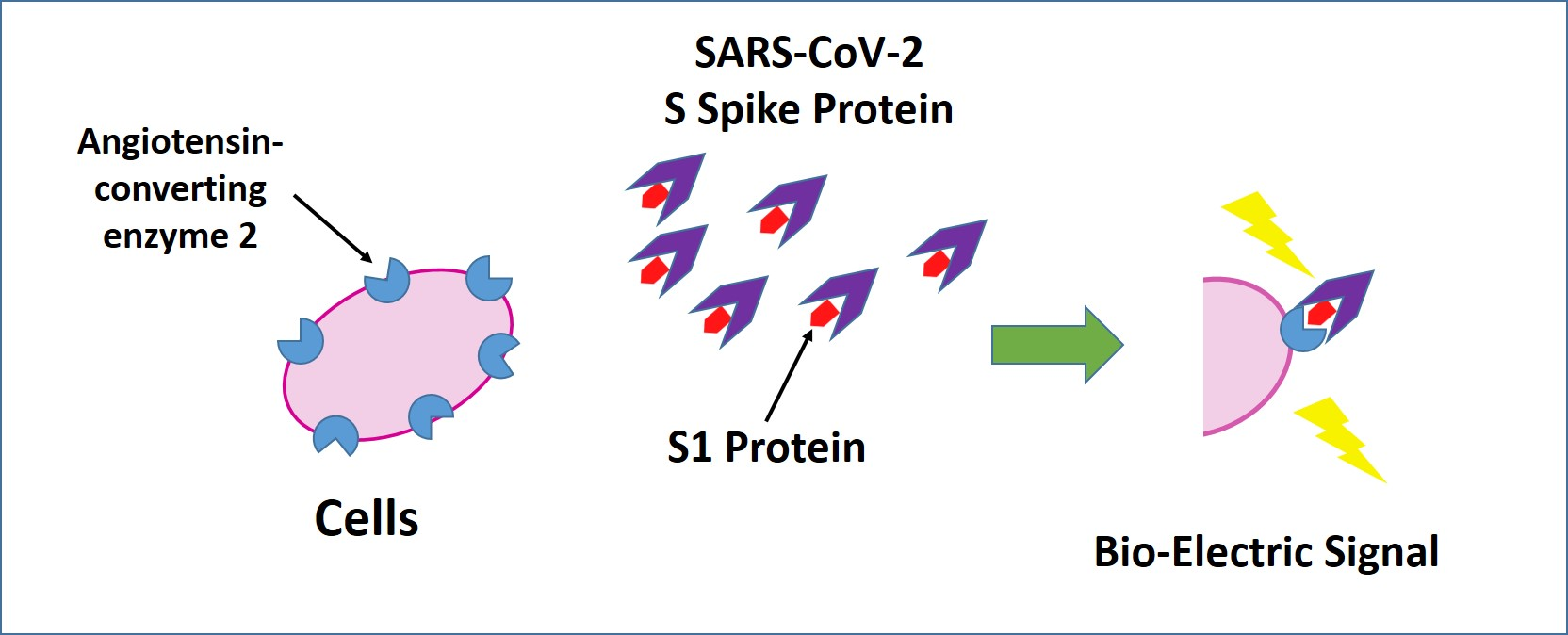
2.2. Development of Bioelectric Recognition Elements: Membrane-Engineered Cells (SK-N-SH/ACE2)

Membraned engineered mammalian cells were used as the bioelectric recognition elements of the sensor. For this purpose, angiotensin-converting enzyme-2 (ACE2, Sigma-Aldrich, SAE0064) was electroinserted into the cell membrane of human neuroblastoma SK-N-SH cells according to Mavrikou et al. [26]. Trypsin-EDTA was used for the cells enzymatic detachment and 2.5 × 10⁶/mL cell populations were resuspended in phosphate-buffered saline (PBS) (pH 7.4). Then, various ACE2 concentrations (750, 1500, and 2250 Units/mL) were added, and the mixtures of cells suspensions with the respective enzyme concentrations were incubated at 4 °C for a period of 20 min. Afterwards, the mixture was transferred to specific cuvettes and subjected to electroporation (Eppendorf Eporator, Eppendorf AG, Hamburg, Germany) by receiving two electric pulses at 1800 V/cm. Immediately after the electric pulses applications, the cells were transferred to a Petri dish (60 mm × 15 mm) containing culture medium and placed in the incubator overnight. Eventually, the membrane-engineered cells were collected from the Petri dish after mechanical detachment and were resuspended in phosphate-buffered saline in Eppendorf tubes. A custom-made extracellular collagen-based (approx. 0.5% w/v) hydrogel was used as a supporting matrix for cell 3D cultures [30]. The bio-electric interactions of ACE2/SK-N-SH cells with the spike S1 protein were then recorded either in cell suspensions (50 × 103 cells per sensor) or in three-dimensional (3D) cell cultures (25 × 103 cells per sensor).

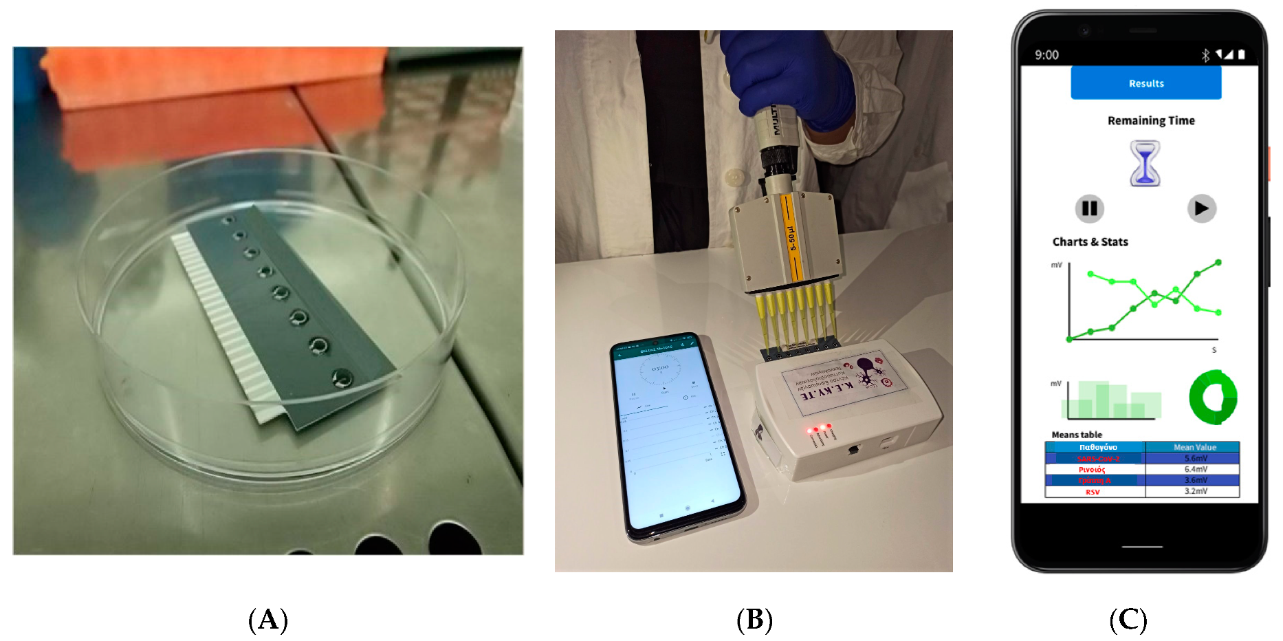
2.3. Biosensor System Assembly-Membrane Potential Recordings

The recordings of bio-electric signals produced by membrane-engineered cells as a response to interactions with analyte anions, according to the principle of the Bioelectric Recognition Assay (BERA) have been previously validated in several studies [31,32].
Thus, an eight-channel configuration custom-made potentiometer in order to measure the alterations of the membrane-engineered cells’ potential. A disposable carbon screen printed multichannel electrode cassette (working electrode: carbon, reference: Ag/AgCl) (www.embiodiagnostics.eu (accessed on 2 November 2021), Cyprus) was integrated into the measurement device (Figure 1A).
Upon the addition of the sample to the top of each carbon electrode containing membrane-engineered cells either in suspensions or 3D cultures (Figure 1B), potentiometric measurements derived by the cell responses were recorded for 3 min in Volts (360 values, sampling rate 2 Hz). Furthermore, the recorded sensors’ data were uploaded via a tablet/Bluetooth communication to a cloud server in order to be further analyzed (Figure 1C) [26].

2.4. Patient Enrollment to Small Scale Clinical Trial, Clinical Examination and Collection of Specimens

All volunteers (17) participating in the study were tested with RT-PCR (Real-Time Polymerase Chain Reaction) on nasopharyngeal swabs for SARS-CoV-2 antigen collected, by a standardized procedure, using Dacron swabs (FIRATMED, 8870000244, Albacete, Spain). At the time of hospital admission and prior to the sample collection for the antigen detection with the biosensor. Seven patients and 10 healthy volunteers consented to have two replicate specimens collected (Table S1). The specimens’ collection for antigen testing was conducted at 3 December 2020. Each specimen was transported in a sterile centrifuge tube (Greiner 15 mL centrifuge tubes) containing 10 mL of PBS (Phosphate-Buffered Saline) at pH 7.4 (Gibco PBS, 10010023, Thermo Fisher Scientific, Waltham, MA USA). Initially, specimens were stored on ice for transport to the clinical laboratory and then they were kept at −80 °C, until the analysis [33,34].

2.5. Experimental Outline and Data Interpretation

The present investigation was implemented in four experimental stages. Initially, we studied the optimum enzyme’s (ACE2) concentration for the biosensor set-up in membrane-engineered cells suspensions. At the second step, we investigated the range of S1 spike protein detection. For this purpose, we used standard spike S1 protein concentrations (2 fg/mL, 20 fg/mL, 200 fg/mL, 2 pg/mL, 20 pg/mL, 200 pg/mL and 2 ng/mL) in PBS and in spiked SARS-CoV-2 negative patient nasopharyngeal samples. Then, we tested the standard S1 solutions and spiked samples against the membrane-engineered cells in 3D cultures. Then, we assessed the biosensor’s cross-reactivity with SARS-CoV-2 nucleocapsid (Z03488, GenScript Biotech B.V., Leiden, The Netherlands) PBS solutions (200 fg/mL, 2 pg/mL, 20 pg/mL, 200 pg/mL and 2 ng/mL). Finally, in the fourth experimental step, we evaluated the BERA biosensor based on 3D membrane-engineered cell culture against SARS-CoV-2 negative and positive patient nasopharyngeal samples. Each sample was tested twice, using a set of eight individual sensors in each application (n = 16). The biosensors’ measurements are presented as a ratio to control while the clinical samples (patients and healthy donors) are expressed as follows:
Normalized   Biosensor   Response = Contol     Sample Control
The results are presented as the mean ± SD whereas the differences between the means were tested for statistical significance using an analysis of variance and Student’s t-test.
Biosensor’s limit of detection was determined by measuring progressively increasing concentrations of the analyte (2 fg/mL–200 ng/mL). The empirical method that we used defines the LOD value as the lowest concentration at which statistical significance (p value) was higher than 0.01 (** p < 0.01) [35,36].

3. Results

3.1. Membrane Engineering of SK-N-SH Cells with ACE2 Triggers Distinct Bioelectric Alterations after Interaction with the SARS-CoV-2 Spike S1 Protein

A 10 μL sample volume of several concentrations of the S1 protein (20 fg/mL, 200 fg/mL, 2 pg/mL, 20 pg/mL, 200 pg/mL and 2 ng/mL) was applied to a population of 50 × 103 membrane-engineered cells in suspension. Upon exposure to increasing concentrations of SARS-CoV-2 spike S1 protein, the SK-N-SH/ACE2 membrane-engineered cells demonstrated differential responses depending on the ACE-2 concentration used. The lowest ACE2 concentration (Figure 2A) presented statistically significant differences after application of 20 fg/mL and 20 pg/mL S1 protein when compared to the control (plain PBS solution). Overall, the bioelectric signal produced was relatively weak in comparison with the cellular responses obtained with the 1500 Units/mL (Figure 2B) and 2250 Units/mL (Figure 2C) of ACE2. A semi-linear concentration dependent pattern was observed with the use of 1500 Units/mL of ACE2 for cell membrane engineering. The limit of S1 detection was very low, down to 20 fg/mL, and very strong signals were obtained even at the highest concentrations used (2 ng/mL). In contrast, when 2250 Units/mL of ACE2 were used for increasing cell specification the results obtained displayed a decrease in the cell membrane potential. This effect, i.e., the decline of the biosensor response above low target analyte concentrations when the biorecognition element has been electroinserted at low or high concentrations (750/2250 Units/mL) is a frequent, yet not fully investigated phenomenon in molecular recognition through membrane engineering [37,38]. It is possibly due to either low level of responses at low ACE2 concentrations (even at higher S1 concentrations) or due to regional ion fluctuations of the cell membranes during the interaction with the analyte [39].

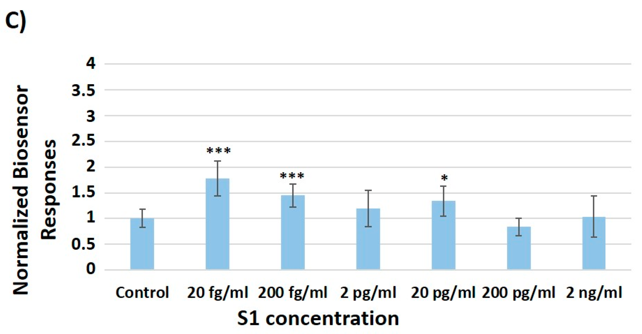
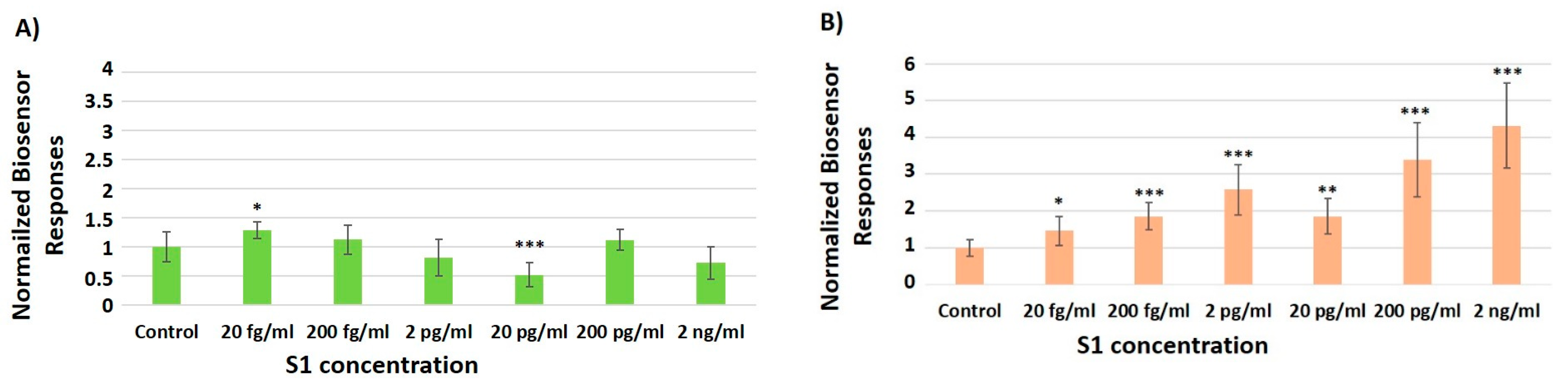
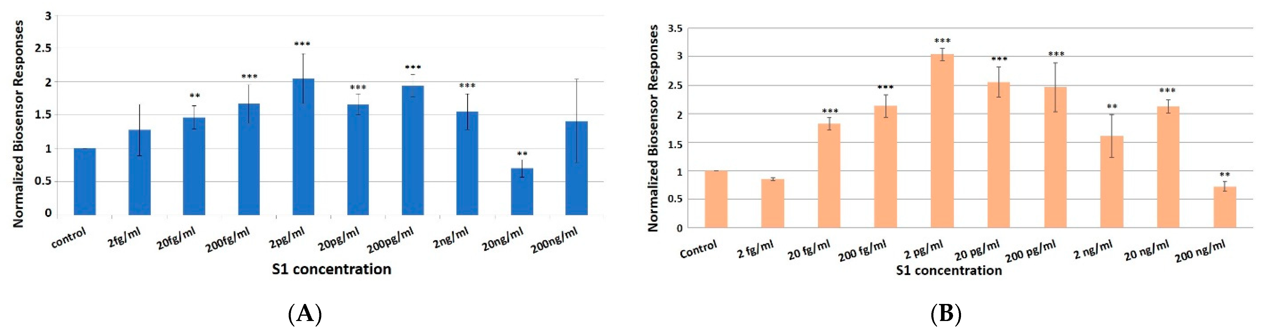
3.2. Concentration-Dependent Responses towards Increasing Concentrations of the SARS-CoV-2 Spike S1 Protein

Membrane-engineered cells with 1500 Units/mL ACE2 enzyme immobilized in a collagen-based biomatrix [22] were treated with escalating concentrations (2 fg/mL–200 ng/mL) of the SARS-CoV-2 spiked S1 protein either in PBS or nasopharyngeal solutions (from healthy donors).
The cell–protein interaction produced an obvious and very fast response within a 3 min interval. The bioelectric alterations of the membrane-engineered 3D cell cultures after exposure to S1 spike protein either in PBS (Figure 3A) or in nasopharyngeal solutions (Figure 3B), were distinctly detectable. More specifically, a linear pattern was observed upon the treatment with increasing concentrations (20 fg–2 pg/mL) of S1 protein. The lower and upper limits of detection for S1 protein in PBS solutions was ranging from 20 fg to 2 ng/mL (above control values), whereas in nasopharyngeal solutions, the range was between 20 fg and 20 ng/mL (above control values).
The biosensor response against increasing S1 concentrations, in particular within the linear range of response, amounted to approx. 150–200% of control samples for PBS spiked solutions and approx. 180–300% of control for nasopharyngeal spiked solutions.

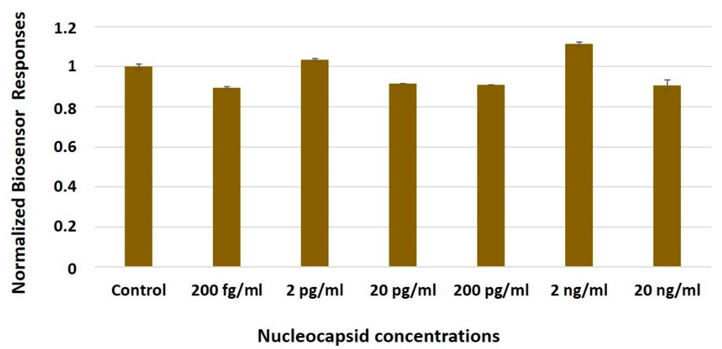
3.3. The Biosensor Response Is Selective for the SARS-CoV-2 Spike S1 Protein

The membrane-engineered SK-N-SH/ACE2 cell-based biosensor was also evaluated against several concentrations of the SARS-CoV-2 nucleocapsid (NC) protein (200 fg/mL–20 ng/mL) in PBS solutions (Figure 4). The bio-electric responses produced following the application of each NC concentration were either slightly lower or upper than the zero concentration of the control samples. Contrary to the sensor’s response against the S1 antigen, changes in the biosensor potential against increasing nucleocapsid concentrations (200 fg–20 ng/mL) ranged only between approx. 90 and 110% of the control PBS solutions, i.e., no essential response was observed against the non-target protein. That is to say, no cross reactions were observed, regarding these specific SARS-CoV-2 proteins.

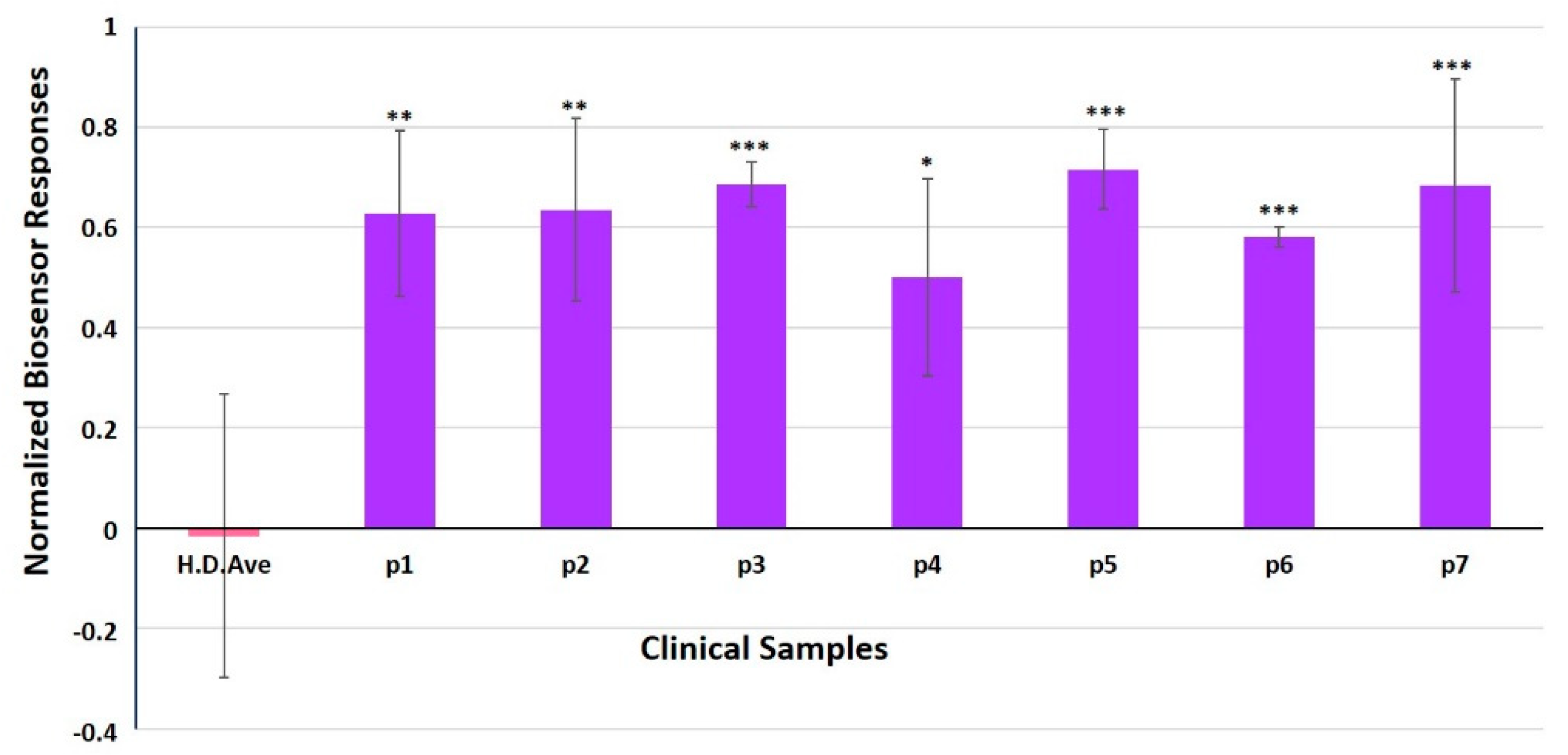
3.4. Evaluation of SK-N-SH/ACE2 Biosensor Performance for the Ultra-Rapid Detection of the SARS-CoV-2 S1 Spike Protein Antigen in Clinical Samples by the Membrane-Engineered Cells in Suspension and 3D Conditions in Comparison with RT-PCR Results

Membrane-engineered cells in 3D cultures were used to validate the biosensors accuracy and specificity in comparison with the standard RT-PCR technique. Samples from healthy donors (10) and COVID-19 patients (7) were used, all of which had previously been tested with RT-PCR. The biosensor successfully identified all negative samples with no false-positive results. The normalized cell biosensor’s responses against specimens derived from patients are presented in Figure 5. The biosensor recognized all positive samples, four of them with statistical significance p < 0.001, two with statistical significance p < 0.01, and one specimen with statistical significance p < 0.05.

4. Discussion

In the present study, we investigated the possible use of ACE2 as a biorecognition element for the detection of the SARS-CoV-2 S1 spike protein combined with an established bioelectric biosensor based on membrane-engineered cells. This approach was compared with using human chimeric anti-S1 antibodies as the S1-binding agents on membrane-engineered cells, a method already used for developing a clinically validated biosensor for the detection of the coronavirus S1 antigen [26,29,30].
Comparing the performance of the ACE2-based biosensor with the anti-S1-based biosensor, their patterns of response against increasing S1 antigen concentrations are similar, with a concentration-dependent linear pattern being observed after the administration of the SARS-CoV-2 spike S1 protein in the concentration range of 20 fg–2 pg/mL. This pattern was observed both in spiked PBS and nasopharyngeal control samples, demonstrating the specific and medium-independent response of the biosensor. This is probably due to the fact that both the anti-S1 antibody and ACE2 bind with the S1 protein in a highly selective and specific manner and similar affinities (Kd = 12.4 nM and 14.7 nM, respectively) [40,41].
On the other hand, using ACE2 as the biorecognition element resulted in 87.9% successful characterization of the positive samples, compared to 92.8% success with the anti-S1-based biosensor. This could be attributed to the binding ability of ACE2 to both S1 and S2 subunits (the second one inducing fusion of the viral envelope with cellular membranes) [18], as well as due to ACE2 availing both over high- and low-affinity S1 binding domains, which could be equally represented in membrane-engineered cells [42]. Furthermore, the novel ACE2-based biosensor exhibited essentially zero cross-reactivity against the non-target nucleocapsid protein, as demonstrated by the sensor’s response against it (~90–110% of control) compared with the response against the S1 antigen (~150–200% of control/~180–300% of control for PBS or nasopharyngeal spiked solutions, respectively).
Compared with the recent, albeit few, methods utilizing ACE2 as a sensing element for the SARS-CoV-2 spike S1 antigen [24,25], our approach allows for a faster and considerably more sensitive detection (LOD = 20 fg/mL) of the antigenic protein, at least at the proof-of-concept stage. In addition, based on our previously established results with cell immobilization in a hydrogel-based extracellular matrix [30], a shelf life of the membrane-engineered cell test consumable for at least two weeks can be achieved. On the other hand, ACE2 may represent a critical step in the further improvement of the principle of molecular identification through membrane engineering, since, contrary to the anti-S1 antibody, ACE2 is already a membrane-based receptor protein, availing over a hydrophobic region which could greatly facilitate electroinsertion into the cell membrane [43].
In order to ameliorate our study’s efficiency, the clinical samples were selected according to specific matching criteria commonly utilized (age, sex, etc.). As indicated by Table S1, all patients had a positive RT-PCR test performed in a period of 6–38 days before sample collection. Furthermore, patient 4 had been positive for SARS-CoV-2 38 days prior to the specimen collection, indicating that the biosensor is able to detect the disease even at very low concentrations of the virus, with statistical significance p < 0.05.
The primary goal of our study was the assessment of this novel methodological approach to develop a screening tool for the coronavirus antigen. However, since ACE2 is the main point of entry for SARS-CoV-2 virions, its interaction with the viral spike protein S1 subunits is a key target in COVID-19 therapeutic research. In other words, ACE2 inhibition is a likely therapeutic approach [44,45]. Therefore, the experimental principle presented here could probably be used to identify potential inhibitors of SARS-CoV-2 entry into the cells by assaying the inhibition of S1 binding to the ACE2 receptor. A few studies have been already reported in this line of research. For example, Yang et al. [46] used atomic force microscopy to investigate the mechanism of S-protein/ACE2 interaction, both on ACE2-covered gold surfaces and ACE2-expressing A549 cells. They determined that the S1-RBD has a strong intrinsic affinity for ACE2 (~120 nM), possibly extended to the whole-virus level. More recently, Kiew et al. [47] applied an electrochemical impedance spectroscopy (EIS)-based biosensing platform having a recombinant ACE2-coated palladium electrode as the core sensing element for screening potential pharmacological inhibitors against S1-ACE2 interaction. The panel of tested compounds comprised FDA-approved peptide analogs computationally predicted to promote hydrogen bonding interactions with ACE2, including ramipril, perindopril and cilazapril. The recording of binding of inhibitors at concentrations as low as 0.1 μg/mL was achieved.

5. Conclusions

In this report, we demonstrated the feasibility and clinical applicability of a biosensor for the detection of the SARS-CoV-2 S1 spike protein antigen based on the recording of changes of the bioelectric properties of mammalian cells membrane-engineered with ACE2. A linear pattern was observed at very low concentrations, between 20 fg/mL and 2 pg/mL, of S1 antigen in both PBS solutions and nasopharyngeal spiked samples. Moreover, the biosensors efficiency of using ACE2 as the membrane-bound biorecognition element was found similar to anti-S1 human chimeric antibody (sensitivity for anti-S1 = 92.7%, whereas for ACE2 = 88.7%), the partial hydrophobic, membrane-based nature of the ACE2 receptor could possibly render it more suitable for mass-manufacturing membrane-engineered cells. In addition, binding of ACE2 to both S1 and S2 subunits could demonstrate a significant recognition efficiency of structural moieties of SARS-CoV-2 particles, thus leading in high rates of successful characterization of positive clinical samples (87.9% accuracy in comparison with PCR). As a complete system, the novel biosensor utilizes the already tested, clinically validated and commercially ready-to-use testing platform, composed of (a) the membrane-engineered cells immobilized in a proprietary biomatrix for extended shelf-life on the electrode array (b) the bespoke read-out device and (c) the user interface and classification software operable on a smartphone or tablet. Therefore, we plan to proceed with its immediate validation in a considerably larger trial, including both nasopharyngeal and saliva samples. In addition, we plan to explore the application of this novel approach for high-throughput screening of potential pharmacological inhibitors of the ACE2 receptor–S1 RBD interaction.

Supplementary Materials

The following are available online at https://www.mdpi.com/article/10.3390/chemosensors9120341/s1, Table S1. Brief presentation of patients’ personal and medical history.

Author Contributions

Conceptualization, S.K.; methodology, S.K. and S.M.; formal analysis, S.K., S.M., P.B., N.R., A.K., A.M., O.N., E.K., J.P., G.P.C., C.K.-G. and V.T.; investigation, S.M., A.T., K.H. and S.K.; data curation, S.M. and V.T.; writing—original draft preparation, S.K., V.T., K.H. and S.M.; writing—review and editing, S.K.; supervision, S.K. All authors have read and agreed to the published version of the manuscript.

Funding

This research received no external funding.

Institutional Review Board Statement

The study was conducted according to the guidelines of the Declaration of Helsinki, and approved by the Ethics Committee of the Agricultural University of Athens (protocol code 35 and date of approval 10 June 2021).

Informed Consent Statement

Informed consent was obtained from all subjects involved in the study.

Data Availability Statement

The data presented in this study are available on request from the corresponding author. The data are not publicly available due to medical confidentiality and privacy.

Acknowledgments

The authors acknowledge the administrative support of Evangelos Koukounasoulis and Stavros Ousios.

Conflicts of Interest

The authors declare no conflict of interest.

References

  1. Wang, Q.; Zhang, Y.; Wu, L.; Niu, S.; Song, C.; Zhang, Z.; Lu, G.; Qiao, C.; Hu, Y.; Yuen, K.-Y.; et al. Structural and Functional Basis of SARS-CoV-2 Entry by Using Human ACE2. Cell 2020, 181, 894–904.e899. [Google Scholar] [CrossRef] [PubMed]
  2. Li, F.; Li, W.; Farzan, M.; Harrison, S.C. Structure of SARS Coronavirus Spike Receptor-Binding Domain Complexed with Receptor. Science 2005, 309, 1864–1868. [Google Scholar] [CrossRef]
  3. Li, W.; Moore, M.J.; Vasilieva, N.; Sui, J.; Wong, S.K.; Berne, M.A.; Somasundaran, M.; Sullivan, J.L.; Luzuriaga, K.; Greenough, T.C.; et al. Angiotensin-converting enzyme 2 is a functional receptor for the SARS coronavirus. Nature 2003, 426, 450–454. [Google Scholar] [CrossRef] [Green Version]
  4. Lan, J.; Ge, J.; Yu, J.; Shan, S.; Zhou, H.; Fan, S.; Zhang, Q.; Shi, X.; Wang, Q.; Zhang, L.; et al. Structure of the SARS-CoV-2 spike receptor-binding domain bound to the ACE2 receptor. Nature 2020, 581, 215–220. [Google Scholar] [CrossRef] [Green Version]
  5. Zhang, H.; Penninger, J.M.; Li, Y.; Zhong, N.; Slutsky, A.S. Angiotensin-converting enzyme 2 (ACE2) as a SARS-CoV-2 receptor: Molecular mechanisms and potential therapeutic target. Intensive Care Med. 2020, 46, 586–590. [Google Scholar] [CrossRef] [Green Version]
  6. Service, R.F. Fast, cheap tests could enable safer reopening. Science 2020, 369, 608–609. [Google Scholar] [CrossRef]
  7. Luo, K.; Lei, Z.; Hai, Z.; Xiao, S.; Rui, J.; Yang, H.; Jing, X.; Wang, H.; Xie, Z.; Luo, P.; et al. Transmission of SARS-CoV-2 in Public Transportation Vehicles: A Case Study in Hunan Province, China. Open Forum Infect. Dis. 2020, 7, ofaa430. [Google Scholar] [CrossRef] [PubMed]
  8. Morales-Narváez, E.; Dincer, C. The impact of biosensing in a pandemic outbreak: COVID-19. Biosens. Bioelectron. 2020, 163, 112274. [Google Scholar] [CrossRef] [PubMed]
  9. Udugama, B.; Kadhiresan, P.; Kozlowski, H.N.; Malekjahani, A.; Osborne, M.; Li, V.Y.C.; Chen, H.; Mubareka, S.; Gubbay, J.B.; Chan, W.C.W. Diagnosing COVID-19: The Disease and Tools for Detection. ACS Nano 2020, 14, 3822–3835. [Google Scholar] [CrossRef] [Green Version]
  10. Kaushik, A. Manipulative magnetic nanomedicine: The future of COVID-19 pandemic/endemic therapy. Expert Opin. Drug Deliv. 2021, 18, 531–534. [Google Scholar] [CrossRef]
  11. Kilic, T.; Weissleder, R.; Lee, H. Molecular and Immunological Diagnostic Tests of COVID-19: Current Status and Challenges. iScience 2020, 23, 101406. [Google Scholar] [CrossRef]
  12. Malik, Y.S.; Kumar, N.; Sircar, S.; Kaushik, R.; Bhat, S.; Dhama, K.; Gupta, P.; Goyal, K.; Singh, M.P.; Ghoshal, U.; et al. Coronavirus Disease Pandemic (COVID-19): Challenges and a Global Perspective. Pathogens 2020, 9, 519. [Google Scholar] [CrossRef]
  13. Turner, A.J.; Hiscox, J.A.; Hooper, N.M. ACE2: From vasopeptidase to SARS virus receptor. Trends Pharmacol. Sci. 2004, 25, 291–294. [Google Scholar] [CrossRef] [PubMed]
  14. Babcock, G.J.; Esshaki, D.J.; Thomas, W.D., Jr.; Ambrosino, D.M. Amino acids 270 to 510 of the severe acute respiratory syndrome coronavirus spike protein are required for interaction with receptor. J. Virol. 2004, 78, 4552–4560. [Google Scholar] [CrossRef] [PubMed] [Green Version]
  15. Wong, S.K.; Li, W.; Moore, M.J.; Choe, H.; Farzan, M. A 193-amino acid fragment of the SARS coronavirus S protein efficiently binds angiotensin-converting enzyme 2. J. Biol. Chem. 2004, 279, 3197–3201. [Google Scholar] [CrossRef] [Green Version]
  16. Zhang, Y.; Zheng, N.; Hao, P.; Cao, Y.; Zhong, Y. A molecular docking model of SARS-CoV S1 protein in complex with its receptor, human ACE2. Comput. Biol. Chem. 2005, 29, 254–257. [Google Scholar] [CrossRef]
  17. Tang, T.; Bidon, M.; Jaimes, J.A.; Whittaker, G.R.; Daniel, S. Coronavirus membrane fusion mechanism offers a potential target for antiviral development. Antivir. Res. 2020, 178, 104792. [Google Scholar] [CrossRef]
  18. Saponaro, F.; Rutigliano, G.; Sestito, S.; Bandini, L.; Storti, B.; Bizzarri, R.; Zucchi, R. ACE2 in the Era of SARS-CoV-2: Controversies and Novel Perspectives. Front. Mol. Biosci. 2020, 7, 271. [Google Scholar] [CrossRef] [PubMed]
  19. Harmer, D.; Gilbert, M.; Borman, R.; Clark, K.L. Quantitative mRNA expression profiling of ACE 2, a novel homologue of angiotensin converting enzyme. FEBS Lett. 2002, 532, 107–110. [Google Scholar] [CrossRef] [Green Version]
  20. Tipnis, S.R.; Hooper, N.M.; Hyde, R.; Karran, E.; Christie, G.; Turner, A.J. A Human Homolog of Angiotensin-converting Enzyme: Cloning and Functional Expression as a Captopril-Insensitive Carboxypeptidase *. J. Biol. Chem. 2000, 275, 33238–33243. [Google Scholar] [CrossRef] [Green Version]
  21. Devaux, C.A.; Lagier, J.-C.; Raoult, D. New Insights Into the Physiopathology of COVID-19: SARS-CoV-2-Associated Gastrointestinal Illness. Front. Med. 2021, 8, 640073. [Google Scholar] [CrossRef]
  22. Zhao, Y.; Zhao, Z.; Wang, Y.; Zhou, Y.; Ma, Y.; Zuo, W. Single-cell RNA expression profiling of ACE2, the receptor of SARS-CoV-2. Am. J. Respir. Crit. Care Med. 2020, 202, 756–759. [Google Scholar] [CrossRef] [PubMed]
  23. de Andrade, J.; Gonçalves, P.F.B.; Netz, P.A. Why Does the Novel Coronavirus Spike Protein Interact so Strongly with the Human ACE2? A Thermodynamic Answer. ChemBioChem 2021, 22, 865–875. [Google Scholar] [CrossRef] [PubMed]
  24. Vezza, V.J.; Butterworth, A.; Lasserre, P.; Blair, E.O.; MacDonald, A.; Hannah, S.; Rinaldi, C.; Hoskisson, P.A.; Ward, A.C.; Longmuir, A.; et al. An electrochemical SARS-CoV-2 biosensor inspired by glucose test strip manufacturing processes. Chem. Commun. 2021, 57, 3704–3707. [Google Scholar] [CrossRef] [PubMed]
  25. de Lima, L.F.; Ferreira, A.L.; Torres, M.D.T.; de Araujo, W.R.; de la Fuente-Nunez, C. Minute-scale detection of SARS-CoV-2 using a low-cost biosensor composed of pencil graphite electrodes. Proc. Natl. Acad. Sci. USA 2021, 118, e2106724118. [Google Scholar] [CrossRef]
  26. Mavrikou, S.; Moschopoulou, G.; Tsekouras, V.; Kintzios, S. Development of a Portable, Ultra-Rapid and Ultra-Sensitive Cell-Based Biosensor for the Direct Detection of the SARS-CoV-2 S1 Spike Protein Antigen. Sensors 2020, 20, 3121. [Google Scholar] [CrossRef]
  27. Kintzios, S. Molecular Identification through Membrane Engineered Cells. World Intellectual Property Organization, WO2007/083170Al, 26 July 2007. [Google Scholar]
  28. Kokla, A.; Blouchos, P.; Livaniou, E.; Zikos, C.; Kakabakos, S.E.; Petrou, P.S.; Kintzios, S. Visualization of the membrane engineering concept: Evidence for the specific orientation of electroinserted antibodies and selective binding of target analytes. J. Mol. Recognit. 2013, 26, 627–632. [Google Scholar] [CrossRef]
  29. Apostolou, T.; Loizou, K.; Hadjilouka, A.; Inglezakis, A.; Kintzios, S. Newly Developed System for Acetamiprid Residue Screening in the Lettuce Samples Based on a Bioelectric Cell Biosensor. Biosensors 2020, 10, 8. [Google Scholar] [CrossRef] [Green Version]
  30. Mavrikou, S.; Tsekouras, V.; Hatziagapiou, K.; Paradeisi, F.; Bakakos, P.; Michos, A.; Koutsoukou, A.; Konstantellou, E.; Lambrou, G.I.; Koniari, E.; et al. Clinical Application of the Novel Cell-Based Biosensor for the Ultra-Rapid Detection of the SARS-CoV-2 S1 Spike Protein Antigen: A Practical Approach. Biosensors 2021, 11, 224. [Google Scholar] [CrossRef]
  31. Kintzios, S.; Pistola, E.; Konstas, J.; Bem, F.; Matakiadis, T.; Alexandropoulos, N.; Biselis, I.; Levin, R. The application of the bioelectric recognition assay for the detection of human and plant viruses: Definition of operational parameters. Biosens. Bioelectron. 2001, 16, 467–480. [Google Scholar] [CrossRef]
  32. Kintzios, S.; Pistola, E.; Panagiotopoulos, P.; Bomsel, M.; Alexandropoulos, N.; Bem, F.; Ekonomou, G.; Biselis, J.; Levin, R. Bioelectric recognition assay (BERA). Biosens. Bioelectron. 2001, 16, 325–336. [Google Scholar] [CrossRef]
  33. Kline, A.; Putnam, N.E.; Youn, J.-H.; East, A.; Das, S.; Frank, K.M.; Zelazny, A.M. Dacron swab and PBS are acceptable alternatives to flocked swab and viral transport media for SARS-CoV-2. Diagn. Microbiol. Infect. Dis. 2021, 99, 115209. [Google Scholar] [CrossRef]
  34. Clerici, B.; Muscatello, A.; Bai, F.; Pavanello, D.; Orlandi, M.; Marchetti, G.C.; Castelli, V.; Casazza, G.; Costantino, G.; Podda, G.M. Sensitivity of SARS-CoV-2 Detection With Nasopharyngeal Swabs. Front. Public Health 2021, 8, 593491. [Google Scholar] [CrossRef]
  35. Armbruster, D.; Tillman, M.; Hubbs, L.M. Limit of Detection (LOD)/Limit of Quantitation (LOQ): Comparison of the empirical and the statistical methods exemplified with GC-MS assays of abused drugs. Clin. Chem. 1994, 40, 1233–1238. [Google Scholar] [CrossRef] [PubMed]
  36. Shrivastava, A. Methods for the determination of limit of detection and limit of quantitation of the analytical methods. Chron. Young Sci. 2011, 2, 21–25. [Google Scholar] [CrossRef]
  37. Moschopoulou, G.; Kintzios, S. Application of “membrane-engineering” to bioelectric recognition cell sensors for the ultra-sensitive detection of superoxide radical: A novel biosensor principle. Anal. Chim. Acta 2006, 573–574, 90–96. [Google Scholar] [CrossRef] [PubMed]
  38. Shin, S.; Choi, M.; Shim, J.; Park, S. Hook effect detection and detection-range-controllable one-step immunosensor for inflammation monitoring. Sens. Actuators B Chem. 2020, 304, 127408. [Google Scholar] [CrossRef]
  39. Moschopoulou Georgia, K.S. Membrane engineered Bioelectric Recognition Cell sensors for the detection of subnanomolar concentrations of superoxide: A novel biosensor principle. In Proceedings of the International Conference on Instrumental Methods of Analysis (IMA), Crete, Greece, 1–5 October 2005. [Google Scholar]
  40. Joyce, M.G.; Sankhala, R.S.; Chen, W.-H.; Choe, M.; Bai, H.; Hajduczki, A.; Yan, L.; Sterling, S.L.; Peterson, C.E.; Green, E.C.; et al. A Cryptic Site of Vulnerability on the Receptor Binding Domain of the SARS-CoV-2 Spike Glycoprotein. bioRxiv 2020. [Google Scholar] [CrossRef] [Green Version]
  41. Wrapp, D.; Wang, N.; Corbett, K.S.; Goldsmith, J.A.; Hsieh, C.L.; Abiona, O.; Graham, B.S.; McLellan, J.S. Cryo-EM structure of the 2019-nCoV spike in the prefusion conformation. Science 2020, 367, 1260–1263. [Google Scholar] [CrossRef] [Green Version]
  42. Prabakaran, P.; Xiao, X.; Dimitrov, D.S. A model of the ACE2 structure and function as a SARS-CoV receptor. Biochem. Biophys. Res. Commun. 2004, 314, 235–241. [Google Scholar] [CrossRef]
  43. Warner, F.J.; Smith, A.I.; Hooper, N.M.; Turner, A.J. What’s new in the renin-angiotensin system? Cell. Mol. Life Sci. CMLS 2004, 61, 2704–2713. [Google Scholar] [CrossRef]
  44. Han, Y.; Král, P. Computational Design of ACE2-Based Peptide Inhibitors of SARS-CoV-2. ACS Nano 2020, 14, 5143–5147. [Google Scholar] [CrossRef] [PubMed] [Green Version]
  45. Li, S.R.; Tang, Z.J.; Li, Z.H.; Liu, X. Searching therapeutic strategy of new coronavirus pneumonia from angiotensin-converting enzyme 2: The target of COVID-19 and SARS-CoV. Eur. J. Clin. Microbiol. Infect. Dis. 2020, 39, 1021–1026. [Google Scholar] [CrossRef] [PubMed]
  46. Yang, J.; Petitjean, S.J.L.; Koehler, M.; Zhang, Q.; Dumitru, A.C.; Chen, W.; Derclaye, S.; Vincent, S.P.; Soumillion, P.; Alsteens, D. Molecular interaction and inhibition of SARS-CoV-2 binding to the ACE2 receptor. Nat. Commun. 2020, 11, 4541. [Google Scholar] [CrossRef] [PubMed]
  47. Kiew, L.-V.; Chang, C.-Y.; Huang, S.-Y.; Wang, P.-W.; Heh, C.-H.; Liu, C.-T.; Cheng, C.-H.; Lu, Y.-X.; Chen, Y.-C.; Huang, Y.-X.; et al. Development of flexible electrochemical impedance spectroscopy-based biosensing platform for rapid screening of SARS-CoV-2 inhibitors. Biosens. Bioelectron. 2021, 183, 113213. [Google Scholar] [CrossRef] [PubMed]
Figure 1. Biosensor’s set-up. Carbon screen-printed multi-electrode (8×) cassettes were prepared with 3D membrane-engineered cell cultures (A). The electrode strip was attached to a portable custom-made potentiometer connected to a smartphone device via Bluetooth for measurements’ recording immediately after the sample addition (B). The bio-electric signal produced was presented as a voltage vs. time graph on the screen of a smartphone (C).
Chemosensors 09 00341 g001
Figure 2. Distinct response of SK-N-SH/ACE2 membrane-engineered cells against the SARS-CoV-2 spike S1 protein. Cells were membrane-engineered with different enzyme concentrations ((A): 750 Units/mL, (B): 1500 Units/mL, (C): 2250 Units/mL). Control: response of membrane-engineered cells in plain PBS solution. ***: statistically significant different results (p < 0.001); **: statistically significant different results (p < 0.01); *: statistically significant different results (p < 0.05); Results (mean ± SD) are expressed as ratio to control (n = 16).
Chemosensors 09 00341 g002aChemosensors 09 00341 g002b
Figure 3. Normalized (ratio to control) cell biosensor’s responses in 3D cultures against the SARS-CoV-2 spike S1 protein in PBS (A) and spiked nasopharyngeal solutions (B). SK-N-SH/ACE2 cells membrane-engineered with 1500 Units/mL of ACE2 were used as the biorecognition elements. ***: statistically significant different results (p < 0.001); **: statistically significant different results (p < 0.01). Results (mean ± SD) are presented after 3 min (columns) of sample–cell interaction and are expressed as ratio to control (n = 16).
Chemosensors 09 00341 g003
Figure 4. Biosensor cross-response against the SARS-CoV-2 nucleocapsid (NC) protein in the 200 fg/mL–20 ng/mL concentration range. Membrane-engineered SK-N-SH/ACE2 cells were utilized as the biorecognition element. Results are presented after three minutes (brown columns) of sample–cell interaction and are expressed as ratio to control (n = 16).
Chemosensors 09 00341 g004
Figure 5. Normalized cell biosensor’s responses in 3D cultures against clinical samples from patients (n = 7) and healthy donors (mean of 10 samples—pink column. Results are expressed as normalized biosensor responses ([control–sample response]/control). Significant differences (Student’s T-test) between normalized biosensor responses * < 0.05, ** < 0.01, *** < 0.001.
Chemosensors 09 00341 g005
Publisher’s Note: MDPI stays neutral with regard to jurisdictional claims in published maps and institutional affiliations.

