Communication

Development of a Textile Solid-Phase Extraction Method Employing the Marine Sulfated Polysaccharide Ulvan for the Extraction and Analysis of Cationic Dyes

1
Department of Nanobiotechnology, Institute of Soil Biology and Biochemistry, Biology Centre CAS, Na Sadkach 7, 370 05 Ceske Budejovice, Czech Republic
2
Regional Centre of Advanced Technologies and Materials, Czech Advanced Technology and Research Institute, Palacky University, Slechtitelu 27, 783 71 Olomouc, Czech Republic
3
Department of Magnetism, Institute of Experimental Physics, SAS, Watsonova 47, 040 01 Kosice, Slovakia
4
Section of Pharmacognosy and Chemistry of Natural Products, Department of Pharmacy, National and Kapodistrian University of Athens, Panepistimiopolis Zografou, 15771 Athens, Greece
*
Authors to whom correspondence should be addressed.
Chemosensors 2025, 13(2), 75; https://doi.org/10.3390/chemosensors13020075
Submission received: 23 December 2024 / Revised: 8 February 2025 / Accepted: 14 February 2025 / Published: 19 February 2025
(This article belongs to the Section (Bio)chemical Sensing)

Abstract

:
Textile solid-phase extraction (TSPE) employing an acrylic nonwoven textile impregnated with the sulfated, negatively charged marine polysaccharide ulvan was employed for the extraction of four positively charged dyes, namely safranin O, methylene blue, malachite green and crystal violet, from aqueous solutions. The extracted dyes were analyzed by image analysis of the dyed textile using a smartphone application. Dyes within the concentration range of 0.1–2.0 mg/L were efficiently analyzed, demonstrating that ulvan-based TSPE is a highly efficient preconcentration method for the analysis of cationic dyes.

1. Introduction

The determination of various types of contaminants in potable water and wastewater is of high importance since their presence can pose significant risks to human health and the environment. Among contaminants, organic dyes, heavily used in various industrial sectors, such as textiles, plastics, food, cosmetics, pharmaceutics, leather and paper, are regarded as toxic not only in high concentrations but, in several cases, even in trace amounts [1].
Up to now, several analytical methods have been employed for the determination of dyes in water samples, most frequently based on high-pressure liquid chromatography (HPLC) coupled to various detectors, such as fluorescence, ultraviolet and diode array, as well as hyphenated techniques such as liquid chromatography–mass spectrometry (LC-MS) and tandem mass spectrometry (LC-MSn). In addition, gas chromatography (GC), capillary electrophoresis and spectrophotometric methods have been used for the determination of dyes [1]. These methods often offer high sensitivity but require separation and/or preconcentration, demanding the use of expensive instrumentation and chemical reagents, as well as long processing times for the pretreatment of samples. Such pretreatment methods include liquid–liquid extraction (LLE) and solid-phase extraction (SPE), with various sorbents (e.g., C18, polystyrene-divinylbenzene polymers, activated charcoal, zeolite, diatomite) having been employed for the enrichment of organic dyes prior to their analysis [1,2]. Currently, SPE procedures are routinely used, with special attention paid to the development and application of advanced materials for the efficient extraction of dyes [3].
New alternative methods, such as magnetic solid-phase extraction (MSPE) and textile solid-phase extraction (TSPE), have been developed and studied for the extraction of organic dyes [4,5]. In the latter, pieces of woven or nonwoven textiles are loaded with an appropriate ion exchange or affinity ligand to enable the entrapment of the target analyte. For the quantification of dyes, two basic strategies can be employed, namely the elution of the dyes from the textile adsorbent and subsequent photometric or chromatographic analysis or elution-free assays employing image analysis of the colored textile pieces [5,6].
Recently, nonwoven textiles modified with the positively charged polysaccharide chitosan were used for the extraction of negatively charged organic dyes [6]. Herein, in order to develop a TSPE procedure suitable for the extraction of positively charged dyes, an appropriate acidic polysaccharide, namely ulvan, was employed for nonwoven textile modification.
Ulvan is an anionic sulfated polysaccharide, representing the main constituent of the cell walls of green algae of the genus Ulva [7,8,9]. It is a highly complex biopolymer consisting mainly of sulfated rhamnose, xylose and glucuronic and iduronic acids and presenting structural similarity to glycosaminoglycan [7,8,9]. Exhibiting a broad spectrum of bioactivities, such as antioxidant, anti-inflammatory, antiviral, antibacterial, antitumor and anti-hyperlipidemic activities, ulvan has attracted considerable attention over the years as a valuable biopolymer for the development of nanofibrous scaffolds, 2D and 3D membranes, polyelectrolyte complexes and hybrid biomaterials, among others [7,8,9,10,11]. Its intriguing physicochemical properties and biological activities render ulvan a promising candidate for applications in various fields, including the biomedical, functional food, food packaging, agrochemical, aquaculture and water treatment sectors [8,9,10,11,12].
As an anionic polysaccharide, due to the presence of negatively charged sulfate and carboxylate groups, ulvan can form polyelectrolyte complexes with positively charged macromolecules [8,9], while the hydroxyl and carboxyl groups on its chains promoting its metal-chelating action can serve as a potential adsorbent for heavy metal ions or dyes [8,13,14,15]. Therefore, the modification of nonwoven textiles with ulvan could enable the development of a TSPE method for the preconcentration of cationic dyes from aqueous solutions.
In the current study, the low-cost and easy-to-perform extraction of four cationic dyes (safranin O, methylene blue, malachite green and crystal violet) was achieved using a TSPE method employing ulvan. The dyes adsorbed onto the ulvan-modified textile pieces were subsequently analyzed using elution-free smartphone-based image analysis of photos of textiles with the extracted dyes.

2. Materials and Methods

2.1. Materials

Agarose, safranin O (Basic Red 2; C.I. 50240) and methylene blue (Basic Blue 9; C.I. 52015) were provided by Sigma-Aldrich (St. Louis, MO, USA). Crystal violet (Basic Violet 3; C.I. 42555) was from Loba Feinchemie GmbH (Fischamend, Austria), whereas malachite green (Basic Green 4, C.I. 42000) was purchased from Erba Lachema s.r.o. (Brno, Czech Republic). Nonwoven textile (Bastelfilz, white 100% acrylic felt, 10 × 30 cm, 150 g/m2) was obtained from Max Bringmann KG-folia (Wendelstein, Germany). A customized photo box, fitted with LED strip lights for consistent illumination, was used to take photographs with a Redmi Note 10 smartphone, utilizing the Color Picker application for portable image analysis.

2.2. Isolation and Characterization of Ulvan

Specimens of the green alga Ulva rigida were collected from Palaiochora in Chania, Crete (Greece), in May 2021 and meticulously cleaned from epiphytes. Subsequently, the algal biomass was washed using seawater and freshwater, air-dried and milled into small pieces. Distilled water (3 L) was added to the milled algal biomass (150 g), and the suspension was heated at 121 °C for 20 min in an autoclave. The hot aqueous extract was filtered using a cotton cloth, and the filtrate was maintained at room temperature to cool down. Subsequently, 96% v/v ethanol was added in a volume quadruple to the volume of the filtrate. The resulting mixture was stored at 4 °C overnight to facilitate the precipitation of the polysaccharide. The precipitate was filtered using a cotton cloth and was washed thoroughly with ethanol under sonication. Afterwards, it was filtered under vacuum and subjected to freeze-drying overnight in order to obtain ulvan as a beige-white powder (34.3 g), which was milled before its use. The characterization of ulvan was performed as previously reported [16]. The obtained ulvan had a molecular weight distribution centered at approx. 1020 kDa, containing 47.8% sulfate and 39.9% carbohydrates; among carbohydrates, rhamnose and uronic acids accounted for 25.7% and 17.7%, respectively. The FT-IR spectrum of ulvan revealed characteristic absorption bands at 3349, 1611 and 1031 cm−1 attributed to the –OH hydroxyl groups, –C=O carboxylic groups and C–O–C ether glycosidic linkage stretching vibrations, respectively, while the absorption bands of the S=O and C–O–S groups were observed at 1207 and 846 cm−1, respectively.

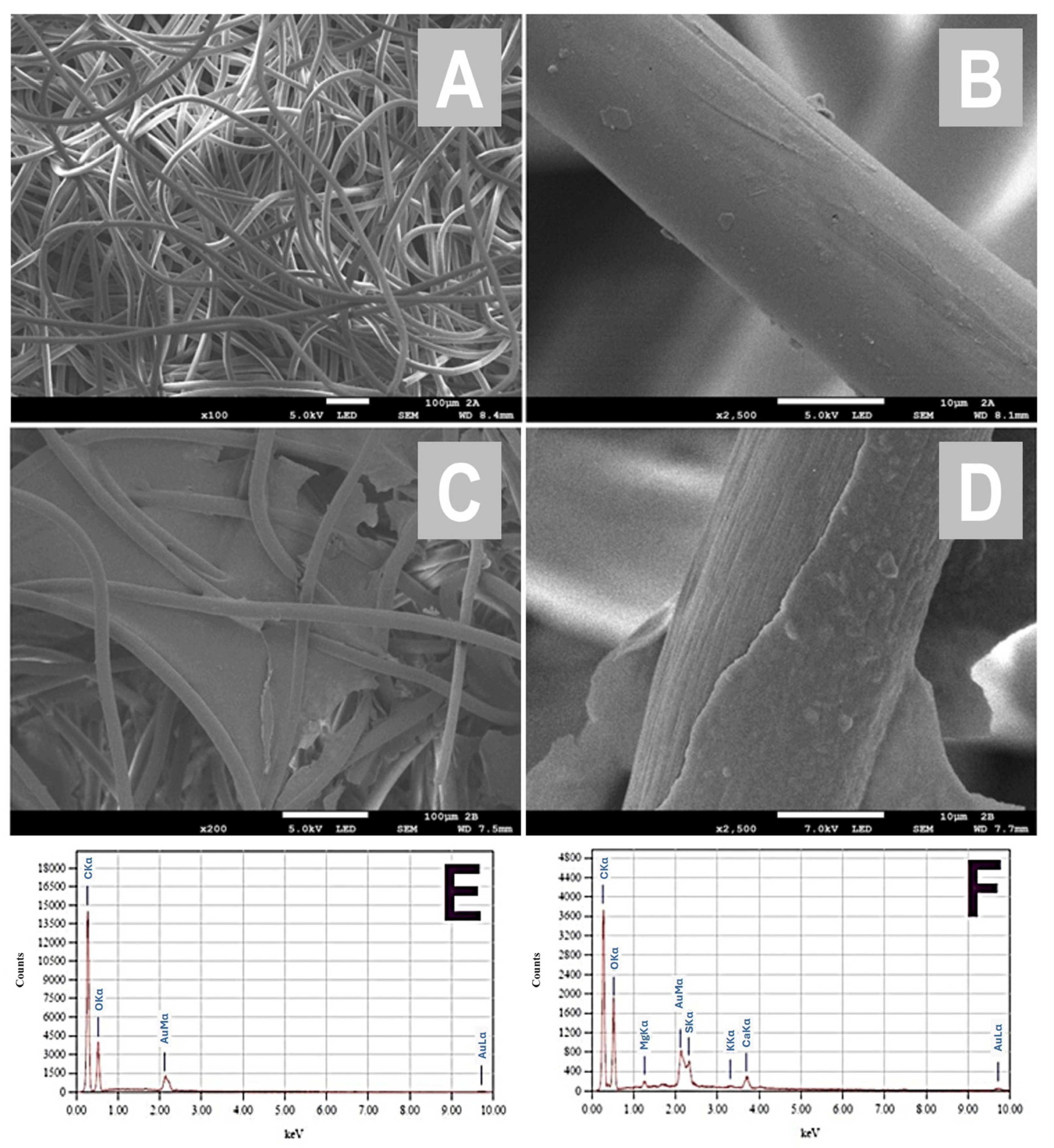
2.3. Optimized Preparation of Ulvan-Modified Nonwoven Textile

Nonwoven acrylic textile sheets were cut into squares (2 × 2 cm) which were subsequently heated at 100 °C in 1% (w/v) ulvan and 1% (w/v) agarose solution in distilled water for 2 min in a standard domestic microwave oven (700 W, 2450 MHz). The ulvan-modified textile squares were placed in Petri dishes or on plastic sheets and dried at room temperature. Subsequently, the original ulvan-modified textile squares were cut to obtain four 1 × 1 cm squares.

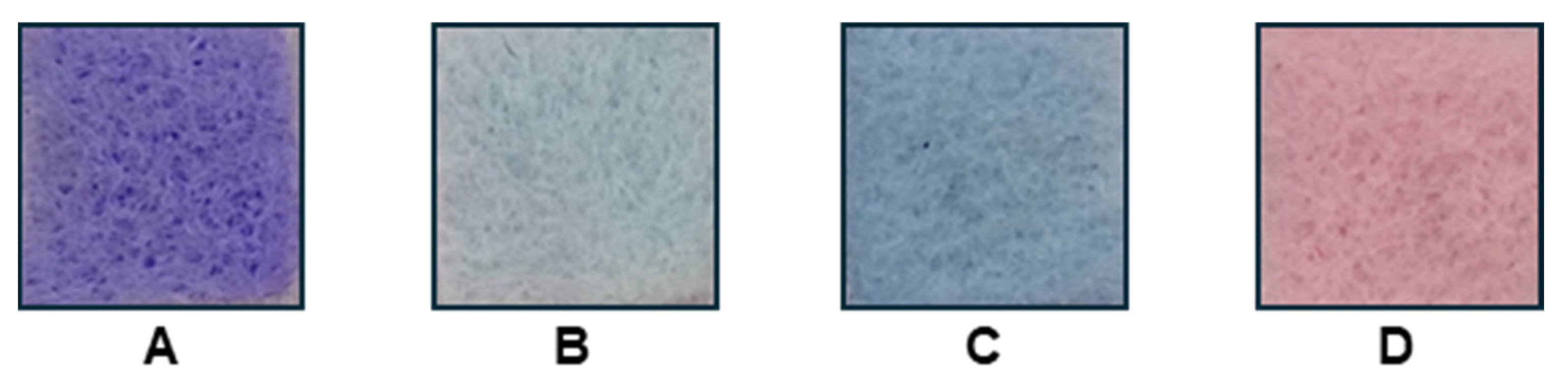
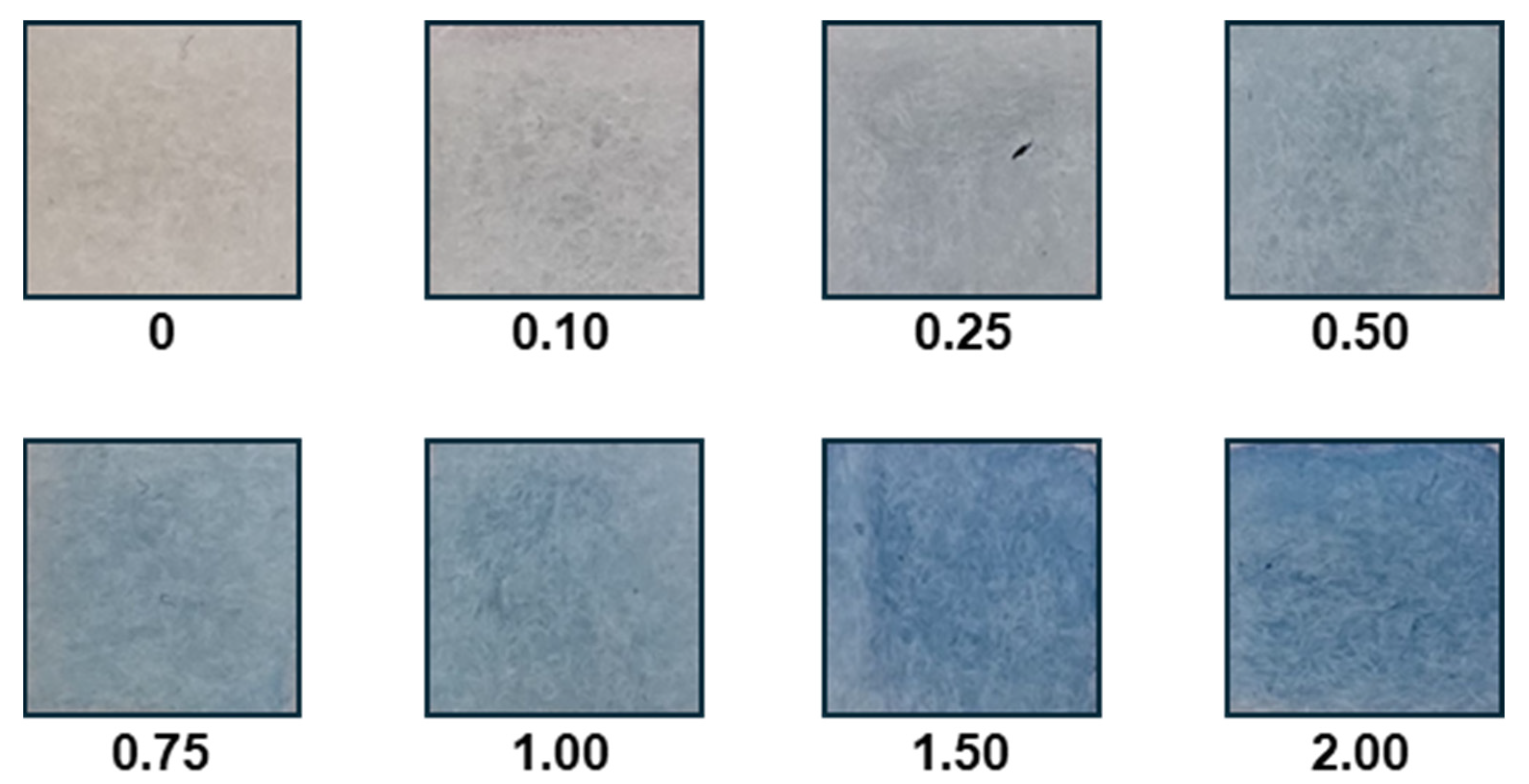
2.4. Textile Solid-Phase Extraction

Ulvan-modified textile squares (1 × 1 cm) were placed into 50 mL of aqueous dye solutions in 150 mL beakers; the dye concentration was in the range 0.1–2.0 mg/L. The extraction was performed for 60 min at 20 °C under mixing using a Unimax 1010 horizontal shaker (Heidolph, Schwabach, Germany). Then, the textile squares with the adsorbed dye were removed from the solution using a laboratory spoon or tweezers, then rinsed with water, and a set of textile squares was immediately photographed in an illuminated photo box using a smartphone.

2.5. Image Analysis

Image analysis of textile square photographs was performed using a Redmi Note 10 smartphone (Xiaomi, Beijing, China) with the installed Color Picker application (Version 7.7.0); the detailed procedure also including illustrative photographs was recently described [17]. Color Picker data for all available color spaces (HSL, HSV, CMY, CMYK, RGB, RYB, CIE XYZ and CIE LAB) [18] were measured and used for the calculations of average values; three textile solid-phase extraction experiments, followed by image analysis, were conducted for all investigated dye solutions.

2.6. Other Procedures

Both untreated and ulvan/agarose-modified textiles were analyzed using scanning electron microscopy (SEM) and energy-dispersive X-ray spectroscopy (EDS). The samples were coated by an approximately 20 nm gold thin film and were examined under a Jeol-7900F SEM microscope (JEOL Ltd., Tokyo, Japan) at an acceleration voltage of 5.0 or 7.0 kV. EDS was performed at an acceleration voltage of 15.0 kV on the gold-coated samples.

3. Results

TSPE can be efficiently used for the preconcentration of target analytes from diluted solutions [6]. In order to extract positively charged molecules, the textile substrate has to be modified by a negatively charged ligand, such as the negatively charged marine polysaccharide ulvan. Due to the fact that ulvan is a water-soluble polysaccharide, its combination with the water-insoluble neutral polysaccharide agarose was employed for nonwoven textile modification. In preliminary experiments, various concentrations of ulvan (1–2%, w/v) and agarose (1–3%, w/v) were evaluated for optimal textile fiber coating, and it was concluded that a mixture of 1% agarose and 1% ulvan solution in distilled water can be successfully used.
Scanning electron microscopy (SEM) was used to verify the modification of the synthetic textile fibers (typical diameters between 10 and 20 µm) with ulvan/agarose (Figure 1). The ulvan/agarose modification of the nonwoven textiles resulted in the surface modification of individual fibers (Figure 1C), as well as the formation of an ulvan/agarose film in between the fibers (Figure 1D). Energy-dispersive X-ray spectroscopy (EDS) analysis confirmed the presence of C and O in the untreated textile (Figure 1E); in the ulvan-modified textile, an additional sulfur peak (2.3 keV) confirming the presence of sulfur groups in ulvan was found. Additional small peaks indicate the presence of Mg, K and Ca, which is in agreement with published data [19] (Figure 1F). The Au signal in the EDS spectra was due to the gold coating of the samples.
Four cationic organic dyes, namely safranin O, methylene blue, malachite green and crystal violet, were efficiently bound to the ulvan-modified textile, as documented in Figure 2. Both the untreated nonwoven textile and the agarose-modified textile exhibited a low nonspecific adsorption of the tested basic dyes at the concentrations used for the experiments after immersion into the dye solutions and subsequent washing with water, which resulted in the slight coloration of the ulvan-free textiles (Figure S1 in Supplementary Materials). Even though the adsorption of the dyes may also depend on the type of textile used, in this case 100% acrylic felt nonwovens, it can be readily observed that treatment with ulvan significantly increased the adsorption of the cationic dyes used in the current study.
In subsequent experiments, the four model organic cationic dyes were extracted on the basis of electrostatic interactions using the ulvan-modified textile (1 × 1 cm) from 50 mL aqueous solutions at 20 °C; the incubation time was 60 min, and the dye concentration ranged from 0 to 2.0 mg/L. The range of concentrations tested was arbitrarily selected based on preliminary experiments, as well as the fact that the presence of dyes in water even at concentrations lower than 1 ppm is considered unacceptable, even though several studies have reported the presence of dyes in wastewater streams in amounts higher than 1 mg/L [20]. Digital images of all analyzed textile squares after dye extraction were acquired under standard illumination in a photo box (Figure 3) to ensure consistent conditions for the subsequent image analysis.
Smartphone-based assays have recently become popular due to their ease of use and applicability, combined with their minimal cost [21,22]. Several color spaces, including RGB, CMY, CMYK, HSL, HSV, RYB, CIE LAB and CIE XYZ, are available for the description of individual dyes. In the current study, smartphone-based image analysis using the Color Picker application was conducted to identify a correlation between the dye concentration and the corresponding color space value. The Color Picker application performs the analysis of the selected picture area, which is a great advantage for TSPE where small differences in color intensities on different textile adsorbent areas can be expected.
The analysis of the ulvan-modified textile adsorbents after the extraction of the dyes using the smartphone-based application Color Picker resulted in the sequence of color space data, which were analyzed afterwards. A strong correlation between the dye concentration and specific color space values was observed. Figure 4 illustrates the concentration dependences of values R and G (from RGB color space), X (from CIE XYZ color space) and C (from CMY color space) for methylene blue. For the investigated dyes, quadratic equations were required to achieve the highest precision in fitting the experimental data. Table 1 provides a summary of the calculated equations, coefficients of determination (R2) and relative standard deviations (RSDs), all computed using Excel. In each instance, the data were obtained from three independent extraction experiments and subsequent analyses.
As it can be observed, the reproducibility of dye adsorption followed by image analysis was confirmed, with the relative standard deviation for the respective color space values ranging from 0 to 16.59% across three independent measurements (Table 1).
To support the applicability of the TSPE method employing ulvan-modified textiles towards the determination of cationic dyes in wastewaters, we compared the adsorption of crystal violet dissolved in pond water from the Zdrahanka pond, Haklovy Dvory near Ceske Budejovice (Czech Republic), to that of crystal violet dissolved in distilled water, in both cases to a final concentration of 1 mg/L. As it could clearly be observed (Figure S2 in Supplementary Materials), crystal violet was efficiently bound in both cases to the ulvan-modified textiles. As the next step, further investigation of the applicability of our method using actual wastewater samples from various industrial sectors is planned for the near future.

4. Conclusions

The current study revealed that low-cost textile materials modified with the acidic marine polysaccharide ulvan can be employed for the determination of cationic organic dyes from water samples and/or aqueous solutions. This rapid, inexpensive and elution-free method utilizes smartphone-based image analysis of the photographs of the colored textile pieces. This very simple TSPE method requires only a smartphone with an installed application (such as the freeware Color Picker), allowing for the fast detection and determination of cationic organic dyes that can be used in various applications, including in water quality monitoring, industrial effluent analysis and environmental research and technology. Furthermore, the method’s elution-free nature eliminates the need for additional chemical reagents and the use of expensive instrumentation, reducing both the cost and environmental impact of the analysis.

Supplementary Materials

The following supporting information can be downloaded at https://www.mdpi.com/article/10.3390/chemosensors13020075/s1, Figure S1: Images of the interaction of the used dyes with the untreated nonwoven textile (top), the agarose-modified nonwoven textile (middle) and the ulvan-modified textile (bottom); Figure S2: Images of the interaction of crystal violet with the ulvan-modified textile.

Author Contributions

S.K., V.R. and E.I. collected the macroalga, isolated and characterized ulvan and contributed to the drafting of the manuscript; I.S. and J.P. performed the analytical experiments, analyzed the data and wrote the manuscript. All authors reviewed and corrected the manuscript. All authors have read and agreed to the published version of the manuscript.

Funding

This research was supported by the projects No. APVV-22-0060 MAMOTEX and ITMS 313011T548 MODEX (Slovak Research and Development Agency, Ministry of Education, Science, Research and Sport of the Slovak Republic) and the ERDF project No. CZ.02.1.01/0.0/0.0/17_048/0007399 (Ministry of Education, Youth and Sports of the Czech Republic). This research was partially funded by the research projects MARINOVA (grant number 70/3/14684) and BioNP (grant number 70/3/14685).

Data Availability Statement

The original contributions presented in this study are included in the article/Supplementary Materials. Further inquiries can be directed to the corresponding authors.

Acknowledgments

This work was carried out in the frame of the COST Action CA18238—European transdisciplinary networking platform for marine biotechnology (Ocean4Biotech). The authors thank the Special Account for Research Grants of the National and Kapodistrian University of Athens for funding to cover the publication costs.

Conflicts of Interest

The authors declare no conflicts of interest.

References

  1. Tkaczyk, A.; Mitrowska, K.; Posyniak, A. Synthetic organic dyes as contaminants of the aquatic environment and their implications for ecosystems: A review. Sci. Total Environ. 2020, 717, 137222. [Google Scholar] [CrossRef]
  2. Mosaffa, E.; Patel, R.I.; Banerjee, A.; Basak, B.B.; Oroujzadeh, M. Comprehensive analysis of cationic dye removal from synthetic and industrial wastewater using a semi-natural curcumin grafted biochar/poly acrylic acid composite hydrogel. RSC Adv. 2024, 14, 7745–7762. [Google Scholar] [CrossRef] [PubMed]
  3. Rathi, B.S.; Kumar, P.S.; Vo, D.-V.N. Critical review on hazardous pollutants in water environment: Occurrence, monitoring, fate, removal technologies and risk assessment. Sci. Total Environ. 2021, 797, 149134. [Google Scholar] [CrossRef] [PubMed]
  4. Safarikova, M.; Safarik, I. Magnetic solid-phase extraction. J. Magn. Magn. Mater. 1999, 194, 108–112. [Google Scholar] [CrossRef]
  5. Safarik, I.; Prochazkova, J. Color catcher sheets for the construction of low-cost, planar optical sensors. Fibers Polym. 2024, 25, 4099–4107. [Google Scholar] [CrossRef]
  6. Safarik, I.; Mullerova, S.; Pospiskova, K. Semiquantitative determination of food acid dyes by magnetic textile solid phase extraction followed by image analysis. Food Chem. 2019, 274, 215–219. [Google Scholar] [CrossRef] [PubMed]
  7. Kidgell, J.T.; Magnusson, M.; de Nys, R.; Glasson, C.R.K. Ulvan: A systematic review of extraction, composition and function. Algal Res. 2019, 39, 101422. [Google Scholar] [CrossRef]
  8. Kraithong, S.; Bunyameen, N.; Theppawong, A.; Ke, X.; Lee, S.; Zhang, X.; Huang, R. Potentials of Ulva spp.-derived sulfated polysaccharides as gelling agents with promising therapeutic effects. Int. J. Biol. Macromol. 2024, 273, 132882. [Google Scholar] [CrossRef] [PubMed]
  9. Anisha, G.S.; Augustianath, T.; Padmakumari, S.; Singhania, R.R.; Pandey, A.; Patel, A.K. Ulvan from green macroalgae: Bioactive properties advancing tissue engineering, drug delivery systems, food industry, agriculture and water treatment. Bioresour. Technol. Rep. 2023, 22, 101457. [Google Scholar] [CrossRef]
  10. Lakshmi, D.S.; Sankaranarayanan, S.; Gajaria, T.K.; Li, G.; Kujawski, W.; Kujawa, J.; Navia, R. A short review on the valorization of green seaweeds and ulvan: Feedstock for chemicals and biomaterials. Biomolecules 2020, 10, 991. [Google Scholar] [CrossRef]
  11. Sulastri, E.; Zubair, M.S.; Lesmana, R.; Mohammed, A.F.A.; Wathoni, N. Development and characterization of ulvan polysaccharides-based hydrogel films for potential wound dressing applications. Drug Des. Devel. Ther. 2021, 15, 4213–4226. [Google Scholar] [CrossRef] [PubMed]
  12. Wang, H.; Cao, Z.; Yao, L.; Feng, T.; Song, S.; Sun, M. Insights into the edible and biodegradable ulvan-based films and coatings for food packaging. Foods 2023, 12, 1622. [Google Scholar] [CrossRef] [PubMed]
  13. Chi, Y.; Li, H.; Fan, L.; Du, C.; Zhang, J.; Guan, H.; Wang, P.; Li, R. Metal-ion-binding properties of ulvan extracted from Ulva clathrata and structural characterization of its complexes. Carbohydr. Polym. 2021, 272, 118508. [Google Scholar] [CrossRef] [PubMed]
  14. Wahlström, N.; Steinhagen, S.; Toth, G.; Pavia, H.; Edlund, U. Ulvan dialdehyde-gelatin hydrogels for removal of heavy metals and methylene blue from aqueous solution. Carbohydr. Polym. 2020, 249, 116841. [Google Scholar] [CrossRef] [PubMed]
  15. Zhao, S.; Gao, B.; Yue, Q.; Wang, Y.; Li, Q.; Dong, H.; Yan, H. Study of Enteromorpha polysaccharides as a new-style coagulant aid in dye wastewater treatment. Carbohydr. Polym. 2014, 103, 179–186. [Google Scholar] [CrossRef]
  16. Kikionis, S.; Koromvoki, M.; Tagka, A.; Polichronaki, E.; Stratigos, A.; Panagiotopoulos, A.; Kyritsi, A.; Karalis, V.; Vitsos, A.; Rallis, M.; et al. Ulvan-based nanofibrous patches enhance wound healing of skin trauma resulting from cryosurgical treatment of keloids. Mar. Drugs 2022, 20, 551. [Google Scholar] [CrossRef]
  17. Safarik, I.; Prochazkova, J. Semiquantitative color catcher and smartphone-based analysis of synthetic food dyes in alcohol containing beverages. Talanta 2023, 262, 124686. [Google Scholar] [CrossRef]
  18. Kahu, S.Y.; Raut, R.B.; Bhurchandi, K.M. Review and evaluation of color spaces for image/video compression. Color. Res. Appl. 2019, 44, 8–33. [Google Scholar] [CrossRef]
  19. Ibrahim, M.I.A.; Amer, M.S.; Ibrahim, H.A.H.; Zaghloul, E.H. Considerable production of ulvan from Ulva lactuca with special emphasis on its antimicrobial and anti-fouling properties. Appl. Biochem. Biotechnol. 2022, 194, 3097–3118. [Google Scholar] [CrossRef] [PubMed]
  20. Alsukaibi, A.K.D. Various approaches for the detoxification of toxic dyes in wastewater. Processes 2022, 10, 1968. [Google Scholar] [CrossRef]
  21. Nelis, J.L.D.; Tsagkaris, A.S.; Dillon, M.J.; Hajslova, J.; Elliott, C.T. Smartphone-based optical assays in the food safety field. TrAC Trends Anal. Chem. 2020, 129, 115934. [Google Scholar] [CrossRef] [PubMed]
  22. Bui, T.H.; Thangavel, B.; Sharipov, M.; Chen, K.; Shin, J.H. Smartphone-based portable bio-chemical sensors: Exploring recent advancements. Chemosensors 2023, 11, 468. [Google Scholar] [CrossRef]
Figure 1. SEM images of untreated nonwoven textile (A,B), and ulvan/agarose-modified nonwoven textile (C,D). EDS of untreated nonwoven textile (E) and textile modified with ulvan/agarose (F). The Au signal in the EDS spectra is due to the gold coating of the samples.
Chemosensors 13 00075 g001
Figure 2. Images of the interaction of the used dyes with the ulvan-modified textile. (A)—crystal violet; (B)—malachite green; (C)—methylene blue; (D)—safranin O. Extraction was performed for 60 min at a dye concentration of 2.0 mg/L.
Chemosensors 13 00075 g002
Figure 3. Examples of ulvan-modified nonwoven textile squares after methylene blue extraction from aqueous solutions (50 mL; extraction time 60 min; concentrations of the original dye solutions (mg/L) are shown).
Chemosensors 13 00075 g003
Figure 4. Dependence of values of R and G (from RGB), X (from CIE XYZ) and C (from CMY) on the concentration of methylene blue (incubation time 60 min, volume 50 mL).
Chemosensors 13 00075 g004
Table 1. Examples of calculated equations, coefficients of determination (R2) and relative standard deviations (RSDs) for selected color space values. Dye concentrations were in the range of 0.1–2.0 mg/L.
DyeColor SpaceValueEquation and CoefficientsR2RSD [%]
Crystal violetRGBRy = 30.407x2 − 96.734x + 147.030.93530–13.05
CMYCy = −11.717x2 + 37.378x + 42.6170.93450–14.43
Malachite greenRGBRy = 9.6046x2 − 39.641x + 177.380.97520–4.72
CMYCy = −3.5776x2 + 15.304x + 30.4250.97190–9.95
Methylene blueRGBRy = 21.329x2 − 83.615x + 177.130.98820–4.28
RGBGy = 8.9023x2 − 40.215x + 170.490.98510.46–2.24
CMYCy = −8.3361x2 + 32.997x + 30.2540.98790–8.08
CIE XYZXy = 6.7135x2 − 23.838x + 39.2720.97891.21–7.85
Safranin ORGBGy = 19.136x2 − 60.789x + 168.930.97670.92–6.10
LABLy = −5.8564x2 + 21.573x + 4.7460.97387.52–16.59
Disclaimer/Publisher’s Note: The statements, opinions and data contained in all publications are solely those of the individual author(s) and contributor(s) and not of MDPI and/or the editor(s). MDPI and/or the editor(s) disclaim responsibility for any injury to people or property resulting from any ideas, methods, instructions or products referred to in the content.

