Abstract
Hepatitis C represents a critical global health crisis, causing approximately 1.4 million deaths annually. Although 98% of cases are treatable, only about 20% of infected individuals know their hepatitis C virus (HCV) status, highlighting the urgent need for rapid and more efficient diagnostic management. Viral genetic material can be detected in serum or plasma within just one week of exposure, making it the most reliable marker and the gold standard for active HCV infection diagnosis. In this study, a biosensor was developed to detect conserved nucleotide sequences of HCV using a 3D surface electrode composed of poly-L-lysine (PLL) and carbon nanotubes (CNTs). PLL is a positively charged biocompatible polymer rich in amine groups, attractive for the immobilization of proteins, DNA, and other biomolecules. PLL was employed to construct a 3D surface with vertically aligned CNTs, achieving a high electron transfer rate. Cyclic voltammetry technique and scanning electron microscopy (SEM) were used to characterize the sensor platform, and analytical responses were measured by differential pulse voltammetry. This HCV biosensor detected the hybridization event by a significant reduction in DPV peaks in the presence of the ferri/ferrocyanide redox probe, without any intercalator agents. DNA responses were observed in phosphate-buffered saline (PBS) and cDNA-spiked serum samples, demonstrating its analytical specificity. These findings represent advances in analytical tools that can effectively address the challenges of timely diagnosis for asymptomatic HCV carriers.
1. Introduction
Chronic hepatitis C virus (HCV) infection is a significant global public health concern, affecting an estimated 50 million people worldwide, with approximately 1 million new infections reported each year [1,2,3]. HCV infection often remains undiagnosed for decades, as it can remain asymptomatic and lead to irreversible liver damage. This bloodborne virus is primarily transmitted through contaminated blood including unsafe transfusions, injection drug use, sexual contact, and vertical transmission [4]. In 2022, approximately 242,000 deaths were attributed to hepatitis C, primarily due to liver cirrhosis and hepatocellular carcinoma (HCC) [3]. Early detection and intervention are critical to avoid disease progression, reduce the risk of transmission, and improve long-term clinical outcomes. Consequently, the World Health Organization (WHO) recommends the routine screening of blood donors and targeted testing of high-risk populations. Currently, HCV diagnosis involves a two-step approach: initial serological tests detect anti-HCV antibodies, indicating past or present exposure, while nucleic acid tests (NATs) are used to confirm the presence of HCV RNA, indicating active infection. Although NAT is widely regarded as the diagnostic gold standard for detecting HCV RNA due to its sensitivity and specificity, it has many drawbacks. These include high operational costs, labor-intensive roadmaps, and susceptibility to contamination, which is difficult for application in clinical routine, especially in resource-limited countries [5,6]. Therefore, there is a great need for diagnostic methods that are rapid, cost-effective, and capable of easily detecting HCV RNA with high specificity and sensitivity. Electrochemical biosensors have been demonstrated as attractive analytical tools to meet these demands.
One major technical limitation in developing RNA biosensors is the instability of single-stranded RNA, which is susceptible to hydrolysis and environmental degradation. As a result, single-stranded DNA (ssDNA) analogs are often used in sensor development and optimization due to their greater chemical stability and structural similarity to RNA targets [7,8,9]. The development of sensitive and selective biosensors for nucleic acid detection has led to the integration of various biorecognition elements to conductive electrode platforms including short ssDNA, hairpin loops, and aptamers. DNA-modified electrodes can selectively hybridize with complementary nucleic acid targets, enabling the detection of specific sequences associated with viruses, genes, or microbial contaminants [10,11]. Among these technologies, electrochemical DNA biosensors have gained significant attention due to their high sensitivity, selectivity, portability, low sample volume requirements, and compatibility with miniaturized and point-of-care formats [12,13]. These sensors rely on detecting hybridization events between immobilized ssDNA probes and complementary target sequences via electrochemical signal transduction. However, one of the main challenges in this field is achieving stable and functional probe immobilization while maintaining accessibility for hybridization and preserving the electrochemical performance of the sensor [14,15].
To circumvent this challenge, researchers have explored a variety of immobilization strategies based on covalent or non-covalent interactions involving functional groups such as amino, carboxyl, and thiol groups. Amino-functionalized DNA probes, for example, can form stable amide bonds with carboxyl electrode surfaces, thereby ensuring robust attachment, while they still maintain their flexibility for effective hybridization. Nanomaterials, such as carbon nanotubes (CNTs) and graphene derivatives, have further revolutionized biosensor platforms due to their high surface-to-volume ratio, excellent electrical conductivity, and ability to be chemically modified for biomolecule immobilization [16]. Multi-walled carbon nanotubes (CNTs) are widely used to improve biosensor performance thanks to their ability to enhance electron transfer kinetics and serve as nanostructured scaffolds for high-density probe loading. Carboxylation of CNTs through acid treatment introduces reactive sites suitable to form covalent bonds with amine-modified DNA probes, increasing the immobilization efficiency and electrochemical responsiveness [17]. In this study, the ssDNA probe was modified at its 5′ end with an amine group to enable covalent attachment to carboxylated CNTs [18]. This strategy preserves the conformational freedom of the probe and facilitates specific hybridization with the target RNA.
To improve biosensor stability, a poly-L-lysine (PLL) film was electrodeposited onto the electrode surface, serving as a biocompatible and positively charged matrix for CNT ,attachment. PLL contains abundant amino groups that form stable amide linkages with the carboxyl groups on CNTs, resulting in a well-organized and electrically conductive three-dimensional (3D) nanostructured interface [19,20]. This 3D architecture promotes efficient charge transfer, improves mechanical stability, and increases the density of immobilized probes. Furthermore, electrostatic interactions between PLL and CNTs contribute to the stable anchoring of nanomaterials onto the electrode surface [21,22].
The specific target for hybridization in this biosensor is a sequence from the conserved non-structural protein 5B (NS5B) gene of HCV. NS5B functions as an RNA-dependent RNA polymerase (RdRP), which is essential for HCV genome replication [23,24,25]. Due to its high sequence conservation across HCV genotypes, NS5B represents an ideal biomarker for detection of the virus. Here, a specific nucleotide sequence with 20 pb from the NS5B gene was established, which was analyzed using the Basic Local Alignment Search Tool (BLAST ,Version 2.16.0+). Results showed that the sequence was unique and HCV specific, distinguishing from other viruses from Flaviviridae family like Dengue, Zika, West Nile, and Yellow Fever.
Electrochemical detection was carried out using differential pulse voltammetry (DPV), a highly sensitive technique that allows for the direct monitoring of hybridization events by measuring changes in current without the need for fluorescent labels or enzymes [26,27,28]. While traditional sandwich assays often rely on intercalating dyes or labeled DNA for signal amplification, these components may degrade over time, limiting assay stability and reproducibility [6,29,30,31]. In contrast, non-intercalant DPV-based biosensing offers simplicity, robustness, and rapid response, making it ideal for point-of-care applications.
Recent advances in DPV-based electrochemical sensors have demonstrated their ability to detect viral nucleic acids with high precision and rapid turnaround, paving the way for compact and low-cost diagnostic devices [28,32]. These sensors offer several operational advantages including ease of fabrication, real-time signal monitoring, low detection limits, and minimal sample preparation. As such, electrochemical DNA biosensors are emerging as a competitive alternative to optical methods in the field of molecular diagnostics [33,34]. In this context, the objective of the present study was to develop an electrochemical DNA biosensor for the sensitive and specific detection of HCV RNA based on a three-dimensional PLL/CNT nanocomposite interface. The biosensor utilizes a 5′- amino-functionalized cDNA probe complementary to the conserved NS5B gene sequence, covalently immobilized on carboxylated CNTs anchored to an electrodeposited PLL matrix. This novel sensing architecture enhances immobilized probe density, hybridization efficiency, and signal stability, enabling the direct and amplification-free detection of HCV. This nanoplatform presents promising rapid, low-cost, and accurate point-of-care diagnostics, with broad applications in early detection, viral surveillance, and the management of hepatitis C in public health.
2. Materials and Methods
2.1. Reagents
Poly-L-lysine (PLL) was obtained from Sigma Aldrich (St. Louis, MO, USA). Multi-walled carbon nanotubes functionalized with carboxylic groups (CNTs) (average diameter of ~10 nm, average length of 1–2 mm, and 95% purity) were acquired from DropSens (Oviedo, Spain). Glycine, dimethylformamide (DMF), N-ethyl-N’-(3-dimethylaminopropyl) carbodiimide, (EDC), N-hydroxysuccinimide (NHS), potassium ferrocyanide (K4Fe(CN)6), and potassium ferricyanide (K3Fe (CN)6) were also obtained from Sigma-Aldrich™ (St. Louis, MO, USA). Ultrapure water obtained from a Milli-Q water purification system (Billerica, MA, USA) (18 MΩ) was utilized to prepare all solutions.
All synthetic oligonucleotides were purchased from DNA Express Biotecnologia Ltd.a (São Paulo, Brazil), with the following sequences:
- Probe single stranded DNA: (AminoC6) 5′gggtaccgcccttgcgagtc 3′ (probe ssDNA);
- Complementary ssDNA: 5′ gactcgcaagggcggtaccc 3′ (cDNA);
- Non-complementary ssDNA: 5′gggtaccgcccttgcgagtc3′ (ncDNA).
The probe ssDNA was immobilized on the nanostructured platform, and the cDNA was used to mimic HCV RNA for optimization purposes. The ncDNA was used as the negative control in these experiments.
The probe ssDNA used was established using Basic Local Alignment Search Tool (BLAST), which finds regions of similarity between biological DNA/RNA sequences. The sequence used in this work aligned with sequences from different strains of the Hepacivirus hominis NS5B gene with 100% certainty.
The pool control serum sample used for the analytical responses was obtained from human blood samples collected by venipuncture from five healthy volunteers and tested for HCV negative. This study was conducted under the Helsinki Declaration. All protocols were approved by the Research Ethics Committee of Fiocruz/Aggeu Magalhães Research Center, Recife, Brazil according to protocol number 63441516.6.0000.5190. The tested spiked HCV pool serum samples were prepared by diluting the serum in 50% of PBS. Spiked samples were obtained by adding a known amount of the ssDNA and maintaining identical concentrations and volumes of complementary (cDNA) and non-complementary strands (ncDNA) to minimize the matrix effects.
2.2. Apparatus
The electrochemical studies were performed by using a portable potentiostat/galvanostat Ivium Compact Stat (Eindhoven, The Netherlands) connected to a microcomputer and controlled by Ivium Soft software, version 4.3. A three-electrode cell was used, comprising a glassy carbon electrode (GCE) (Ø = 3 mm) as the working electrode, a helical platinum wire as the counter electrode, and Ag/AgCl (KCl saturated) as the reference electrode. Electrochemical analyses were carried out using an electrochemical cell (10 mL) and conducted at room temperature (24 °C ± 1).
The structural characterization was accomplished using Fourier transform infrared spectroscopy in attenuated total reflectance mode (ATR FT-IR) with a Bruker IFS 66 model FTIR spectrometer (Billerica, MA, USA). The spectra were acquired in the range of 4000 cm−1 to 500 cm−1. Scanning electron microscopy (SEM) assays were performed using a JEOL—JSM 5600LV scanning electron microscope at the Technological Integration Center/Aggeu Magalhães Research Center (CPqAM)—FIOCRUZ.
2.3. Preparation of the DNA Biosensor
Before the modification process, the glassy carbon electrode (GCE) underwent mechanical polishing using alumina slurry with particle sizes of 1 and 0.5 µm. This polishing was conducted for 5 min, followed by thorough rinsing with ultrapure water to ensure the complete removal of any leftover alumina particles, resulting in a pristine surface. The PLL film was synthesized according to the methodology described by Silva et. al. (2016) [21]. Briefly, 0.05 mmol L−1 of PLL was prepared in phosphate-buffered saline (PBS) solution and maintained at a physiological pH of 7.4 (0.01 mol L−1). The deposition of PLL onto the GCE surface was realized by the cyclic voltammetry (CV) technique, sweeping from −2.0 to +2.0 V at a controlled scan rate of 0.05 V s−1 for a total of 15 cycles, following established protocols to achieve a uniform layer (Supplementary Figure S1a,b).
After the PLL film deposition, CNT dispersion was introduced via drop casting of 5 µL on the modified GCE surface. The electrode was subsequently placed in a controlled oven at 40 °C for 1 h for solvent evaporation. This step was repeated three times to enhance the layer’s thickness and uniformity. The CNT dispersion consisted of 1 mg of carbon nanotubes prepared in 1 mL of dimethylformamide (DMF), which was sonicated in an ultrasonic bath for 2 h to achieve a black and well-homogeneous solution.
2.4. Immobilization of the Probe ssDNA and Analytical Responses
Single-stranded DNA (ssDNA) sequences functionalized with amine groups were immobilized on the nanoelectrode surface. This process involved an incubation of a 100 pmol L−1 aliquot of probe ssDNA with the CNT/PLL nanoplatform for a duration of one hour in a moist chamber at room temperature (24 °C ± 1 °C). To facilitate the formation of a stable covalent amide bond, the carboxyl groups present on the carbon nanotubes were subjected to activation through a mixture of 20 mM 1-ethyl-3-(3-dimethylaminopropyl) carbodiimide (EDC) and 50 mM N-hydroxysuccinimide (NHS) in phosphate-buffered saline (PBS) at a pH of 7.4. This activation consisted of pipetting 5 µL of this solution onto the electrode surface for 30 min, which was kept in a moist chamber at room temperature (24 °C ± 1 °C) to ensure optimal conditions for the reaction.
Analytical responses of the HCV nanoelectrode were assessed by incubating one aliquot (5 µL) of cDNA and ncDNA samples at a concentration of 100 pmol L−1 for a duration of 30 min in a moist chamber at room temperature (24 °C ± 1 °C) using samples in PBS buffer (n = 5) and human serum (n = 5). The human serum samples were obtained via the Fiocruz/Aggeu Magalhães Research Center. This study was conducted under the Helsinki Declaration as revised in 2013. All protocols were approved by the Research Ethics Committee of Fiocruz/Aggeu Magalhães Research Center, Recife, Brazil according to protocol number 63441516.6.0000.5190.
To investigate the interactions between cDNA and the interface of the single-stranded DNA-modified glassy carbon electrode (ssDNA-GCE) differential pulse voltammetry (DPV) was used as the analytical technique, as illustrated in Figure 1. During the measurements, the window potential was maintained between 0.0 and +0.5 V at a frequency of 10 Hz, with a pulse amplitude of 10 mV and a step potential of 0.0025 V. This analysis was conducted in the presence of the redox couple K3Fe(CN)6/K4Fe(CN)6 at a 5 mmol L−1 concentration, which was prepared using a 0.1 mol L−1 potassium chloride (KCl) solution to provide an optimal ionic environment. In addition to DPV, the stepwise modifications of the biosensor were monitored using CV, which served to validate the results obtained by DPV. The CV analysis was performed within a potential range extending from −0.2 to +0.6 V, maintaining a scan rate of 0.05 V s−1 in 5 mmol L−1 K3Fe(CN)6/K4Fe(CN)6 solution prepared in 0.1 mol L−1 KCl.
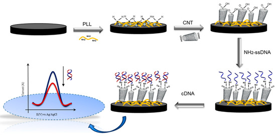
Figure 1.
Stepwise representation of the immunosensor assembly steps and analytical response.
3. Results and Discussion
3.1. Characterization of CNT/PLL/GCE
ATR-FTIR has proven to be an invaluable technique for the analysis of biosensor surfaces, allowing for the characterization of chemical bonds and functional groups present on the surfaces of various materials. The spectral data obtained from the electrode surface during each stage of immunosensor development are depicted in Figure 2, showcasing three distinct curves: curve I corresponds to carbon nanotubes, curve II represents poly-L-lysine (PLL), and curve III shows the CNT/PLL nanoplatform. In curve I, absorption bands indicative of carboxylic acid groups at 3350 cm−1 could be observed, which correlate to the stretching vibrations of hydroxyl (OH) groups. Additionally, a pronounced band at 1734 cm−1 signifies the molecular stretching of the carbonyl (C=O) group, implying the presence of the CNTs [35,36]. Curve II revealed an absorption band at 1623 cm−1, characteristic of the vibrational modes associated with primary amine groups (-NH2), corresponding to primary amino stretches and bending vibrations found within the PLL polymer matrix, as noted by García-Barrera et al. [37]. Finally, in curve III, a peak around 1570 cm−1 was observed, which is probably associated with the presence of CNTs by the C=C stretch of conjugated (sp2). Under these conditions, it is probable that the CNTs are attached to PLL by a strong electrostatic interaction, attributed to the high pKa of PLL [21] which promotes positively charged −NH3+ amine groups even in neutral pH as well as the carboxyl groups of CNTs, to stay negatively charged with their dispersion in DMF.
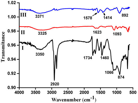
Figure 2.
FTIR spectra of the GCE modified with (I) CNT, (II) PLL, and (III) CNT/PLL.
3.2. Characterizations of the CNT/PLL Sensor Platform
Stepwise modifications of the GCE surface were investigated through CV and scanning electron microscopy (SEM). In Figure 3a, the GCE surface is depicted following the electrodeposition of the PLL film, revealing an array of diverse plate shapes. Among these, the hexagonal and cubic agglomerated structures stand out as predominant features, characteristic of PLL’s unique morphology [38]. In Figure 3b, the micrograph presents a topography attributed to the incorporation of carboxylated carbon nanotubes on the electrode surface. Here, the anchoring effect becomes evident, as the bond formed between the PLL and the carboxylated CNTs yields non-spaghetti-like configurations that illustrate this interaction. In contrast, the bright dots scattered across the surface indicate the presence of vertically aligned CNTs, while the dark coloration likely reflects the depth and distribution of CNTs across the GCE surface.
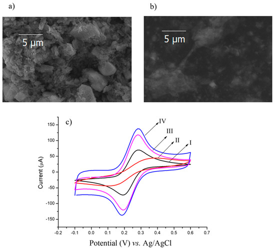
Figure 3.
SEM micrographs of: (a) PLL/GCE and (b) CNT/PLL/GCE. (c) CV curves of: (I) GCE; (II) PLL/GCE; (III) CNT/GCE; (IV) CNT/PLL/GCE. CV was performed in K3Fe(CN)6/K4Fe(CN)6 (0.005 mol L−1) prepared in a KCl solution (0.1 mol L−1).
The CV profiles illustrated in Figure 3c, specifically curve IV, validate the significant enhancement in the electroactive area resulting from the interaction between the PLL and carboxylated CNTs. This interaction results in the expansion of the active surface area compared with bare electrodes. Furthermore, an analysis of the redox peaks in the CV after modifying the GCE with PLL revealed a reduction in these peaks compared with the bare electrode. This decrease can be attributed to the non-conductive properties of PLL; however, this reduction results in symmetrical redox peaks. This phenomenon indicates that the homopolymer, primarily composed of L-lysine residue structures, was successfully preserved during the electrochemical deposition process on the electrode surface [38].
The mechanistic investigations were conducted by subjecting the CNT/PLL/GCE electrode to CVs and varying their scan rates, as depicted in Figure 4a. The resulting voltammograms demonstrated an increase in cathodic and anodic current peaks (Ipc and Ipa) with an increase in the scan rates (ν), which ranged from 10 to 150 mV s−1. This relationship can be confirmed by observing Figure 4b. Mathematical analysis revealed linear correlations between variables according to equations for both the anodic and cathodic peak currents: Ipa (µA) = 32.62ν − 81.11 (r = 0.999), Ipc (µA) = −30.31ν + 64.19 (r = 0.996); respectively. These correlation coefficients (r) were approximately equal to 1.0, which are indicative that the electrochemical reactions occurred on the sensor surface were predominantly controlled by ion transport mechanisms, specifically by diffusion on the interfacial surface. This insight underscores the dynamic changes occurring at the sensing electrode surface [39,40]. Furthermore, by analyzing slope of curves denoted by Log of the current of anodic and cathodic peaks (in modulus) versus Log of scan rates, their values were close to 0.50 (0.57 and 0.53, respectively), confirming that the process was in fact controlled by diffusion, indicating a good stability of the evaluated film (Figure 4c) [39,40].
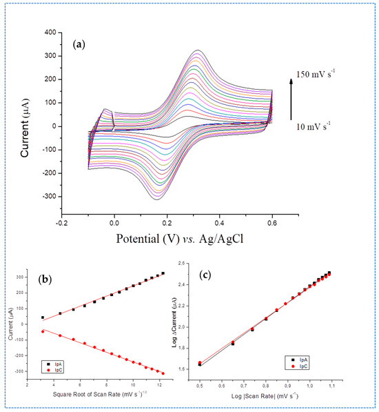
Figure 4.
(a) Voltametric profiles of CNT/PLL/GCE under different scan rates (10, 20, 30, 40, 50, 60, 70, 80, 90, 100, 110, 120, 130, 140 and 150 mV s−1), (b) plots of Ipa and Ipc vs. the square root of scan rate, (c) Log of Ipa and Ipc currents vs. Log of scan rate in modulus. All measurements were performed in K3Fe(CN)6/K4Fe(CN)6 (0.005 mol L−1).
The stability characteristic of the film was also studied by subjecting the CNT/PLL/GCE surface to 20 successive voltametric cycles. CVs were obtained at a potential window of −0.2 to +0.6 V and at 50 mVs−1 scan rate (Supplementary Figure S2). Under these conditions, amplitude of redox peaks remained practically constant, demonstrating an excellent electrochemical stability of this sensor platform. The coefficient of variations recorded were 0.45% and 0.32% for anodic and cathodic peaks in 20 cycles, respectively. This high sensor stability of CNT/PLL/GCE can be attributed to the presence of PLL, which plays a crucial role in anchoring the CNTs through strong electrostatic PLL–CNT interactions.
3.3. Experimental Optimizations
It is well-established that a high concentration of CNTs on the electrode surface can enhance electrocatalytic activity as well as increase the number of immobilized biomolecules, and consequently the analytical sensitivity. However, excess CNTs may reduce the electron charge transfer to the sensor surface by forming aggregates. Additionally, these aggregates can increase the number of randomly aligned CNTs. Herein, the amount of CNT was optimized based on the increase in CV redox peaks by drop casting consecutive layers of 0.5 mg/mL CNT on the electrode surface. According to the CVs, the maximal redox peak amplitude was obtained on the sixth layer, as indicated by a linear trend in the curve (Supplementary Figure S3a).
An investigation into the influence of probe ssDNA concentrations on the sensor platform was conducted to determine the most favorable current response. As demonstrated in Supplementary Figure S3b, a significant increase in current variation corresponding to a decrease in the CV current peaks was observed. A ssDNA probe solution in concentration of 100 pmol L−1 was successively applied to the CNT/PLL film until maximal response was reached. This decrease in current can be attributed to the ssDNA probe immobilized on the sensor platform. The immobilization mechanism can be elucidated through the amide bonds that occur between the amino groups in the amine-ssDNA and carboxyl groups in the CNTs by EDC/NHS chemistry, which hinder the transfer of ions, thereby impacting the overall current response [41].
3.4. Analytical Responses
The analytical performance of the sensor surface was assessed by exposing the electrode to samples containing 100 pmol L−1 of complementary DNA sequence, serving as a positive reference, alongside non-complementary DNA (ncDNA) used as a control, prepared in phosphate buffered-medium. The analytical response was determined by calculating the difference in relative current (RC (%)) before and after incubating with both the positive (n = 5) and control samples (n = 5). In Figure 5, a bar graph illustrates the variation in RC observed during experiments. The graph revealed a decrease in signal following incubation with cDNA (Figure 5a), which was significantly greater than the decrease associated with ncDNA (Figure 5b). This difference can be attributed to the hindrance in electron transfer caused by the hybridization events that occur between cDNA strands. This hybridization effectively increases the diffusion barrier that reduces the current response. Thus, this DNA sensor shows the ability to distinguish between complementary and non-complementary target sequences, even using a short sequence with 20 base pairs [42]. Therefore, a proof-of-concept of the biosensor for the specific detection of HCV was established, and analysis performed with a direct detection of HCV-RNA after DNA amplification, which can be easily achieved using an isothermal amplification technique of nucleic acid in human serum samples.
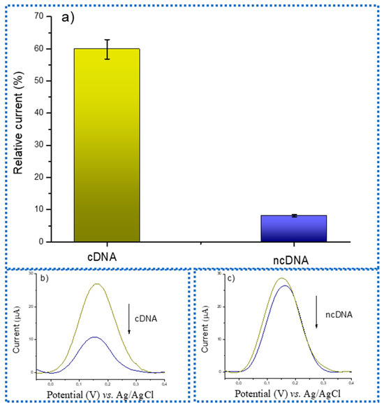
Figure 5.
(a) Bar graph showing the variation in relative current before and after the addition of PBS spiked with cDNA and ncDNA; DPV curves of the analytical detection of (b) cDNA and (c) ncDNA.
Assays to evaluate biosensor sensitivity through analytical curves were carried out by using the DPV protocol previously described in responses to different concentrations of cDNA (Figure 6). Herein, the modified HCV probe-sensors were subjected to successive incubations with an aliquot of 3 µL of cDNA prepared in phosphate-buffered medium. These were maintained in a moist chamber during incubations at room temperature as established in routine protocol, which was concluded with two PBS washes before the measurements. In Figure 6a, the analytical curve derived from the DPV anodic peaks is exhibited in response to concentrations of the HCV-cDNA sequences, with an exponential profile. To obtain the limit of detection (LOD), a linear adjust of the initial curve was obtained with the logarithmic of HCV-cDNA sequence concentrations of the current vs. relative current (Figure 6b), resulting in the equation I (%) = 52.3 Log [HCV-cDNA sequency] −52.1, and with R square of 0.998, confirming a good linearity. Data were processed in Origin Software version 8.5 SR1. The limit of detection (LOD) calculated by the residual standard deviation of the regression line denoted as σ, based on formula LOD = 3.3 σ/S; where S is the slope of the calibration curve [40] showed a value around 1.33 pmol L−1 HCV-cDNA sequences, which is in accordance with the range obtained from previous works [43,44].
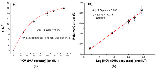
Figure 6.
(a) Responses to spiked samples of HCV-cDNA sequency prepared in PBS buffe; bar errors represent the standard deviation of three replicates measurements. (b) Relative current in the response of the Log HCV-DNA sequence at different concentrations, where I (%) = 100 × (I sample − I blank)/I blank; bar errors represent the coefficient of variation of triplicated measurements.
Studies of selectivity and matrix effect were conducted using DPV in a spiked pool of human serum, under the same conditions as those applied to the buffered samples, distinguishing between complementary and non-complementary (control) sequencies of HCV, as illustrated in Figure 7a. The serum samples exhibited a higher RC variation upon hybridization with cDNA, as depicted in Figure 7b, compared with the ncDNA strand (Figure 7c). However, it is important to highlight that the extent of RC variation observed in the serum samples was less pronounced than that in the buffered samples, as previously described. This reduced discrepancy between RC can be attributed to the complex nature of samples, such as serum and whole blood, which contain many interfering substances that can compromise the accuracy of electrochemical measurements. Despite these challenges, the analytical responses indicated a reduction in DPV peaks by the hybridization event. Furthermore, upon the addition of the control, the results reaffirmed the robustness and efficacy of the proposed DNA biosensor, even within such a complex biological medium.
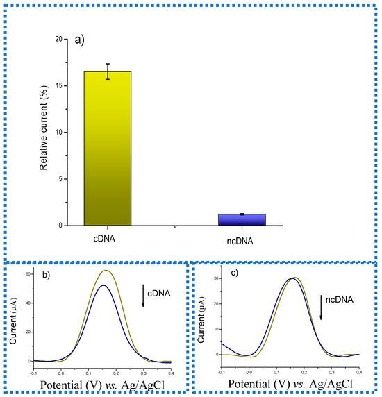
Figure 7.
(a) Bar graph represents the variation in relative current before and after the addition of human serum samples spiked with cDNA and ncDNA. (b) cDNA and (c) ncDNA DPV curves of the analytical detection.
A comparative analysis of this work with the previously described HCV biosensors was carried out, showing that, although other biosensors have achieved a lower LOD than the one described here (Table 1), some authors used strategies such as intercalant agents that have the disadvantage of being more time-consuming in the response time and sometimes non-electrochemically stable in the long-term [44]. Here, a label-free strategy for DNA hybridization detection using the ferri/ferrocyanide redox probe was adopted, similar to other studies highlighted in Table 1. In this work, however, the biosensor demonstrated the ability to discriminate between complementary and non-complementary DNA sequences with a few base (20 pb), differing from genomic DNA, even in complex samples. This selective ability was not possible to compare with other studies since they did not present selective responses or detection in complex samples [45,46]. Finally, Oliveira et al., 2018 [47] and Chittuam at al., 2023 [48] used a practical label-free system for HVC detection; however, they showed a higher LOD than described in this work. The high performance of this biosensor is attributed to the dual functionality of poly-L-lysine (PLL) in anchoring CNTs in a stable manner and enhancing the electron transfer. This characteristic improves the biosensor stability, cost-effectiveness, and specificity, making it more practical for clinical applications.

Table 1.
Electrochemical biosensor for HCV virus detection showing the LOD according to hybridization detection methods.
4. Conclusions
Serological methods for HCV diagnostics can indicate whether there has been previous exposure to the hepatitis virus. However, to effectively prevent the disease spreading, it is crucial to identify active and transmissible infections. Viral RNA can be detected in serum or plasma within one week of exposure, making it the most reliable marker for diagnosing HCV. The proposed DNA sensor was based on a CNT/PLL nanostructured platform and demonstrated both sensitivity and specificity in detecting complementary oligonucleotides that contained the conserved sequence of the hepatitis C virus (HCV). The electrochemical response was recorded after the sensor was incubated with cDNA, confirming hybridization through a decrease in amperometric current measured by DPV. The change in current was considered insignificant in samples containing ncDNA, which indicates the specificity of the test.
Supplementary Materials
The following supporting information can be downloaded at: https://www.mdpi.com/article/10.3390/chemosensors13110379/s1, Figure S1: (a) Deposition curves of PLL by CV; potential window from −2.0 to +2.0 V at 0.05 V s−1 scan rate for 15 cycles. PLL was prepared in phosphate-buffered saline (PBS) solution and maintained at a physiological pH of 7.4 (0.01 mol L−1). (b) CV of PLL film after deposition; Figure S2: CV plot of 20 successive cycles showing the stability of the CNT/PLL/GCE; Figure S3: (a) Plot of current variation vs. number of the CNT deposition layers in concentration of 0.5 mg/mL; (b) Plot of current variation vs. concentration of probe ssDNA.
Author Contributions
Conceptualization, R.F.D.; Methodology, G.M.S., A.P.O.S., and E.K.G.T.; Formal analysis, G.M.S., A.P.O.S., and S.R.B.; Writing—original draft, G.M.S.; Writing—review and editing, A.P.O.S., E.K.G.T., S.R.B., and R.F.D.; Funding acquisition, R.F.D. All authors have read and agreed to the published version of the manuscript.
Funding
The Brazilian National Council for Scientific and Technological Development of Brazil, grant number 440815/2022-4, and FACEPE—Fundação do Amparo a Ciência, grant APQ 1550-313/22. The National Institute of Science and Technology in Bioanalytic (INCTBio).
Institutional Review Board Statement
This study was conducted under the Helsinki Declaration as revised in 2013. All protocols were approved by the Research Ethics Committee of Fiocruz/Aggeu Magalhães Research Center, Recife, Brazil according to protocol number 63441516.6.0000.5190.
Informed Consent Statement
Written informed consent has been obtained from all the individuals who participated in this research.
Data Availability Statement
The original data presented in the study are openly available in https://repositorio.ufpe.br/bitstream/123456789/39628/1/TESE%20Gilv%C3%A2nia%20Marienete%20de%20Santana.pdf and https://repositorio.ufpe.br (accessed on 20 October 2025).
Conflicts of Interest
The authors declare that they have no knowledge of competing financial interests or personal relationships that could have influenced the work reported in this paper.
References
- Antoniou, T.; Pritlove, C.; Shearer, D.; Tadrous, M.; Shah, H.; Gomes, T. Accessing Hepatitis C Direct Acting Antivirals among People Living with Hepatitis C: A Qualitative Study. Int. J. Equity Health 2023, 22, 112. [Google Scholar] [CrossRef]
- Torre, P.; Festa, M.; Sarcina, T.; Masarone, M.; Persico, M. Elimination of HCV Infection: Recent Epidemiological Findings, Barriers, and Strategies for the Coming Years. Viruses 2024, 16, 1792. [Google Scholar] [CrossRef]
- Li, J.; Pang, L.; Liu, Z. Interpretation of the National Action Plan for Eliminating Hepatitis C as a Public Health Threat (2021–2030). China CDC Wkly. 2022, 4, 627–630. [Google Scholar] [PubMed]
- Adane, T.; Getawa, S. The Prevalence and Associated Factors of Hepatitis B and C Virus in Hemodialysis Patients in Africa: A Systematic Review and Meta-Analysis. PLoS ONE 2021, 16, e0251570. [Google Scholar] [CrossRef] [PubMed]
- Kanokudom, S.; Poovorawan, K.; Nilyanimit, P.; Suntronwong, N.; Aeemjinda, R.; Honsawek, S.; Poovorawan, Y. Comparison of Anti-HCV Combined with HCVcAg (Elecsys HCV Duo Immunoassay) and Anti-HCV Rapid Test Followed by HCV RNA Analysis Using QRT-PCR to Identify Active Infection for Treatment. PLoS ONE 2024, 19, e0313771. [Google Scholar] [CrossRef] [PubMed]
- El-Said, W.A.; Choi, J. High Selective Spectroelectrochemical Biosensor for HCV-RNA Detection Based on a Specific Peptide Nucleic Acid. Spectrochim. Acta A Mol. Biomol. Spectrosc. 2019, 217, 288–293. [Google Scholar] [CrossRef]
- Kornienko, I.V.; Aramova, O.Y.; Tishchenko, A.A.; Rudoy, D.V.; Chikindas, M.L. RNA Stability: A Review of the Role of Structural Features and Environmental Conditions. Molecules 2024, 29, 5978. [Google Scholar] [CrossRef]
- Roux, C.; Ramos-Hue, M.; Audonnet, M.; Duviau, M.-P.; Nouaille, S.; Carpousis, A.J.; Laguerre, S.; Hajnsdorf, E.; Cocaign-Bousquet, M.; Girbal, L. RNA Stability Is Regulated by Both RNA Polyadenylation and ATP Levels, Linking RNA and Energy Metabolisms in Escherichia coli. mBio 2025, 16, e02680-24. [Google Scholar] [CrossRef]
- Tani, H. Recent Advances and Prospects in RNA Drug Development. Int. J. Mol. Sci. 2024, 25, 12284. [Google Scholar] [CrossRef]
- Hua, Y.; Ma, J.; Li, D.; Wang, R. DNA-Based Biosensors for the Biochemical Analysis: A Review. Biosensors 2022, 12, 183. [Google Scholar] [CrossRef]
- Kim, J.H.; Suh, Y.J.; Park, D.; Yim, H.; Kim, H.; Kim, H.J.; Yoon, D.S.; Hwang, K.S. Technological Advances in Electrochemical Biosensors for the Detection of Disease Biomarkers. Biomed. Eng. Lett. 2021, 11, 309–334. [Google Scholar] [CrossRef]
- Antipchik, M.; Korzhikova-Vlakh, E.; Polyakov, D.; Tarasenko, I.; Reut, J.; Öpik, A.; Syritski, V. An Electrochemical Biosensor for Direct Detection of Hepatitis C Virus. Anal. Biochem. 2021, 624, 114196. [Google Scholar] [CrossRef]
- Mahardika, I.H.; Naorungroj, S.; Khamcharoen, W.; Kin, S.; Rodthongkum, N.; Chailapakul, O.; Shin, K. Point-of-Care Testing (POCT) Devices for DNA Detection: A Comprehensive Review. Adv. NanoBioMed Res. 2023, 3, 2300058. [Google Scholar] [CrossRef]
- González-Fernández, E.; Staderini, M.; Avlonitis, N.; Murray, A.F.; Mount, A.R.; Bradley, M. Effect of Spacer Length on the Performance of Peptide-Based Electrochemical Biosensors for Protease Detection. Sens. Actuators B Chem. 2018, 255, 3040–3046. [Google Scholar] [CrossRef]
- Milić, J.V. Multifunctional Layered Hybrid Perovskites. J. Mater. Chem. C Mater. 2021, 9, 11428–11443. [Google Scholar] [CrossRef]
- Mohd Nurazzi, N.; Asyraf, M.R.M.; Khalina, A.; Abdullah, N.; Sabaruddin, F.A.; Kamarudin, S.H.; Ahmad, S.; Mahat, A.M.; Lee, C.L.; Aisyah, H.A.; et al. Fabrication, Functionalization, and Application of Carbon Nanotube-Reinforced Polymer Composite: An Overview. Polymers 2021, 13, 1047. [Google Scholar] [CrossRef] [PubMed]
- Sezer, N.; Koç, M. Oxidative Acid Treatment of Carbon Nanotubes. Surf. Interfaces 2019, 14, 1–8. [Google Scholar] [CrossRef]
- Garrett, D.J.; Flavel, B.S.; Shapter, J.G.; Baronian, K.H.R.; Downard, A.J. Robust Forests of Vertically Aligned Carbon Nanotubes Chemically Assembled on Carbon Substrates. Langmuir 2010, 26, 1848–1854. [Google Scholar] [CrossRef]
- Daza Millone, M.A.; Ramirez, E.A.; Chain, C.Y.; Crivaro, A.; Romanin, D.; Rumbo, M.; Docena, G.; Cocco, M.D.; Pedano, M.L.; Fainstein, A.; et al. SPR Biosensing MUA/Poly-L-Lysine Platform for the Detection of 2,4-Dinitrophenol as Small Molecule Model System. J. Nanomater. 2016, 2016, 5432656. [Google Scholar] [CrossRef]
- Sahiner, N. Single Step Poly(L-Lysine) Microgel Synthesis, Characterization and Biocompatibility Tests. Polymer 2017, 121, 46–54. [Google Scholar] [CrossRef]
- Silva, P.M.S.; Lima, A.L.R..; Silva, B.V.M.; Coelho, L.C.B.B.; Dutra, R.F.; Correia, M.T.S. Cratylia Mollis Lectin Nanoelectrode for Differential Diagnostic of Prostate Cancer and Benign Prostatic Hyperplasia Based on Label-Free Detection. Biosens. Bioelectron. 2016, 85, 171–177. [Google Scholar] [CrossRef] [PubMed]
- Sun, X.; Shao, H.; Xiang, K.; Yan, Y.; Yu, X.; Li, D.; Wu, W.; Zhou, L.; So, K.-F.; Ren, Y.; et al. Poly(Dopamine)-Modified Carbon Nanotube Multilayered Film and Its Effects on Macrophages. Carbon 2017, 113, 176–191. [Google Scholar] [CrossRef]
- Adeboyejo, K.; Grosche, V.R.; José, D.P.; Ferreira, G.M.; Shimizu, J.F.; King, B.J.; Tarr, A.W.; Soares, M.M.C.N.; Ball, J.K.; McClure, C.P.; et al. Simultaneous Determination of HCV Genotype and NS5B Resistance Associated Substitutions Using Dried Serum Spots from São Paulo State, Brazil. Access Microbiol. 2022, 4, 000326. [Google Scholar] [CrossRef] [PubMed]
- Malik, A.A.; Chotpatiwetchkul, W.; Phanus-umporn, C.; Nantasenamat, C.; Charoenkwan, P.; Shoombuatong, W. StackHCV: A Web-Based Integrative Machine-Learning Framework for Large-Scale Identification of Hepatitis C Virus NS5B Inhibitors. J. Comput. Aided Mol. Des. 2021, 35, 1037–1053. [Google Scholar] [CrossRef]
- Sabariegos, R.; Albentosa-González, L.; Palmero, B.; Clemente-Casares, P.; Ramírez, E.; García-Crespo, C.; Gallego, I.; de Ávila, A.I.; Perales, C.; Domingo, E.; et al. Akt Phosphorylation of Hepatitis C Virus NS5B Regulates Polymerase Activity and Hepatitis C Virus Infection. Front. Microbiol. 2021, 12, 754664. [Google Scholar] [CrossRef]
- da Fonseca Alves, R.; Franco, D.L.; Cordeiro, M.T.; de Oliveira, E.M.; Fireman Dutra, R.A.; Del Pilar Taboada Sotomayor, M. Novel Electrochemical Genosensor for Zika Virus Based on a Poly-(3-Amino-4-Hydroxybenzoic Acid)-Modified Pencil Carbon Graphite Electrode. Sens. Actuators B Chem. 2019, 296, 126681. [Google Scholar] [CrossRef]
- Ferrari, L.; Rovati, L.; Fabbri, P.; Pilati, F. Photobleaching Effects in Organic Thin Film Sensing Probes. In Proceedings of the 2012 IEEE International Instrumentation and Measurement Technology Conference Proceedings, Graz, Austria, 13–16 May 2012; pp. 1235–1239. [Google Scholar]
- Savas, S.; Sarıçam, M. A Novel PCR-Free Ultrasensitive GQD-Based Label-Free Electrochemical DNA Sensor for Sensitive and Rapid Detection of Francisella Tularensis. Micromachines 2024, 15, 1308. [Google Scholar] [CrossRef]
- Kim, S.K.; Oh, Y.H.; Ko, D.H.; Sung, H.; Oh, H.B.; Hwang, S.H. Nanoparticle-Based Visual Detection of Amplified DNA for Diagnosis of Hepatitis C Virus. Biosensors 2022, 12, 744. [Google Scholar] [CrossRef]
- Fang, C.S.; Kim, K.; Yu, B.; Jon, S.; Kim, M.-S.; Yang, H. Ultrasensitive Electrochemical Detection of MiRNA-21 Using a Zinc Finger Protein Specific to DNA–RNA Hybrids. Anal. Chem. 2017, 89, 2024–2031. [Google Scholar] [CrossRef]
- Liu, L.; Wang, X.; Ma, Q.; Lin, Z.; Chen, S.; Li, Y.; Lu, L.; Qu, H.; Su, X. Multiplex Electrochemiluminescence DNA Sensor for Determination of Hepatitis B Virus and Hepatitis C Virus Based on Multicolor Quantum Dots and Au Nanoparticles. Anal. Chim. Acta 2016, 916, 92–101. [Google Scholar] [CrossRef]
- Trotter, M.; Borst, N.; Thewes, R.; von Stetten, F. Review: Electrochemical DNA Sensing—Principles, Commercial Systems, and Applications. Biosens. Bioelectron. 2020, 154, 112069. [Google Scholar] [CrossRef]
- Ferapontova, E.E. DNA Electrochemistry and Electrochemical Sensors for Nucleic Acids. Annu. Rev. Anal. Chem. 2018, 11, 197–218. [Google Scholar] [CrossRef] [PubMed]
- Hai, X.; Li, Y.; Zhu, C.; Song, W.; Cao, J.; Bi, S. DNA-Based Label-Free Electrochemical Biosensors: From Principles to Applications. TrAC Trends Anal. Chem. 2020, 133, 116098. [Google Scholar] [CrossRef]
- Benkaddour, A.; Jradi, K.; Robert, S.; Daneault, C. Grafting of Polycaprolactone on Oxidized Nanocelluloses by Click Chemistry. Nanomaterials 2013, 3, 141–157. [Google Scholar] [CrossRef] [PubMed]
- Östergren, I.; Darmadi, I.; Lerch, S.; da Silva, R.R.; Craighero, M.; Paleti, S.H.K.; Moth-Poulsen, K.; Langhammer, C.; Müller, C. A Surface Passivated Fluorinated Polymer Nanocomposite for Carbon Monoxide Resistant Plasmonic Hydrogen Sensing. J. Mater. Chem. A Mater. 2024, 12, 7906–7915. [Google Scholar] [CrossRef]
- García-Barrera, L.J.; Meza-Zamora, S.A.; Noa-Carrazana, J.C.; Delgado-Macuil, R.J. Chemometric Analysis Using Infrared Spectroscopy and PCA-LDA for Early Diagnosis of Fusarium Oxysporum in Tomato. J. Plant Dis. Prot. 2024, 131, 1609–1626. [Google Scholar] [CrossRef]
- Morsin, M.; Nafisah, S.; Sanudin, R.; Razali, N.L.; Mahmud, F.; Soon, C.F. The Role of Positively Charge Poly-L-Lysine in the Formation of High Yield Gold Nanoplates on the Surface for Plasmonic Sensing Application. PLoS ONE 2021, 16, e0259730. [Google Scholar] [CrossRef]
- Mendonça, P.D.; Santos, L.K.B.; Foguel, M.V.; Rodrigues, M.A.B.; Cordeiro, M.T.; Gonçalves, L.M.; Marques, E.T.A.; Dutra, R.F. NS1 Glycoprotein Detection in Serum and Urine as an Electrochemical Screening Immunosensor for Dengue and Zika Virus. Anal. Bioanal. Chem. 2021, 413, 4873–4885. [Google Scholar] [CrossRef]
- Saadati, A.; Hassanpour, S.; Bahavarnia, F.; Hasanzadeh, M. A Novel Biosensor for the Monitoring of Ovarian Cancer Tumor Protein CA 125 in Untreated Human Plasma Samples Using a Novel Nano-Ink: A New Platform for Efficient Diagnosis of Cancer Using Paper Based Microfluidic Technology. Anal. Methods 2020, 12, 1639–1649. [Google Scholar] [CrossRef]
- Liang, G.; Man, Y.; Li, A.; Jin, X.; Liu, X.; Pan, L. DNAzyme-Based Biosensor for Detection of Lead Ion: A Review. Microchem. J. 2017, 131, 145–153. [Google Scholar] [CrossRef]
- Dias, A.C.M.S.; Gomes-Filho, S.L.R.; Silva, M.M.S.; Dutra, R.F. A Sensor Tip Based on Carbon Nanotube-Ink Printed Electrode for the Dengue Virus NS1 Protein. Biosens. Bioelectron. 2013, 44, 216–221. [Google Scholar] [CrossRef]
- El-Sheikh, S.M.; Osman, D.I.; Ali, O.I.; Shousha, W.G.; Shoeib, M.A.; Shawky, S.M.; Sheta, S.M. A Novel Ag/Zn Bimetallic MOF as a Superior Sensitive Biosensing Platform for HCV-RNA Electrochemical Detection. Appl. Surf. Sci. 2021, 562, 150202. [Google Scholar] [CrossRef]
- Chaibun, T.; Karunaithas, S.; Ngamdee, T.; Wasitthankasem, R.; Lapchai, S.; Poovorawan, Y.; Yin, L.S.; Lertanantawong, B. Highly Sensitive and Specific Electrochemical Biosensor for Direct Detection of Hepatitis C Virus RNA in Clinical Samples Using DNA Strand Displacement. Sci. Rep. 2024, 14, 23792. [Google Scholar] [CrossRef]
- Roohizadeh, A.; Ghaffarinejad, A.; Salahandish, R.; Omidinia, E. Label-Free RNA-Based Electrochemical Nanobiosensor for Detection of Hepatitis C. Curr. Res. Biotechnol. 2020, 2, 187–192. [Google Scholar] [CrossRef]
- Sheta, S.M.; El-Sheikh, S.M.; Osman, D.I.; Salem, A.M.; Ali, O.I.; Harraz, F.A.; Shousha, W.G.; Shoeib, M.A.; Shawky, S.M.; Dionysiou, D.D. A Novel HCV Electrochemical Biosensor Based on a Polyaniline@Ni-MOF Nanocomposite. Dalton Trans. 2020, 49, 8918–8926. [Google Scholar] [CrossRef] [PubMed]
- Oliveira, D.A.; Silva, J.V.; Flauzino, J.M.R.; Sousa, H.S.; Castro, A.C.H.; Moço, A.C.R.; Soares, M.M.C.N.; Madurro, J.M.; Brito-Madurro, A.G. Carbon Nanomaterial as Platform for Electrochemical Genosensor: A System for the Diagnosis of the Hepatitis C in Real Sample. J. Electroanal. Chem. 2019, 844, 6–13. [Google Scholar] [CrossRef]
- Chittuam, K.; Jampasa, S.; Vilaivan, T.; Tangkijvanich, P.; Chuaypen, N.; Avihingsanon, A.; Sain, M.; Panraksa, Y.; Chailapakul, O. Electrochemical Capillary-Driven Microfluidic DNA Sensor for HIV-1 and HCV Coinfection Analysis. Anal. Chim. Acta 2023, 1265, 341257. [Google Scholar] [CrossRef]


Disclaimer/Publisher’s Note: The statements, opinions and data contained in all publications are solely those of the individual author(s) and contributor(s) and not of MDPI and/or the editor(s). MDPI and/or the editor(s) disclaim responsibility for any injury to people or property resulting from any ideas, methods, instructions or products referred to in the content. |
© 2025 by the authors. Licensee MDPI, Basel, Switzerland. This article is an open access article distributed under the terms and conditions of the Creative Commons Attribution (CC BY) license (https://creativecommons.org/licenses/by/4.0/).