Article

Water-Soluble Photoluminescent Ag Nanoclusters Stabilized by Amphiphilic Copolymers as Nanoprobe for Hypochlorite Detection

1
Shandong Provincial Key Laboratory of Detection Technology for Tumor Markers, College of Medicine, Linyi University, Linyi 276005, China
2
Beijing Key Laboratory for Bioengineering and Sensing Technology, School of Chemistry and Biological Engineering, University of Science and Technology Bejing, Beijing 100083, China
*
Authors to whom correspondence should be addressed.
Chemosensors 2024, 12(8), 166; https://doi.org/10.3390/chemosensors12080166
Submission received: 17 July 2024 / Revised: 10 August 2024 / Accepted: 15 August 2024 / Published: 17 August 2024

Abstract

:
Luminescent Ag nanoclusters (Ag NCs) are a promising probe material for sensing and bioimaging applications. However, the intrinsic obstacle of poor water stability and photostability greatly restrict their practical application in biological systems. Herein, we report the intracellular hypochlorite (ClO) detection with amphiphilic copolymer-modified luminescent Ag NCs with good biocompatibility and photostability. The Ag NCs were synthesized by using chemically inert hydrophobic ligands and then modified with an amphiphilic (1,2-distearoyl-sn-glycero-3-phosphoethanolamine-N-[methoxy(poly(ethylene glycol))-2000]) (DSPE-PEG-2000) and sodium dodecyl sulfonate (SDS) for phase transfer. It was found that the approach of the removal of organic solvents during the phase transfer has remarkable influences on the properties of the Ag NCs, including their size, luminescence property, and aqueous stability. Furthermore, the silver core of Ag NCs could be oxidatively damaged by ClO, thereby causing photoluminescence (PL) quenching. The ClO-induced PL quenching was specific over the other common reactive oxygen species (ROS) as well as some common interferences. Finally, they have been successfully applied as a fluorescent nanoprobe for detecting exogenous and endogenous ClO in living cells.

1. Introduction

In daily life and production, hypochlorous acid (HClO) has been widely used to disinfect surfaces as well as water, fruits, and vegetables for health. However, excessive amounts of ClO could backfire. In human bodies, ClO, as a highly reactive oxygen species (hROS), can be endogenously produced by neutrophils that are the first line of defense of the innate immune system to kill the invading bacteria and fungi based on nicotinamide adenine dinucleotide phosphate (NADPH) oxidase and myeloperoxidase (MPO). The NADPH oxidase at the plasma membrane serves to reduce molecular oxygen to generate superoxide radical (O2), which further gives H2O2, a common weakly reactive oxygen species (wROS). In the presence of H2O2, the heme-containing MPO is responsible for the catalytic oxidation of halides (mainly Cl due to their high abundance, i.e., 100–140 mM) ions to produce highly cytotoxic hypohalites including hypochlorite and hypochlorous acid [1,2,3]. However, inappropriate stimulation of oxidant formation by MPO, including possible overbalance of hypochlorite generation during inflammation, can induce and exacerbate various oxidative stress-related chronic diseases such as atherosclerosis, periodontitis, arthritis, neuron degeneration diseases, and even cancers [2,4,5,6]. Therefore, it is of great importance to develop approaches for sensitive and selective detection of ClO.
Recently, ultrasmall luminescent metallic nanoclusters (NCs) have been considered as attractive nanomaterials for the development of various luminescent nanoprobes [7,8,9,10,11,12]. Metallic NCs typically have a core–shell structure that consists of a metal(0) core and a metal(I)–ligand complex shell. The luminescence of metallic NCs might originate from the metal(0) cores according to the free-electron (Jellium) model, emitting short-lifetime fluorescence via a radiative transition between states of the same electronic spin multiplicities (S1→S0) [13,14,15,16], and also have the chance to be from the metal(I)–ligand complexes, especially those that are often associated with intramolecular aurophilic interactions, showing long-lifetime and stronger phosphorescence via a radiative transition between states of different electronic spin multiplicities (T1→S0) [17,18,19]. The ligand shell of metallic NCs, as the outer layer, directly interacts with the external systems. Therefore, the chemistry of the metallic core and the surface ligands is critical for the design of luminescent metallic NCs-based sensors. Recently, the fluorescent sensors of ClO have been developed using luminescent metallic NCs, including Au NCs, Ag NCs, and Cu NCs [20,21,22,23,24]. Those sensors have been basically based on the ClO-induced oxidation of the reductive moieties of the pristine metallic NCs to generate the agglomeration of the NCs, resulting in the quenching-characteristic signal changes. These reductive moieties have included the metal(0) atoms of the NCs’ cores and the thiolate groups or amine groups of NCs’ ligands. However, these reductive moieties were often susceptible to all high ROS, thereby largely weakening the reliability of those NCs for the specific detection of ClO.
In the present study, we report the amphiphilic copolymer-engineered luminescent Ag NCs (namely DSPE-PEG-SDS@Ag NCs) for the sensitive and selective detection of ClO (Scheme 1). To this end, the photoluminescent Ag NCs were synthesized using chemically inert hydrophobic ligands in hexane and purified by column chromatography, and then the dual-component amphiphilic copolymers of DSPE-PEG-2000 [25,26] and SDS were introduced as the encapsulant to produce hydrophilic DSPE-PEG-SDS@Ag NCs for biosensing applications. The introduction of the amphiphilic copolymer composite, i.e., DSPE-PEG-SDS, to encapsulate the Ag NCs can provide multiple effects. For the internal, the hydrophobic alkyl tails of the DSPE-PEG-SDS could offer a sufficient hydrophobic driving force for the self-assembly of the hydrophobic Ag NCs inside, meanwhile the hydrophilic part of the DSPE-PEG-SDS could endow whole assemblies with high water dispersity. For the external, the amphiphilic copolymer-engineered ligand shell of the formed DSPE-PEG-SDS@Ag NCs assemblies could build a zone like a “time gate” to delay the arrival of ROS to the innermost silver cores, so that the short-lived wROS and hROS cannot have access to the innermost luminescent DSPE-PEG-SDS@Ag NCs. On the other hand, the long-lived wROS are incapable of oxidizing the DSPE-PEG-SDS@Ag NCs, even though they can penetrate through the so-called “time gate” zone. Due to that, ClO is long-lived and belongs to hROS; it can react with the innermost DSPE-PEG-SDS@Ag NCs by penetrating through the so-called “time gate”. Then, the amphiphilic copolymer-engineered luminescent DSPE-PEG-SDS@Ag NCs can selectively detect ClO.

2. Materials and Methods

2.1. Chemicals

Silver tetrafluoroborate (AgBF4) and 3,5-bis(trifluoromethyl)benzenethiol(C8H4F6S) were purchased from Alpha Essar Chemical Reagent Co. (Tianjin, China). Sodium borohydride (NaBH4), tri-phenylphosphine (PPh3), dichloromethane (CH2Cl2), tri-ethylamine ((C2H5)3N), and n-Hexane (C6H14) were purchased from Sinopharm Chemical Reagents (Shanghai, China). 1,2-Distearoyl-sn-glycero-3-phosphoethanolamine-N-[methoxy(polyethylene glycol)-2000] (DSPE-PEG-2000, 98%) were purchased from McLean Biotech (Shanghai, China), and sodium dodecyl sulfate (SDS, 98%) were from Aladdin Reagent Company (Shanghai, China). All other chemicals of at least analytical reagent were obtained from Beijing Chemical Corporation (Beijing, China). All solutions were freshly prepared with deionized water.

2.2. Synthesis of Luminescent Ultrasmall Ag NCs and Phase Transfer

AgBF4 (20 mg), PPh3 (6 mg), 3,5-bis(trifluoromethyl)benzenethiol (8 μL), triethylamine (80 μL), and NaBH4 (40 mg) were added to 1 mL of ice-water solution and reacted at 0 °C for 12 h. Then, the organic phase and impurities were washed with water and partitioned with n-hexane, followed by storing in a refrigerator at 4 °C. Subsequently, organic solvent extraction and column chromatography separation were carried out for purification. To this end, a silica gel column was used as the stationary phase, and the mixture of n-hexane to dichloromethane with a ratio of 5:2 was used as the mobile phase, followed by rotary evaporation. Finally, the obtained luminescent Ag NCs were kept in dichloromethane in a refrigerator at 4 °C.
To prepare the water-soluble DSPE-PEG-SDS@Ag NCs, a simplified microemulsion self-assembly was applied. In brief, 3 mg of DSPE-PEG-2000 was added into 500 μL of the as-synthesized Ag NCs solution (1 mg/mL), followed by sonicating for 5 min at room temperature, and then dichloromethane was removed by rotary evaporation. Afterwards, 10 mL of aqueous solution containing 30 mg SDS was added, followed by sonicating until the solution became transparent and clear. Finally, the obtained products were dialyzed in deionized water using a filter bag with a molecular weight cutoff (MWCO) of 100–500 Da (Solarbio, Beijing, China) for 24 h, producing the water-soluble DSPE-PEG-SDS@Ag NCs. Finally, the obtained water-soluble DSPE-PEG-SDS@Ag NCs were kept in a refrigerator at 4 °C.

2.3. Detection of ClO

Firstly, 80 μL of 0.5 mg/mL of the as-prepared DSPE-PEG-SDS@Ag NCs was mixed with 10 μL of 100 mM PBS solution (pH = 7.4). Then, 10 μL of different concentrations of NaClO was added and allowed to react for 1 min at room temperature. Then, the emission spectra of the as-prepared DSPE-PEG-SDS@Ag NCs, excited at 428 nm, were collected on a fluorescence spectrophotometer.

2.4. Evaluation of Cytotoxicity of the As-Prepared DSPE-PEG-SDS@Ag NCs

Methyl thiazolyl tetrazolium (MTT) assay was applied to evaluate the cytotoxicity of the as-prepared DSPE-PEG-SDS@Ag NCs. Firstly, HeLa cells were cultured in DMEM medium containing 10% fetal bovine serum (FBS), 100 U/mL of penicillin, and 100 μg/mL of streptomycin, and passaged at 37 °C in 5% CO2. Cells in the logarithmic growth phase were added to 96-well culture plates with 100 μL wells (cell density of 5 × 10⁵ cells/mL per well). Next, the HeLa cells were transfected with the as-prepared DSPE-PEG-SDS@Ag NCs and incubated for 24 h. Subsequently, the cells were washed with PBS (pH 7.4, 10 mM) three times, and 100 μL of DMEM medium containing 0.5 mg/mL MTT was added into each well and incubated at 37 °C for 4 h, followed by adding 100 μL of dimethyl sulfoxide (DMSO). Finally, the corresponding absorbance was measured at 492 nm. Cell viability was calculated by the formula: Cellular Viability = {(Experimental group OD490 nm − Blank group OD490 nm)/(Control group OD490 nm − Blank group OD490 nm)} × 100%.

2.5. Cellular Imaging

HeLa cells were seeded into the confocal dish for 24 h and then the DSPE-PEG-SDS@Ag NCs with a final concentration of 300 μg/mL were added and allowed to react for 6 h at 37 °C. After that, the cells were washed with 1X PBS (10 mM, pH 7.4) at least three times. Then, the cells were treated with 100 μM ClO for 30 min, washed with PBS buffer, and imaged on a confocal laser scanning fluorescence microscope (CLSF). The control group was incubated with the DMEM medium without ClO for the same time. For endogenous ClO imaging, the HeLa cells were treated with lipo-polysaccharides (LPS, 1 μg/mL) for 12 h, then rinsed three times with DMEM, followed by incubating with phorbol myristate acetate (PMA, 10 nM) for 30 min, and further incubating with the DSPE-PEG-SDS@Ag NCs for 6 h. Finally, the cells were washed with 1X PBS three times, and then their fluorescence images were recorded.

2.6. Instrumentation

UV-vis absorption was recorded on a Lambda 900 UV-Vis NIR spectrophotometer (Platinum Elmer, Waltham, USA). PL spectra were obtained on an F-4500 spectrometer (Hitachi, Tokyo, Japan). X-ray photoelectron spectroscopy (XPS) measurements were recorded with an AXIS-ULTRA DLD electron spectrometer (Kratos, Manchester, UK). Transmission electron microscopy (TEM) characterizations were carried out using a JEM-2200FS Field Emission TEM (JEOL, Tokyo, Japan). PL lifetimes were measured using a FLS 980 steady/transient state fluorescence spectrometer (EI, Edinburgh, UK). For PL lifetime measurements, the excitation wavelength was 428 nm, and the PL decay at 650 nm was recorded. The zeta potential was measured on a dynamic light scattering (DLS) analyzer (Zetasizer Nano-ZS90, Malvern, UK). Fluorescence images were obtained by a confocal laser scanning fluorescence microscope (FV1200, Olympus, Tokyo, Japan).

3. Results

3.1. Characterizations of the DSPE-PEG-SDS@Ag NCs

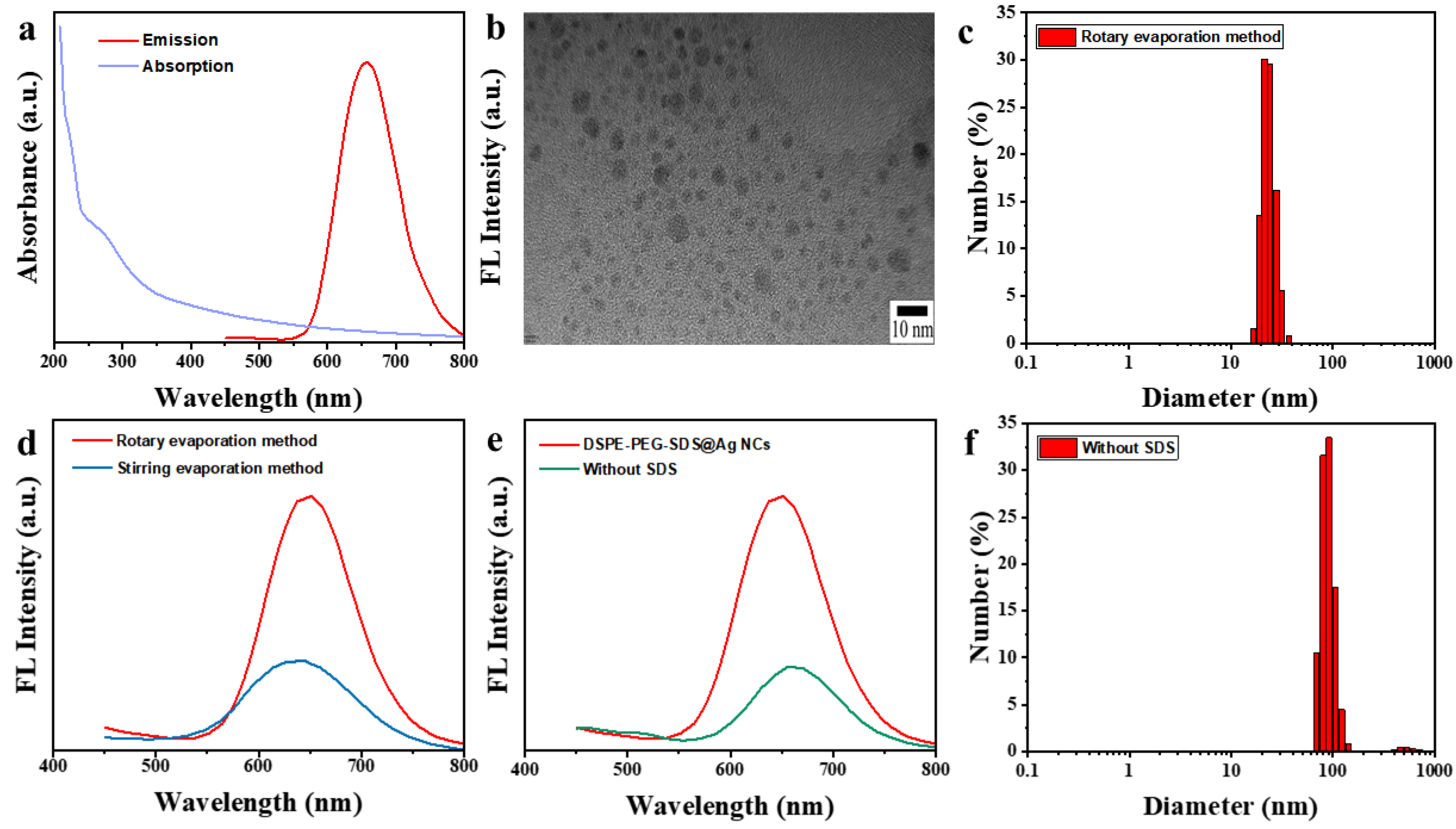
Firstly, the pristine Ag NCs were prepared by reducing Ag+ precursor with NaBH4 in the mixed hydrophobic solvents. The PL spectrum of the pristine Ag NCs (Supplementary Materials: Figure S1) showed an emission peak at 650 nm, which is supportive for its red emission under 365 nm UV light. Their mean lifetime was 10.12 ns, as displayed in Figure S2. Then, the poorly water-soluble Ag NCs are encapsulated by the amphiphilic copolymers of the DSPE-PEG and SDS to form the water-soluble nano-assemblies DSPE-PEG-SDS@Ag NCs, which retain the luminescent property of the pristine Ag NCs. As shown in Figure 1a, the UV-vis absorption spectrum of DSPE-PEG-SDS@Ag NCs showed a wide optical absorption band. They displayed a strong emission peak at 650 nm. The quantum yield (QY) calibrated with Ru(bpy)32+ was ~1.19%. The PL lifetime was measured to be 6.53 ns (Figure S2), indicating typical fluorescence characteristics. These observations clearly indicate that the PL properties of the pristine Ag NCs could be well preserved after phase transfer into an aqueous solution. It is worth noting that we unexpectedly found that the approach of the removal of organic solvents during the phase transfer had a remarkable influence on the DSPE-PEG-SDS@Ag NCs, including the sizes, luminescent properties, and sensing performance. During phase transfer process, the original organic solvents need to be removed mainly via evaporation approaches, including stirring evaporation and rotary evaporation. When the stirring evaporation approach was applied to remove the organic solvents, it took at least 12 h, and the resultant NPs had large sizes, as shown in Figure S3a, and their mean hydrodynamic diameter measured with dynamic light scattering (DLS) method was as large as 150 nm (Figure S3b). On the contrary, the rotary evaporation approach only needed several minutes to remove the solvents and the as-produced DSPE-PEG-SDS@Ag NCs had a small size of 5.26 nm, as shown in Figure 1b, just slightly larger than that of the pristine Ag NCs (Figure S4). And, their mean hydrodynamic diameter was measured to be 21.33 nm (Figure 1c), which was a size suitable for cellular endocytosis. Moreover, the PL intensity of the latter (Figure 1d, red line) was stronger than that of the former (Figure 1d, blue line). Therefore, the rotary evaporation approach was chosen in the following study. In addition, the auxiliary use of SDS during the amphiphilic copolymers-induced phase transfer process was found to be valuable. As revealed, no use of SDS led to the weakened emission (Figure 1e) and increased particle sizes (Figure 1f).
To confirm the stability of the Ag NCs in the process of encapsulation, the Ag valent state of the DSPE-PEG-SDS@Ag NCs was measured by XPS. As shown in Figure 2a, the peaks of the binding energy (BE) for Ag 3d could be deconvoluted into two components centered at Ag(0) BE (374.18 eV for Ag 3d3/2 and 368.08 eV for Ag 3d5/2) and Ag(I) BE (373.53 eV for Ag 3d3/2 and 367.48 eV for Ag 3d5/2), indicating that the silver existed in the forms of both Ag(0) and Ag(I) [27]. As a control, the Ag valent state of the pristine Ag NCs was found to be mainly Ag(0), as shown in Figure S5. Therefore, during the self-assembly-driven phase transfer, the conformation of the ligand shell might be affected to some extent by the interactions between the organic ligands and the amphiphilic copolymers, resulting in the further exposure of partial surfaces of the Ag NCs to the surplus ligands, causing further oxidation of the Ag cores. As revealed by the aforementioned ns-level lifetime, the luminescence of the DSPE-PEG-SDS@Ag NCs should be fluorescence originated from spatial confinement of free electrons in ultrasmall Ag core and be remarkably affected by the electron-donating ability of the ligands, rather than the so-called ligand’s aggregation-induced emissive phosphorescence. Thus, further oxidation of the Ag cores could weaken the luminescence of the Ag NCs. In addition, the photostability of the DSPE-PEG-SDS@Ag NCs was examined by measuring their luminescence intensity under various pH and salt concentration conditions. It was found that they could retain their PL intensity in a wide pH value range from 4.0 to 10.0 and at a salt concentration as high as 200 mM, as shown in Figure 2b,c, indicating excellent aqueous solubility and luminescence stability. The observed luminescence stability also dropped a hint that the amphiphilic polymer-engineered ligand shell had a stable conformation in aqueous solutions, showing little effect on the luminescence of the Ag NCs.

3.2. Response of the DSPE-PEG-SDS@Ag NCs to ClO

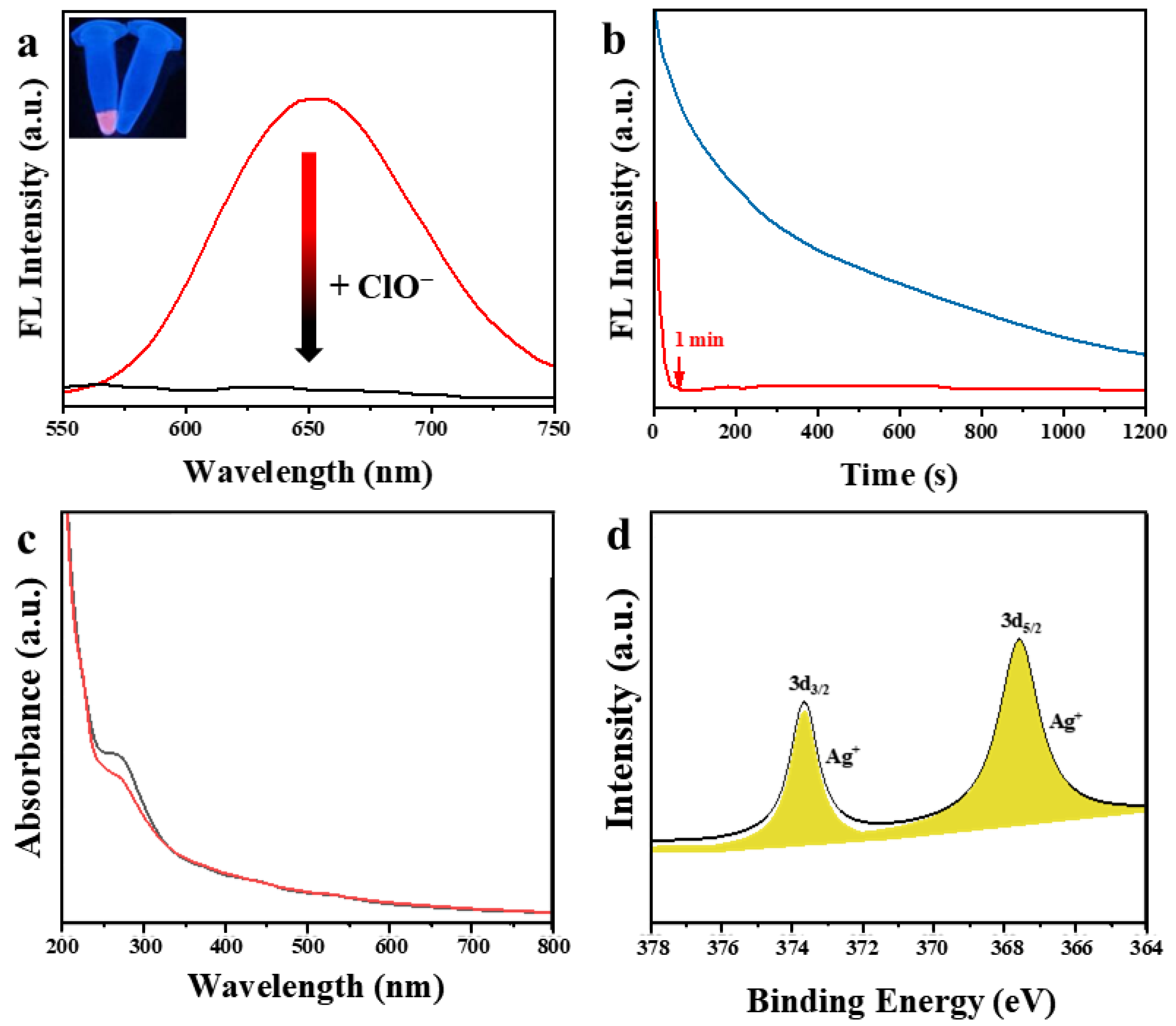
Interestingly, the PL of the as-formed DSPE-PEG-SDS@Ag NCs could be quenched by ClO. As shown in the inset of Figure 3a, in the presence of ClO, the PL of the DSPE-PEG-SDS@Ag NCs solution was quenched. Accordingly, the PL peak (red line) of the as-formed DSPE-PEG-SDS@Ag NCs at 650 nm disappeared (black line) and the quenching reaction proceeded rapidly. As shown in Figure 3b, the quenching could be completed within 1 min (red line). It is worth noting that the DSPE-PEG-SDS@Ag NCs prepared via slow evaporation showed an obviously sluggish response to ClO, as shown in Figure 3b (blue line). The effect of ClO on the DSPE-PEG-SDS@Ag NCs was further studied through the UV-vis absorption spectrum. As displayed in Figure 3c, the absorbance peak at 260 nm in the UV-vis spectrum of the DSPE-PEG-SDS@Ag NCs became weaker in the presence of ClO, which indicated the DSPE-PEG-SDS@Ag NCs might be oxidized by ClO. The ratiocination of the oxidation of the DSPE-PEG-SDS@Ag NCs could be proved by the following XPS measurement. As shown in Figure 3d, only Ag(I) component centered at Ag(I) BE (373.63 eV for Ag 3d3/2 and 367.58 eV for Ag 3d5/2) could be found in the XPS spectrum of the DSPE-PEG-SDS@Ag NCs after PL quenching, indicating that ClO caused full oxidation of Ag(0) atoms in the DSPE-PEG-SDS@Ag NCs. In addition, from the lifetime data, it can be seen that the lifetime of the oxidized DSPE-PEG-SDS@Ag NCs was 3.43 ns (Figure S2), shorter than that of the original DSPE-PEG-SDS@Ag NCs, indicating a possible dynamic quenching mechanism [28].
The PL intensity of the DSPE-PEG-SDS@Ag NCs showed a linear response relationship against the ClO concentration. As shown in Figure 4a,b, the I/I0 decreased linearly (R2 = 0.994) with increasing the concentration of ClO over the range of 1–100 μM. I0 and I correspond to the fluorescence intensity of the DSPE-PEG-SDS@Ag NCs in the absence and presence of ClO, respectively. The limit of detection (LOD) of the DSPE-PEG-SDS@Ag NCs for ClO was 0.67 μM (S/N = 3). More importantly, this method showed excellent selectivity to ClO over other common ROS including H2O2, OH, and ONOO, as well as some common metallic ions, as shown in Figure 4c. Compared with previous ClO sensors, the sensitivity of this method is comparable to most other studies, but has a better selectivity and response time than other methods (Table S1). As mentioned above, the ligand shell of the DSPE-PEG-SDS@Ag NCs, containing amphiphilic copolymers in an aqueous solution, formed the hydrophobic/hydrophilic dual permeability zone. External species had to struggle through such a blocking zone to have access to the innermost Ag core. Obviously, it is detrimental for short-lived ROS that are mainly free radicals, including superoxide radicals (O2), hydroperoxyl radicals (HO2*), hydroxyl radicals (HO*), and other free radicals such as peroxyl radicals (ROO*) [29]. Compared with these free radicals with μs-/ms-level lifetimes and nonradical ONOO with ms-level lifetime, hypochlorite’s lifetime is hour-level [30], providing enough time to permeate through the ligand shell to reach the Ag core. Meanwhile, ClO is strongly oxidative, and thus could oxidize the Ag core to quench its luminescence. On the other hand, H2O2 as a non-radical oxidant also has a long lifetime; however, it is poorly reactive. It has been reported that H2O2 does not oxidize most intracellular molecules readily, including lipids, DNA, and proteins, unless the latter have hyper-reactive thiol groups or methionine residues [31]. In fact, this is the reason that the thiolated ligand-encapsulated metal nanoclusters can often be used as the “turn-off” type fluorescent sensor for the detection of H2O2 [32,33,34]. However, those metal nanoclusters-based sensors might suffer from the interferences from hROS, especially nonradical strong oxidants. In this study, no hyper-reactive molecules including thiol groups or methionine residues were used as ligands to prepare the DSPE-PEG-SDS@Ag NCs; therefore, the sensing performance of the DSPE-PEG-SDS@Ag NCs was not affected by H2O2, as shown in Figure 4c.

3.3. Cellular Imaging Using the DSPE-PEG-SDS@Ag NCs

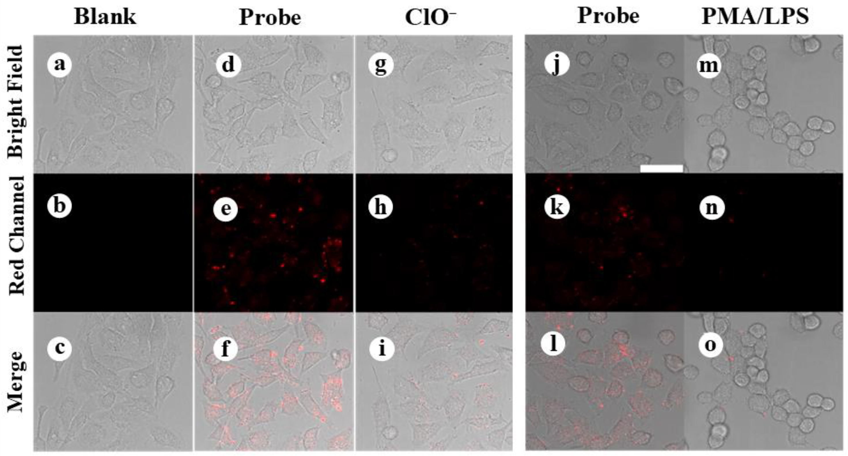
Encouraged by the high sensitivity and selectivity of the DSPE-PEG-SDS@Ag NCs for sensing ClO, we further applied the DSPE-PEG-SDS@Ag NCs for visualizing ClO in living cells using HeLa cells as a model. Firstly, the cytotoxicity of the DSPE-PEG-SDS@Ag NCs was tested by MTT assay. As shown in Figure S6, the cell viabilities were close to 100% as the concentration of the DSPE-PEG-SDS@Ag NCs ranged from zero to 450 μg/mL, confirming the low toxicity of the DSPE-PEG-SDS@Ag NCs to HeLa cells at a relatively high concentration, which implies the potential for cellular imaging applications or other biomedical applications. As for live cell imaging, firstly, HeLa cells were incubated with the DSPE-PEG-SDS@Ag NCs for 8 h, which was used as a control, and then treated using 150 μM ClO for 30 min. Then, the cells were washed with PBS buffer before imaging with a confocal laser scanning microscope (CLSM). As shown in Figure 5, compared with the blank group (Figure 5a–c), a visible fluorescence signal (Figure 5d–f) was observed after incubating HeLa cells with the DSPE-PEG-SDS@Ag NCs. This indicates that the DSPE-PEG-SDS@Ag NCs had good cell penetration capacity and maintained their emission characteristics inside the cells and could be used a fluorescent nanoprobe. For exogenous ClO detection, after the cells were treated with ClO, the fluorescence of the DSPE-PEG-SDS@Ag NCs was dramatically weakened (Figure 5g–i), indicating the DSPE-PEG-SDS@Ag NCs could be used as a fluorescent nanoprobe for the visual recognition of ClO in living cells. Then, the monitoring of endogenous ClO using the DSPE-PEG-SDS@Ag NCs as the fluorescent nanoprobe was investigated. It has been well known that endogenous ClO can be generated when the cells are treated with lipopolysaccharide (LPS) and phorbol myristate acetate (PMA) [35,36]. As shown in Figure 5, after treated with LPS- and PMA, the HeLa cells showed nearly invisible fluorescent signals (Figure 5m–o), indicating that the fluorescence of the DSPE-PEG-SDS@Ag NCs could be effectively quenched by endogenous ClO. These results indicated that the DSPE-PEG-SDS@Ag NCs could be used as a fluorescence nanoprobe for the visual recognition of ClO in living cells.

4. Conclusions

In summary, this study has demonstrated the use of amphiphilic copolymer-engineered luminescent Ag NCs for the sensitive and selective detection of intracellular ClO. With the help of the amphiphilic copolymer composite, i.e., DSPE-PEG-SDS, the luminescent Ag NCs capped with chemically inert hydrophobic ligands could be facilely transferred into aqueous solutions and remained their photoluminescent ability. It was found that the approach of the removal of organic solvents during the phase transfer has remarkable influences on the properties of DSPE-PEG-SDS@Ag NCs, such as the sizes, luminescent properties and sensing performance. Compared with slow evaporation, rotary evaporation spent less time and generated more advantageous DSPE-PEG-SDS@Ag NCs, including stronger emission and smaller particle sizes. In addition, the auxiliary use of SDS during the amphiphilic copolymers-induced phase transfer process was also found to be valuable in this sense. The DSPE-PEG-SDS@Ag NCs had an excellent aqueous stability. Further reaction with ClO resulted in full oxidation of the Ag core of the DSPE-PEG-SDS@Ag NCs and their emission quenching. Interestingly, the quenching of the emission of the DSPE-PEG-SDS@Ag NCs was specific toward ClO over other common hROS and wROS as well as some common interferences. Further cellular testing indicated the utility of the DSPE-PEG-SDS@Ag NCs as fluorescent nanoprobes for the fluorescent imaging of exogenous and endogenous ClO in living cells. This study demonstrates the promising potential of amphiphilic copolymer-engineered luminescent Ag NCs for the development of nanosensors or nanoprobes.

Supplementary Materials

The following supporting information can be downloaded at https://www.mdpi.com/article/10.3390/chemosensors12080166/s1, Figure S1: PL spectrum of pristine Ag NCs; Figure S2: PL decay profiles of the pristine Ag NCs, the DSPE-PEG-SDS@Ag NCs, and the DSPE-PEG-SDS@Ag NCs after reacting with ClO, respectively. Figure S3: TEM image and hydrodynamic size distribution histogram of DSPE-PEG-SDS@Ag NCs prepared under the slow evaporation method. Figure S4: TEM image of the pristine Ag NCs. Figure S5: XPS characterization of the pristine Ag NCs. Figure S6: Cell viabilities. Table S1. Comparison of the proposed sensor with previously reported nanomaterials-based hypochlorite sensors [20,22,24,37,38,39,40,41,42,43].

Author Contributions

Conceptualization, validation, data curation, writing—original draft preparation, X.L.; synthesis of silver nanoclusters and characterizations, Q.D.; assistance in cell experiments, Y.C.; funding acquisition, S.Z.; project administration, P.S. All authors have read and agreed to the published version of the manuscript.

Funding

This work was supported by National Natural Science Foundation of China (Grant No. 22274068, 22076073, 22304069), Major Basic Research Project of Natural Science Foundation of Shandong Province (No. ZR2023ZD27), Natural Science Foundation of Shandong Province (No. ZR2023MB139), and the Key R&D Projects of Linyi City (2022022).

Institutional Review Board Statement

Not applicable.

Informed Consent Statement

Not applicable.

Data Availability Statement

Data are contained within the article or Supplementary Materials.

Conflicts of Interest

The authors declare that they have no known competing financial interests or personal relationships that could have appeared to influence the work reported in this paper.

References

  1. Mutze, S.; Hebling, U.; Stremmel, W.; Wang, J.; Arnhold, J.; Pantopoulos, K.; Mueller, S. Myeloperoxidase-derived hypochlorous acid antagonizes the oxidative stress-mediated activation of iron regulatory protein 1. J. Biol. Chem. 2003, 278, 40542–40549. [Google Scholar] [CrossRef]
  2. Yang, Y.T.; Whiteman, M.; Gieseg, S.P. HOCl causes necrotic cell death in human monocyte derived macrophages through calcium dependent calpain activation. Biochim. Biophys. Acta 2012, 1823, 420–429. [Google Scholar] [CrossRef]
  3. Zhang, R.; Song, B.; Yuan, J. Bioanalytical methods for hypochlorous acid detection: Recent advances and challenges. Trends Anal. Chem. 2018, 99, 1–33. [Google Scholar] [CrossRef]
  4. Yang, J.; Chen, Z.; Yang, Y.; Zheng, B.; Zhu, Y.; Wu, F.; Xiong, H. Visualization of endogenous hypochlorite in drug-induced liver injury mice via a bioluminescent probe combined with firefly luciferase mRNA-loaded lipid nanoparticles. Anal. Chem. 2024, 96, 6978–6985. [Google Scholar] [CrossRef]
  5. Davies, M.J. Myeloperoxidase-derived oxidation mechanisms of biological damage and its prevention. J. Clin. Biochem. Nutr. 2010, 48, 8–19. [Google Scholar] [CrossRef] [PubMed]
  6. Jeitner, T.M.; Kalogiannis, M.; Krasnikov, B.F.; Gomolin, I.; Peltier, M.R.; Moran, G.R. Linking inflammation and parkinson disease: Hypochlorous acid generates parkinsonian poisons. Toxicol. Sci. 2016, 151, 388–402. [Google Scholar] [CrossRef] [PubMed]
  7. Wang, T.; Tan, H.S.; Wang, A.J.; Li, S.S.; Feng, J.J. Fluorescent metal nanoclusters: From luminescence mechanism to applications in enzyme activity assays. Biosens. Bioelectron. 2024, 257, 116323. [Google Scholar] [CrossRef] [PubMed]
  8. Xiao, Y.; Wu, Z.; Yao, Q.; Xie, J. Luminescent metal nanoclusters: Biosensing strategies and bioimaging applications. Aggregate 2021, 2, 114–132. [Google Scholar] [CrossRef]
  9. Wang, J.; Li, J.; Li, M.; Ma, K.; Wang, D.; Su, L.; Zhang, X.; Tang, B.Z. Nanolab in a cell: Crystallization-induced in situ self-assembly for cancer theranostic amplification. J. Am. Chem. Soc. 2022, 144, 14388–14395. [Google Scholar] [CrossRef] [PubMed]
  10. Yang, M.; Zhu, L.; Yang, W.; Xu, W. Nucleic acid-templated silver nanoclusters: A review of structures, properties, and biosensing applications. Coordin. Chem. Rev. 2023, 491, 215247. [Google Scholar] [CrossRef]
  11. Shu, T.; Su, L.; Wang, J.; Lu, X.; Liang, F.; Li, C.; Zhang, X. Value of the debris of reduction sculpture: Thiol etching of Au nanoclusters for preparing water-soluble and aggregation-induced emission-active Au(I) Complexes as phosphorescent copper ion sensor. Anal. Chem. 2016, 88, 6071–6077. [Google Scholar] [CrossRef]
  12. Zhang, C.; Gao, X.; Chen, W.; He, M.; Yu, Y.; Gao, G.; Sun, T. Advance of gold nanoclusters for bioimaging. iScience 2022, 25, 105022. [Google Scholar] [CrossRef]
  13. Kang, X.; Zhu, M. Tailoring the photoluminescence of atomically precise nanoclusters. Chem. Soc. Rev. 2019, 48, 2422–2457. [Google Scholar] [CrossRef] [PubMed]
  14. Lin, H.; Song, X.; Chai, O.J.H.; Yao, Q.; Yang, H.; Xie, J. Photoluminescent characterization of metal nanoclusters: Basic parameters, methods, and applications. Adv. Mater. 2024, 36, e2401002. [Google Scholar] [CrossRef]
  15. Wu, Z.; Jin, R. On the ligand’s role in the fluorescence of gold nanoclusters. Nano Lett. 2010, 10, 2568–2573. [Google Scholar] [CrossRef]
  16. Deng, G.; Malola, S.; Yuan, P.; Liu, X.; Teo, B.K.; Hakkinen, H.; Zheng, N. Enhanced surface ligands reactivity of metal clusters by bulky ligands for controlling optical and chiral properties. Angew. Chem. Int. Ed. 2021, 60, 12897–12903. [Google Scholar] [CrossRef] [PubMed]
  17. Zhu, C.; Xin, J.; Li, J.; Li, H.; Kang, X.; Pei, Y.; Zhu, M. Fluorescence or phosphorescence? The metallic composition of the nanocluster kernel does matter. Angew. Chem. Int. Ed. 2022, 61, e202205947. [Google Scholar] [CrossRef] [PubMed]
  18. Pniakowska, A.; Kumaranchira Ramankutty, K.; Obstarczyk, P.; Peric Bakulic, M.; Sanader Marsic, Z.; Bonacic-Koutecky, V.; Burgi, T.; Olesiak-Banska, J. Gold-doping effect on two-photon absorption and luminescence of atomically precise silver ligated nanoclusters. Angew. Chem. Int. Ed. 2022, 61, e202209645. [Google Scholar] [CrossRef] [PubMed]
  19. Ishii, W.; Okayasu, Y.; Kobayashi, Y.; Tanaka, R.; Katao, S.; Nishikawa, Y.; Kawai, T.; Nakashima, T. Excited state engineering in Ag29 nanocluster through peripheral modification with silver(I) complexes for bright near-infrared photoluminescence. J. Am. Chem. Soc. 2023, 145, 11236–11244. [Google Scholar] [CrossRef] [PubMed]
  20. Cao, X.; Cheng, S.; You, Y.; Zhang, S.; Xian, Y. Sensitive monitoring and bioimaging intracellular highly reactive oxygen species based on gold nanoclusters@nanoscale metal-organic frameworks. Anal. Chim. Acta. 2019, 1092, 108–116. [Google Scholar] [CrossRef] [PubMed]
  21. Li, Y.; Yi, S.; Lei, Z.; Xiao, Y. Amphiphilic polymer-encapsulated Au nanoclusters with enhanced emission and stability for highly selective detection of hypochlorous acid. RSC Adv. 2021, 11, 14678–14685. [Google Scholar] [CrossRef] [PubMed]
  22. Jia, M.; Mi, W.; Guo, S.; Yang, Q.Z.; Jin, Y.; Shao, N. Peptide-capped functionalized Ag/Au bimetal nanoclusters with enhanced red fluorescence for lysosome-targeted imaging of hypochlorite in living cells. Talanta 2020, 216, 120926. [Google Scholar] [CrossRef] [PubMed]
  23. Lin, X.; Tian, M.; Cao, C.; Shu, T.; Wang, J.; Wen, Y.; Su, L.; Zhang, X. Strongly phosphorescent and water-soluble gold(I)-silver(I)-cysteine nanoplatelets via versatile small biomolecule cysteine-assisted synthesis for intracellular hypochlorite detection. Biosens. Bioelectron. 2021, 193, 113571. [Google Scholar] [CrossRef] [PubMed]
  24. Li, Y.; He, Y.; Ge, Y.; Song, G.; Zhou, J. Smartphone-assisted visual ratio-fluorescence detection of hypochlorite based on copper nanoclusters. Spectrochim. Acta A Mol. Biomol. Spectrosc. 2021, 255, 119740. [Google Scholar] [CrossRef] [PubMed]
  25. Kastantin, M.; Missirlis, D.; Black, M.; Ananthanarayanan, B.; Peters, D.; Tirrell, M. Thermodynamic and kinetic stability of dspe-peg 2000 micelles in the presence of bovine serum albumin. J. Phys. Chem. B 2010, 114, 12632–12640. [Google Scholar] [CrossRef] [PubMed]
  26. Kastantin, M.; Ananthanarayanan, B.; Karmali, P.; Ruoslahti, E.; Tirrell, M. Effect of the lipid chain melting transition on the stability of DSPE-PEG(2000) micelles. Langmuir 2009, 25, 7279–7286. [Google Scholar] [CrossRef]
  27. Chen, Z.; Walsh, A.G.; Wei, X.; Zhu, M.; Zhang, P. Site-specific electronic properties of [Ag25SR18] nanoclusters by X-ray spectroscopy. Small 2021, 17, e2005162. [Google Scholar] [CrossRef]
  28. Zhang, M.; Lv, Q.; Yue, N.; Wang, H. Study of fluorescence quenching mechanism between quercetin and tyrosine-H2O2-enzyme catalyzed product. Spectrochim. Acta A Mol. Biomol. Spectrosc. 2009, 72, 572–576. [Google Scholar] [CrossRef]
  29. Zhang, Y.; Demokritou, P.; Ryan, D.K.; Bello, D. Comprehensive assessment of short-lived ROS and H2O2 in laser printer emissions: Assessing the relative contribution of metal oxides and organic constituents. Environ. Sci. Technol. 2019, 53, 7574–7583. [Google Scholar] [CrossRef]
  30. Abdul-Baki, A.A. A selected literature review of hypochlorite chemistry and definition of terms. J. Seed Technol. 1979, 1, 43–56. [Google Scholar]
  31. Rhee, S.G. H2O2-a necessary evil for cell signaling. Science 2006, 312, 1882–1883. [Google Scholar] [CrossRef]
  32. Li, H.; Wu, Y.; Xu, Z.; Wang, Y. In situ anchoring Cu nanoclusters on Cu-MOF: A new strategy for a combination of catalysis and fluorescence toward the detection of H2O2 and 2,4-DNP. Chem. Eng. J. 2024, 479, 147508. [Google Scholar] [CrossRef]
  33. Mi, W.; Tang, S.; Jin, Y.; Shao, N. Au/Ag bimetallic nanoclusters stabilized by glutathione and lysozyme for ratiometric sensing of H2O2 and hydroxyl radicals. ACS. Appl. Nano Mater. 2021, 4, 1586–1595. [Google Scholar] [CrossRef]
  34. Yue, G.; Li, S.; Liu, W.; Ding, F.; Zou, P.; Wang, X.; Zhao, Q.; Rao, H. Ratiometric fluorescence based on silver clusters and N, Fe doped carbon dots for determination of H2O2 and UA: N, Fe doped carbon dots as mimetic peroxidase. Sens. Actuat. B-Chem. 2019, 287, 408–415. [Google Scholar] [CrossRef]
  35. Gui, L.; Yan, J.; Zhao, J.; Wang, S.; Ji, Y.; Liu, J.; Wu, J.; Yuan, K.; Liu, H.; Deng, D.; et al. Hypochlorite activatable ratiometric fluorescent probe based on endoplasmic reticulum stress for imaging of atherosclerosis. Biosens. Bioelectron. 2023, 240, 115660. [Google Scholar] [CrossRef] [PubMed]
  36. Huang, T.; Yan, S.; Yu, Y.; Xue, Y.; Yu, Y.; Han, C. Dual-Responsive Ratiometric fluorescent probe for hypochlorite and peroxynitrite detection and imaging in vitro and in vivo. Anal. Chem. 2022, 94, 1415–1424. [Google Scholar] [CrossRef]
  37. Gopu, C.L.; Shanti Krishna, A.; Sreenivasan, K. Fluorimetric detection of hypochlorite using albumin stabilized gold nanoclusters. Sens. Actuat. B-Chem. 2015, 209, 798–802. [Google Scholar] [CrossRef]
  38. Zhao, G.; Lv, C.-C.; Yang, X.-K.; Zhao, X.; Xie, F. Levonorgestrel protected Au10 cluster for hypochlorite sensing in living organisms. Anal. Chim. Acta 2024, 1320, 343033. [Google Scholar] [CrossRef] [PubMed]
  39. Yang, G.; Wang, Y.; Li, Y.; Yan, C.; Liu, Z.; Wang, H.; Peng, H.; Du, J.; Zheng, B.; Guo, Y. Synthesis of vitamin B1-stabilized gold nanoclusters with high quantum yields for application as sensors. ACS Appl. Nano Mater. 2022, 5, 17234–17242. [Google Scholar] [CrossRef]
  40. Tang, Q.; Yang, T.; Huang, Y. Copper nanocluster-based fluorescent probe for hypochlorite. Microchim. Acta 2015, 182, 2337–2343. [Google Scholar] [CrossRef]
  41. Meng, Y.; Zhang, Z.; Zhao, H.; Jiao, Y.; Li, J.; Shuang, S.; Dong, C. Facile synthesis of multifunctional carbon dots with 54.4% orange emission for label-free detection of morin and endogenous/exogenous hypochlorite. J. Hazard. Mater. 2022, 424, 127289. [Google Scholar] [CrossRef] [PubMed]
  42. Gao, Y.; Liu, Y.; Zhang, H.; Lu, W.; Jiao, Y.; Shuang, S.; Dong, C. One-pot synthesis of efficient multifunctional nitrogen-doped carbon dots with efficient yellow fluorescence emission for detection of hypochlorite and thiosulfate. J. Mater. Chem. B 2022, 10, 8910–8917. [Google Scholar] [CrossRef] [PubMed]
  43. Gu, Y.; Zheng, X.; Chen, Z.; Teng, R.; Zhang, Y.; Li, H.; Ding, C.; Huang, Y. Fluorescent-colorimetric dual signal ratio sensor with AuNRs@UCNPs superstructure nanoprobe for accurate hypochlorite detection. Sens. Actuat. B-Chem. 2024, 419, 136384. [Google Scholar] [CrossRef]
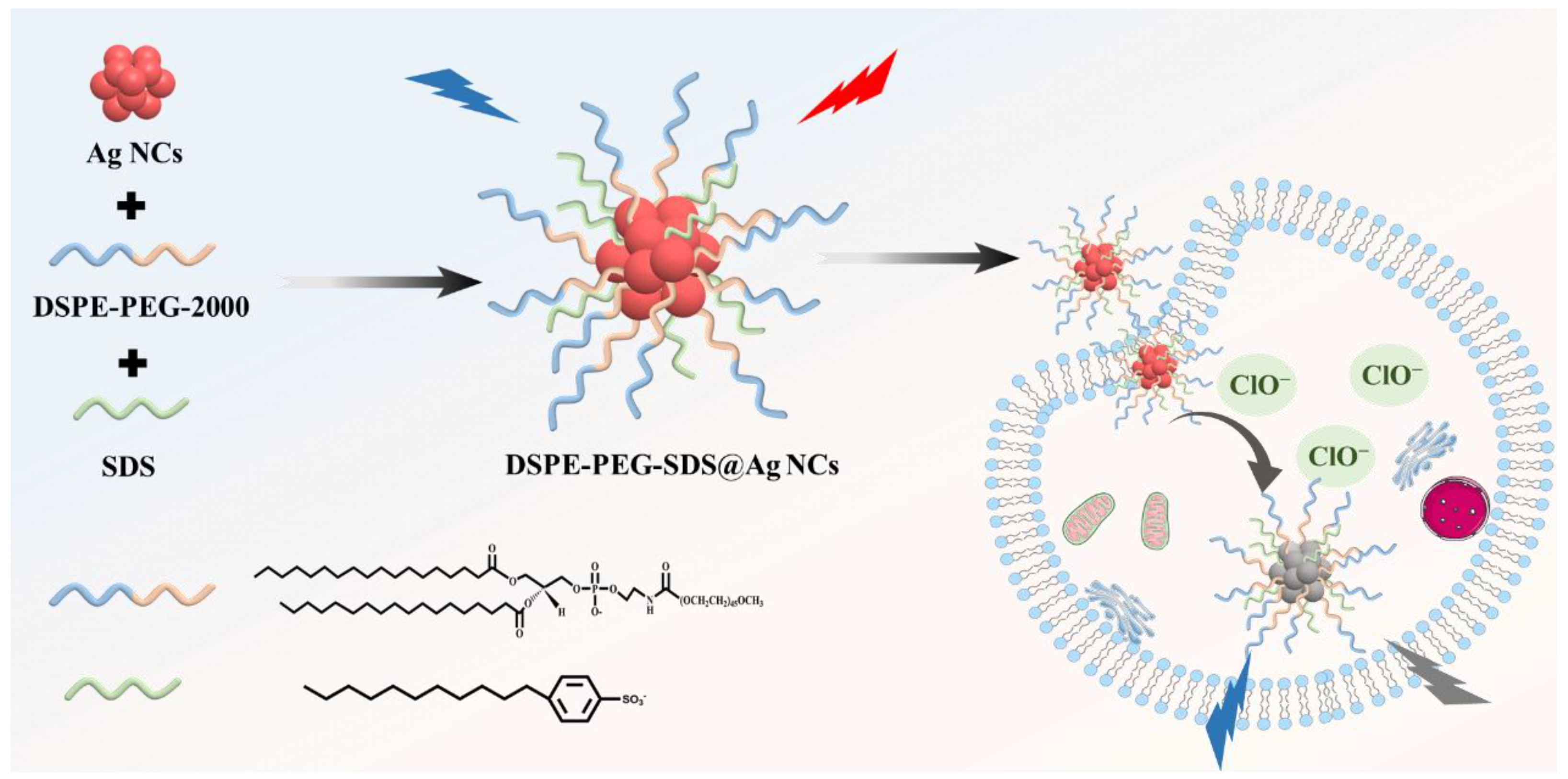
Scheme 1. Phase transfer of the luminescent Ag NCs using amphiphilic copolymers for the detection of intracellular ClO.
Chemosensors 12 00166 sch001
Figure 1. (a) Emission curves (red line) and absorption spectrum (purple line) of the DSPE-PEG-SDS@Ag NCs. (b) TEM image of the DSPE-PEG-SDS@Ag NCs prepared via rotary evaporation, and (c) their hydrodynamic size distribution histogram measured by DLS. (d) Emission curves of the rotary evaporation (red line) and slow evaporation (blue line)-produced DSPE-PEG-SDS@Ag NCs. (e) Emission curves of DSPE-PEG-SDS@Ag NCs (red line) and without SDS (green line), and (f) their hydrodynamic size distribution histogram without SDS.
Chemosensors 12 00166 g001
Figure 2. (a) XPS spectra of Ag 3d of the DSPE-PEG-SDS@Ag NCs. (b) PL intensity of the DSPE-PEG-SDS@Ag NCs at different pH values and (c) at different salt concentrations.
Chemosensors 12 00166 g002
Figure 3. (a) PL curves of the DSPE-PEG-SDS@Ag NCs in the absence (red line) and presence of 100 μM ClO (black line) at an excitation wavelength of 428 nm. Inset: photographs of the DSPE-PEG-SDS@Ag NCs in the absence (left) and presence of hypochlorite (right) under 365 nm UV light. (b) Time-dependent ClO-induced quenching of the PL of the DSPE-PEG-SDS@Ag NCs (red line) and the DSPE-PEG-SDS@Ag NCs prepared via slow evaporation (blue line). (c) UV-vis absorption curves of the DSPE-PEG-SDS@Ag NCs in the absence (black line) and presence of 100 μM ClO (red line). (d) XPS spectra of Ag 3d of the DSPE-PEG-SDS@Ag NCs after reacting with ClO.
Chemosensors 12 00166 g003
Figure 4. (a) PL curves of the DSPE-PEG-SDS@Ag NCs excited at 420 nm in the presence of different concentration of ClO ranging from 0 to 100 μM. (b) Linear relationship between PL intensity and the concentration of ClO. (c) Selectivity of the DSPE-PEG-SDS@Ag NCs towards ClO. All possible interferences were given to be 100 μM.
Chemosensors 12 00166 g004
Figure 5. Fluorescence microscopy images of HeLa cells. (ac) Th control; (df) the cells incubated with the as-formed DSPE-PEG-SDS@Ag NCs nanoprobes; (gi) the cells incubated with the nanoprobes followed by treated with exogenous ClO (150 μM); (jo) the cells treated with LPS and PMA prior to incubation with the nanoprobes. Top images: bright field images. Middle images: fluorescence confocal images of the red channel. Bottom images: merge images of bright field and red channel.
Chemosensors 12 00166 g005
Disclaimer/Publisher’s Note: The statements, opinions and data contained in all publications are solely those of the individual author(s) and contributor(s) and not of MDPI and/or the editor(s). MDPI and/or the editor(s) disclaim responsibility for any injury to people or property resulting from any ideas, methods, instructions or products referred to in the content.