Review

Defect Engineering in Transition Metal Dichalcogenide-Based Gas Sensors

by
Xiaqing Fu
1,†,
Zirui Qiao
2,†,
Hangyu Zhou
3 and
Dan Xie
4,*
1
School of Microelectronics, Shanghai University, Shanghai 201800, China
2
Department of Chemistry, Tsinghua University, Beijing 100084, China
3
China Academy of Safety Science and Technology, Beijing 100012, China
4
School of Integrated Circuits, Tsinghua University, Beijing 100084, China
*
Author to whom correspondence should be addressed.
These authors contributed equally to this work.
Chemosensors 2024, 12(6), 85; https://doi.org/10.3390/chemosensors12060085
Submission received: 13 April 2024 / Revised: 15 May 2024 / Accepted: 20 May 2024 / Published: 21 May 2024
(This article belongs to the Special Issue Emerging 2D Materials for Sensing Applications)

Abstract

:
Since the discovery of innovative two-dimensional (2D) materials, significant efforts have been dedicated to exploring their intriguing properties and emerging applications. Among all candidates, transition metal dichalcogenides (TMDs) have proven to be exceptional for gas sensing, while defects engineering has been introduced to modify the pristine TMDs for better gas sensing performances. In this review, we systematically summarize types of defects, advanced characterization techniques, and state-of-the-art controllable synthetic methods. Various types of defects in TMDs can induce diverse changes in chemical and electron structures, which are closely correlated with gas sensing ability. Therefore, connections between defects and gas sensing mechanisms and performances have been addressed based on both defect categories and electron affinity of gases. This review will be a guide for researchers in defective materials and open up the field of precisely synthesis chemistry and deepen the understanding of the underlying effects of defects in other 2D materials.

1. Introduction

As promising materials, a family of two-dimensional (2D) materials is steadily gaining interest for their novel properties, which can be roughly divided into several categories: transition metal dichalcogenides (TMDs, which have a general chemical formula of MX2), graphene and its derivatives, violet/black phosphorus, MXene, metal–organic frameworks, metal–oxide nanosheets and other 2D materials [1]. Most of these 2D materials possess unique properties (i) High surface-area-to-volume ratio and large specific area [2]. (ii) Tunable size-related electronic structure and bandwidth [3]. (iii) Thickness can be controlled at the atomic level by designed approaches for specific 2D materials [4]. Taking into account all these unique properties, 2D materials are especially suitable for ultra-scaled electronic devices while maintaining the desired performance [5,6], and applications in sensing, catalysis, energy storage, etc [3]. Among all the application fields, sensing, especially efficient gas sensing based on 2D materials, can straightforwardly solve the existing challenges in environmental monitoring, workplace safety supervision, breath analysis, food quality assessment and counter-terrorism efforts [7], which have aroused wide research interests in designing and fabricating new generation high-performance gas sensors.
As a member of 2D materials, TMDs have already been extensively used in photodetectors [8]. Furthermore, due to the unique electrical characteristics generated from their atomically thin 2D structure [9], one of the most exciting features of TMD sensors is their exceptional ability to detect gas molecules at room temperature, especially for MoS2, MoSe2, and WS2 [10,11,12]. However, pristine TMDs usually face unsatisfactory sensing performance, such as low sensitivity and long response time due to their naturally inert, dangling bonds-free surface and long recovery time due to their randomly distributed surface defects during common fabrication flow such as CVD. These undesirable merits can be greatly optimized by controllably introducing/designing desirable defects into TMD structures. Defects can be regarded as alterations in any form within the intrinsic structure, such as vacancy, heteroatom substitution, grain boundary, edge site, etc. Even though several techniques have been developed to introduce different types of defects into TMDs, controllable and orienteering modification is still difficult to achieve,-based the origin of defects lacks systemic mechanism study. Here, in this review, we will first briefly introduce common defect types in TMDs, then several promising defect characterization methods. After that, some methods which can controllably introduce defects will be introduced, and finally, the TMDs’ gas sensing performances and mechanisms will be discussed (Figure 1).

2. Defects in TMDs

Since defects in TMDs can significantly influence their performances as in common semiconductors like silicon, we will summarize types, characterizations and synthesis methods of defects in TMDs in this section.

2.1. Defect Types

2.1.1. Point Defects

Point defects are the simplest and the most common defects in 2D materials and can be roughly divided into vacancies, self-interstitials and substitution. Though point defects can be classified, the origin of the former two defects (or, intrinsic defects) is complex and unique in specific 2D material, which will lead to extra charges or electron–hole recombination and increased oxygen affinity, while the last type (extrinsic defects) is usually caused by intentionally introducing foreign atoms to replace original atoms in host lattice [13]. It is worth noting that, rather than substituting onto the lattice, foreign atoms can be adsorbed to the surface, which is also an extrinsic defect.
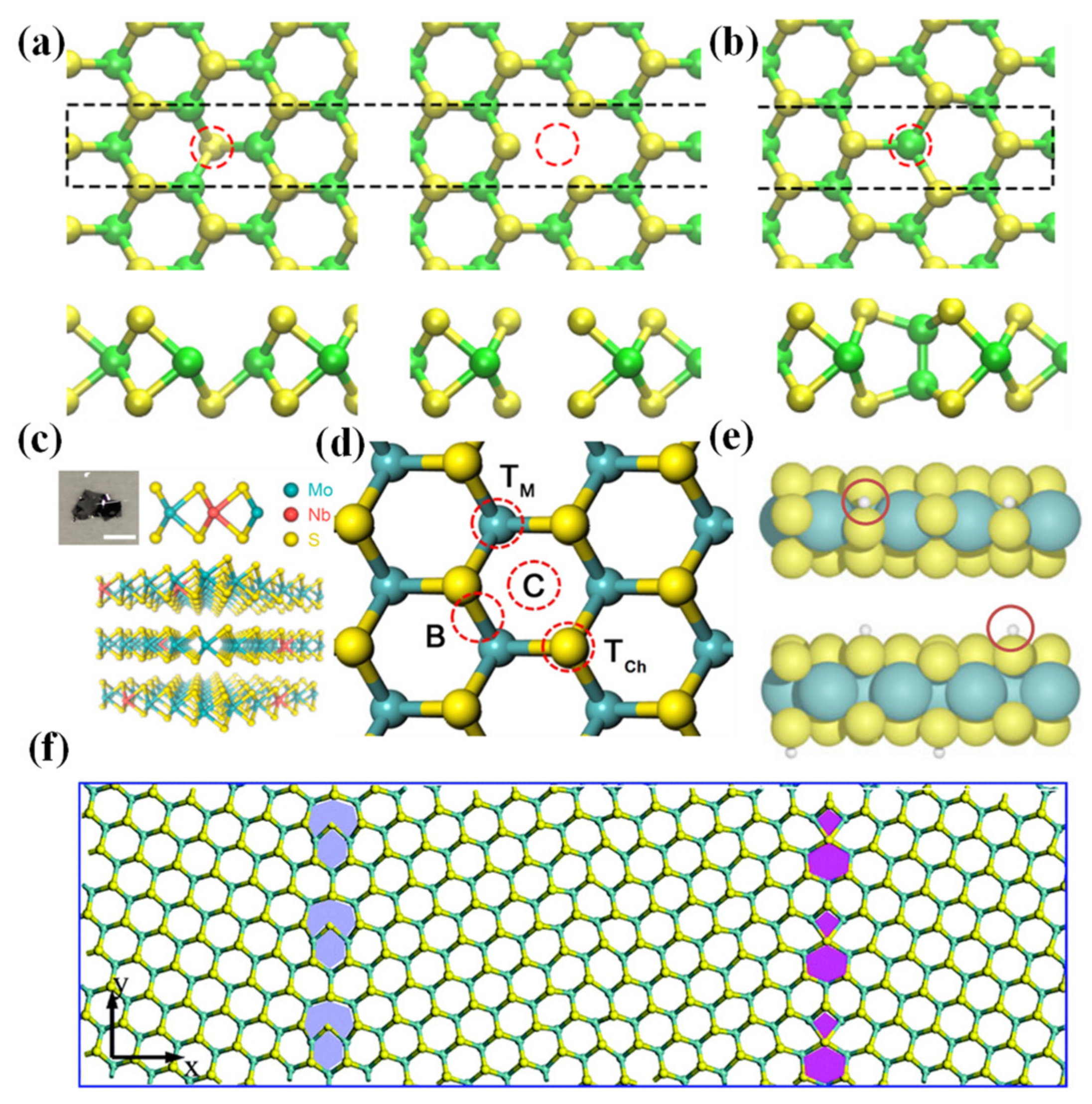
In TMDs (MX2), X vacancies are the most common defects due to the energetical stability of such defects. X (sulfur, selenium, or tellurium atoms) vacancies typically induce unpaired electrons in surrounding M atoms, leading to localized states (also known as unpaired electrons) within the bandgap and higher than the Fermi level, which in turn provide n-type doping and will also act as scattering centers causing low electron mobility [14]. A schematic view of both Mo vacancy and S vacancy in MoS2 can be seen in Figure 2a [15]. M (transition metal atoms) vacancies occur less frequently due to higher forming energy, causing many different states in the bandgap, higher or lower than the Fermi level, which in turn introduces diverse doping characteristics [16]. Meanwhile, the electro states introduced by self-interstitials either M or X atoms are highly dependent on specific interstitial positions [17]. The chemical structure of typical self-interstitial defects is modeled in MoS2 as Mo–Mo interstitial pairs (Figure 2b) [15]. For the extrinsic defects in TMDs, most of them originate from doping foreign atoms, which can typically replace lattice atoms or occupy the vacancy sites (Figure 2c, [18]). Since typical doping ions possess hundreds of KeV kinetic energy, which is destructive for atomically thin TMDs, some specific doping methods, such as electrostatic doping [19], charge transfer doping [20], chemical element substitutions [21,22], and photoinduced doping [23], have been invented to dope TMD material. While considering adsorbed atoms instead of directly replacing host atoms, 1H phase TMDs have four positions available to an adsorbent: above the metal atoms (TM), above the chalcogen atoms (TCh), on a metal–chalcogen bond (B), and above or within the center of hexagonal voids (C) [24] (Figure 2d, [25]). For stacking layers, 2H phase TMDs, the TM and TCh sites are equivalent to intercalated species due to the 2H phase’s stacking sequence [15]. Since adatoms are predicted to be quite mobile even at room temperature [26], further research on adatom mobility in TMDs may lead to insights into the kinetics, stability of TMD synthesis at elevated temperatures and electronic properties for future applications.

2.1.2. Edge Sites

Edge sites are also prominent defects in 2D material, which have been proven to be active sites arising from magnetic order in graphene. In synthetic TMDs, single-crystalline islands often have sharp edges with triangular shapes, which is the preferred edge morphology possessing low energy. The surface energies are a function of the sulfur vapor potential μS. When μS is low while synthesizing TMDs (i.e., CVD) [27], distorted hexagonal shapes will be adopted in TMDs rather than triangular shapes (Figure 2e, [28]).

2.1.3. Grain Boundaries

Grain boundaries, which separate two domains of different lattice orientations, can be different in different 2D materials. In TMDs, due to the tri-layer structure (X-M-X), when atoms are dislocated or removed, the ringed motifs can be highly dependent on grain boundary angle [29,30,31]. When gain boundaries have low angles with high strain, X (i.e., sulfur) atoms can be mobile under quite low accelerating voltage and be easily dislocated [32]. Grain boundaries can also affect the electronic properties of TMDs. For example, depending on the atomic structures in MoS2 (The defects in the mirror boundary are molybdenum-rich; the defects in the tilt boundary are sulfur-rich), grain boundaries can be either sulfur-deficient or molybdenum-deficient and in turn locally n-dope or p-dope the material [33] (Figure 2f shows MoS2 film with the S4|6 and S6|8 mirror grain boundaries [34]).
Figure 2. (a) Schematic top and side views of MoS2 with single-S vacancy (left); single-Mo vacancy (right). (b) Top and side view of Mo–Mo split interstitial in MoS2. Reproduced with permission [15]. Copyright 2015, Physical Review B. (c) Cross-section illustration of van der Waals coupled MoS2: Nb layers where Nb dopants replace the Mo host atoms in a substitutional manner. A digital photograph inset shows a synthesized MoS2:Nb crystal with a scale bar of 3 m. Reproduced with permission [18]. Copyright 2014, American Chemical Society. (d) Top view of four positions available to an adsorbent in TMDs. Reproduced with permission [25]. Copyright 2016, IOP Publishing Ltd. (e) Side views of Mo-edge (top) and S-edge (bottom) in MoS2. Reproduced with permission [28]. Copyright 2015, Tsinghua University Press and Springer-Verlag Berlin Heidelberg. (f) MoS2 film with the S4|6 and S6|8 grain boundaries. Reproduced with permission [34]. Copyright 2020, Royal Society of Chemistry.
Chemosensors 12 00085 g002

2.2. Characterization

Since defects can play an important role in a material’s performance, it is necessary to accurately recognize defects in materials by different characterizations. This section will focus on some well-developed characterization methods for ultrasmall defects in TMDs.

2.2.1. Microscope

Microimaging is the most straightforward technique for defect characterization because of its ability to directly visualize the alignment of atoms. Advanced microscopes which can be used to characterize defects at an atomic level usually include a high-resolution transmission electron microscope (HRTEM), scanning transmission electron microscope (STEM) and scanning tunneling microscope (STM).
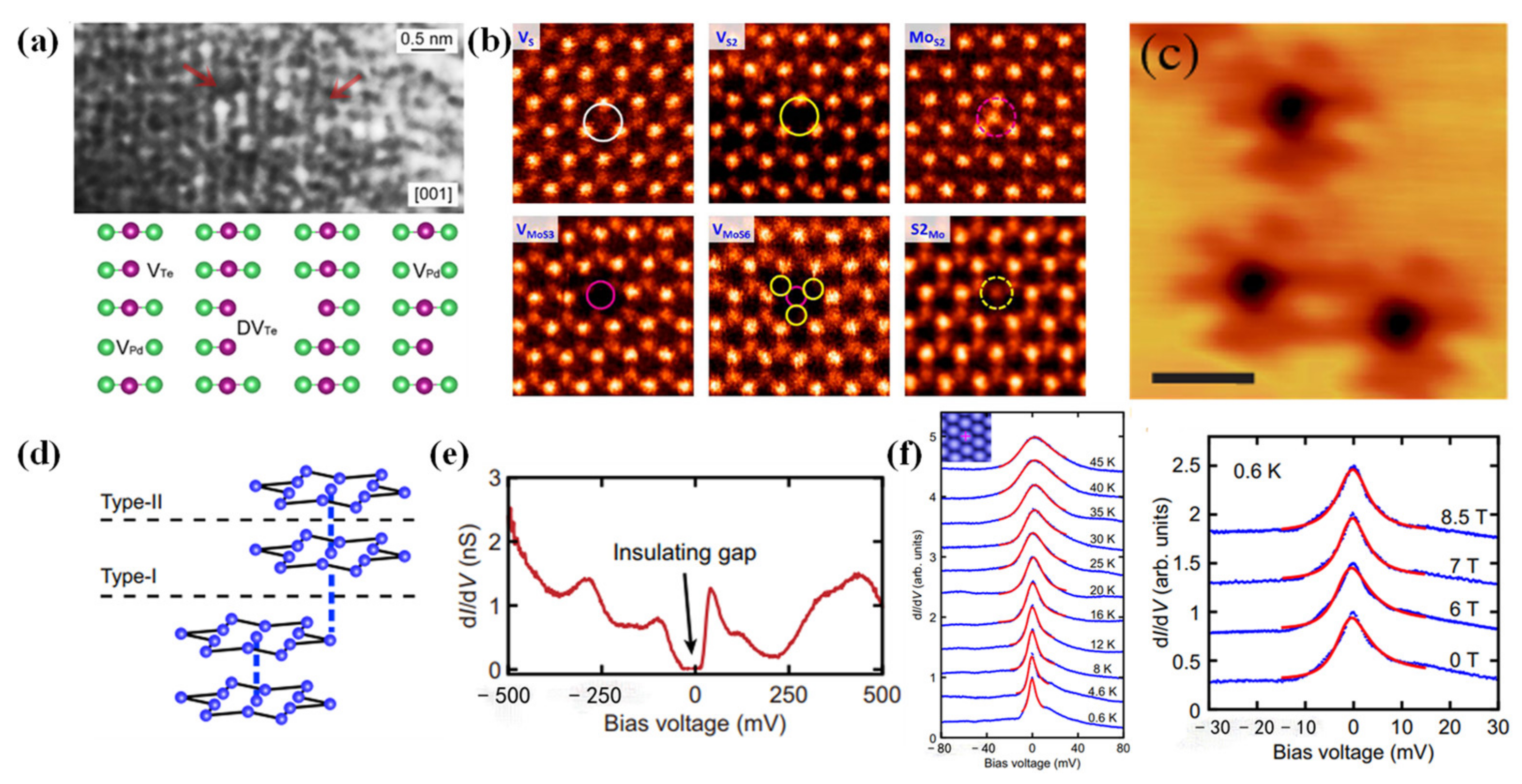
The Traditional Transmission electron microscope (TEM) mainly collects either transmitted or scattered electrons to form an image and it cannot reach atomic resolution, which means that defects cannot be observed. Nevertheless, HRTEM adopts an interference image generated by all diffraction and transmission electrons due to phase differences. Therefore, it has a higher resolution and can be used for investigating atomic information. Most recently, Zuo et al. [35] proposed a two-step method (including HNO3 etching and electrochemical exfoliation) to fabricate PdTe2-based catalysts with three different types of vacancies (d-PdTex), including single Pd vacancy, single Te vacancy and double Te vacancies. The HRTEM successfully revealed three types of defects in one magnified image (Figure 3a). HRTEM has also been used to validate defects in other 2D materials, such as Se vacancies in MoSe2 [36], sulfur defects in MoS2 and a phase transformation from the 2H-to-1T near extended sulfur vacancies when MoS2 was exposed to high electron beam doses [37] and plasma-induced single S vacancy in WS2 [38].
STEM combines the advantages of SEM and TEM, adopting the point by point scanning method of SEM and collecting both scattering/diffracting electrons that penetrate the sample. Therefore, STEM also has a higher resolution than TEM and a better image of the sample’s interior nanostructure than SEM. The intensity of each point is a function of the quantity and number of atoms overlapping in the vertical direction there, making it capable of clearly characterizing point defects in 2D materials [39]. Recently, Zhang et al. [40] proposed a one-step way to simultaneously control the 1T/2H phase ratio and defect density in MoS2 monolayers through the in situ strain induced by NaCl additives. During the experiment, two phases of boundary around NaCl particles can be clearly recognized by STEM. Zhou et al. successfully imaged six types of point defects in MoS2 (Figure 3b, [27]). A detailed study of dominant defects in MoS2 was conducted, showing that the main defects will change from S vacancies in mechanical exfoliation and chemical vapor deposition samples to Mo antisite in physical vapor deposition samples. Extrinsic point defects such as Nb substitutions at the Mo site in MoS2 were found by Gao et al. [41]. Vacancies such as Te double vacancy in 2H-MoTe2 have been revealed [13], and the agglomeration of Se vacancies into line defects in monolayer MoSe2 can also be seen by STEM [42].
STM makes use of ultrathin tip(s) and the sample’s surface as electrodes, giving opportunities for atomic-level imaging. When the tip–sample distance is less than 1 nm, under the effect of an electric field provided by STM, electrons will travel through the potential barrier between electrodes and form a current, which is exponential to the tip–sample distance. By combining the first principal calculation and STM images, S vacancies, M vacancies and S adatoms can be identified in MoS2 [43]. Sulfur passivation has been studied in zigzag MoS2 triangular islands using STM, identifying states at the Fermi level at the edges [44]. Also, by taking advantage of STM in measuring different currents according to the different surface morphology of materials, the most common intrinsic defect in WSe2, single tungsten vacancies which leads to p-type doping, rather than X vacancies in other TMDs, was first found by Zhang et al. [45] (Figure 3c). Moreover, STM not only identifies the defect structures, but also can identify their magnetic properties. Recently, Shen et al. [46] adopted STM to further explore the Type-II 1T-TaS2 surface (Type-II means surface terminates in the middle of the dimerization of two Star-of-David clusters) (Figure 3d). After Pb intercalation, both dark SD clusters (Pb atom beneath cluster) and bright SD clusters (no Pb atom beneath the cluster) can be observed on the surface of Type-II 1T-TaS2. An insulating gap of ~50 mV was found on a spot within a small area without dark SD clusters, just as the insulating gap shown in pristine Type-II 1T-TaS2′s surface. However, a transition from ~50 mV insulating gap to a sharp zero bias peak within a much larger area consisting of both bright and dark clusters can be found according to the dI/dV spectra from STM. The formation of a strong zero bias peak was believed to be Kondo screening of the unpaired electrons in Star-of-David clusters and was demonstrated by conducting the temperature and magnetic field-dependent STM dI/dV measurements (Figure 3f). Fitting temperature-dependent ZBP with Kondo expression yields a Kondo temperature at 37 K and no clear splitting of the Kondo resonance peak was discovered up to 8.5 T external magnetic field. Shen et al. further explained the reason for the transmission from the insulating bandgap in pristine Type-II TaS2 to the Kondo resonance peak on the Pb intercalated 1T-TaS2. Mott-insulating gap observed on the pristine Type-II 1T-TaS2 surface results from the weak in-plane Coulomb interaction between unpaired localized electrons in clusters and a few itinerant electrons within the underlying layer. However, with the intercalation of Pb atoms, charge transfer between the intercalated atoms and the host 1T-TaS2 layer is generated, as suggested by calculations. Consequently, more itinerant electrons from the Pb intercalation beneath the surface layer can interact with the Mott-localized electrons in the top layer, leading to the emergence of a sharp Kondo resonance peak in the dI/dV spectrum. The foreign atoms in layered narrow-electronic-band materials aroused various new phenomena in electronic states and band structures, which would be potential candidates for sensing applications.
Figure 3. (a) The HRTEM image at the atomic scale shows the defects, as illustrated in the atomic model: Pd (green) and Te (purple). Reproduced under the terms of the CC-BY license [35]. Copyright 2023, American Chemical Society. (b) Atomic resolution STEM-ADF images of various intrinsic point defects present in monolayer CVD MoS2. Reproduced with permission [27]. Copyright 2013, American Chemical Society. (c) Close-up STM topographic images showing point defects in WSe2 at −1.2 v. Reproduced with permission [45]. Copyright 2017, Physical Review Letters. (d) Schematic showing the alternating interlayer stacking of the SD clusters in 1T-TaS2 which results in two inequivalent cleavage planes. (e) dI/dV spectrum taken on a spot within one area without dark SD clusters. (f) temperature (left) and magnetic field-dependent dI/dV curves (right). In the left image, blue lines are the dI/dV spectra measured at different temperatures on the bright SD cluster. The red lines are the thermally convolved Fano fits to the ZBP at each temperature. In the right image, blue lines are the dI/dV spectra measured with different external magnetic fields at 0.6 K. The red lines are the thermally convolved Fano fits to the ZBP at the measured magnetic fields. Reproduced under the terms of the CC-BY license [46]. Copyright 2022, The Author(s).
Chemosensors 12 00085 g003

2.2.2. Raman Spectrum

Raman spectrum is a scattering spectrum, with different frequencies from incident lights analyzed to obtain information about molecular vibration and rotation. Therefore, Raman spectra can be used to count layers of 2D material, track the electronic band structure, and investigate how various perturbations [47,48,49] such as strain, doping, electric and magnetic fields, and functional groups. For example, in MoS2, both Raman spectra of pristine monolayer and defective MoS2 have been investigated, while Pristine monolayer MoS2 has two prominent Raman-active peaks (E′, and A′1, corresponding to in-plane vibration and out-of-plane vibration of Mo-S bond, respectively). After defects are introduced in MoS2, remarkable changes in peaks can be observed: (i) the positions of two prominent Raman-active peaks shift in opposite directions (Usually caused by vacancies with E′ peak redshift, A′1 peak blueshift) [50], (ii) two prominent Raman-active peaks will broaden (Usually caused by both vacancies and implantations) [51,52,53], (iii) defect-activated peaks might appear [53]. Substitution in TMDs can also be distinguished by observing Raman peaks shift [54] (can be treated as vacancies since substitution atoms cannot fully restore the bonding strength in pristine material). For example, in Parkin et al.’s experiment [50], monolayer MoS2 was electron beam irradiated to generate defects. As the irradiation dose increased, the E′ peak continued red-shifted while the A1′ peak was initially slightly blue-shifted then red-shifted back almost to its original position (Figure 4a). The redshift of the E′ peak usually results from the dissociation of the Mo–S bond at vacancy sites. In simple terms, Mo-S bond dissociation leads to fewer in-plane vibrations, so the E′ mode vibration is weakened (redshift). On the contrary, fewer in-plane vibrations lead to more out-of-plane vibrations, thus strengthening A1′ mode vibration (blueshift). Parkin et al. performed an energy dispersive spectrometer (EDS) to further determine which type of vacancy (Mo vacancy or S vacancy) has been introduced into the MoS2 sample. According to the measured EDS intensity of sulfur and molybdenum as a function of electron dose (Figure 4b), Mo’s intensity nearly kept constant while S intensity dropped as electron dose density increased. Later, by exposing the sample to the atmosphere and being intact, no holes were observed, which finally demonstrated that S vacancies were indeed the major defects in MoS2 after electron beam irradiation. The reason why A1′ shifted nearly back to its original position as the electron dose increased has also been explained by Parkin et al. It was believed that, when electron dose as well as S vacancy concentration increased, line defects tended to form, possibly contributing to the decrease in Mo’s out-of-plane vibration and the decrease in blueshift of the A1′ peak.

2.2.3. EPR

Electron paramagnetic resonance (EPR), also called electron spin resonance (ESR), is one of the most promising tools for analyzing and characterizing lattice defects in materials for their unpaired electrons in defects such as vacancies. The magnetic moments of unpaired electrons cause an identifiable peak to emerge in EPR spectra at a particular g factor. The intensity of the peak is related to the vacancy concentration, and the g factor varies in different types of vacancies, thus EPR could both qualitative and quantitative unveil the defective structure of TMDs. Wang et al. [55] adopted an H2O2 etching method to introduce homogeneously distributed single S-vacancies onto the MoS2 nanosheet surface. Both STEM and EPR were used for characterization. From STEM’s image, single S vacancies can be seen (Figure 4c). EPR was further adopted to reveal the proportional relation between dangling bonds and etching time, as well as further verify the single-atom state of these S-vacancies (Figure 4d), and signal intensity at g = 2.009 continues to increase as the etching time increases, which means there are no agglomerate S-vacancies. Moreover, a hydrothermal technique was used to create a series of multilayered MoS2 with varying amounts of sulfur defects [56]. The manufactured samples observed EPR spectra show variations in the sulfur vacancy density, which are thought to be proportionate to the Mo–S dangling bonds in the MoS2 slabs. An increase in vacancy density in annealing samples (WS2) was further demonstrated by EPR spectra [57]. Since different coordination has a great influence on EPR’s signal, we summarized some examples in the following table (Table 1). Note that the commonly used EPR approach is sample-averaging, and that other defect existences, such as cracks, or chemical bond expansion, may also cause a similar signal [58]. Moreover, the EPR signal can be greatly disturbed by high temperatures, so it is better to use EPR at a low temperature and combine other characterization methods to have a better understanding of a certain material. Additionally, in gas sensing, aside from characterizing gas sensing materials, there are more applications for EPR in gas detection [59]. For example, EPR can be used for directly detecting O2 due to its triplet ground state, which can form magnetic coupling with specially designed probes thus influencing the dynamics of probes. EPR can also be used for sensing intermediates formed during the gas adsorption process by combining in situ heating/irradiation/atmosphere control units and the spin trapping technique. In general, EPR has a desirable prospect in the gas sensing domain due to its wide usage.

2.2.4. PL Spectroscopy

PL spectroscopy mainly compares incident photons and the photons emitted from electron–hole pairs in material to detect defects in materials, especially in TMDs. In detecting vacancies in TMDs, since vacancies cause localized states within the bandgap, TMDs such as MoS2 with S vacancies have two peak signals (A exciton peak [68], or so-called free exciton peak and bound exciton peak) in PL spectra. A excitons (X0, X0 or X0+, the latter two types of three-body exciton peak is close to and lower than X0 peak) result from the coulomb interaction between one electron in the conduction band and one hole in the valence band. Bound exciton (Xb) results from exciton bound to localized states within the bandgap caused by vacancies [68]. It is worth noting that Xb peak possesses a nonlinear dependence on incident laser power, will be saturated at high laser power and will vanish when temperature is higher than 100 K, while A exciton (X0, X0 or X0+) has a linear dependence on laser power and will not be vanished or saturated. Though Xb peak in PL spectra can be saturated, it can still have a sublinear dependence on the density of defects and a linear dependence on the density of defects after adopting the normalization method (IXb/IX0) [69], which means that Xb peak can be used as a standard approach to characterize defects’ density in TMDs. For instance, He et al. [53] made use of Au ion irradiation to introduce defects in monolayer MoS2 with controllable defect density. XPS was first used to define the defects after irradiation (Figure 4e). A decreasing trend of the S/Mo ratio from 2.0 to lower was observed with the increase in ion fluence, which demonstrated the main defects after irradiation were S vacancies. The PL was adopted to further characterize the monolayer MoS2. The A exciton peak showed a great decrease (Figure 4f) as the ion fluence increased, probably due to the increased density of the vacancies, which could lead to more mid-gap states in MoS2. In the meantime, the bound exciton peak will come into being at low temperatures theoretically. Therefore, He et al. continued to conduct PL at 80 K, and the bound exciton peak of irradiated MoS2 can be seen in Figure 4g, which demonstrated the existence of large mid-gap states caused by vacancies. PL spectroscopy can also be used to investigate edges and grain boundaries in TMDs. WS2′s edges (bare sulfur, bare tungsten, sulfur, oxygen passivated tungsten) have also been investigated to support edge-localized states at the Fermi level [70]. As for grain boundaries, different PL intensities in tilt (sulfur-rich, locally p-type doing) and mirror boundaries (molybdenum-rich, locally n-type doping) have been demonstrated due to their dependence on charge density [30].
Figure 4. (a) Raman spectra of monolayer MoS2 after irradiation with different doses. (b) Measured EDS intensity of S and Mo as a function of electron dose. Reproduced with permission [50]. Copyright 2016, American Chemical Society. (c) STEM shows single-S vacancies in MoS2. (d) EPR signal of MoS2, signal intensity increases as etching time increases. Reproduced with permission [55]. Copyright 2020, American Chemical Society. (e) S/Mo ratio drops from 2.0 in pristine MoS2 to lower showed in XPS image. (f) A-exciton peak intensity of MoS2 under different ion fluence. (g) Low-temperature PL map shows a clear bound exciton peak (Xb) in MoS2 with S vacancies. Reproduced with permission [53]. Copyright 2018, American Chemical Society.
Chemosensors 12 00085 g004

2.3. Controllable Introduction of Defects into TMDs

Though defects in 2D materials such as vacancies or charged impurities might act as scattering centers that will hinder electron mobility, eliminating unfavorable defects and introducing beneficial defects are strategies (or, defect engineering) to modulate the electronic structure and improve the sensing performances. What is more, some commonly used methods such as plasma irradiation can cause local damage to TMDs. Under this scenario, a controllable introduction of defects is necessary; therefore, the following part will introduce several methods that can be used in introducing defects and are well-controlled.

2.3.1. Electron Beam Irradiation

Electron beam irradiation (EBI) is a promising way to introduce controllable defects [71,72,73]. Its basic principle is that, when an energetic electron penetrates 2D material, it will collide with atoms (including both nuclei and electrons surrounding the nuclei). Only small energy will be transferred from incident electrons to nuclei through collision, so a huge amount of energy (normally tens of KeV, usually refers to knock-on threshold energy) is needed for incident electrons to displace an atom out of its lattice. For electron–electron collision, much lower energy is needed to stimulate ionization or bond breaking, which can also be utilized to engineer defects in TMDs after a long time of exposure under irradiation. In a word, by controlling the dose, energy and irradiation area of the electron beam, both electron–nuclei collision and electron–electron collision can be controllably achieved, therefore the expected defects will come into being (Figure 5a, [74]).
EBI can introduce point defects in TMDs, and the anion vacancy can be generated in monolayer TMDs under the e-beam at a voltage much lower than the energy needed to displace an atom, which probably proceeds via ionization damage by electron–electron collision or is catalyzed by surface contaminants [75]. EBI can also produce grain boundaries in TMDs. In particular, atom dislocations caused by EBI can form grain boundaries, and the formation process is quite complex and depends on the structure of dislocations and components of TMDs. For instance, in MoS2, 5|7, 4|4, 4|6, 4|8, and 6|8 fold rings can all form different grain boundary structures. EBI can also bring structural transformation in TMDs. It has been demonstrated that at both room temperature and high temperature, chalcogen loss-driven transformations of tin dichalcogenide sheets lead to mixed monochalcogenides and dichalcogenides at first, which finally will be fully converted to highly anisotropic orthorhombic monochalcogenides. (Figure 5b, [76]). Also, electron beam irradiation can trigger the transformation between the semiconducting 2H phase and to metallic 1T phase in MoS2, which involves lattice-plane gliding in the irradiation region [77].

2.3.2. Scanning Tunneling Microscope (STM) Realized Atomic Manipulation
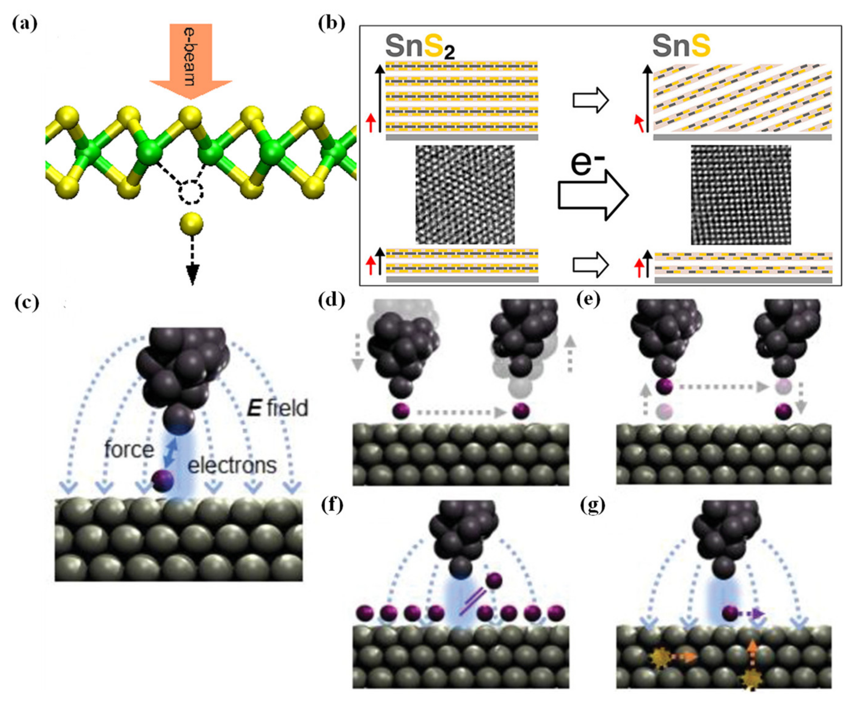
STM is a tool for defects engineering because it can image defects with atomic resolution and allow manipulating defects individually. In fact, STM allows vertical and lateral manipulation, extraction of adatoms and manipulation of adsorbates or subsurface defects. The basic principle of manipulating individual atoms lies in the interaction between the tip and atom on the sample’s surface [78,79]. When manipulating atoms, van der Waals force, electric force, charge and energy transfer from tip to atom by tunneling electrons can all play important roles depending on the tip-sample distance, bias voltage and tunneling current (Figure 5c–g, [80]). Reversible manipulation of individual vacancies was successfully demonstrated in PdSe2 [81], a pentagonal layered TMD. In the experiment, the STM tip was used to “write” and “erase” near-surface vacancies by controlling Se vacancies migrations at negative sample bias and positive sample bias. In the writing process, negatively charged Se vacancies can be attracted toward the tip by a negative electric field. In the erasing process, a positive electric field was applied and Se vacancies were neutral, so the hot electrons might play a role in making Se vacancies migrate deeper into bulk by inelastic scattering. Carbon radical ion (CRI) in WS2 as an effective spin-1/2 system was demonstrated by firstly using a positively biased STM tip to desorb hydrogen atoms on carbon atoms from frequently found carbon–hydrogen (CH) complexes at S sites [82]. The hydrogen desorption by the STM tip is likely a resonant process where a defect state tunneling into an unoccupied CH weakens the C–H bond, which can be supported by DFT calculations that the defect states exhibited a local anti-bonding character with a nodal plane between the carbon and hydrogen atom. To mention that, after desorption, Cx with dangling bonds were formed, which could be used as a reactive site for other gas atoms or molecules. What is more, this desorption process was also found to be reversible at a high bias tip and the whole CHs could even be removed to form S vacancies.
Figure 5. (a) Schematic view of atom sputtering under electron beam irradiation in TMDs. Reproduced with permission [74]. Copyright 2012, Physical Review Letters. (b) Structural transformation of SnS2 under electron beam irradiation. Reproduced with permission [76]. Copyright 2016, American Chemical Society. (c) Physical mechanisms between tip atoms and surface atoms during STM defects manipulation. Schematic of (d) lateral manipulation, (e) vertical manipulation, (f) extraction of adatoms with bias pulses, and (g) field-induced manipulation of adsorbates or subsurface defects. Reproduced with permission [80]. Copyright 2019, WILEY-VCH Verlag GmbH & Co. KGaA, Weinheim.
Chemosensors 12 00085 g005

2.3.3. Bottom-Up Synthesis Method to Engineer Defects

The most commonly used method to synthesize TMDs is CVD, which can cause unintentional defects [83,84]. This part will introduce several methods which were adopted in CVD to controllably introduce defects.
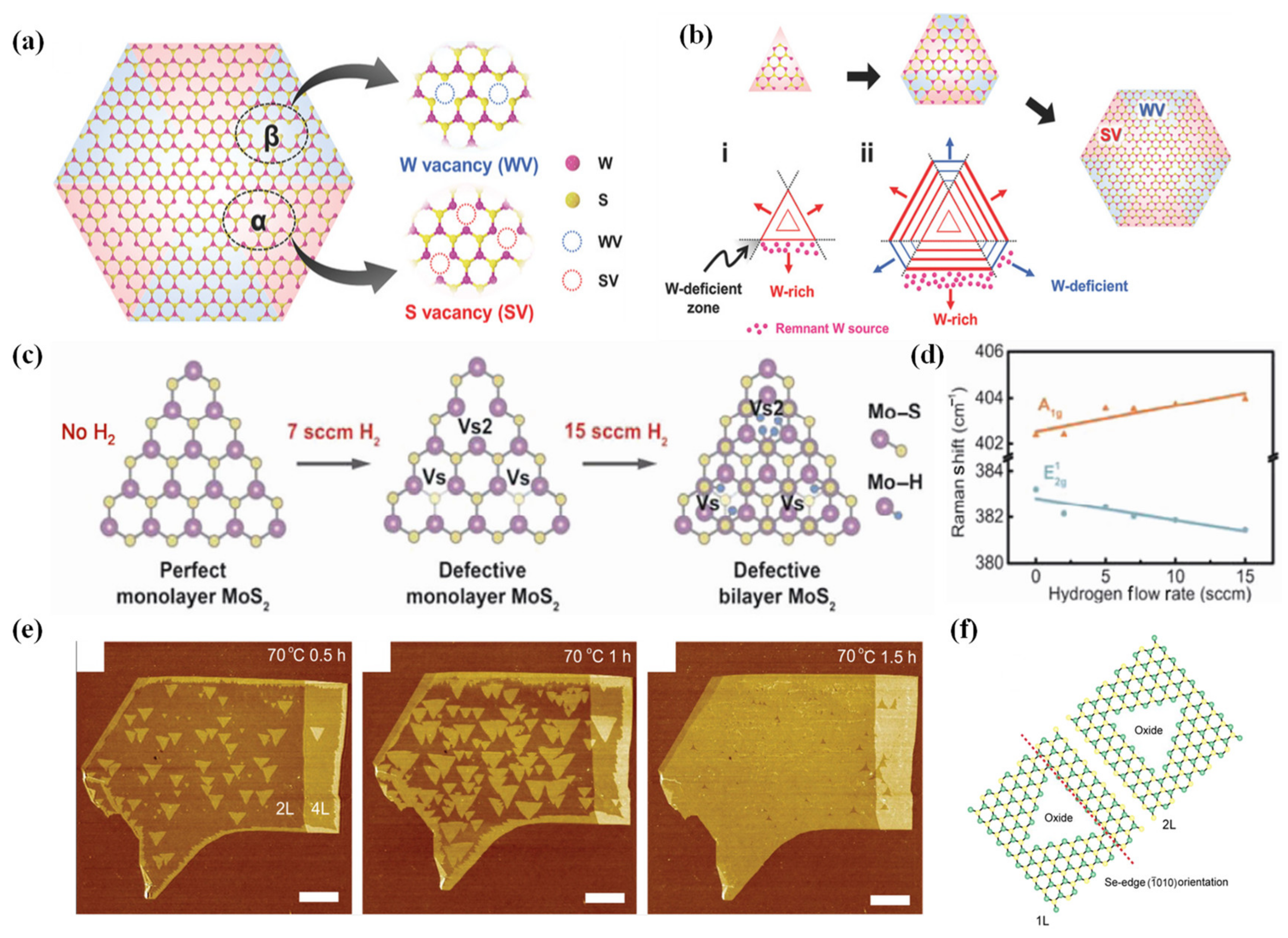
Jeong et al. [85] proposed a controllable way to synthesize single-crystalline WS2 with heterogeneous defective domains (W vacancies domain and S vacancies domain, see Figure 6a) in order to investigate the influence of W vacancy. During the experiment, 2H-monolayer WS2 single crystals were fabricated by the W-precursor-based CVD method, and by changing the solution ratio, both non-defect triangular WS2 (precursor: sodium cholate: medium solution = 1:6:1) and hexagonal WS2 with defective domains (precursor: sodium cholate: medium solution = 2:6:1) can be realized. Note that the author also provided a possible growth mechanism of heterogeneous defect formation in hexagonal WS2 as follows (Figure 6b): At first, the small triangular WS2 flake was fabricated, as flake size increases, remnant W-precursors accumulate near the triangular facet edges while corners of the triangular flake become W-precursors absence due to the facet growth direction-preferred accumulation. Therefore, the corners of triangular have a low concentration of W source while triangular facets have enough W source. The deficiency of W in corners will lead to truncated facets as the triangular facets grow further, thus a hexagon-shaped WS2 is produced by the development of the domains along the truncated facets that emerge from the three corners. The final WS2 has domains that alternate in six aspect directions.
Wu et al. [86] came up with a method to synthesize defective MoS2 free of local damages by dosing Hydrogen into the chamber during a normal CVD process, that is, an Ar/H2 mixture transporting the vapor precursor at a steady 100 sccm flow rate was adopted. Wu et al. varied the H2 flow rate of 0, 2, 5, 7, 10, and 15 sccm while adjusting Ar flow rate to maintain a constant flow rate. According to HRTEM on all samples, when the hydrogen flow was raised from 0 to 7 sccm, the produced MoS2 samples went from having a single crystal with a perfect lattice to having a single crystal with several defects (sulfur vacancy and sulfur divacancies both increased as H2 flow rate increased, and the density of the defect is controlled directly by the concentration of hydrogen). When the hydrogen flow was increased to 10 and 15 sccm, samples then turned into faulty bilayer crystals (Figure 6c,d).

2.3.4. Oxidation by Ozone Treatment

Oxidation treatment is another method to engineer TMDs, which can be achieved by both UV-ozone treatment and laser irradiation. However, laser irradiation needs high energy and will cause unwanted point defects inside the material, yet UV-ozone treatment needs less energy and is a more controllable method without causing damage to the origin lattice. UV radiation has the ability to split the connection between oxygen molecules, releasing two O atoms. O atoms can then oxidize 2D materials due to their strong oxidizing ability. It has been widely studied in introducing defects in TMDs. Self-limiting oxidized with an underlying perfect hexagonal lattice but hole-doped WSe2 was demonstrated by Yamamoto et al. [87]. Oxidized multilayer WSe2 exhibits higher carrier concentration and mobility due to the p-type doping and lower degree of interfacial defects (Figure 6e,f). The oxidation behavior of monolayer WS2 and WSe2 flakes by UV-O3 treatment was further conducted by Kang et al. [88]. The experiment shows that oxidation caused by ozone can lead to n-type doping in WS2 and p-type doping in WSe2 due to the formation of semiconducting WO3. Liang et al. demonstrated controllable low-temperature (60 °C) partial surface oxidation in 2D PdSe2 restricted to the top chalcogen atom layer with ozone’s oxidation, which finally caused a p-type doping PdSe2 [89]. A better catalytic activity due to oxygen incorporation in the basal plane of PdSe2 was also demonstrated.
Figure 6. (a) Schematic view of heterogeneous defect domains for W vacancies and S vacancies in single-crystalline hexagonal WS2. (b) Schematic view of possible growth mechanism of heterogeneous defects in WS2. Red and blue regions represent facet edges for S vacancies and W vacancies domains. (i) shows initial triangular shape WS2 flake with W-rich facet edges and W-deficient corners, (ii) shows growing domains with W vacancies or S vacancies. Reproduced with permission [85]. Copyright 2017, WILEY-VCH Verlag GmbH & Co. KGaA, Weinheim. (c) Schematic view of the role of H2 in introducing S vacancies in MoS2 at different sccm during the CVD process. (d) Peak location of the A1g (blue dots) and E1 2g (orange triangles) Raman modes as a function of H2 flow rate, which demonstrates H2′s controllability in introducing defects during the CVD process. Reproduced with permission [86]. Copyright 2018, Tsinghua University Press and Springer-Verlag GmbH Germany, part of Springer Nature. (e) AFM images of thin WSe2 flakes on SiO2 after O3 exposure at 70 °C for (left) 0.5, (middle) 1, and (right) 1.5 h. The number of layers (NL with N = 1 to 6) of each flake is indicated. Triangular islands are the oxidized regions. (f) Schematic drawing of hexagonal lattices of 1 L and 2 L WSe2 with triangular oxides. The red dashed line indicates a selenium zigzag edge ( 10 ¯ 10) orientation. Reproduced with permission [87]. Copyright 2015, American Chemical Society.
Chemosensors 12 00085 g006

3. Gas Sensing Performance and Mechanism

3.1. Introduction of Gas Sensing Mechanism
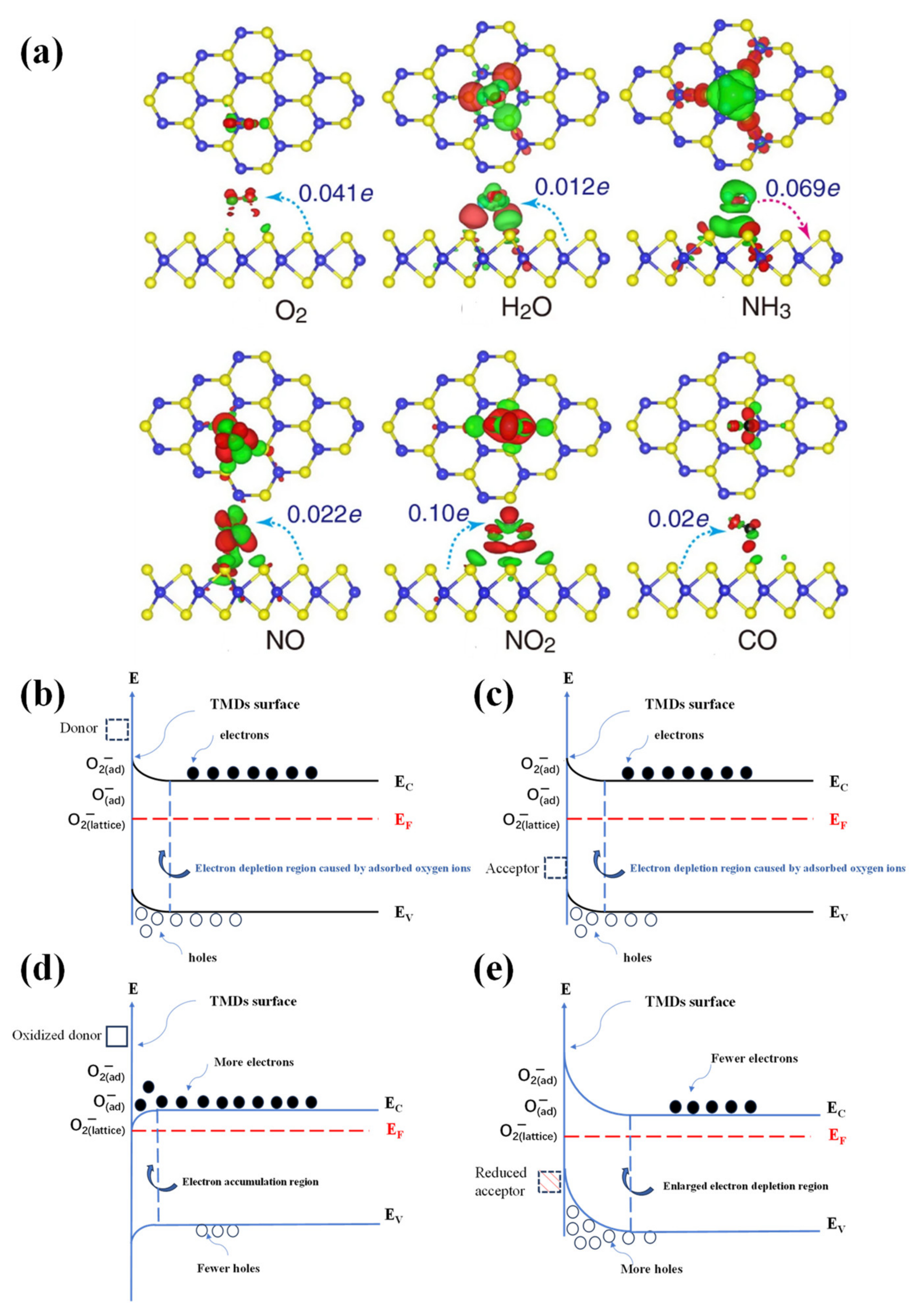
For TMD-based gas sensors, the sensing mechanism mainly relies on charge transfer (molecular doping) [90]. When TMDs are exposed to the reactive gas, electron transfer occurs directly between the physically adsorbed [91] (long-range forces dominate, such as van der Waals force, London force, hydrogen bonding interaction) or chemically adsorbed (stronger short-range force dominates: chemical bonds formed between the TMDs and the gas) gas and TMDs. The direction and intensity of electron transfer depend on two widely believed mechanisms which are suitable for all 2D layered materials [92,93]: (i) If the highest occupied molecular orbital (HOMO) in gas is higher than Fermi level (or the Dirac point) in 2D material, electrons will move from gas molecules to material; if the lowest unoccupied molecular orbital (LUMO) is lower than Fermi level in 2D material, electrons will move from material to gas. (ii) Charge transfer is also partially determined by orbitals hybridization, that is, the mixing of the HOMO and LUMO with 2D material’s orbitals. This mixing scales with the overlap of the interacting orbitals and the inverse of their energy difference. Typically, a smaller distance between the adsorbed gas molecule and the adsorption site (defective site) means stronger hybridization, which will cause larger changes in the electronic properties. Figure 7a shows the charge transfer mechanism due to the gas adsorption on the TMDs. Since the charge transfer amount and direction are quite different, different TMDs can be utilized to distinguish different gases according to this mechanism [94].
Though in most cases detecting reactive gas does not need oxygen’s participation for TMDs, the role of oxygen functional groups should not be neglected in certain cases. When the TMDs are exposed to the air, oxygen molecules can be adsorbed at the materials’ surface by extracting free electrons from the conduction band and forming an electron-depletion layer at the surface (Figure 7b,c, taking n-type MoS2 as an example). The formed ionized oxygen species vary according to the working temperature. Typically, O2(ads) occurs when the temperature is lower than 100 °C; O(ads) occurs at the range of 100~300 °C; O(lattice)2− occurs when the temperature is higher than 300 °C [95]. Next, when the TMDs are exposed to the reactive oxidizing gas or reducing gas, the detecting mechanism either relies on simply transferring electrons between oxygen ions and gas molecules or can be the combining effect of both charge transfer and the adsorbed oxygen ions [95,96]. First taking reducing gas CO as an example, CO typically reacts with adsorbed oxygen ions on the TMDs surface as follows: 2CO + O2→2CO2 + e, electrons will be transferred to TMDs, forming the electron accumulation region (Figure 7d). In the case of oxidizing gas, such as NO2, NO2 can directly capture electrons from the acceptor level from TMDs (NO2 + e→NO2) and electrons from adsorbed oxygen ions (NO2 + O2 + 2e→NO2 + 2O), and the reduced products (NO2) can again react with S vacancies to release holes (NO2 + Vs++→NO2 + h+), thus greatly reducing electrons and enlarging the electron depletion region (Figure 7e).
Figure 7. (a) Charge transfer process and density difference plots for O2, H2O, NH3, NO, NO2, and CO interacting with monolayer MoS2. Reproduced under the terms of the CC-BY license [94]. Copyright 2013, Yue et al.; licensee Springer. (b,c) The development of the space charge region and the corresponding surface band bending for n-type TMDs with adsorbed oxygen ions: situation of the band structure in the absence of occupied surface states (donor and acceptor). (d) The accumulation region associated with an oxidized donor state. (e) The depletion region associated with a reduced acceptor state.
Chemosensors 12 00085 g007
Since electrons are transferred between gas and TMDs, changes in the conductivity of the gas sensor can be observed. For instance, in MoS2, WS2 and WSe2 [97], O2, H2O and NO2 act as electron receptors, which means that electrons will transfer from TMDs to gas while NH3 act as an electron donator, thus electrons will transfer from gas to sensor. Since MoS2, and WS2 are both typically n-type materials, the conductivity in the former condition will decrease but increase in the latter scenario. WSe2 is typically a p-type material, so its conductivity will increase in the former condition and decrease in the latter condition, which is opposite to the n-type material.
Normally speaking, there are several main indicators for TMD-based-gas sensors, namely sensitivity, recovery ability, selectivity, etc. Sensitivity mainly depends on the strength of gas adsorption onto the TMD surface, which can be described by the equation: Ead = ETMD/gas − (ETMD+ Egas) [98] where Ead is the adsorption energy, Egas/TMD is the energy of the optimized structure of the gas molecule adsorbed on the TMD. ETMD and Egas, in turn, are the energies of the pristine TMD layer and of the isolated gas molecule. The smaller Ead, the smaller energy is needed to support the adsorption of the gas on the sensor. When Ead < 0, the corresponding reaction is exothermic and spontaneous, which means the gas can be easily and strongly adsorbed on the TMD surface, that is, a higher sensitivity for this gas sensor. Normally speaking, for most pristine, perfect 2D materials, the gas will only be physically (weakly) adsorbed on the surface due to their inert, dangling bonds-free surface [96], which is attributed to less sensitivity than chemically adsorbed functionalized materials (e.g., defective 2D material). However, a relatively weak connection between the gas and sensing material means a better recovery ability, which is an advantage for low-cost, mass production of future gas sensors. Meanwhile, for defective materials such as WSe2, experiments have revealed a slow recovery due to strong adsorption between NO2 and surface defective sites [99,100]. According to the expression of recovery time [101]: τ =   υ 1 e x p E a d k B T , where Ead is the adsorption energy, υ is the operational frequency, T is the temperature of operation, and kB is the Boltzmann constant, it is clear that there is a trade-off between high sensitivity and short recovery time and it is an alternative way to optimize Ead between the range of strong physisorption and weak chemisorption. For selectivity, the amount of charge transferred between different 2D materials and gases is also different due to the different extent of the mixing orbitals, which reflects the selectivity of gas-sensing materials [102].

3.2. Connection between Defects and Sensing Mechanism

3.2.1. Vacancies

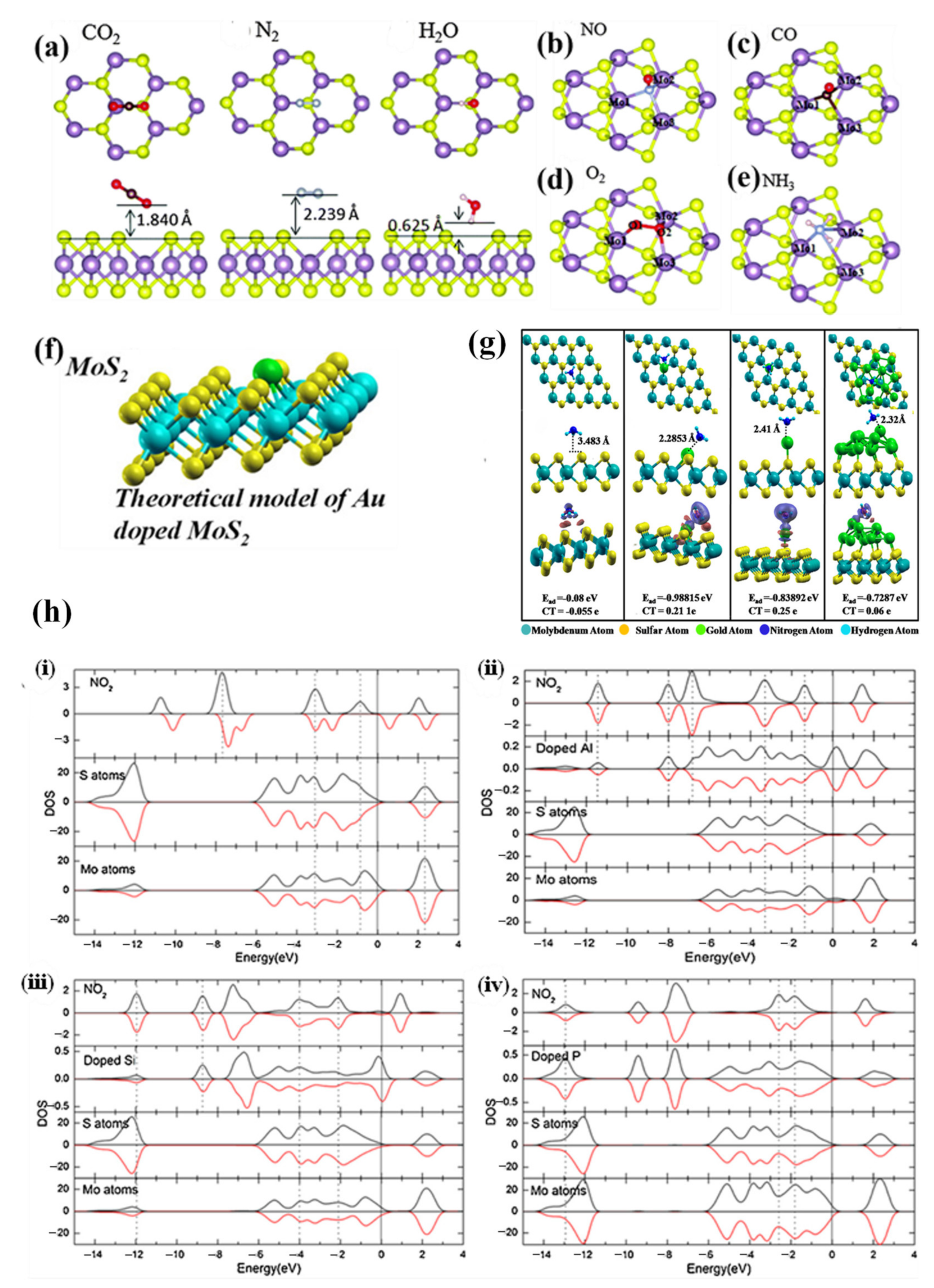
Vacancies, as common defects in TMDs, can play important roles in TMD-based gas sensors as reactive sites (or, gas adsorption sites), which makes TMDs become more sensitive for certain gases. According to former first principal calculation on pristine MoS2 monolayer’s detection of CO, CO2, NH3, NO, NO2, CH4, H2O, N2, O2 and SO2 [103], it can be found that all the gases’ adsorption energy Ead is quite large, indicating only weak forces (physically adsorbed) are formed between gases and pristine MoS2 slabs, leading to low sensitivities. Later experiments have been further conducted on monolayer MoS2 with sulfur vacancy [104], which shows a smaller Ead for CO2, H2O, N2, CO, NO, O2 and NH3. Furthermore, researchers found that CO, NO, O2 and NH3 molecules can adsorb at the S vacancy site of MoS2 (chemically adsorbed, shown in Figure 8a–e) with a moderate recovery time. The bond length changes of CO, NO and O2 molecules after adsorption are much larger than those of the CO2, H2O and N2 molecules, demonstrating reliable molecular activation. This experiment clearly showed that S vacancy in MoS2 not only strengthens long-range force (physically adsorbed condition), but also attracts certain gas molecules to be chemically adsorbed at vacancy sites, which means much higher sensitivities for these gases. Additionally, Annanouch et al. [94] found that the NO2 can react with both S vacancies and adsorbed oxygen ions in MoS2, combining the high oxygen affinity on vacancy sites, it is believed that the vacancies play an important role in achieving extremely high NO2 response (6600%). Other TMDs have also been explored. For example, in WS2 [105], it has been found that W vacancy (VW) could significantly enhance NO adsorption than intrinsic WS2 (Ead = −0.882 eV in VW WS2 and Ead = −0.140 eV in intrinsic WS2). The absolute charge transfer amount of VW WS2 under this scenario is enhanced to 0.005 e, which is five times higher than that of intrinsic WS2 (0.001 e). What is more, though this strong adsorption leads to a long τ at 300 K, it is still much shorter than previously reported heterojunction structures [106,107] in 2020 and 2021. In SnS2 [108], Qin et al. successfully demonstrated rapid response and high sensitivity to NH3 at room temperature due to rich sulfur vacancies which possess high binding energies and serve as main gas adsorption sites due to their high activity, catalysis and dissociation characteristics. Also, PtSe2 with vacancy defect exhibits better sensing for NO2 and NO [109].

3.2.2. Substitution

Substitution, which is commonly caused by doping foreign atoms, can also influence TMDs for detecting gases. The doping atoms can influence the sensitivity and selectivity of certain TMDs for several reasons as follows: (i) Dopants can commonly occupy the intrinsic vacancies in TMDs [110] and chemically adsorb the target gas molecules, which leads to an enhancement in sensitivity of the gas sensor. For instance, an Au-doped MoS2 [111] (Figure 8f) shows better sensitivity for detecting NH3 (Figure 8g) due to a weak chemisorption of NH3 on Au dopants at S vacancy sites. Note that, even in sulfur vacancy MoS2, the adsorption energy of NH3 Ead equals −0.407 eV, while in Au-MoS2, Ead equals −0.839 eV. The charge transfer in Au-MoS2 reaches up to 0.25 e, which is 4 times higher than that of the pristine MoS2 counterpart. Thus, larger adsorption energy and associated greater charge transfer for Au-MoS2 materials are attributed to the experimentally observed high response of Au-MoS2 to NH3. (ii) Certain dopant atoms can enhance orbital hybridization between TMDs and gas molecules, which leads to an improvement in charge transfer. Luo et al. [112] found in Al-doped MoS2, the Al-3p impurity state and Mo-4d state around the Fermi level have strong hybridization, which can enhance charge transfer between dopants and MoS2. Furthermore, according to the projected density of states (PDOS), Al can either be a charge transfer link between target gas NO2 and MoS2 or directly enhance orbital hybridization between MoS2 and NO2 at a wide range of energy, which demonstrates the ability of dopants to improve charge transfer (−0.5 of Al-MoS2 compared to −0.02 in pristine MoS2) in the gas sensor (Figure 8h). A similar phenomenon has also been found in Si-doped and P-doped MoS2 (Figure 8h). Panigrahi et al. [113] further demonstrated that strong hybridization of the Ge state with that of N and O states near the Fermi energy in MoSe2–Ge can enhance binding of NO. Also, doping MoTe2 with Sb can enhance charge transfer due to the strong hybridization of the dopant Sb state with that of N and O states near the Fermi energy of NO.
Figure 8. (a) The most favorable configurations for CO2, N2 and H2O molecules physically adsorbed on defective MoS2 (S vacancy) from the top and side view. H, C, N and O atoms are shown in white, brown, silver and red, respectively. (be) Schematic view of NO, CO, O2, NH3 chemically adsorbed on MoS2 at S vacancy site. Reproduced with permission [104]. Copyright 2016, Royal Society of Chemistry. (f) Theoretical model of Au doped MoS2 at S vacancy. (g) The most stable adsorption configurations of NH3 adsorbed on pristine MoS2 sheet, MoS2 sheet with Au atom doped at S-vacancy site, MoS2 sheet containing Au attached on top of S and nanocomposite of MoS2 with Au13 cluster. Compared to pristine MoS2, the later three types of MoS2 all have a shorter length between adsorbed gas molecule and MoS2 surface. Reproduced with permission [111]. Copyright 2021, American Chemical Society. (h) PDOS of adsorbed NO2 molecule, doped Al, Si, P atom, and all S, Mo atoms for (i) undoped MoS2, (ii) Al-MoS2, (iii) Si-MoS2, and (iv) P-MoS2. Reproduced with permission [112]. Copyright 2015, Elsevier B.V. All rights reserved.
Chemosensors 12 00085 g008

3.2.3. Grain Boundaries

Grain boundaries (GBs), as described in Section 2.1.3, are also an important type of defect in TMDs. However, the influence of the grain boundaries on gas sensing for TMDs is quite “indirect” and unclear according to current research. On the one hand, regulating the GBs will cause changes in other properties such as surface area, which might serve as the descriptor for gas sensing performance. On the other hand, few of the previous studies have focused on unveiling the underlying mechanism of GBs, though it is an effective method to improve metal oxide-based gas sensors by building inner-grain boundaries thus forming a conductive percolative network [114], whether this conclusion is valid for TMDs is still to be proved. Guo et al. [64] found that, by doping SnSe2 with In, the grain size will continue to shrink as In concentration increases due to the substitution of In3+ into the SnSe2 lattice, which in turn prevents SnSe2 grain’s growth (Figure 9a, left). Since the trend in surface area change is precisely the opposite of the trend in grain size change (Figure 9a, right), this suggests that In3+ doping can inhibit the growth of SnSe2 grains and boost a larger specific area. Thus, a larger number of GBs will also come into being. According to the experiment, smaller grain size or more GBs and larger specific areas are indeed important in improving In-doped SnSe2 in detecting SO2. Choi et al. [115] also found that by doping Nb into MoSe2, the grain size will decrease as doping concentration increases, and larger surface area (smaller grain size, more GBs) in turn contributes to better sensing performance of NO2. Though the experiments above do not directly focus on increasing the quantity of GBs, the increased number of GBs indeed has improved gas sensing performances. However, GBs also might have a negative effect on gas sensing. For instance, due to dense GBs in synthesized MoS2 film, Kim et al. [116] found that edge sites (which will be discussed in the next part) could not be efficiently utilized to improve gas sensing performance of responsivity and recovery. Also, Choi et al. [115] found that continuously increasing the Nb dopant concentration in MoSe2 will increase GBs but decrease the gas response, and the main reason is the formation of NbSe2 lattice in heavily doped MoSe2, which is rarely responsive to gas molecules. Therefore, the influence of GBs on gas sensing for TMDs still remains to be studied further.

3.2.4. Edge Site

Maximally exposing the edges on the TMD surfaces is a favorable way to enhance the gas sensing sensitivity (See schematic view of gas adsorption on edge sites and basal plane in Figure 9b), since most edge sites are catalytically more active than basal planes because of (i) increased dangling bonds, (ii) high d-orbital electron density [117] and (iii) adsorption energy of the edges of TMDs is much lower than that of 2D plane [118]. All these merits together promote the adsorption of gas and reaction kinetics, which in turn further promotes the charge transfer amount and adsorption rate. Therefore, it is a promising way to maximumly expose edge sites in a TMD material in order to obtain the best gas-sensing performance. However, since edges have high surface energy, it is quite difficult to form a large number of edge sites on the surface, so researchers have turned their sights on exposing more edge sites while fabricating the materials. For instance, Cho et al. [117] reported that by adopting a rapid sulfurization method in a single CVD process synthesizing vertically aligned MoS2, higher NO2 sensing properties than the horizontally aligned MoS2 can be observed (Figure 9c). NO2′s adsorption energy Ead has been demonstrated to be higher in edge sites according to density functional theory calculations, and the adsorption directly relies on the density of exposed edge sites according to the experiment. More experiments have been then conducted in order to fabricate a vertically aligned TMD structure. Shim et al. first proposed a facile approach to synthesize vertically aligned MoS2 using a nanostructured platform by first fabricating vertically ordered SiO2 on the patterned platinum-interdigitated electrode on substrates using the glancing angle deposition (GLAD) method [119]. Then the MoS2 layers encapsulating nanorods were synthesized by thermolysis of [(NH4)2MoS4] solution precursor spin-coated on nanorods during the CVD process. Highly porous nanorods enable a large number of edge sites exposed during the synthesizing process. The vertically aligned MoS2 with edge sites showed excellent response to NO2 and reversible response without degradation at 100 °C. Suh et al. used a similar method that Shim et al. adopted to fabricate vertically aligned WS2 [120]. GLAD was used to fabricate vertical SiO2 nanorods on Si substrate and WCl6 precursor was spin coated on nanorods for later fabrication of WS2 in CVD. This WS2 gas sensor showed excellent sensitivity, selectivity and desirable recovery time even at room temperature. Other methods have also been put forward in order to maximumly expose edge sites. Cha et al. [121] synthesized WS2 nanoflakes anchored to multichannel carbon nanofibers (Figure 9d) for NO2 gas sensors at room temperature. The WS2 nanoflakes with increased edge sites effectively improved the NO2 sensing characteristics. Therefore, the edge-abundant structure is important for superior-performance TMD-based gas sensors. Koo et al. [122] fabricated few-layered WS2 nanoplates anchored on Co, N-doped hollow carbon nanocages (WS2_Co-N-HCNCs) (Figure 9e), realizing a large number of edge sites with highly porous structures. The WS2_Co-N-HCNCs exhibited high resistance changes and stable response to NO2 and recovery even at room temperature during the experiment.
Figure 9. (a) The relation between grain sizes of the SI0–SI4 samples and doping concentrations (left), the relation between Brunner–Emmet–Teller (BET) measured surface area and doping concentration (right). Reproduced with permission [64]. Copyright 2022, Royal Society of Chemistry. (b) Schematic comparison between gas adsorption mechanism on edge sites and the basal plane of MoS2. (c) Schematic view of vertically aligned MoS2 with exposed edge sites. Reproduced with permission [117]. Copyright 2015, American Chemical Society. (d) Multitubular carbon nanofiber with monolayers of WS2, and WS2 nanoflakes uniformly anchored to multitubular carbon nanofiber. Reproduced with permission [121]. Copyright 2017, Royal Society of Chemistry. (e) Schematic view of the synthesis process of WS2_Co-N-HCNCs. Reproduced with permission [122]. Copyright 2018, WILEY-VCH Verlag GmbH & Co. KGaA, Weinheim.
Chemosensors 12 00085 g009

4. Conclusions and Outlook

In general, this paper starts by summarizing common defects in TMDs, including point defects, grain boundaries and edge sites. Then, several characterization techniques capable of identifying defects at the atomic level are introduced, such as STM, Raman spectrum, PL spectrum, EPR, etc. Since defects can greatly influence the physical and chemical properties of materials and most intrinsic defects are randomly arranged, some controllable methods for defects engineering are listed, including electron beam irradiation, STM manipulation, bottom-up synthesizing methods and ozone treatment. Then, gas sensing performance and mechanism are discussed. Charge transfer as the main mechanism for TMDs with two widely believed explanations are stressed, while the role of adsorbed oxygen ions is discussed, and three important indicators of gas sensor, namely sensitivity, recovery time and selectivity are introduced with several description models, such as adsorption energy Ead, dominant force between gas molecules and sensor’s surface, charge transfer analysis etc. Last but not least, the connection between defects (vacancies, substitutions, grain boundaries, edge sites) and sensing performances are covered. In most cases, defects in TMDs can greatly enhance their performances. According to the summary in Table 2 displayed as follows, vacancies and edge sites can act as active adsorption sites: SnS2 with rich S vacancies on the surface shows a rapid response time (NH3)~16 s [108], edge enriched MoS2 shows extremely high response (NO2)~6600% [94]; substitutions can enhance orbital hybridizations thus increasing charge transfer [112], Ce doped SnS2 shows significant response to NO2 at 500 ppb with a response value of 1.67 at 100 °C [123]. Nevertheless, grain boundaries are found to have both positive and negative effects on gas sensing in different conditions: a proper Nb doping concentration in SnSe2 can inhibit grain growth and lift its response (NO2) to 2000% [64], but an excessive Nb doping can also decrease the response [115]. It can also be found that the TMD-based gas sensors we found and listed here are still chemoresistive gas sensors, while different defect types have quite different influences for different TMDs, and even for the same defect type engineering, responses to the same gas have huge differences. Therefore, different TMDs and different methods of achieving defect engineering should be considered individually.
Despite the optimized performances, there are still several challenges for defective TMD-based gas sensors.
First, the LOD range of ppb should be decreased to ppt, as some TMD-based gas sensors are still limited at the ppm range. This goal can be achieved with the help of photoactivation or heating the device. However, an extra light source means higher power consumption and is more inconvenient. A heating device is an alternative way; however, many TMDs are not stable and will degrade in ambient air conditions, and so developing a suitable package technique is necessary to protect TMD-based gas sensors while maintaining their gas sensing performance.
Second, defective TMDs, especially vacancies and edge sites, are quite sensitive and in most cases the adsorption energy for the target gas can be extremely low, the recovery time will be greatly prolonged, and the reaction rate constant will be reduced when the TMDs are exposed in the large, continuous gas flow. Under this scenario, the slow desorption rate of gases on the defective sites will cause fewer and fewer available sites for target gas, leading to performance degradation. It is important to find ways to introduce desirable amounts of defects in TMDs or design defects that the gas can be weakly chemisorbed on to achieve a balance between sensitivity and recovery. Additionally, field effect transistors (FET), p–n junction, and Schottky junction-based gas sensors have emerged as promising candidates for high-performance sensing applications since a highly selective performance can be achieved in these devices by tuning the external electrical field, and Schottky barrier height. It is a promising way to fabricate these kinds of devices by using the defective TMDs mentioned in this review to further improve their sensing performances, including their sensitivity, recovery and LOD.
What is more, since the selective discrimination of the target gas from the ambient atmosphere including vapors and aerosols is one of the tough issues for chemoresistance gas sensors, there is an emerging trend to utilize machine learning and quantitative algorithms to distinguish multiple gases in the ambient condition, which helps TMD-based- gas sensors in achieving a better selectivity in mixed gas conditions. It is also worth noting that gasistor (gas sensor + memristor) is an alternative way to improve the sensor’s selectivity. Recently, traditional metal oxides such as TiO2 and SnO2 have been used to fabricate gasistors and have promising performances. However, there is no research now focusing on using TMDs as the material to fabricate gasistor. We believe that the use of TMDs in gasistor has great prospects for future research and development.
Last but not least, aside from several in situ techniques we mentioned in this review, more straightforward, advanced techniques are needed to conduct further studies in TMD gas sensing. Operando techniques like diffuse reflectance infrared Fourier transform spectroscopy can detect the changes in the concentration of vacancies; UV-Vis spectroscopy provides valuable information about the electronic structure of the sensing material as well as the oxidation state and size, which helps us understand the mechanism, especially when both charge transfer and adsorbed oxygen ions play roles in gas sensing processes. Theoretical calculations also play an important role in illustrating the gas sensing mechanism and enhancing performance for those TMD-based gas sensors by using different models, such as adsorption energy calculation and charge transfer amount analysis. There are also limitations to these currently used theoretical methods, and the actual situations are typically more complicated than those situations conceived in the ideal models. Therefore, more work is needed to optimize and further develop the theoretical calculation methods to obtain a more accurate understanding of gas sensing mechanisms as well as their performances.

Funding

The project funded by the National Natural Science Foundation of China (No. 52072204), National Key Research and Development Program of China (2023YFE0109100) and the Science Foundation of China Academy of Safety Science and Technology (2023JBKY17).

Institutional Review Board Statement

Not applicable.

Informed Consent Statement

Not applicable.

Data Availability Statement

Not applicable.

Conflicts of Interest

All authors declare no conflict of interest.

References

  1. Zhang, L.; Khan, K.; Zou, J.; Zhang, H.; Li, Y. Recent advances in emerging 2D material-based gas sensors: Potential in disease diagnosis. Adv. Mater. Interfaces 2019, 6, 1901329. [Google Scholar] [CrossRef]
  2. Yang, S.; Jiang, C.; Wei, S.-H. Gas sensing in 2D materials. Appl. Phys. Rev. 2017, 4, 021304. [Google Scholar] [CrossRef]
  3. Liu, Z.; Qiao, Z.; Li, C.-Y.; Sun, Y. Recent progress in multifunctional gas sensors based on 2D materials. Chemosensors 2023, 11, 483. [Google Scholar] [CrossRef]
  4. Sun, J.; Giorgi, G.; Palummo, M.; Sutter, P.; Passacantando, M.; Camilli, L. A scalable method for thickness and lateral engineering of 2D materials. ACS Nano 2020, 14, 4861–4870. [Google Scholar] [CrossRef]
  5. Wu, F.; Tian, H.; Shen, Y.; Hou, Z.; Ren, J.; Gou, G.; Sun, Y.; Yang, Y.; Ren, T.-L. Vertical MoS2 transistors with sub-1-nm gate lengths. Nature 2022, 603, 259–264. [Google Scholar] [CrossRef]
  6. Liu, L.; Kong, L.; Li, Q.; He, C.; Ren, L.; Tao, Q.; Yang, X.; Lin, J.; Zhao, B.; Li, Z. Transferred van der Waals metal electrodes for sub-1-nm MoS2 vertical transistors. Nat. Electron. 2021, 4, 342–347. [Google Scholar] [CrossRef]
  7. Zhao, J.; Wang, H.; Cai, Y.; Zhao, J.; Gao, Z.; Song, Y.-Y. The Challenges and Opportunities for TiO2 Nanostructures in Gas Sensing. ACS Sens. 2024, 9, 1644–1655. [Google Scholar] [CrossRef]
  8. Aftab, S.; Hegazy, H.H. Emerging Trends in 2D TMDs Photodetectors and Piezo-Phototronic Devices. Small 2023, 19, 2205778. [Google Scholar] [CrossRef]
  9. Lee, E.; Yoon, Y.S.; Kim, D.-J. Two-dimensional transition metal dichalcogenides and metal oxide hybrids for gas sensing. ACS Sens. 2018, 3, 2045–2060. [Google Scholar] [CrossRef] [PubMed]
  10. Wang, X.; Gu, D.; Li, X.; Lin, S.; Zhao, S.; Rumyantseva, M.N.; Gaskov, A.M. Reduced graphene oxide hybridized with WS2 nanoflakes based heterojunctions for selective ammonia sensors at room temperature. Sens. Actuators B—Chem. 2019, 282, 290–299. [Google Scholar] [CrossRef]
  11. Agrawal, A.V.; Kumar, R.; Venkatesan, S.; Zakhidov, A.; Yang, G.; Bao, J.; Kumar, M.; Kumar, M. Photoactivated mixed in-plane and edge-enriched p-type MoS2 flake-based NO2 sensor working at room temperature. ACS Sens. 2018, 3, 998–1004. [Google Scholar] [CrossRef]
  12. Zhang, S.; Nguyen, T.H.; Zhang, W.; Park, Y.; Yang, W. Correlation between lateral size and gas sensing performance of MoSe2 nanosheets. Appl. Phys. Lett. 2017, 111, 161603. [Google Scholar] [CrossRef]
  13. Seo, S.-Y.; Yang, D.-H.; Moon, G.; Okello, O.F.; Park, M.Y.; Lee, S.-H.; Choi, S.-Y.; Jo, M.-H. Identification of point defects in atomically thin transition-metal dichalcogenide semiconductors as active dopants. Nano Lett. 2021, 21, 3341–3354. [Google Scholar] [CrossRef]
  14. Edelberg, D.; Rhodes, D.; Kerelsky, A.; Kim, B.; Wang, J.; Zangiabadi, A.; Kim, C.; Abhinandan, A.; Ardelean, J.; Scully, M. Approaching the intrinsic limit in transition metal diselenides via point defect control. Nano Lett. 2019, 19, 4371–4379. [Google Scholar] [CrossRef]
  15. Komsa, H.-P.; Krasheninnikov, A.V. Native defects in bulk and monolayer MoS2 from first principles. Phys. Rev. B 2015, 91, 125304. [Google Scholar] [CrossRef]
  16. Ma, Y.; Dai, Y.; Guo, M.; Niu, C.; Lu, J.; Huang, B. Electronic and magnetic properties of perfect, vacancy-doped, and nonmetal adsorbed MoSe2, MoTe2 and WS2 monolayers. Phys. Chem. Chem. Phys. 2011, 13, 15546–15553. [Google Scholar] [CrossRef]
  17. Onofrio, N.; Guzman, D.; Strachan, A. Novel doping alternatives for single-layer transition metal dichalcogenides. J. Appl. Phys. 2017, 122, 185102. [Google Scholar] [CrossRef]
  18. Suh, J.; Park, T.-E.; Lin, D.-Y.; Fu, D.; Park, J.; Jung, H.J.; Chen, Y.; Ko, C.; Jang, C.; Sun, Y. Doping against the native propensity of MoS2: Degenerate hole doping by cation substitution. Nano Lett. 2014, 14, 6976–6982. [Google Scholar] [CrossRef]
  19. Chen, J.-W.; Lo, S.-T.; Ho, S.-C.; Wong, S.-S.; Vu, T.-H.-Y.; Zhang, X.-Q.; Liu, Y.-D.; Chiou, Y.-Y.; Chen, Y.-X.; Yang, J.-C. A gate-free monolayer WSe2 pn diode. Nat. Commun. 2018, 9, 3143. [Google Scholar] [CrossRef]
  20. Mouri, S.; Miyauchi, Y.; Matsuda, K. Tunable photoluminescence of monolayer MoS2 via chemical doping. Nano Lett. 2013, 13, 5944–5948. [Google Scholar] [CrossRef]
  21. Li, M.; Yao, J.; Wu, X.; Zhang, S.; Xing, B.; Niu, X.; Yan, X.; Yu, Y.; Liu, Y.; Wang, Y. P-type doping in large-area monolayer MoS2 by chemical vapor deposition. ACS Appl. Mater. Interfaces 2020, 12, 6276–6282. [Google Scholar] [CrossRef]
  22. Yang, L.; Majumdar, K.; Liu, H.; Du, Y.; Wu, H.; Hatzistergos, M.; Hung, P.; Tieckelmann, R.; Tsai, W.; Hobbs, C. Chloride molecular doping technique on 2D materials: WS2 and MoS2. Nano Lett. 2014, 14, 6275–6280. [Google Scholar] [CrossRef]
  23. Seo, S.-Y.; Park, J.; Park, J.; Song, K.; Cha, S.; Sim, S.; Choi, S.-Y.; Yeom, H.W.; Choi, H.; Jo, M.-H. Writing monolithic integrated circuits on a two-dimensional semiconductor with a scanning light probe. Nat. Electron. 2018, 1, 512–517. [Google Scholar] [CrossRef]
  24. Ataca, C.; Ciraci, S. Functionalization of single-layer MoS2 honeycomb structures. J. Phys. Chem. C 2011, 115, 13303–13311. [Google Scholar] [CrossRef]
  25. Lin, Z.; Carvalho, B.R.; Kahn, E.; Lv, R.; Rao, R.; Terrones, H.; Pimenta, M.A.; Terrones, M. Defect engineering of two-dimensional transition metal dichalcogenides. 2D Mater. 2016, 3, 022002. [Google Scholar] [CrossRef]
  26. Dolui, K.; Rungger, I.; Pemmaraju, C.D.; Sanvito, S. Possible doping strategies for MoS2 monolayers: An ab initio study. Phys. Rev. B 2013, 88, 075420. [Google Scholar] [CrossRef]
  27. Zhou, W.; Zou, X.; Najmaei, S.; Liu, Z.; Shi, Y.; Kong, J.; Lou, J.; Ajayan, P.M.; Yakobson, B.I.; Idrobo, J.-C. Intrinsic structural defects in monolayer molybdenum disulfide. Nano Lett. 2013, 13, 2615–2622. [Google Scholar] [CrossRef]
  28. Wang, H.; Tsai, C.; Kong, D.; Chan, K.; Abild-Pedersen, F.; Nørskov, J.K.; Cui, Y. Transition-metal doped edge sites in vertically aligned MoS2 catalysts for enhanced hydrogen evolution. Nano Res. 2015, 8, 566–575. [Google Scholar] [CrossRef]
  29. Zou, X.; Liu, Y.; Yakobson, B.I. Predicting dislocations and grain boundaries in two-dimensional metal-disulfides from the first principles. Nano Lett. 2013, 13, 253–258. [Google Scholar] [CrossRef]
  30. Lehtinen, O.; Komsa, H.-P.; Pulkin, A.; Whitwick, M.B.; Chen, M.-W.; Lehnert, T.; Mohn, M.J.; Yazyev, O.V.; Kis, A.; Kaiser, U. Atomic scale microstructure and properties of Se-deficient two-dimensional MoSe2. ACS Nano 2015, 9, 3274–3283. [Google Scholar] [CrossRef]
  31. Lin, J.; Pantelides, S.T.; Zhou, W. Vacancy-induced formation and growth of inversion domains in transition-metal dichalcogenide monolayer. ACS Nano 2015, 9, 5189–5197. [Google Scholar] [CrossRef]
  32. Azizi, A.; Zou, X.; Ercius, P.; Zhang, Z.; Elías, A.L.; Perea-López, N.; Stone, G.; Terrones, M.; Yakobson, B.I.; Alem, N. Dislocation motion and grain boundary migration in two-dimensional tungsten disulphide. Nat. Commun. 2014, 5, 4867. [Google Scholar] [CrossRef]
  33. Van Der Zande, A.M.; Huang, P.Y.; Chenet, D.A.; Berkelbach, T.C.; You, Y.; Lee, G.-H.; Heinz, T.F.; Reichman, D.R.; Muller, D.A.; Hone, J.C. Grains and grain boundaries in highly crystalline monolayer molybdenum disulphide. Nat. Mater. 2013, 12, 554–561. [Google Scholar] [CrossRef]
  34. Liu, X.; Yu, Z.G.; Zhang, G.; Zhang, Y.-W. Remarkably high thermal-driven MoS2 grain boundary migration mobility and its implications on defect healing. Nanoscale 2020, 12, 17746–17753. [Google Scholar] [CrossRef]
  35. Zuo, Y.; Antonatos, N.; Děkanovský, L.; Luxa, J.; Elliott, J.D.; Gianolio, D.; Šturala, J.; Guzzetta, F.; Mourdikoudis, S.; Regner, J. Defect engineering in two-dimensional layered PdTe2 for enhanced hydrogen evolution reaction. ACS Catal. 2023, 13, 2601–2609. [Google Scholar] [CrossRef]
  36. Meng, Y.; Ling, C.; Xin, R.; Wang, P.; Song, Y.; Bu, H.; Gao, S.; Wang, X.; Song, F.; Wang, J. Repairing atomic vacancies in single-layer MoSe2 field-effect transistor and its defect dynamics. NPJ Quantum Mater. 2017, 2, 16. [Google Scholar] [CrossRef]
  37. Patra, T.K.; Zhang, F.; Schulman, D.S.; Chan, H.; Cherukara, M.J.; Terrones, M.; Das, S.; Narayanan, B.; Sankaranarayanan, S.K. Defect dynamics in 2-D MoS2 probed by using machine learning, atomistic simulations, and high-resolution microscopy. ACS Nano 2018, 12, 8006–8016. [Google Scholar] [CrossRef]
  38. Chow, P.K.; Jacobs-Gedrim, R.B.; Gao, J.; Lu, T.-M.; Yu, B.; Terrones, H.; Koratkar, N. Defect-induced photoluminescence in monolayer semiconducting transition metal dichalcogenides. ACS Nano 2015, 9, 1520–1527. [Google Scholar] [CrossRef]
  39. Liu, J. Scanning transmission electron microscopy and its application to the study of nanoparticles and nanoparticle systems. Microscopy 2005, 54, 251–278. [Google Scholar] [CrossRef]
  40. Zhang, H.; Poh, E.T.; Lim, S.X.; Zhang, Y.; Qin, H.; Xie, H.; He, C.; Sow, C.H. In situ strain-induced phase transition and defect engineering in CVD-synthesized atomically thin MoS2. 2D Mater. 2023, 10, 035018. [Google Scholar] [CrossRef]
  41. Gao, J.; Kim, Y.D.; Liang, L.; Idrobo, J.C.; Chow, P.; Tan, J.; Li, B.; Li, L.; Sumpter, B.G.; Lu, T.-M. Transition-metal substitution doping in synthetic atomically thin semiconductors. Adv. Mater. 2016, 28, 0935–9648. [Google Scholar] [CrossRef]
  42. Lee, C.-H.; Lin, Y.-H.; Chang, S.-H.; Tai, C.-D.; Liu, S.-J.; Chu, Y.; Wang, C.-J.; Hsu, M.-Y.; Chang, H.; Chang, G.-J. Local sustained delivery of acetylsalicylic acid via hybrid stent with biodegradable nanofibers reduces adhesion of blood cells and promotes reendothelialization of the denuded artery. Int. J. Nanomed. 2014, 9, 311–326. [Google Scholar]
  43. Santosh, K.; Longo, R.C.; Addou, R.; Wallace, R.M.; Cho, K. Impact of intrinsic atomic defects on the electronic structure of MoS2 monolayers. Nanotechnology 2014, 25, 375703. [Google Scholar]
  44. Lauritsen, J.V.; Kibsgaard, J.; Helveg, S.; Topsøe, H.; Clausen, B.S.; Lægsgaard, E.; Besenbacher, F. Size-dependent structure of MoS2 nanocrystals. Nat. Nanotech. 2007, 2, 53–58. [Google Scholar] [CrossRef]
  45. Zhang, S.; Wang, C.-G.; Li, M.-Y.; Huang, D.; Li, L.-J.; Ji, W.; Wu, S. Defect structure of localized excitons in a WSe2 monolayer. Phys. Rev. Lett. 2017, 119, 046101. [Google Scholar] [CrossRef]
  46. Shen, S.; Wen, C.; Kong, P.; Gao, J.; Si, J.; Luo, X.; Lu, W.; Sun, Y.; Chen, G.; Yan, S. Inducing and tuning Kondo screening in a narrow-electronic-band system. Nat. Commun. 2022, 13, 2156. [Google Scholar] [CrossRef]
  47. Chen, Y.; Meng, L.; Zhao, W.; Liang, Z.; Wu, X.; Nan, H.; Wu, Z.; Huang, S.; Sun, L.; Wang, J. Raman mapping investigation of chemical vapor deposition-fabricated twisted bilayer graphene with irregular grains. Phys. Chem. Chem. Phys. 2014, 16, 21682–21687. [Google Scholar] [CrossRef]
  48. Ferrari, A.C.; Meyer, J.C.; Scardaci, V.; Casiraghi, C.; Lazzeri, M.; Mauri, F.; Piscanec, S.; Jiang, D.; Novoselov, K.S.; Roth, S. Raman spectrum of graphene and graphene layers. Phys. Rev. Lett. 2006, 97, 187401. [Google Scholar] [CrossRef]
  49. Wu, Z.T.; Zhao, W.W.; Chen, W.Y.; Jiang, J.; Nan, H.Y.; Guo, X.T.; Liang, Z.; Chen, Y.M.; Chen, Y.F.; Ni, Z.H. The influence of chemical solvents on the properties of CVD graphene. J. Raman Spectrosc. 2015, 46, 21–24. [Google Scholar] [CrossRef]
  50. Parkin, W.M.; Balan, A.; Liang, L.; Das, P.M.; Lamparski, M.; Naylor, C.H.; Rodríguez-Manzo, J.A.; Johnson, A.C.; Meunier, V.; Drndic, M. Raman shifts in electron-irradiated monolayer MoS2. ACS Nano 2016, 10, 4134–4142. [Google Scholar] [CrossRef] [PubMed]
  51. Mignuzzi, S.; Pollard, A.J.; Bonini, N.; Brennan, B.; Gilmore, I.S.; Pimenta, M.A.; Richards, D.; Roy, D. Effect of disorder on Raman scattering of single-layer MoS2. Phys. Rev. B 2015, 91, 195411. [Google Scholar] [CrossRef]
  52. Nipane, A.; Karmakar, D.; Kaushik, N.; Karande, S.; Lodha, S. Few-layer MoS2 p-type devices enabled by selective doping using low energy phosphorus implantation. ACS Nano 2016, 10, 2128–2137. [Google Scholar] [CrossRef]
  53. He, Z.; Zhao, R.; Chen, X.; Chen, H.; Zhu, Y.; Su, H.; Huang, S.; Xue, J.; Dai, J.; Cheng, S. Defect engineering in single-layer MoS2 using heavy ion irradiation. ACS Appl. Mater. Interfaces 2018, 10, 42524–42533. [Google Scholar] [CrossRef]
  54. Windom, B.C.; Sawyer, W.; Hahn, D.W. A Raman spectroscopic study of MoS2 and MoO3: Applications to tribological systems. Tribol. Lett. 2011, 42, 301–310. [Google Scholar] [CrossRef]
  55. Wang, X.; Zhang, Y.; Si, H.; Zhang, Q.; Wu, J.; Gao, L.; Wei, X.; Sun, Y.; Liao, Q.; Zhang, Z. Single-atom vacancy defect to trigger high-efficiency hydrogen evolution of MoS2. J. Am. Chem. Soc. 2020, 142, 4298–4308. [Google Scholar] [CrossRef]
  56. Li, L.; Qin, Z.; Ries, L.; Hong, S.; Michel, T.; Yang, J.; Salameh, C.; Bechelany, M.; Miele, P.; Kaplan, D. Role of sulfur vacancies and undercoordinated Mo regions in MoS2 nanosheets toward the evolution of hydrogen. ACS Nano 2019, 13, 6824–6834. [Google Scholar] [CrossRef]
  57. Ding, X.; Liu, T.; Ahmed, S.; Bao, N.; Ding, J.; Yi, J. Enhanced ferromagnetism in WS2 via defect engineering. J. Alloys Compd. 2019, 772, 740–744. [Google Scholar] [CrossRef]
  58. Wang, X.; Wu, J.; Zhang, Y.; Sun, Y.; Ma, K.; Xie, Y.; Zheng, W.; Tian, Z.; Kang, Z.; Zhang, Y. Vacancy defects in 2D transition metal dichalcogenide electrocatalysts: From aggregated to atomic configuration. Adv. Mater. 2023, 35, 2206576. [Google Scholar] [CrossRef]
  59. Li, W.; Qiao, Z.; Liu, Z. Behind the gas sensors: Revealing sensing mechanism with advanced magnetic resonance technology. J. Mater. Chem. A 2023, 11, 19281–19297. [Google Scholar] [CrossRef]
  60. Yang, H.; Du, Z.; Yang, Y.; Li, X.; Wu, Q.; Tang, J.; Wang, X.; Zeng, D. Ag intercalated SnS2 with S vacancy and expanded interlayer for enhancing NO2 sensing. Sens. Actuators B—Chem. 2023, 393, 134140. [Google Scholar] [CrossRef]
  61. Wu, R.; Hao, J.; Wang, T.; Zheng, S.; Wang, Y. Carbon-doping-induced energy-band modification and vacancies in SnS2 nanosheets for room-temperature ppb-level NO2 detection. Inorg. Chem. Front. 2021, 8, 5006–5015. [Google Scholar] [CrossRef]
  62. Tang, H.; Gao, C.; Yang, H.; Sacco, L.; Sokolovskij, R.; Zheng, H.; Ye, H.; Vollebregt, S.; Yu, H.; Fan, X. Room temperature ppt-level NO2 gas sensor based on SnOx/SnS nanostructures with rich oxygen vacancies. 2D Mater. 2021, 8, 045006. [Google Scholar] [CrossRef]
  63. Li, X.; Liu, W.; Huang, B.; Liu, H.; Li, X. Layered SnSe2 microflakes and SnSe2/SnO2 heterojunctions for low-temperature chemiresistive-type gas sensing. J. Mater. Chem. C 2020, 8, 15804–15815. [Google Scholar] [CrossRef]
  64. Guo, X.; Shi, Y.; Ding, Y.; He, Y.; Du, B.; Liang, C.; Tan, Y.; Liu, P.; Miao, X.; He, Y. Indium-doping-induced selenium vacancy engineering of layered tin diselenide for improving room-temperature sulfur dioxide gas sensing. J. Mater. Chem. A 2022, 10, 22629–22637. [Google Scholar] [CrossRef]
  65. Kumar, R.R.; Habib, M.R.; Khan, A.; Chen, P.-C.; Murugesan, T.; Gupta, S.; Anbalagan, A.K.; Tai, N.-H.; Lee, C.-H.; Lin, H.-N. Sulfur monovacancies in liquid-exfoliated MoS2 nanosheets for NO2 gas sensing. ACS Appl. Nano Mater. 2021, 4, 9459–9470. [Google Scholar] [CrossRef]
  66. Tian, X.; Cui, X.; Yao, B.; Wang, S.; Li, H.; Chen, T.; Xiao, X.; Wang, Y. Ultrasensitive room-temperature flexible ammonia gas sensor based on Au-functionalized polypyrrole wrapped enriched edge sulfur vacancies MoS2 nanosheets. Sens. Actuators B—Chem. 2023, 395, 134449. [Google Scholar] [CrossRef]
  67. Zheng, S.; Yin, D.; Zhang, S.; Wang, Y.; Li, J.; Wang, Z.; Yuan, Y.; Tsai, H.-S.; Hao, J. Tailoring selenium vacancies in MoSe2 through oxygen passivation for room-temperature NO2 sensing enhancement. J. Mater. Chem. A 2023, 11, 18755–18764. [Google Scholar] [CrossRef]
  68. Ross, J.S.; Wu, S.; Yu, H.; Ghimire, N.J.; Jones, A.M.; Aivazian, G.; Yan, J.; Mandrus, D.G.; Xiao, D.; Yao, W. Electrical control of neutral and charged excitons in a monolayer semiconductor. Nat. Commun. 2013, 4, 1474. [Google Scholar] [CrossRef]
  69. Wu, Z.; Luo, Z.; Shen, Y.; Zhao, W.; Wang, W.; Nan, H.; Guo, X.; Sun, L.; Wang, X.; You, Y. Defects as a factor limiting carrier mobility in WSe2: A spectroscopic investigation. Nano Res. 2016, 9, 3622–3631. [Google Scholar] [CrossRef]
  70. Mak, K.F.; He, K.; Lee, C.; Lee, G.H.; Hone, J.; Heinz, T.F.; Shan, J. Tightly bound trions in monolayer MoS2. Nat. Mater. 2013, 12, 207–211. [Google Scholar] [CrossRef]
  71. Sun, L.; Banhart, F.; Warner, J. Two-dimensional materials under electron irradiation. MRS Bull. 2015, 40, 29–37. [Google Scholar] [CrossRef]
  72. Xu, T.; Yin, K.; Sun, L. In-situ study of electron irradiation on two-dimensional layered materials. Chin. Sci. Bull. 2017, 62, 2919–2930. [Google Scholar] [CrossRef]
  73. Rummeli, M.H.; Ta, H.Q.; Mendes, R.G.; Gonzalez-Martinez, I.G.; Zhao, L.; Gao, J.; Fu, L.; Gemming, T.; Bachmatiuk, A.; Liu, Z. New frontiers in electron beam–driven chemistry in and around graphene. Adv. Mater. 2019, 31, 1800715. [Google Scholar] [CrossRef] [PubMed]
  74. Komsa, H.-P.; Kotakoski, J.; Kurasch, S.; Lehtinen, O.; Kaiser, U.; Krasheninnikov, A.V. Two-dimensional transition metal dichalcogenides under electron irradiation: Defect production and doping. Phys. Rev. Lett. 2012, 109, 035503. [Google Scholar] [CrossRef] [PubMed]
  75. Mendes, R.G.; Pang, J.; Bachmatiuk, A.; Ta, H.Q.; Zhao, L.; Gemming, T.; Fu, L.; Liu, Z.; Rümmeli, M.H. Electron-driven in situ transmission electron microscopy of 2D transition metal dichalcogenides and their 2D heterostructures. ACS Nano 2019, 13, 978–995. [Google Scholar] [CrossRef] [PubMed]
  76. Sutter, E.; Huang, Y.; Komsa, H.-P.; Ghorbani-Asl, M.; Krasheninnikov, A.; Sutter, P. Electron-beam induced transformations of layered tin dichalcogenides. Nano Lett. 2016, 16, 4410–4416. [Google Scholar] [CrossRef] [PubMed]
  77. Lin, Y.-C.; Dumcenco, D.O.; Huang, Y.-S.; Suenaga, K. Atomic mechanism of the semiconducting-to-metallic phase transition in single-layered MoS2. Nat. Nanotech. 2014, 9, 391–396. [Google Scholar] [CrossRef]
  78. Bartels, L.; Meyer, G.; Rieder, K.-H.; Velic, D.; Knoesel, E.; Hotzel, A.; Wolf, M.; Ertl, G. Dynamics of electron-induced manipulation of individual CO molecules on Cu (111). Phys. Rev. Lett. 1998, 80, 2004. [Google Scholar] [CrossRef]
  79. Stroscio, J.A.; Celotta, R.J. Controlling the dynamics of a single atom in lateral atom manipulation. Science 2004, 306, 242–247. [Google Scholar] [CrossRef]
  80. Ko, W.; Ma, C.; Nguyen, G.D.; Kolmer, M.; Li, A.P. Atomic-scale manipulation and in situ characterization with scanning tunneling microscopy. Adv. Funct. Mater. 2019, 29, 1903770. [Google Scholar] [CrossRef]
  81. Oyedele, A.D.; Yang, S.; Liang, L.; Puretzky, A.A.; Wang, K.; Zhang, J.; Yu, P.; Pudasaini, P.R.; Ghosh, A.W.; Liu, Z. PdSe2: Pentagonal two-dimensional layers with high air stability for electronics. J. Am. Chem. Soc. 2017, 139, 14090–14097. [Google Scholar] [CrossRef] [PubMed]
  82. Cochrane, K.A.; Lee, J.-H.; Kastl, C.; Haber, J.B.; Zhang, T.; Kozhakhmetov, A.; Robinson, J.A.; Terrones, M.; Repp, J.; Neaton, J.B. Spin-dependent vibronic response of a carbon radical ion in two-dimensional WS2. Nat. Commun. 2021, 12, 7287. [Google Scholar] [CrossRef] [PubMed]
  83. Xu, E.; Liu, H.; Park, K.; Li, Z.; Losovyj, Y.; Starr, M.; Werbianskyj, M.; Fertig, H.; Zhang, S. p-Type transition-metal doping of large-area MoS2 thin films grown by chemical vapor deposition. Nanoscale 2017, 9, 3576–3584. [Google Scholar] [CrossRef]
  84. Han, H.-V.; Lu, A.-Y.; Lu, L.-S.; Huang, J.-K.; Li, H.; Hsu, C.-L.; Lin, Y.-C.; Chiu, M.-H.; Suenaga, K.; Chu, C.-W. Photoluminescence enhancement and structure repairing of monolayer MoSe2 by hydrohalic acid treatment. ACS Nano 2016, 10, 1454–1461. [Google Scholar] [CrossRef]
  85. Jeong, H.Y.; Jin, Y.; Yun, S.J.; Zhao, J.; Baik, J.; Keum, D.H.; Lee, H.S.; Lee, Y.H. Heterogeneous defect domains in single-crystalline hexagonal WS2. Adv. Mater. 2017, 29, 1605043. [Google Scholar] [CrossRef]
  86. Wu, K.; Li, Z.; Tang, J.; Lv, X.; Wang, H.; Luo, R.; Liu, P.; Qian, L.; Zhang, S.; Yuan, S. Controllable defects implantation in MoS2 grown by chemical vapor deposition for photoluminescence enhancement. Nano Res. 2018, 11, 4123–4132. [Google Scholar] [CrossRef]
  87. Yamamoto, M.; Dutta, S.; Aikawa, S.; Nakaharai, S.; Wakabayashi, K.; Fuhrer, M.S.; Ueno, K.; Tsukagoshi, K. Self-limiting layer-by-layer oxidation of atomically thin WSe2. Nano Lett. 2015, 15, 2067–2073. [Google Scholar] [CrossRef] [PubMed]
  88. Kang, M.; Yang, H.I.; Choi, W. Oxidation of WS2 and WSe2 monolayers by ultraviolet-ozone treatment. J. Phys. D Appl. Phys. 2019, 52, 505105. [Google Scholar] [CrossRef]
  89. Liang, Q.; Zhang, Q.; Gou, J.; Song, T.; Arramel; Chen, H.; Yang, M.; Lim, S.X.; Wang, Q.; Zhu, R. Performance improvement by ozone treatment of 2D PdSe2. ACS Nano 2020, 14, 5668–5677. [Google Scholar] [CrossRef]
  90. Kim, Y.; Sohn, I.; Shin, D.; Yoo, J.; Lee, S.; Yoon, H.; Park, J.; Chung, S.-m.; Kim, H. Recent Advances in Functionalization and Hybridization of Two-Dimensional Transition Metal Dichalcogenide for Gas Sensor. Adv. Eng. Mater. 2024, 26, 2301063. [Google Scholar] [CrossRef]
  91. Ou, J.Z.; Ge, W.; Carey, B.; Daeneke, T.; Rotbart, A.; Shan, W.; Wang, Y.; Fu, Z.; Chrimes, A.F.; Wlodarski, W. Physisorption-based charge transfer in two-dimensional SnS2 for selective and reversible NO2 gas sensing. ACS Nano 2015, 9, 10313–10323. [Google Scholar] [CrossRef] [PubMed]
  92. Leenaerts, O.; Partoens, B.; Peeters, F. Adsorption of H2O, NH3, CO, NO2, and NO on graphene: A first-principles study. Phys. Rev. B 2008, 77, 125416. [Google Scholar] [CrossRef]
  93. Zeng, Y.; Lin, S.; Gu, D.; Li, X. Two-dimensional nanomaterials for gas sensing applications: The role of theoretical calculations. Nanomaterials 2018, 8, 851. [Google Scholar] [CrossRef] [PubMed]
  94. Yue, Q.; Shao, Z.; Chang, S.; Li, J. Adsorption of gas molecules on monolayer MoS2 and effect of applied electric field. Nanoscale Res. Lett. 2013, 8, 425. [Google Scholar] [CrossRef] [PubMed]
  95. Annanouch, F.E.; Alagh, A.; Umek, P.; Casanova-Chafer, J.; Bittencourt, C.; Llobet, E. Controlled growth of 3D assemblies of edge enriched multilayer MoS2 nanosheets for dually selective NH3 and NO2 gas sensors. J. Mater. Chem. C 2022, 10, 11027–11039. [Google Scholar] [CrossRef]
  96. Kim, J.-H.; Sakaguchi, I.; Hishita, S.; Ohsawa, T.; Suzuki, T.T.; Saito, N. Self-heated CO gas sensor based on Au-decorated Sb-implanted WS2 nanosheets. Sens. Actuators B—Chem. 2023, 382, 133501. [Google Scholar] [CrossRef]
  97. Kim, Y.; Lee, S.; Song, J.G.; Ko, K.Y.; Woo, W.J.; Lee, S.W.; Park, M.; Lee, H.; Lee, Z.; Choi, H. 2D transition metal dichalcogenide heterostructures for p-and n-type photovoltaic self-powered gas sensor. Adv. Funct. Mater. 2020, 30, 2003360. [Google Scholar] [CrossRef]
  98. Sik Hwang, W.; Remskar, M.; Yan, R.; Protasenko, V.; Tahy, K.; Doo Chae, S.; Zhao, P.; Konar, A.; Seabaugh, A.; Jena, D. Transistors with chemically synthesized layered semiconductor WS2 exhibiting 105 room temperature modulation and ambipolar behavior. Appl. Phys. Lett. 2012, 101, 013107. [Google Scholar] [CrossRef]
  99. Wu, Y.; Joshi, N.; Zhao, S.; Long, H.; Zhou, L.; Ma, G.; Peng, B.; Oliveira, O.N., Jr.; Zettl, A.; Lin, L. NO2 gas sensors based on CVD tungsten diselenide monolayer. Appl. Surf. Sci. 2020, 529, 147110. [Google Scholar] [CrossRef]
  100. Ko, K.Y.; Park, K.; Lee, S.; Kim, Y.; Woo, W.J.; Kim, D.; Song, J.-G.; Park, J.; Kim, H. Recovery improvement for large-area tungsten diselenide gas sensors. Acs. Appl. Mater. Inter. 2018, 10, 23910–23917. [Google Scholar] [CrossRef]
  101. Pitt, I.G.; Gilbert, R.G.; Ryan, K.R. Application of transition-state theory to gas-surface reactions: Barrierless adsorption on clean surfaces. J. Phys. Chem. 1994, 98, 13001–13010. [Google Scholar] [CrossRef]
  102. Wang, B.; Gu, Y.; Chen, L.; Ji, L.; Zhu, H.; Sun, Q. Gas sensing devices based on two-dimensional materials: A review. Nanotechnology 2022, 33, 252001. [Google Scholar] [CrossRef]
  103. Zhao, S.; Xue, J.; Kang, W. Gas adsorption on MoS2 monolayer from first-principles calculations. Chem. Phys. Lett. 2014, 595, 35–42. [Google Scholar] [CrossRef]
  104. Li, H.; Huang, M.; Cao, G. Markedly different adsorption behaviors of gas molecules on defective monolayer MoS2: A first-principles study. Phys. Chem. Chem. Phys. 2016, 18, 15110–15117. [Google Scholar] [CrossRef]
  105. Cui, Z.; Yang, K.; Shen, Y.; Yuan, Z.; Dong, Y.; Yuan, P.; Li, E. Toxic gas molecules adsorbed on intrinsic and defective WS2: Gas sensing and detection. Appl. Surf. Sci. 2023, 613, 155978. [Google Scholar] [CrossRef]
  106. Shen, J.; Yang, Z.; Wang, Y.; Xu, L.-C.; Liu, R.; Liu, X. The gas sensing performance of borophene/MoS2 heterostructure. Appl. Surf. Sci. 2020, 504, 144412. [Google Scholar] [CrossRef]
  107. Raad, N.H.; Manavizadeh, N.; Frank, I.; Nadimi, E. Gas sensing properties of a two-dimensional graphene/h-BN multi-heterostructure toward H2O, NH3 and NO2: A first principles study. Appl. Surf. Sci. 2021, 565, 150454. [Google Scholar] [CrossRef]
  108. Qin, Z.; Xu, K.; Yue, H.; Wang, H.; Zhang, J.; Ouyang, C.; Xie, C.; Zeng, D. Enhanced room-temperature NH3 gas sensing by 2D SnS2 with sulfur vacancies synthesized by chemical exfoliation. Sens. Actuators B—Chem. 2018, 262, 771–779. [Google Scholar] [CrossRef]
  109. Norouzzadeh, E.; Mohammadi, S.; Moradinasab, M. First principles characterization of defect-free and vacancy-defected monolayer PtSe2 gas sensors. Sens. Actuators A—Phys. 2020, 313, 112209. [Google Scholar] [CrossRef]
  110. Wang, S.; Robertson, A.; Warner, J.H. Atomic structure of defects and dopants in 2D layered transition metal dichalcogenides. Chem. Soc. Rev. 2018, 47, 6764–6794. [Google Scholar] [CrossRef]
  111. Burman, D.; Raha, H.; Manna, B.; Pramanik, P.; Guha, P.K. Substitutional doping of MoS2 for superior gas-sensing applications: A proof of concept. ACS Sens. 2021, 6, 3398–3408. [Google Scholar] [CrossRef] [PubMed]
  112. Luo, H.; Cao, Y.; Zhou, J.; Feng, J.; Cao, J.; Guo, H. Adsorption of NO2, NH3 on monolayer MoS2 doped with Al, Si, and P: A first-principles study. Chem. Phys. Lett. 2016, 643, 27–33. [Google Scholar] [CrossRef]
  113. Panigrahi, P.; Hussain, T.; Karton, A.; Ahuja, R. Elemental substitution of two-dimensional transition metal dichalcogenides (MoSe2 and MoTe2): Implications for enhanced gas sensing. ACS Sens. 2019, 4, 2646–2653. [Google Scholar] [CrossRef] [PubMed]
  114. Ferroni, M.; Carotta, M.; Guidi, V.; Martinelli, G.; Ronconi, F.; Richard, O.; Van Dyck, D.; Van Landuyt, J. Structural characterization of Nb–TiO2 nanosized thick-films for gas sensing application. Sens. Actuators B—Chem. 2000, 68, 140–145. [Google Scholar] [CrossRef]
  115. Choi, S.Y.; Kim, Y.; Chung, H.-S.; Kim, A.R.; Kwon, J.-D.; Park, J.; Kim, Y.L.; Kwon, S.-H.; Hahm, M.G.; Cho, B. Effect of Nb doping on chemical sensing performance of two-dimensional layered MoSe2. ACS Appl. Mater. Interfaces 2017, 9, 3817–3823. [Google Scholar] [CrossRef] [PubMed]
  116. Kim, C.; Park, J.C.; Choi, S.Y.; Kim, Y.; Seo, S.Y.; Park, T.E.; Kwon, S.H.; Cho, B.; Ahn, J.H. Self-formed channel devices based on vertically grown 2D materials with large-surface-area and their potential for chemical sensor applications. Small 2018, 14, 1704116. [Google Scholar] [CrossRef] [PubMed]
  117. Cho, S.-Y.; Kim, S.J.; Lee, Y.; Kim, J.-S.; Jung, W.-B.; Yoo, H.-W.; Kim, J.; Jung, H.-T. Highly enhanced gas adsorption properties in vertically aligned MoS2 layers. ACS Nano 2015, 9, 9314–9321. [Google Scholar] [CrossRef] [PubMed]
  118. Duan, Y.; Feng, S.; Zhang, K.; Qiu, J.; Zhang, S. Vertical few-layer WSe2 nanosheets for NO2 sensing. Acs. Appl. Nano. Mater. 2021, 4, 12043–12050. [Google Scholar] [CrossRef]
  119. Shim, Y.-S.; Kwon, K.C.; Suh, J.M.; Choi, K.S.; Song, Y.G.; Sohn, W.; Choi, S.; Hong, K.; Jeon, J.-M.; Hong, S.-P. Synthesis of numerous edge sites in MoS2 via SiO2 nanorods platform for highly sensitive gas sensor. Acs Appl. Mater. Interfaces 2018, 10, 31594–31602. [Google Scholar] [CrossRef]
  120. Suh, J.M.; Kwon, K.C.; Lee, T.H.; Kim, C.; Lee, C.W.; Song, Y.G.; Choi, M.-J.; Choi, S.; Cho, S.H.; Kim, S. Edge-exposed WS2 on 1D nanostructures for highly selective NO2 sensor at room temperature. Sens. Actuators B—Chem. 2021, 333, 129566. [Google Scholar] [CrossRef]
  121. Cha, J.-H.; Choi, S.-J.; Yu, S.; Kim, I.-D. 2D WS2-edge functionalized multi-channel carbon nanofibers: Effect of WS2 edge-abundant structure on room temperature NO2 sensing. J. Mater. Chem. A 2017, 5, 8725–8732. [Google Scholar] [CrossRef]
  122. Koo, W.T.; Cha, J.H.; Jung, J.W.; Choi, S.J.; Jang, J.S.; Kim, D.H.; Kim, I.D. Few-layered WS2 nanoplates confined in Co, N-doped hollow carbon nanocages: Abundant WS2 edges for highly sensitive gas sensors. Adv. Funct. Mater. 2018, 28, 1802575. [Google Scholar] [CrossRef]
  123. Yang, H.; Li, X.; Wu, Q.; Su, H.; Ma, C.; Wang, X.; Xie, C.; Zeng, D. Synergetic effect of highly active Ce sites and interlayer engineering induced by Ce doping of SnS2 to enhance gas sensing of NO2. Sens. Actuators B—Chem. 2023, 376, 133033. [Google Scholar] [CrossRef]
  124. Sibi, S.L.; Rajkumar, M.; Manoharan, M.; Mobika, J.; Priya, V.N.; Kumar, R.R. Humidity activated ultra-selective room temperature gas sensor based on W doped MoS2/RGO composites for trace level ammonia detection. Anal. Chim. Acta 2024, 1287, 342075. [Google Scholar] [CrossRef] [PubMed]
  125. Zhang, L.; Liang, Y.; Yu, L.; Wang, H.; Yin, M. Activating and modifying the basal planes of MoS2 for superior NO2 sensing at room temperature. Sens. Actuators B—Chem. 2022, 359, 131539. [Google Scholar] [CrossRef]
  126. Alagh, A.; Annanouch, F.E.; Umek, P.; Bittencourt, C.; Sierra-Castillo, A.; Haye, E.; Colomer, J.F.; Llobet, E. CVD growth of self-assembled 2D and 1D WS2 nanomaterials for the ultrasensitive detection of NO2. Sens. Actuators B—Chem. 2021, 326, 128813. [Google Scholar] [CrossRef]
  127. Alagh, A.; Annanouch, F.E.; Al Youssef, K.; Bittencourt, C.; Gueell, F.; Martínez-Alanis, P.R.; Reguant, M.; Llobet, E. PdO and PtO loaded WS2 boosts NO2 gas sensing characteristics at room temperature. Sens. Actuators B—Chem. 2022, 364, 131905. [Google Scholar] [CrossRef]
  128. Jaiswal, J.; Das, A.; Chetry, V.; Kumar, S.; Chandra, R. NO2 sensors based on crystalline MoSe2 porous nanowall thin films with vertically aligned molecular layers prepared by sputtering. Sens. Actuators B—Chem. 2022, 359, 131552. [Google Scholar] [CrossRef]
Figure 1. Introduction of this review. Section 1, introduction; Section 2, including defects types in TMDs, characterization methods for defects, and controllable introduction of defects and Section 3, including gas sensing mechanism for TMD-based gas sensor, connection between defects types and gas sensing will be discussed.
Chemosensors 12 00085 g001
Table 1. Summary of several TMD-based gas sensing materials and the corresponding EPR signal.
Sensing MaterialsTarget GasTypes of DefectsEPR SignalRef.
Ag-doped SnS2NO2S vacancyg = 2.004[60]
SnS2NO2S vacancyg = 2.003[61]
SnOx/SnSNO2O vacancyg = 2.000[62]
SnSe2NO2Se vacancyg = 2.002[63]
In-doped SnSe2SO2Se vacancyg = 2.003[64]
MoS2NO2S vacancyg = 2.004[65]
MoS2/PPyNH3S vacancyg = 2.002[66]
MoSe2NO2Se vacancyg = 2.0019[67]
Table 2. Summary of several defective TMDs with corresponding performance.
Sensing MaterialsTarget GasLODResponseMechanismRef.
W-MoS2/RGO50 ppm NH31.32 ppm42.3% aCharge transfer[124]
Zn -MoS25 ppm NO28.1 ppb368% bCharge transfer[125]
Edge-enriched MoS210 ppm NO21 ppm6600% bCharge transfer
Surface-adsorbed oxygen ions
[94]
Au-MoS2500 ppm NH3/150% aCharge transfer[111]
Au-decorated Sb-WS2 NSs50 ppm CO41 ppb3.9 cSurface-adsorbed oxygen ions[95]
Edge-enriched WS20.8 ppm NO2<5 ppb~5600% bCharge transfer[126]
Edge-enriched WS21 ppm NO2100 ppb18% bCharge transfer[122]
Grain boundaries-enriched WS2100 ppm H25 ppb30% bCharge transfer[127]
Edge-enriched MoSe210 ppm NO2/−78.3% bCharge transfer[128]
Grain boundaries-enriched MoSe23 ppm NO2/8% bCharge transfer[115]
Edge-exposed WSe21 ppm NO24 ppb34.6% bCharge transfer[118]
S vacancies SnS2500 ppm NH3/4.2 dCharge transfer[108]
Ce-SnS2500 ppb NO2/1.67 cSurface-adsorbed oxygen ions[123]
Grain boundaries-enriched SnSe210 ppm NO2300 ppb2000% bCharge transfer[64]
Number a = (Ra − Rg)/Rg × 100; Number b = [(Rg − Ra)/Ra × 100]; Number c = Ra/Rg; Number d = Ig/Ia; LOD: limitation of detection.
Disclaimer/Publisher’s Note: The statements, opinions and data contained in all publications are solely those of the individual author(s) and contributor(s) and not of MDPI and/or the editor(s). MDPI and/or the editor(s) disclaim responsibility for any injury to people or property resulting from any ideas, methods, instructions or products referred to in the content.