Review

Emerging Role of Biosensors and Chemical Indicators to Monitor the Quality and Safety of Meat and Meat Products

by
Pramod Kumar Nanda
1,†,
Dipanwita Bhattacharya
2,†,
Jyotishka Kumar Das
1,
Samiran Bandyopadhyay
1,
Daniel Ekhlas
3,
Jose M. Lorenzo
4,5,
Premanshu Dandapat
1,
Laura Alessandroni
6,7,
Arun K. Das
1,* and
Mohammed Gagaoua
7,*
1
Eastern Regional Station, ICAR-Indian Veterinary Research Institute, 37 Belgachia Road, Kolkata 700 037, India
2
Department of Livestock Products Technology, Faculty of Veterinary and Animal Sciences, Banaras Hindu University, Varanasi 221 005, India
3
Teagasc Food Research Centre, Ashtown, D15 KN3K Dublin, Ireland
4
Centro Tecnológico de la Carne de Galicia, Avd. Galicia n° 4, Parque Tecnológico de Galicia, San Cibrao das Viñas, 32900 Ourense, Spain
5
Área de Tecnología de los Alimentos, Facultad de Ciencias de Ourense, Universidad de Vigo, 32004 Ourense, Spain
6
Chemistry Interdisciplinary Project (CHIP), School of Pharmacy, University of Camerino, Via Madonna delle Carceri, 62032 Camerino, Italy
7
Food Quality and Sensory Science Department, Teagasc Food Research Centre, Ashtown, D15 KN3K Dublin, Ireland
*
Authors to whom correspondence should be addressed.
These authors contributed equally to this work.
Chemosensors 2022, 10(8), 322; https://doi.org/10.3390/chemosensors10080322
Submission received: 16 July 2022 / Revised: 5 August 2022 / Accepted: 9 August 2022 / Published: 11 August 2022

Abstract

:
The meat industry requires prompt and effective control measures to guarantee the quality and safety of its products and to avert the incidence of foodborne illnesses and disease outbreaks. Although standard microbiological methods and conventional analytical techniques are employed to monitor the quality and safety, these procedures are tedious and time-consuming, require skilled technicians, and sophisticated instruments. Therefore, there is an urgent need to develop simple, fast, and user-friendly hand-held devices for real-time monitoring of the quality of meat and meat products in the supply chain. Biosensors and chemical indicators, due to their high sensitivity, specificity, reproducibility, and stability, are emerging as promising tools and have the potential for monitoring and controlling the quality (freshness and sensory traits such as tenderness) and safety (metabolites, contaminants, pathogens, drug residues, etc.) of muscle foods. In this review, the application of biosensors in the meat industry and their emerging role in the quantification of key meat quality components are discussed. Furthermore, the role of different biosensors to identify and detect contaminants, adulterants, pathogens, antibiotics, and drug residues in meat and meat products is also summarized.

1. Introduction

Meat and meat products are highly perishable and prone to different chemical, enzymatic, microbial, or environmental degradation over time from processing to storage. Emerging microbial hazards directly affecting safety issues of raw and processed meat products are Shiga-toxin-producing Escherichia coli O157:H7, Salmonella Enteritidis, Salmonella Typhimurium, Campylobacter jejuni, Listeria monocytogenes, and Yersinia enterocolitica. These microbial hazards may enter the meat production chain at different processing stages or during storage, resulting in meat-borne outbreaks. Therefore, the quality and safety aspects of meat are a forefront issue to be maintained at each level, from the farm to the fork. This is essential not only to protect the consumer’s interest but also to reduce the risk of zoonotic outbreaks or meat-associated food poisoning. Generally, ante- and postmortem inspection, visual observation, palpation, and incision are some of the traditional and time-honored methods used during meat inspection. But these procedures are not sufficient to offer full protection to consumers and may even increase the chances of cross-contamination of the products. To overcome this problem, many laboratories use conventional analytical techniques such as high-performance liquid chromatography, gas chromatography, real-time polymerase chain reaction (PCR), or enzyme-linked immunosorbent assay (ELISA) to assess the quality of fresh meat and meat products. Although some of the analytical tests and procedures, namely sensory analysis, chemical tests, and microbial culture-based methods are somewhat useful, they are time- and cost-consuming, and require trained personnel and expensive equipment for sample analyses. Moreover, these methods are not sensitive enough to immediately detect quality and safety issues in meat. As food safety control is an urgent matter for public health protection, the development of user-friendly testing devices that can help in the early detection of any kind of traditional or emerging hazards in the supply chain is required for the safety of public health [1]. The drawbacks of conventional quality and safety detection methods for meat and meat products have resulted in recent advancements in the development of quick, user-friendly, and real-time monitoring devices for meat safety and quality assurance. Therefore, research on biosensors is gaining momentum, driven by consumers’ needs and preferences regarding meat food safety issues.
In recent years, biosensors or indicator sensors have been used as monitoring devices to trace different hazards, either in raw meat or during different product processing steps that indicate the quality of the product. Research and developments have progressed over time to drive the application of food biosensors at industrial or commercial levels. Freshness indicators, time-temperature integrators, microbial spoilage biosensors, nanosensors, barcodes, RFID (Radio Frequency Identification) tags, etc. are different successful applications of biosensors [2,3]. Moreover, intelligent packaging with different kinds of biosensors or labeling is now available. These smart packaging systems can communicate with the internal or external environment of the packaged meat product. This is beneficial as customers can easily understand and judge the freshness and quality of meat or meat products by real-time optical screening before buying the products.
The basic difference between a sensor and an indicator is that a sensor consists of a receptor and a transducer, whereas an indicator is simple, cost-effective, and communicates through direct visual aid. With recent advancements, biosensors are opening new possibilities to trace different hazards in food safety chains. Biosensors cannot only detect traditional, new, or emerging hazards but also natural toxins and anti-nutrients, pesticides, antibiotics, and the extent of glycolysis in the meat chain system. Moreover, these sensors can also analyze the nutrient content (proteins, fatty acids, vitamin B complex), pH, color parameters or natural drip losses, freshness, tenderness, and more. Novel biosensing technologies are now available as point of care (PoC) devices for pathogen detection by which meat inspection and safety parameters can be monitored from farm to slaughterhouse [1].
Biosensors are analytical devices incorporating biological materials, intimately associated with or within a physiochemical transducer or transducing microsystems, which may be optical, electrochemical, thermometric, piezoelectric or magnetic [4]. These are recognized as functional tools for assessing the quality of meat and meat products due to their specificity, sensitivity, linear response range, reproducibility, short response and recovery time, and ultimately due to their operational stability [5]. Presently, advanced biosensors are being developed with extended applications of nanotechnology to revolutionize the existing quality and safety detection protocols [6]. Furthermore, different types of nanomaterials, such as carbon nanotubes, graphite, and graphene, with a high surface area and several available active sites, may provide added biocompatibility to these sensors. In the near future, these advanced nano biosensors may play a crucial role in the rapid and accurate detection of any kind of physical, chemical, or microbiological hazards in meat preservation systems [7,8]. Although many articles on biosensors have been published recently, an updated and comprehensive review on the usefulness of biosensors in meat and meat products is highly desired. This review aims to give an overview of the recent advancements in biosensors and their applications in the meat industry (Figure 1). The research developments and further scopes in indicator- or sensor-based smart meat packaging are additionally described. Furthermore, the applicability of biosensors at the commercial level and future possibilities as well as constraints are briefly highlighted.

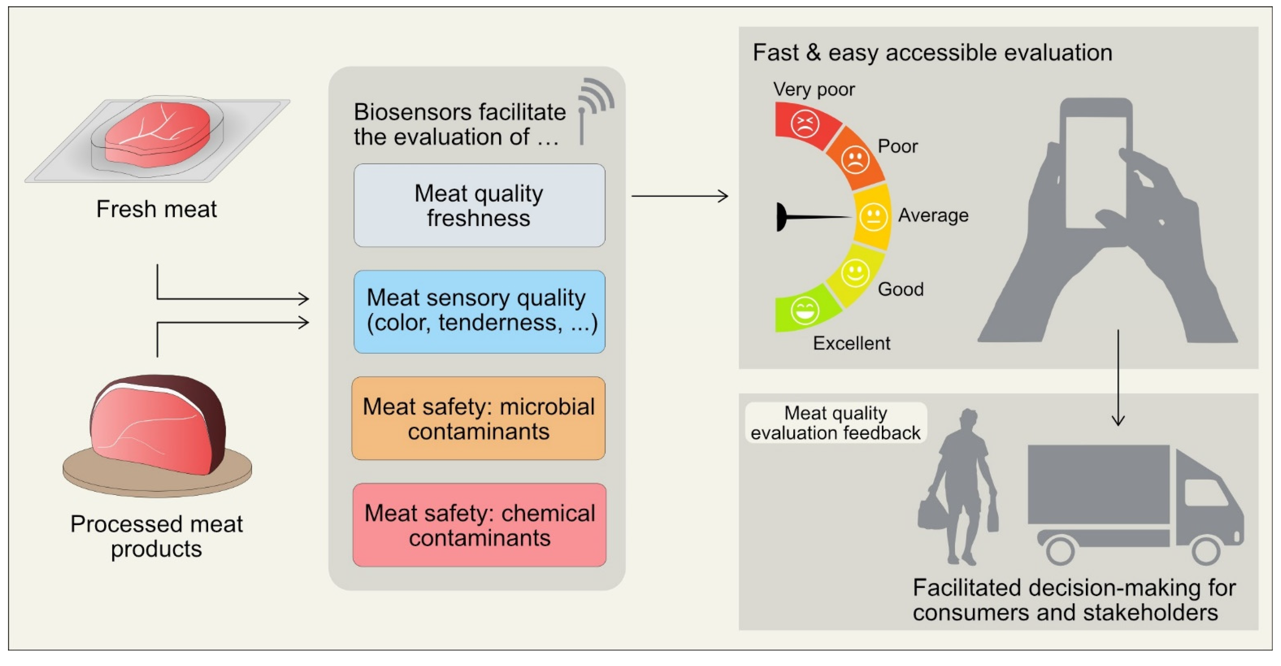
2. The Potential Use of Biosensors for Muscle Foods

The production of high-quality meat and the supply of safer meat products are now considered essential prerequisites in the meat processing sector. This is due to growing awareness among consumers, requesting quality meat products that are safe and available at an affordable price with a longer shelf-life [9]. Hence, meat producers or processors are putting more emphasis on safe and good manufacturing practices, suitable product packaging and labeling, and uniformity in maintaining the quality control methods to satisfy consumers and regulatory authorities [6]. Although conventional methods for analyzing meat quality are available, such as detection of microbial pathogens, antibiotic and drug residues, adulterants and contaminants, toxins, heavy metals, pesticides, etc., these methods are cost- and time-consuming, and require sophisticated instruments to yield results [5,10]. Hence, meat processors and researchers are in constant search of simple, low-cost, and easy-to-operate analytical techniques or methods that use portable instruments for quantification of various key meat quality components and to efficiently detect contaminants with great accuracy.
In this context, biosensors could play a vital role in meat safety and quality analysis due to their portability, high sensitivity, and specificity. In general, a biosensor is an analytical sensing device that comprises two main elements, like a transducer and a biorecognition element, with supporting components [11]. The main advantages of using biosensors in the food system are that they are inexpensive, easy to operate, and require less time for analysis of samples, which can detect a wide spectrum of food samples, either quantitatively or semi-quantitatively [12]. Hence, when biologically active components (analytes or groups of analytes) such as glucose, lactate, antibodies, drug residues, receptors, bacterial cells, toxins, or enzymes interact with the biorecognition (sensing) element, the attached transducer converts the biological interaction between the sensing element and analyte to meaningful and measurable electrical signals [13]. The interpretation of the resulting electrical signals is carried out by a signal processor. A schematic diagram depicting the basic principles of a biosensor is presented in Figure 2.
The use and choice of any biorecognition element(s) is crucial for real-time analysis of target substrates or biologically active components of interest [14]. As shown in Figure 2, biosensors are classified in diversified ways based on the biorecognition elements (enzyme, aptamer, whole cell, nano, immunosensors, antibody-based), transducers (electrochemical, optical, mass-based/gravimetric) and also based on label-free (surface plasmon resonance-SPR, mass spectrometry, acoustic wave, etc.) and label-based (fluorescence, chemiluminescence, etc.) detection techniques [15]. As this review focuses on the quality and safety of meat and meat products, the advantages and disadvantages of transducer-based biosensors are summarized in Table 1. A good number of research and review articles, elaborately discussing the materials used in biosensors, their stability and stabilization, performance in different environments including validation and calibration, are already available [5,15,16,17,18,19], and hence are not extensively covered in this review.

3. Application of Biosensors in Assessing Meat Quality

3.1. Biosensors to Assess Meat Freshness

The fresh appearance of food or its freshness is an important quality attribute that largely influences the purchase decisions of consumers. In the case of meat and meat products, freshness indicates the quality and safety of the product, which is important from both the producers’ and consumers’ point of view. The most common and popular quantitative and qualitative attributes used to evaluate the freshness of meat and meat products are pH, visual appearance, and meaty aroma [20]. Metabolites (organic acid, biogenic amines, glucose, sulfur, carbon dioxide, etc.) originating from microbial growth and chemical components generated due to oxidative changes (lipid and protein) during storage alter the freshness and quality of muscle foods [21,22]. Microbial metabolites and oxidative chemical compounds react with the indicator, which irreversibly affects the visual indication of the freshness and quality of the product. Therefore, a thorough understanding of the metabolites that can impact specific quality changes of food products over time is an essential prerequisite for the development and customization of indicators [23]. Many laboratory methods, including the measurement of total volatile basic nitrogen (TVB-N), pH, and culture-based bacterial quantification, are available, which are precise and mostly non-destructive but require more time to yield results [24]. Therefore, it is important to find new methods for detection of meat freshness to ensure a supply of quality and safe meat food to consumers [20].
Meat freshness as an indicator of product quality has an inherent relationship with aging as well as spoilage of meat. Therefore, during prolonged preservation, both monitoring of aging and spoilage of meat need to be evaluated. In that sense, the single important marker indicating meat freshness is hypoxanthine (Hx), a degradation product of ATP (adenosine triphosphate) [6].
For meat freshness analysis, various biosensors are used. For example, Albelda et al. [25] fabricated a graphene-based amperometric Hx sensor with titanium dioxide (TiO2-G) that was used for the estimation of Hx in pork meat stored under room temperature conditions for 7 days. The authors reported a high correlation coefficient (r = 0.9795) between the conventional enzymatic method and the biosensor, with a detection limit of Hx at 9.5 µM. Therefore, TiO2 -G nanocomposite can be used as a suitable electrode component in biosensors for the evaluation of meat freshness. An innovative amperometric biosensor with Fe3O4/polyaniline nanoparticles was developed for the estimation of xanthine in fish and chicken meat samples [26]. This biosensor with good reproducibility was able to detect xanthine within 8 s due to its quick response time, linear range (R2 = 0.997) and high sensitivity (13.58 µA µM−1 cm−2). The detection limit of the biosensor was 0.1 µM. Similarly, Hernández-Cázares et al. [27] developed and optimized an enzyme-based (amperometric) biosensor coupled with an oxygen electrode for the measurement of pork meat freshness. However, when using high-performance liquid chromatography (HPLC) and the biosensor, these authors found little variation in Hx concentration. In another study, a simple, easy-to-use, low-cost paper-based biosensor having microfluidic properties was fabricated for Hx estimation in meat samples [28]. The biosensor could detect Hx in meat samples within 5 min, having a detection limit of 1.8 mg/L and showing a good linear range of 5–40 mg/L, thus it can be used as an alternative to conventional methods with high accuracy. In a recent study by Garg and Verma [29], a fiber-optic biosensor was used for the estimation of xanthine in chicken meat. The authors concluded that the developed technique was very fast and reliable, requiring a minute volume of sample for detection of xanthine. Hence is suitable for mass-scale screening of samples.
Omanovic-Miklicanin and Valzacchi [30] developed two innovative chemiluminescence biosensors based on enzyme oxidase (putrescine and diamine) to detect the concentration of biogenic amine in meat products that were compared to samples evaluated by HPLC. The authors reported that the biosensors can quantify putrescine in the range of 1–2 mg/L with a detection limit of 0.8–1.3 mg/L, and thus may be preferred over HPLC analysis due to their simple nature of operation and the short-time detection of analytes. The results of Ag/AgCl and platinum electrode-based amperometric biosensors developed to quantify the biogenic amines and hypoxanthine of beef were comparable with conventional liquid chromatography methods [31]. Similarly, a non-destructive method was developed to detect the TVB-N as a freshness indicator in pork meat by combing two sensing components like hyperspectral imaging (HSI) and colorimetric sensors [32], which had a coefficient value of R² = 0.932. The authors stated that integrating two sensors along with back propagation adaptive boosting non-linear data fusion algorithms can potentially monitor the quality and safety of pork meat in real-time. Moreover, an inexpensive novel sensor was developed by applying titanium dioxide-polyaniline composites on the surface of silk fibroin fiber to quantify TVB-N in pork samples [33]. This sensor showed a good correlation with the TVB-N concentration in meat samples (R2 = 0.990).
The use of Bacillus subtilis, a Gram-positive bacterium, as a biosensor to assess meat spoilage has also been reported. The volatile compounds released from spoiled meat can activate the specially identified promoter to drive fluorescent protein formation in B. subtilis [34]. The researchers explained that cell-based biosensors have immense potential on a commercial scale and could be used as a promising tool to evaluate meat quality traits. The various types of biosensors used for the detection of freshness in meat and meat products are summarized in Table 2.

3.2. Biosensors to Evaluate Meat Tenderness

Appearance/color, taste, tenderness, juiciness, among other quality attributes, are the most important quality traits of meat [42,43]. Amongst these, tenderness is one of the major quality attributes and an important quality trait for consumer satisfaction and repurchasing meat [6,44,45]. The tenderization of meat largely depends on the degradation of cytoskeletal proteins responsible for the structural integrity of muscle fibers and many other interconnected pathways, as evidenced recently by Gagaoua et al. [46] using an integromics proteomics approach. The post-mortem muscle changes and variations in tenderness are driven by five mechanisms that are: (a) the degradation of muscle myofibrils by endogenous proteolytic systems during ageing, (b) collagen and cross-linking, (c) sarcomere length and its status during the post-mortem period, (d) intramuscular fat content known as marbling, and (e) the denaturation of muscle proteins during cooking [47]. The activation of the endogenous proteolytic systems (proteases and their inhibitors) is the first key among these mechanisms [48]. Among them, different research studies have proven that calpain proteases are responsible for the changes in muscle/meat that occur during ageing or post-slaughter tenderization. Calpains, cathepsins, caspases, and proteasomes are endogenous proteolytic systems, among many others, that can degrade several myofibrillar proteins, ultimately driving the final tenderness of meat [49,50]. In the skeletal muscles, the calpain system mainly consists of three proteoforms, from which µ-calpain and m-calpain and their specific endogenous inhibitor, calpastatin [51,52], have been extensively studied. Other associated factors like post-mortem glycolysis and pH, which additionally affect meat tenderness, may vary because of different dietary systems, use of growth promoters, and finally due to the stress of animals during transport and slaughter [53,54,55].
Different conventional techniques like sensory taste panels, Warner-Bratzler Shear Force (WBSF) as an instrumental method, ELISA, proteomics, or chromatography are available to evaluate meat tenderness, but these methods are time-consuming, expensive, and difficult to use on a broad scale [43,56]. Amongst these, the WBSF method has been proven to be more accurate in tenderness evaluation compared to other methods [57]. These techniques evaluate the resistance of the meat during cutting without providing a direct determination of meat tenderness [36,58]. In such a scenario, biosensors may play an immense role and act as a measurement tool that converts biochemical information into an analytical signal by using biological ligands as part of a biotransducer.
Geesink et al. [39] developed an immunological biosensor using the SRP system (Biacore Q) for the detection of calpastatin activity in beef meat. The biosensor showed a linear correlation with a conventional enzymatic assay and the correlation coefficient was in the range of r = 0.51–0.99 in several experiments. Based on their findings, Geesink et al. concluded that the Biacore Q is an effective online screening tool to measure calpastatin activity, and thus, may be used as a predictor of meat tenderness for high-throughput applications in the commercial sector as well as in research. Bratcher et al. [40] used a fluorescence resonance energy transfer (FRET) method to estimate calpastatin in an optical biosensor device in stored meat. They found that the 48 h post-mortem would be the most accurate time for grading and classification of meat using the FRET optical biosensor. Zór et al. [36] developed a multi-channel portable electrochemical immunosensing device known as the Tendercheck system, based on antibody-antigen biorecognition and amperometric detection. The device accurately quantified calpastatin. Furthermore, results from this biosensor showed a similar correlation (R2 = 0.62) compared to the WBSF method. More studies are required to develop user-friendly biosensing methods for rapid screening of meat tenderness that will help the meat industry in providing high quality meat products to consumers. The types of biosensors used for the detection of tenderness in meat and meat products are summarized in Table 2.

3.3. Biosensors to Detect Microbial Contaminants in Meat

Contamination of food has become a public health concern, seeking worldwide attention as human consumption of such products may cause food poisoning and disease outbreaks. These foodborne outbreaks cause irreparable damage to human health as well as to the world economy and environment. Microbial contamination in any food product may occur via raw materials or by cross contamination at any point of the food processing system [59]. A continuous microbiological environmental surveillance system is therefore necessary for the early detection of pathogenic organisms in the food chain to assure microbiological safety [60]. For detection of microbial contamination, apart from traditional biochemical and microbiological methods, which include bacterial colony counts, staining, and methylene blue reduction tests; various conventional techniques such as ELISA, PCR, and fluorescence detection are also available [11]. However, most of the most advanced analytical methods require well-equipped laboratories, sophisticated and expensive instruments, and skilled technicians. Furthermore, most of these methods necessitate lengthy sample preparation and processing steps that involve various enrichment and incubation stages, and can take up to 10 days to produce results [61,62,63]. These limitations have created a demand for novel in situ analysis methods that are more sensitive, accurate, fast, and specific than existing methods [7,14,64]. Recently, analytical tools like biosensors with high specificity and sensitivity have become available, which can detect microbiological safe limits, toxins, or their metabolites in different products.
In fact, biosensors are simple, cost-effective, easy-to-handle devices that can rapidly detect pathogens but do not need pre-enrichment methods, unlike nucleic-acid based and immunological methods [65]. Nowadays, various user-friendly biosensors based on optical, electrochemical, photoelectrochemical, and bioluminescence are available [11]. For instance, optical biosensors facilitate real-time monitoring of microbial activities in the food matrix. These biosensors discriminate against microbes in foods either by changing the level of signal or refractive index, or by detecting the concentration of the microbial cells attached to the biorecognition sensing site on the optical transducer surface [17]. Out of different optical analyzing techniques like colorimetric, fluorescence, localized SPR, and chemiluminescence; SPR is widely used as an optical biosensor [66]. In SPR, bioreceptors are immobilized on a metal surface transducer, where electromagnetic radiation of a specific wavelength interacts with the electron cloud of the transducer and thereby generates a strong resonance. When the bacterial cells (target analyte) interact with the metal surface, an observable alteration in the refractive index can be noticed [61]. Generally, it allows reflectance spectroscopy for target pathogen detection.
There are many reports available on the use of optical biosensors that can detect pathogenic organisms in various meat and meat products. In a study, L. monocytogenes in meat was detected up to 3 × 10² CFU/mL by a specially designed fiber optic immunosensor, equipped with a powerful immunomagnetic separation [52]. An aptamer-based fiber-optic biosensor was employed to detect pathogenic L. monocytogenes from other non-pathogenic or pathogenic species in artificially contaminated ready-to-eat (RTE) meat products [53]. Oh et al. [54] identified S. Typhimurium from pork meat up to 4 log CFU/mL within 30 min using localized SPR. Zhang et al. [55] developed a multi-channel SPR biosensor for specific detection of three different foodborne pathogens, namely E. coli O157:H7, S. Enteritidis, and L. monocytogenes, together in naturally contaminated food. In another study, bacteria of the species Shigella sonnei were detected and isolated apart from other enteric organisms such as E. coli and S. Typhimurium, using fluorescent biosensors with basic aptamers [56]. By combining the lateral flow biosensors with multiple cross displacement amplification, Wang et al. [57] reported high specificity and sensitivity of the sensor that could detect Shigella spp. within one hour. However, high costs, quality assurance, stability disputes, sensitivity issues, and instrumentation design are current limitations which need to be addressed prior to the commercialization and wider applications of these optical biosensors.
Based upon the antigen-bioreceptor interactions, different kinds of electrochemical biosensors like amperometric, impedimetric, potentiometric, and conductometric are available [67]. An electrochemical immunosensor prepared with chitosan/gold nanoparticles composite has been reported to provide a wide range of detection limits from 1–5 Log CFU/mL that may be helpful for proper detection of Salmonella contamination [68]. Similarly, Morant-Miñana and Elizalde [69] isolated Campylobacter spp. from chicken meat using an electrochemical genosensor prepared with thin-film gold electrodes. Che et al. [70] isolated C. jejuni from turkey and chicken meat samples using a fluorescence biosensor with a detection limit of 2.1 × 10⁴ CFU/mL. The rapid detection of C. jejuni is important, as it is considered as a major food-borne pathogen, causing diarrhea and fever in consumers.
Currently, nano-based sensors are outstanding in detecting different food-borne pathogens and toxins [61]. Using carbon nanotube-based biosensors, Yamada et al. [62] was able to detect E. coli within 5 min with a detection limit of 2 log CFU/mL. In another study, Muñoz-Berbel et al. [63] could detect E. coli with a detection limit of between 1 and 7 log CFU/mL by the use of impedimetric spectroscopy.
A quartz crystal microbalance (QCM) biosensor is generally characterized by the resonant frequency of quartz crystals with a higher sensitivity for qualification and quantification of microbial whole cells at extremely low levels. By using this QCM technique, C. jejuni (LOD: 1.30 log CFU/mL) and S. Typhimurium (LOD: <100 CFU/mL) were detected in chicken meat samples [71,72,73].
Liu et al. [74] developed an impedance based microfluidic biosensor for the detection of Salmonella serotypes B and D in a RTE turkey matrix. The study reported the detection of a very low concentration of Salmonella (300 cells/mL) within one hour. The selectivity of the sensor was also tested using non-specific binding of different E. coli strains. Besides, high concentrations of inactivated Salmonella and very low levels of alive Salmonella cells can additionally be differentiated by this sensor. Liang et al. [75] prepared a smartphone-based biosensor for preliminary screening of microbial contamination in food matrices. The researchers applied this system, coupled with a digital camera and a gyro sensor of a smartphone, to detect microbial pathogens in ground beef. E. coli K-12 contaminated beef was identified by simply taking a picture with a smartphone, which was subsequently analyzed by an implemented procedure. Mie scatter assays were applied at different angles (15°, 30°, 45°, and 60°) out of which the lower limits were detected (1 log CFU/mL) at a 45°angle, and 2 log CFU/mL at angles of 30° and 60°. This biosensor, integrated with a smartphone, is easy to handle, rapid, cost-effective, and does not need antibodies, microbeads, or any other reagents.
Bioluminescence-based ATP detection is another approach to microbial spoilage detection [11,76]. It is well known that ATP is present in all living microbial cells (bacteria, mold, yeast, and algae) as an activated energy carrier. For the bioluminescence-based ATP detection, ATP is converted to adenosine monophosphate (AMP) with the emission of light. The intensity of light as the result of the breakdown of ATP in a bioluminescence reaction can be quantified using very sensitive photons from light meters placed in an instrument called a luminometer. The more ATP is present, the higher the intensity of light in terms of relative light units will be obtained from the reaction. The ATP bioluminescence assay is a very useful tool, as it takes very little time to give successful results [77]. In a study by Siragusa et al. [78], a rapid ATP assay was developed to measure total bacterial counts in samples obtained from beef and pork carcasses. The results were compared to the conventional bacterial plate count methods, which allowed a positive correlation coefficient of 0.91 for beef and 0.93 for pork carcasses. Cheng et al. [79] combined an ATP bioluminescence assay with functional and suitable magnetic nanoparticles for rapid estimation of E. coli from ground beef, which was artificially contaminated. A detection limit of 1.30 log CFU/mL was observed. However, one weakness of this detection method is that ATP is present in all living microorganisms, including meat. Therefore, ATP should be destroyed in meat prior to performing the bioluminescence assay.
In a recent study by Vizzini et al. [80], a paper-based DNA biosensor was used to detect Campylobacter spp. in chicken meat. The authors reported that the developed biosensor was very cost-effective, portable, and the level of detection (3 pg/µL of DNA) was comparable to available qPCR kits. The types of biosensors used for the detection of various microorganisms in meat and meat products are summarized in Table 3.
Although much progress has been made in the biosensing technologies for the detection of microorganisms in contaminated food to date, more research in this field is needed to address the requirements of industry with more simplified, easy-to-handle, and cost-effective methods. Overall, high sensitivity with good specificity and modern nanotechnology-based biosensors can be an alternative for food-borne pathogen detection.

3.4. Biosensors to Detect Contaminants, Antibiotics, and Drug Residues in Meat and Meat Products

Various contaminants such as toxins, pesticides, antibiotics, veterinary drug residues, and hazardous food additives can enter the food chain system at any processing step and contaminate the whole batch. Many analytical techniques like HPLC, capillary electrophoresis, and mass spectrometry are available for analyzing the samples at the end of the processing steps. However, these procedures are costly, complex, require sophisticated instruments, and the intervention of skilled personnel [100]. Therefore, there is a growing interest in the rapid, reliable, and more sensitive detection of contaminants in the processing steps itself, through the involvement of biosensor technology, which can provide a real-time screening and monitoring of food contaminants, minimizing the unwanted threat to consumers’ health. The types of biosensors used for the detection of various antibiotics and drug residues, adulterants, allergens, and additives in meat and meat products are summarized in Table 4.
According to Erofeeva et al. [118], almost half of all produced antibiotics worldwide are used in the livestock sector. In fact, a wide range of broad-spectrum antibiotics are used in the livestock sector that are effective against both Gram-positive and Gram-negative bacteria. Furthermore, the combined use of veterinary antibiotics for metaphylactic and prophylactic purposes, or as in-feed supplements for growth promotion, outweighs their therapeutic usage by far [119]. The extensive and non-therapeutic use of antimicrobials in animals, either in therapeutic or sub-therapeutic doses, not only aids the development and spread of antimicrobial resistance, but may also result in the accumulation of residual amounts of antimicrobials or their metabolites in food producing animals, which may then be consumed by humans via meat, milk, and eggs [120]. The presence of antimicrobial residues in animal-derived food products beyond maximum permissible limits has become an intensely debated topic and has received much attention in recent years. The growing concern is due to their adverse public health effects, causing hypersensitivity reactions, antimicrobial resistance, disruption of the intestinal flora, and even neurological disorders [121]. It is therefore crucial to monitor the presence of high concentrations of antimicrobial residues in meat products to guarantee food safety [122,123]. In this context, SPR is a widely accepted technique in biosensors to detect such drug residues. By using the SPR technique, chloramphenicol and sulphonamides have been quantified in different meat species such as pork, beef, and chicken [124,125,126]. Cai et al. [117] developed a chemiluminescence based-sensor to check the presence of benzimidazole residues in beef and mutton. This sensor acquires ultrahigh sensitivity (range of detection: 1.5–21 pg/mL) and can detect residues in a short time (18 min).
Mohammad-Razdari et al. [127] designed an aptamer-based electrochemical biosensor to detect sulfadimethoxine (SDM) in beef and chicken meat. The sensor offered adequate sensitivity and better stability with a LOD of 3.7 × 10⁻¹⁶ M. Moreover, the percentage recovery of SDM was comparable with the results of the HPLC analysis. In a study conducted by Stevenson et al. [128], an affinity-based electrochemical biosensor was used to detect ceftiofur residues in turkey meat samples. The biosensor was cost-effective and rapidly detected the antibiotics within 15 min, even at low concentrations (10 ng/mL). A luminescent bacterial biosensor designed by Pikkemaat et al. [108] was able to screen a large number of poultry muscle samples for tetracycline (TET) within three hours, for which approximately 12 h were needed using conventional microbiological assays. Additionally, the sensor was cost-effective and highly sensitive. Comparing the performance of the SPR biosensor with that of LC-MS/MS and GC-MS/MS to detect chloramphenicol (CAP) and its glucuronide residue in poultry muscle, Ferguson et al. [109] showed that the performance of the biosensor was better with detection capabilities of 0.02 µg/kg. In another study, a novel colorimetric aptasensor used for the detection of multiplex antibiotics, namely CAP and TET, was able to detect these antibiotics in chicken meat with a LOD of 7.0 nM and 32.9 nM, respectively [110]. This method is very simple and does not require high-end instruments, so it could be used for on-site screening of samples. The detection of sulfadiazine and acetaminophen in pork and chicken samples has been examined by Sun et al. [111]. This study used an electrochemical sensor that exhibited great accuracy and stability for concurrent determination of both antibiotics, with results comparable to those obtained through HPLC analysis. By using biosensing technology, aminoglycosides, lincosamides, quinolones, and tetracyclines have also been detected in meat samples from different animal species [6,13,112,114].
For identifying bacterial or fungal toxins present in meat or meat products, electrochemical biosensors are beneficial. These contaminants may occur at any stage of the food processing chain, or even during transportation and storage. Toxins are not only harmful from a public health point of view but are also associated with severe economic losses. Several reports/findings are available in this regard. For instance, Staphylococcal enterotoxin B was identified using an electrochemical biosensor in pork and milk [129] and trichothecene (T-2 toxin) in swine meat [89]. In another study, an SPR biosensor was used as a tool for the rapid and real-time analysis of Staphylococcal enterotoxin B in potted meat [87]. Moreover, food additives are widely used components in the food industry to maintain and improve physicochemical, sensory and rheological properties, and to extend the shelf-life of products. Commonly used food additives are nitrite, benzoic acid, monosodium glutamate (MSG), propyl gallate, and food colorants, which are hazardous and undesirable if used beyond the maximum permissible limits. Batra et al. [96] developed an amperometric biosensor and enzyme biosensor (glutamate dehydrogenase and glutamate oxidase) to detect the presence of excessive amounts of MSG in food, which can pose a significant health risk to consumers. Wang et al. [97] discovered a biosensor for the determination of food colorants, especially Amaranth (E123) and Ponceau 4R (E124) in processed food products, based on the use of carbon nanotubes and a polypyrrole (ppy-) composite modified electrode. Another amperometric biosensor was made by Shan et al. [98] to screen for the presence of benzoic acids in processed foods using mushroom tissue, tyrosinase, and polyphenol oxidase as biological recognition compounds. By employing the amperometric biosensor, Dinçkaya et al. [99] estimated the nitrate levels in meat and demonstrated that this method is simple and inexpensive with high accuracy and sensitivity. Moreover, this method could estimate nitrate with an LOD of 2.2 × 10⁻⁹ M and a response time of 10 seconds. According to the aforementioned reports the meat processing industry should focus on using biosensing technologies, which are simple but rapid in response with high sensitivity, to reduce hazards or contaminations in the meat processing chain, to produce safe and quality meat and to gain consumer trust and confidence.
Adulteration of meat is mostly done by the fraudulent addition of cheaper/low quality meat species or non-meat ingredients or by mislabeling actual constituents of commercial products for economic gain. The authenticity of meat and meat products is required to safeguard religious sentiments, comply with the norms of regulation authorities and, above all, to protect the interests of consumers [105,130]. Analytical techniques based on protein and DNA analysis to discriminate meat from different species are not only expensive but also time-consuming and require skilled personnel.
Several reports indicate that the use of SPR and electrochemical biosensors allows discrimination against adulterated meat samples or processed meat products. For example, SPR-based DNA biosensors were employed to identify donkey meat samples in beef sausages. The sensor was specific and had a high sensitivity with a detection LOD of 1.0 nM [101]. Likewise, in another study, an electrochemical DNA biosensor detected pork in food products when added up to 10% in biological samples [102]. Biosensors are also capable of differentiating adulteration of meat from closely related animal species. Using an amperometric PCR-free electrochemical biosensor, beef meat that was adulterated with horse meat (0.5 % w/w) could be obtained within one hour without the involvement of any extraction or amplification of genetic material [103].
Food allergies caused by different allergens are regarded as an emerging public health problem. For example, porcine serum albumin (PSA) is a major allergen in pork and its products, which can cause allergic reactions. Although conventional methods like PCR, ELISA, and mass-spectroscopy are employed for the analysis of food allergens, SPR can rapidly and accurately detect PSA with an LOD of 19.81 ng/mL, similar to the ELISA-based method [105].

4. Sensors and Indicators for Smart Packaging of Meat and Meat Products

Currently, there is no standard and reliable method available to satisfactorily confirm the freshness of meat, except for sensory analysis, chemical, and microbiological tests. Consumers have no other option but to check the ‘use by’ or ‘sell by’ date on printed packaging material [131]. Conventional food packaging systems with expiration dates can protect the food from external environments and delay the spoilage of the product, for a stipulated period. The ever-increasing incidences of meat-borne outbreaks is an indication that expiration dates displayed on packaging material alone are not sufficient in protecting consumers from the threat of spoiled meat and meat products.
To overcome this problem, research on the integration of intelligent materials with traditional packaging systems is gaining interest. This kind of smart packaging system with different indicators or sensors can sense, track, detect, and record the external or internal environment of products and turns out to be a promising tool for the freshness detection of food products [132,133,134,135]. As far as meat and meat products are concerned, intelligent packaging systems can monitor the quality in real-time throughout the supply chain and detect any quality deterioration with the help of chromogenic substances. Several indicators or sensor-based packaging systems, including freshness indicators, gas sensors, time-temperature indicators, pH indicators, barcodes, and RFID tags, are available for the assessment of meat freshness using changing color patterns of the indicator. Out of these, freshness indicators based on pH sensitivity have gained popularity due to their simple applicability and authentic results [136]. In fact, during microbial spoilage, acidic or alkaline metabolites such as NH3, CO2, H2S, TVB-N, including dimethyl amine and trimethyl amine, are released after the decomposition of nutritive components of meat. When these metabolites come into contact with intelligent chromogenic material in the packaged headspace, a chromatic change occurs due to their pH sensitivity. Thus, the chromogenic indicator reacts with microbial metabolites and exhibits color changes as a result of changes in pH [132,134].
Various chemical, and natural chromogenic substances are currently used as freshness indicators of food products by various studies because of their rapid response, color-changing ability, and good stability. Commonly used chemical indicators are methyl, phenol, and bromophenol red, bromophenol blue, bromocresol violet, and bromocresol green. These can be used as non-contact indicators to avoid direct contact between chemical reagents and food, considering the problem of migration of chemical reagents [134]. In a study by Shukla et al. [137], a colorimetric indicator sensor was used with bromophenol blue to examine the quality deterioration in buffalo meat cuts stored for 9 days in a refrigerated condition. The study reported a correlated color response of the indicator sensor from yellow to blue at different levels of spoilage based on the increase in concentrations of TVB-N, indicating the deterioration in meat quality. In a different study, Kuswandi et al. [138] designed a litmus paper-based (red to blue from pH 5.7 to 6.0) sticker type indicator and attached it to the inner side of a plastic-film wrapped polyethylene tray containing beef stored at a conventional temperature. The study reported that the color change of the indicator sensor was driven by the production of biogenic amines, which were produced by microbes inside the packaged product. Although single chemical dye indicators are used, composite or mixed indicators are advantageous compared to the former in regard to detecting the spoilage threshold and freshness levels of the food accurately [139]. Rukchon et al. [140] developed two mixed pH sensitive indicator-based smart packaging containing bromothymol blue and methyl red, and a mixture of bromothymol blue, bromocresol green, and phenol red for observing spoilage of skinless chicken breast stored under refrigerated and modified atmospheric packaging (MAP) conditions, respectively. These chemical indicators responded well to produced CO2 and other microbial metabolites by changing their colors. Chen et al. [141] developed a colorimetric sensor array containing three pH indicators (bromocresol green, bromocresol purple, and neutral red) and nine metalloporphyrins fabricated on a C2 reverse silica-gel flat plate. This array provided an excellent color fingerprint for the detection of biogenic amines from chicken during spoilage. Another prototype freshness indicator was developed by Lee and Shin [131] with a methyl red-cellulose acetate mixer to trace the freshness of beef stored at 20°C for 24 h. The authors observed a positive color change from red to yellow, indicating the production of microbial metabolites [123,131]. Although the chemical chromogenic sensors respond quickly with an assured color change and good stability, the migration of chemical reagents into food can be disadvantageous [134]. Conversely, the natural chromogenic sensors are safe to use and inexpensive compared to chemical sensors.
The commonly used natural chromogenic indicators that can be safely used in smart packaging are anthocyanin, carotenoids, chlorophyll, curcumin, and betaine [142]. Out of these natural freshness indicators, anthocyanin has drawn much attention for its wider spectrum and vibrant color-changing patterns at different pH ranges [143,144]. Anthocyanin is one of the most important flavonoids and can be readily extracted from purple cabbage, roses, pomegranates, blueberries, black grapes, eggplant, and black raspberry. Apart from having a rich matrix, anthocyanins are hydrophilic, non-toxic, and odorless. The color stability of anthocyanin is affected by pH, light, temperature, metal ions, enzymes, UV radiation, gases, and also different chemical forms of anthocyanidin [133,135]. The use of anthocyanin in packaging systems has been reported in different studies. Shukla et al. [145] extracted anthocyanin from rose and red cabbage, which was printed on filter paper to prepare colorimetric sensor-based intelligent packaging. The natural sensor was capable of detecting the release of ammonia, indicated by a color change from red to green at a higher pH. Golasz et al. [146] developed a smart film containing cassava starch and grape anthocyanin, which was applied to pork loin and stored under refrigerated conditions for up to 14 days. A positive correlation was observed between the color change of the film and microbial spoilage. Choi et al. [147] developed another type of intelligent packaging with agar, potato starch, and natural dyes (anthocyanins) extracted from purple sweet potatoes. The study reported a change in the color of the dye from red to green, indicating the spoilage of pork samples.
However, it is important to mention that dye-based indicator sensors have limited practical applications because of their poor mechanical and gas barrier properties and high polarity [133,144]. Making composite films with more than one biopolymer by casting, compression molding, or extrusion method is, therefore, becoming more popular nowadays [135,148,149]. This natural pH-sensitive color changing indicator is incorporated with different biodegradable, biocompatible, and eco-friendly biopolymers, which are also edible, for the preparation of smart packaging to track the freshness of food [135,142]. The selection of appropriate nanomaterials and their applications in biodegradable films, which may improve the structural morphology, including mechanical barrier and thermal properties of the film, plays a crucial role [150]. In this regard, polysaccharide-based biopolymers are more preferable over protein- or lipid-based due to their ability to form cohesive networks with other polymers through covalent or non-covalent bonds [151,152].
Alizadeh-Sani et al. [153] developed a multifunctional halochromic intelligent pack aging material with anthocyanins extracted from saffron petals. The anthocyanins were fabricated into chitosan nanofibers and methyl-cellulose to examine their pH-sensitivity, to ammonia gas, which is produced during the storage of lamb meat. Apart from offering good mechanical and gas barrier properties, the packaging material showed eligibility as a freshness indicator, again changing the color from reddish/pink to violet to green to yellow when exposed to increasing concentrations of ammonia vapor. In another study, Vedove et al. [154] developed smart packaging with cassava starch sheets, using an extrusion process to incorporate anthocyanin as a color indicator to monitor spoilage of beef meat stored at 4°C for 3 days. The study reported a positive correlation with the color change of the film with the production of biogenic amine. Dudnyk et al. [155] developed a similar kind of smart packaging material, containing pectin and red cabbage extract, which was rich in anthocyanins. Anthocyanin was extracted by using a casting method and was incorporated with plasticizers to strengthen the mechanical properties of the film. The film was attached to the headspace of different meat products containers, which showed a similar color change from purple to yellow when volatile nitrogenous substances were produced in association with microbial growth. Zhai et al. [156] successfully developed biogenic amine-sensing bilayer films by using agar, gellan gum, TiO2, and anthocyanin for the detection of NH3, trimethylamine, and dimethylamine while studying the quality deterioration of meat. Zhou et al. [157] developed another double-layered indicator film containing carrageenan, anthocyanin, curcumin, and an emulsified layer of glucomannan, i.e., konjac, and camellia oil to monitor the freshness of chicken meat. This emulsified, intelligent double-layered film performed well in monitoring the freshness of chicken at 25 °C with a noticeable color change.
To monitor the freshness of pork in real-time, a protein-polysaccharide nano-complex colorimetric system was developed by Zhang et al. [148]. In this study, nanocomplexes of anthocyanin-loaded ovalbumin-propylene glycol alginate were incorporated into polyvinyl alcohol/glycerol matrices to provide improved strength and barrier properties to the film. A color change from purplish-red to dark-blue was observed in response to the presence of volatile ammonia, which indicated a decrease in pork meat freshness. Likewise, Niu et al. [158] created intelligent packaging to ensure the safety of pork consumption, which was done by tracing the presence of biogenic amines and microbes. For this, anthocyanin was extracted from colored potatoes (Black King Kong) and fabricated into starch/glycerol/gelatin, which was used for the development of the packaging. This matrix-rich anthocyanin loaded film showed improved physicochemical and antioxidant properties along with exceptional ammonia- and TVB-N-sensing properties, indicated by color change. For example, Chayavanich et al. [159] used a pH-sensitive biocompatible smart film consisting of starch, gelatin, and extracted anthocyanin from red radish for real-time monitoring of meat spoilage. Due to the basicity of the spoiled medium, the pH-sensitive film changed its color from orange to pink and then to bluish-purple in real time which was visible to the naked eye.
Kuntzler et al. [160] developed a different kind of pH-sensitive chromogenic intelligent film with nanofibers of polylactic acid and polyethylene oxide, combined with the microalga Spirulina. Spirulina is known for its natural blue-green color, which contains different pigments such as β-carotene, tocopherols, phycocyanin, phycoerythrin, and chlorophylls, and thus offers a promising role as a pH-sensitive color indicator. The microalga biomass could be encapsulated in the polymer nanofiber successfully and was indicated via a color change from red to green, upon exposure to microbial metabolites in pork meat stored at a refrigerated temperature. Sun et al. [143] developed a double-layer indicator film using anthocyanin from raspberry, which was incorporated into low-acyl gellan. The outer layer of the film matrix was shielded by chitosan. This encapsulated film matrix showed improved mechanical strength, tensile property, high opacity, and water barrier property. The double-layer film not only improved the stability of anthocyanin but also showed a visible positive color changing pattern based on pH variances, indicating alteration of meat quality in refrigerated storage conditions. A nanofiber-based film with curcumin, chitosan, and polyethylene oxide was developed to monitor the freshness of chicken by Yildiz et al. [161]. A color change from bright yellow to reddish yellow in the film implied that meat was producing TVB-N, creating a basic environment, thus ultimately heading for deterioration. In another study, Kanatt [162] utilized a pH-sensitive dye, namely betalain, which was more pH-stable than anthocyanin. Betalain was used to make an intelligent film with gelatin and polyvinyl alcohol. Due to their stable pH-sensitivity and other biological functions, betalains are gaining more attention for their usage as colorimetric dyes. When tested for the packaging of chicken and shrimp, the wrapped film changed its color from red to yellow in association with microbial quality deterioration over time with the production of volatile basic nitrogenous substances.
It can be concluded that natural pigment-based smart or intelligent packaging systems play an important role in critically assessing the internal quality of meat or meat products in stored conditions, apart from providing accurate information related to quality and safety of the products. This may be a good alternative to other types of packaging systems, as they are economical, safer, and user-friendly. However, more research is needed to overcome limitations related to the instability of pH-sensitive pigments in biopolymer matrices and their migration properties in food.

5. Factors Influencing the Analytical Performance of Biosensors

As stated earlier, biosensor technology is based on combining advances in biological detecting elements like a sensor system and a transducer. However, the analytical performance of biosensors depends on several interrelated parameters such as accuracy, selectivity, linearity and range, sensitivity, specificity, reproducibility, limit of detection/quantification, etc. [15]. Further, the biological sensing components (DNA probes, enzymes, antibodies, tissue, cell receptors, etc.) can lose their activity within a short period of time, either due to the biological nature of the molecule or upon exposure to environmental stresses, such as pH, temperature, or ionic strength (these are the main aspects underlying muscle food variability). The performance of biosensors in terms of accuracy, precision, and long-term stability can be improved by standardizing the sensor elements and overcoming the environmental effects. These can be handled, for instance, by strict control of temperature. Maintaining the stability of different bio-elements of biosensor devices should be ensured before commercialization of their application. Application of nanotechnology [163] or their involvement with high-affinity biomolecules can increase the selective and sensitive detection of target analytes. The incorporation of various nanostructured materials, including nanocomposites, nanoparticles, nanotubes and nanowires into sensor structures can improve sensitivity, response time, and efficiency [15,164]. Although nanomaterials have immense potential, the analytical performance (e.g., sensitivity, detection limit, and signal-to-noise ratio) of nanomaterial-based sensors varies considerably due to variation in properties, which needs to be addressed. Strategies such as studying the nanomaterials’ characteristics, their operational conditions, and application of appropriate calibration algorithms are suggested to reduce the inconsistency in analytical response and the properties of sensing devices and to improve the performance and practical application of such sensors [165].

6. Concluding Remarks and Future Perspectives

In the meat industry, the use of bio-sensing technology is growing steadily for real-time monitoring of meat quality and safety in the supply chain, including transport and storage. In fact, biosensors are considered efficient and alternative techniques to conventional methods due to their specificity, sensitivity, and cost-effectiveness. These unique technologies can help detect the purity and freshness of raw meat, followed by the evaluation of the glycolysis extent and tenderness, as well as for the detection of pathogens, adulterants, antibiotics, allergens, drug residues, additives, and other contaminants. Likewise, much progress has also been made in utilizing different natural and chemical indicators in developing smart packaging to trace meat quality and safety on a real-time basis. Although various indicators, including time-temperature, pH, freshness, etc., and sensors are available, 3D printing technology offers new possibilities. There is scope for development of intelligent indicator-based smart packaging technology, which combines 3D-printing approach with indicators to check real-time detection and continuous monitoring of meat freshness/degradation. Efforts should also be made in designing sustainable and environmental-friendly packaging solutions by using edible freshness indicators, such as chromogenic indicator-based smart packaging, to assess the freshness of meat products.
Despite extensive research in this innovative field, not many biosensors are yet available for their application in the meat sector, and still there is a long way to go before conventional methods can be substituted commercially. However, with recent advancements in the field of nanotechnology, nanomaterials are being explored for application in emerging fields of science, including bioengineering that deals with biosensors and bioelectronics. Nanomaterials exhibit high surface area to volume ratio, increased mechanical strength, excellent catalytic activity, good stability, enhanced chemical and biological activities, and better electrical and magnetic properties, which can be exploited in improving the analytical performance of biosensors. Different kinds of functional nanomaterials such as metal and metal oxide nanoparticles, carbon-based nanomaterials, magnetic nanoparticles, polymeric nanoparticles, graphene sheets, and other novel nanomaterials are available that can be incorporated into biosensor technology. However, the key lies in understanding the mechanism of interaction between biomolecules and nanomaterials for the fabrication of biosensors. Therefore, future research work should focus on understanding the novel properties of these nanomaterials and their compatibility with biomolecules to design a new generation of nanomaterial-based biosensors that are not only inexpensive but also portable, easy to operate, and reliable enough. Above all, the designed biosensors should offer lower detection limits, higher sensitivity, and faster response time for real-time monitoring of large numbers of samples and rapid detection of the targets (biological and chemical substances/contaminants) of interest for their wider applications in the food industry, including the meat processing sector.

Author Contributions

Conceptualization, A.K.D. and M.G.; data curation, P.K.N., D.B., J.K.D., A.K.D. and M.G.; writing—original draft preparation, P.K.N., D.B., J.K.D., A.K.D. and M.G; writing—review and editing, P.K.N., D.B., J.K.D., S.B., D.E., J.M.L., P.D., L.A., A.K.D. and M.G.; supervision, A.K.D. and M.G.; project administration, M.G.; funding acquisition, M.G. All authors have read and agreed to the published version of the manuscript.

Funding

This research received no external funding.

Institutional Review Board Statement

Not applicable.

Informed Consent Statement

Not applicable.

Data Availability Statement

Not applicable.

Acknowledgments

Thanks to the Director, ICAR-Indian Veterinary Research Institute (IVRI), Izatnagar, Bareilly, India, and the Station in-charge, Eastern Regional Station, ICAR-IVRI, Kolkata, India for their encouragement in writing this manuscript.

Conflicts of Interest

The authors declare no conflict of interest.

References

  1. Nastasijevic, I.; Mitrovic, R.; Jankovic, S. Biosensors for animal health and meat safety monitoring: Farm-to-slaughterhouse continuum. In Proceedings of the IOP Conference Series: Earth and Environmental Science, Jakarta, Indonesia, 25–26 September 2021; IOP Publishing Ltd.: London, UK, 2021; Volume 854. [Google Scholar]
  2. Ahmed, I.; Lin, H.; Zou, L.; Li, Z.; Brody, A.L.; Qazi, I.M.; Lv, L.; Pavase, T.R.; Khan, M.U.; Khan, S.; et al. An overview of smart packaging technologies for monitoring safety and quality of meat and meat products. Packag. Technol. Sci. 2018, 31, 449–471. [Google Scholar] [CrossRef]
  3. Park, Y.W.; Kim, S.M.; Lee, J.Y.; Jang, W. Application of biosensors in smart packaging. Mol. Cell. Toxicol. 2015, 11, 277–285. [Google Scholar] [CrossRef]
  4. Couletm, P.M.; Blum, L.J. Biosensor Principles and Applications; Marcel Dekker: New York, NY, USA, 1999; Volume 15. [Google Scholar]
  5. Lim, S.A.; Ahmed, M.U. CHAPTER 1: Introduction to Food Biosensors. In Food Chemistry, Function and Analysis; RSC Publishing: London, UK, 2017; Volume 2017, pp. 1–21. ISBN 9781782623618. [Google Scholar]
  6. Sionek, B.; Przybylski, W.; Tambor, K. Biosensors in Evaluation of Quality of Meat and Meat Products—A Review. Ann. Anim. Sci. 2020, 20, 1151–1168. [Google Scholar] [CrossRef]
  7. Sionek, B.; Przybylski, W.; Bańska, A.; Florowski, T. Applications of biosensors for meat quality evaluations. Sensors 2021, 21, 7430. [Google Scholar] [CrossRef]
  8. Sharifi, S.; Vahed, S.Z.; Ahmadian, E.; Dizaj, S.M.; Eftekhari, A.; Khalilov, R.; Ahmadi, M.; Hamidi-Asl, E.; Labib, M. Detection of pathogenic bacteria via nanomaterials-modified aptasensors. Biosens. Bioelectron. 2020, 150, 111933. [Google Scholar] [CrossRef]
  9. Biswas, O.; Kandasamy, P.; Patnaik, S.; Lorenzo, J.M.; Das, A.K. Effect of phytochemicals on quality and safety aspects of meat and meat products. Indian J. Anim. Health 2021, 60, 97–108. [Google Scholar] [CrossRef]
  10. Nelis, J.L.D.; Bose, U.; Broadbent, J.A.; Hughes, J.; Sikes, A.; Anderson, A.; Caron, K.; Schmoelzl, S.; Colgrave, M.L. Biomarkers and biosensors for the diagnosis of noncompliant pH, dark cutting beef predisposition, and welfare in cattle. Compr. Rev. Food Sci. Food Saf. 2022, 21, 2391–2432. [Google Scholar] [CrossRef]
  11. Ali, A.A.; Altemimi, A.B.; Alhelfi, N.; Ibrahim, S.A. Application of biosensors for detection of pathogenic food bacteria: A review. Biosensors 2020, 10, 58. [Google Scholar] [CrossRef]
  12. Kara, P.; Kılıçkaya, O.; Özsöz, M. Electrochemical DNA Biosensors in Food Safety; CRC Press: Boca Raton, FL, USA, 2010; pp. 123–134. [Google Scholar]
  13. Mungroo, N.A.; Neethirajan, S. Biosensors for the detection of antibiotics in poultry industry—A Review. Biosensors 2014, 4, 472–493. [Google Scholar] [CrossRef] [Green Version]
  14. Poghossian, A.; Geissler, H.; Schöning, M.J. Rapid methods and sensors for milk quality monitoring and spoilage detection. Biosens. Bioelectron. 2019, 140, 111272. [Google Scholar] [CrossRef]
  15. Naresh, V.; Lee, N. A review on biosensors and recent development of nanostructured materials-enabled biosensors. Sensors 2021, 21, 1109. [Google Scholar] [CrossRef]
  16. Kour, R.; Arya, S.; Young, S.-J.; Gupta, V.; Bandhoria, P.; Khosla, A. Review—Recent Advances in Carbon Nanomaterials as Electrochemical Biosensors. J. Electrochem. Soc. 2020, 167, 037555. [Google Scholar] [CrossRef]
  17. Ivnitski, D.; Atanassov, P. Biosensors Based on Direct Bioelectrocatalysis for Environmental Monitoring. Biosens. Bioelectron. 1999, 14, 599–624. [Google Scholar] [CrossRef]
  18. Curulli, A. Electrochemical biosensors in food safety: Challenges and perspectives. Molecules 2021, 26, 2940. [Google Scholar] [CrossRef]
  19. Aynalem, B.; Muleta, D. Microbial Biosensors as Pesticide Detector: An Overview. J. Sens. 2021, 2021, 9. [Google Scholar] [CrossRef]
  20. Weng, X.; Luan, X.; Kong, C.; Chang, Z.; Li, Y.; Zhang, S.; Al-Majeed, S.; Xiao, Y. A Comprehensive Method for Assessing Meat Freshness Using Fusing Electronic Nose, Computer Vision, and Artificial Tactile Technologies. J. Sens. 2020, 2020, 14. [Google Scholar] [CrossRef]
  21. Arvanitoyannis, I.S.; Stratakos, A.C. Application of Modified Atmosphere Packaging and Active/Smart Technologies to Red Meat and Poultry: A Review. Food Bioprocess Technol. 2012, 5, 1423–1446. [Google Scholar] [CrossRef]
  22. Vasconcelos, H.; Coelho, L.C.C.; Matias, A.; Saraiva, C.; Jorge, P.A.S.; de Almeida, J.M.M.M. Biosensors for biogenic amines: A review. Biosensors 2021, 11, 82. [Google Scholar] [CrossRef]
  23. Puligundla, P.; Jung, J.; Ko, S. Carbon dioxide sensors for intelligent food packaging applications. Food Control 2012, 25, 328–333. [Google Scholar] [CrossRef]
  24. Vasconcelos, H.; Saraiva, C.; de Almeida, J.M.M.M. Evaluation of the Spoilage of Raw Chicken Breast Fillets Using Fourier Transform Infrared Spectroscopy in Tandem with Chemometrics. Food Bioprocess Technol. 2014, 7, 2330–2341. [Google Scholar] [CrossRef]
  25. Albelda, J.A.V.; Uzunoglu, A.; Santos, G.N.C.; Stanciu, L.A. Graphene-titanium dioxide nanocomposite based hypoxanthine sensor for assessment of meat freshness. Biosens. Bioelectron. 2017, 89, 518–524. [Google Scholar] [CrossRef] [PubMed]
  26. Sadeghi, S.; Fooladi, E.; Malekaneh, M. A nanocomposite/crude extract enzyme-based xanthine biosensor. Anal. Biochem. 2014, 464, 51–59. [Google Scholar] [CrossRef] [PubMed]
  27. Hernández-Cázares, A.S.; Aristoy, M.C.; Toldrá, F. Hypoxanthine-based enzymatic sensor for determination of pork meat freshness. Food Chem. 2010, 123, 949–954. [Google Scholar] [CrossRef]
  28. Mooltongchun, M.; Teepoo, S. A Simple and Cost-effective Microfluidic Paper-Based Biosensor Analytical Device and its Application for Hypoxanthine Detection in Meat Samples. Food Anal. Methods 2019, 12, 2690–2698. [Google Scholar] [CrossRef]
  29. Garg, D.; Verma, N. Fibre-optic biosensor for the detection of xanthine for the evaluation of meat freshness. J. Phys. Conf. Ser. 2020, 1531, 12098. [Google Scholar] [CrossRef]
  30. Omanovic-Miklicanin, E.; Valzacchi, S. Development of new chemiluminescence biosensors for determination of biogenic amines in meat. Food Chem. 2017, 235, 98–103. [Google Scholar] [CrossRef]
  31. Yano, Y.; Kataho, N.; Watanabe, M.; Nakamura, T.; Asano, Y. Evaluation of beef aging by determination of hypoxanthine and xanthine contents: Application of a xanthine sensor. Food Chem. 1995, 52, 439–445. [Google Scholar] [CrossRef]
  32. Li, H.; Chen, Q.; Zhao, J.; Wu, M. Nondestructive detection of total volatile basic nitrogen (TVB-N) content in pork meat by integrating hyperspectral imaging and colorimetric sensor combined with a nonlinear data fusion. LWT-Food Sci. Technol. 2015, 63, 268–274. [Google Scholar] [CrossRef]
  33. Shi, Y.; Li, Z.; Shi, J.; Zhang, F.; Zhou, X.; Li, Y.; Holmes, M.; Zhang, W.; Zou, X. Titanium dioxide-polyaniline/silk fibroin microfiber sensor for pork freshness evaluation. Sens. Actuators B Chem. 2018, 260, 465–474. [Google Scholar] [CrossRef]
  34. Daszczuk, A.; Dessalegne, Y.; Drenth, I.; Hendriks, E.; Jo, E.; Van Lente, T.; Oldebesten, A.; Parrish, J.; Poljakova, W.; Purwanto, A.A.; et al. Bacillus subtilis biosensor engineered to assess meat spoilage. ACS Synth. Biol. 2014, 3, 999–1002. [Google Scholar] [CrossRef]
  35. Dervisevic, M.; Dervisevic, E.; Azak, H.; Çevik, E.; Şenel, M.; Yildiz, H.B. Novel amperometric xanthine biosensor based on xanthine oxidase immobilized on electrochemically polymerized 10-[4H-dithieno(3,2-b:2′,3′-d)pyrrole-4-yl]decane-1-amine film. Sens. Actuators B Chem. 2016, 225, 181–187. [Google Scholar] [CrossRef]
  36. Zór, K.; Castellarnau, M.; Pascual, D.; Pich, S.; Plasencia, C.; Bardsley, R.; Nistor, M. Design, development and application of a bioelectrochemical detection system for meat tenderness prediction. Biosens. Bioelectron. 2011, 26, 4283–4288. [Google Scholar] [CrossRef]
  37. Costa, C.A.B.; Grazhdan, D.; Fiutowski, J.; Nebling, E.; Blohm, L.; Lofink, F.; Rubahn, H.G.; de Oliveira Hansen, R. Meat and fish freshness evaluation by functionalized cantilever-based biosensors. Microsyst. Technol. 2020, 26, 867–871. [Google Scholar] [CrossRef]
  38. Wang, G.; Sun, J.; Yao, Y.; An, X.; Zhang, H.; Chu, G.; Jiang, S.; Guo, Y.; Sun, X.; Liu, Y. Detection of Inosine Monophosphate (IMP) in Meat Using Double-Enzyme Sensor. Food Anal. Methods 2020, 13, 420–432. [Google Scholar] [CrossRef]
  39. Geesink, G.H.; Van Der Palen, J.G.P.; Kent, M.P.; Veiseth, E.; Hemke, G.; Koohmaraie, M. Quantification of calpastatin using an optical surface plasmon resonance biosensor. Meat Sci. 2005, 71, 537–541. [Google Scholar] [CrossRef] [Green Version]
  40. Bratcher, C.L.; Grant, S.A.; Vassalli, J.T.; Lorenzen, C.L. Enhanced efficiency of a capillary-based biosensor over an optical fiber biosensor for detecting calpastatin. Biosens. Bioelectron. 2008, 23, 1674–1679. [Google Scholar] [CrossRef]
  41. Uwimbabazi, E.; Mukasekuru, M.R.; Sun, X. Glucose Biosensor Based on a Glassy Carbon Electrode Modified with Multi-Walled Carbon Nanotubes-Chitosan for the Determination of Beef Freshness. Food Anal. Methods 2017, 10, 2667–2676. [Google Scholar] [CrossRef]
  42. Gagaoua, M.; Bonnet, M.; De Koning, L.; Picard, B. Reverse Phase Protein array for the quantification and validation of protein biomarkers of beef qualities: The case of meat color from Charolais breed. Meat Sci. 2018, 145, 308–319. [Google Scholar] [CrossRef]
  43. Gagaoua, M.; Duffy, G.; Alvarez, C.; Burgess, C.M.; Hamill, R.; Crofton, E.; Botinestean, C.; Ferragina, A.; Cafferky, J.; Mullen, A.M.; et al. Current research and emerging tools to improve fresh red meat quality. Ir. J. Agric. Food Res. 2022. [Google Scholar] [CrossRef]
  44. Listrat, A.; Gagaoua, M.; Andueza, D.; Gruffat, D.; Normand, J.; Mairesse, G.; Picard, B.; Hocquette, J.F. What are the drivers of beef sensory quality using metadata of intramuscular connective tissue, fatty acids and muscle fiber characteristics? Livest. Sci. 2020, 240, 104209. [Google Scholar] [CrossRef]
  45. Gagaoua, M.; Picard, B.; Soulat, J.; Monteils, V. Clustering of sensory eating qualities of beef: Consistencies and differences within carcass, muscle, animal characteristics and rearing factors. Livest. Sci. 2018, 214, 245–258. [Google Scholar] [CrossRef]
  46. Gagaoua, M.; Terlouw, E.M.C.; Mullen, A.M.; Franco, D.; Warner, R.D.; Lorenzo, J.M.; Purslow, P.P.; Gerrard, D.; Hopkins, D.L.; Troy, D.; et al. Molecular signatures of beef tenderness: Underlying mechanisms based on integromics of protein biomarkers from multi-platform proteomics studies. Meat Sci. 2021, 172, 108311. [Google Scholar] [CrossRef]
  47. Purslow, P.P.; Gagaoua, M.; Warner, R.D. Insights on meat quality from combining traditional studies and proteomics. Meat Sci. 2021, 174, 108423. [Google Scholar] [CrossRef]
  48. Gagaoua, M.; Hafid, K.; Boudida, Y.; Becila, S.; Ouali, A.; Picard, B.; Boudjellal, A.; Sentandreu, M.A. Caspases and Thrombin Activity Regulation by Specific Serpin Inhibitors in Bovine Skeletal Muscle. Appl. Biochem. Biotechnol. 2015, 177, 279–303. [Google Scholar] [CrossRef]
  49. Hopkins, G.B.; Geesink, G. Protein degradation post mortem and tenderisation. In Applied Muscle Biology and Meat Science; Du, M., McCormick, R., Eds.; CRC Press: Boca Raton, FL, USA; Taylor & Francis Group: Milton Park, UK, 2009. [Google Scholar]
  50. Ouali, A.; Gagaoua, M.; Boudida, Y.; Becila, S.; Boudjellal, A.; Herrera-Mendez, C.H.; Sentandreu, M.A. Biomarkers of meat tenderness: Present knowledge and perspectives in regards to our current understanding of the mechanisms involved. Meat Sci. 2013, 95, 854–870. [Google Scholar] [CrossRef] [PubMed]
  51. Hyatt, H.W.; Powers, S.K. The Role of Calpains in Skeletal Muscle Remodeling with Exercise and Inactivity-induced Atrophy. Int. J. Sports Med. 2020, 41, 994–1008. [Google Scholar] [CrossRef] [PubMed]
  52. Goll, D.E.; Thompson, V.F.; Li, H.; Wei, W.; Cong, J. The calpain system. Physiol. Rev. 2003, 83, 731–801. [Google Scholar] [CrossRef] [PubMed]
  53. Gagaoua, M.; Troy, D.; Mullen, A.M. The Extent and Rate of the Appearance of the Major 110 and 30 kDa Proteolytic Fragments during Post-Mortem Aging of Beef Depend on the Glycolysing Rate of the Muscle and Aging Time: An LC-MS/MS Approach to Decipher Their Proteome and Associated Pathways. J. Agric. Food Chem. 2021, 69, 602–614. [Google Scholar] [CrossRef]
  54. Gagaoua, M.; Warner, R.D.; Purslow, P.; Ramanathan, R.; Mullen, A.M.; López-Pedrouso, M.; Franco, D.; Lorenzo, J.M.; Tomasevic, I.; Picard, B.; et al. Dark-cutting beef: A brief review and an integromics meta-analysis at the proteome level to decipher the underlying pathways. Meat Sci. 2021, 181, 108611. [Google Scholar] [CrossRef]
  55. Claudia Terlouw, E.M.; Picard, B.; Deiss, V.; Berri, C.; Hocquette, J.F.; Lebret, B.; Lefèvre, F.; Hamill, R.; Gagaoua, M. Understanding the determination of meat quality using biochemical characteristics of the muscle: Stress at slaughter and other missing keys. Foods 2021, 10, 84. [Google Scholar] [CrossRef]
  56. Van Eenennaam, A.L.; Li, J.; Thallman, R.M.; Quaas, R.L.; Dikeman, M.E.; Gill, C.A.; Franke, D.E.; Thomas, M.G. Validation of commercial DNA tests for quantitative beef quality traits. J. Anim. Sci. 2007, 85, 891–900. [Google Scholar] [CrossRef] [Green Version]
  57. Wheeler, T.L.; Shackelford, S.D.; Koohmaraie, M. Technical note: Sampling methodology for relating sarcomere length, collagen concentration, and the extent of postmortem proteolysis to beef and pork longissimus tenderness. J. Anim. Sci. 2002, 80, 982–987. [Google Scholar] [CrossRef]
  58. Boccard, R.; Buchter, L.; Casteels, E.; Cosentino, E.; Dransfield, E.; Hood, D.E.; Joseph, R.L.; MacDougall, D.B.; Rhodes, D.N.; Schön, I.; et al. Procedures for measuring meat quality characteristics in beef production experiments. Report of a working group in the commission of the European communities’ (CEC) beef production research programme. Livest. Prod. Sci. 1981, 8, 385–397. [Google Scholar] [CrossRef]
  59. Das, A.K.; Nanda, P.K.; Das, A.; Biswas, S. Hazards and safety issues of meat and meat products. Food Saf. Hum. Heal. 2019, 145–168. [Google Scholar] [CrossRef]
  60. Pradhan, S.R.; Patra, G.; Nanda, P.K.; Dandapat, P.; Bandyopadhyay, S.; Das, A.K. Comparative Microbial Load Assessment of Meat, Contact Surfaces and Water Samples in Retail Chevon meat Shops and Abattoirs of Kolkata, W.B, India. Int. J. Curr. Microbiol. Appl. Sci. 2018, 7, 158–164. [Google Scholar] [CrossRef]
  61. Zhao, X.; Lin, C.W.; Wang, J.; Oh, D.H. Advances in rapid detection methods for foodborne pathogens. J. Microbiol. Biotechnol. 2014, 24, 297–312. [Google Scholar] [CrossRef] [Green Version]
  62. Rajapaksha, P.; Elbourne, A.; Gangadoo, S.; Brown, R.; Cozzolino, D.; Chapman, J. A review of methods for the detection of pathogenic microorganisms. Analyst 2019, 144, 396–411. [Google Scholar] [CrossRef]
  63. Batani, G.; Bayer, K.; Böge, J.; Hentschel, U.; Thomas, T. Fluorescence in situ hybridization (FISH) and cell sorting of living bacteria. Sci. Rep. 2019, 9, 1–13. [Google Scholar] [CrossRef] [Green Version]
  64. Weng, X.; Neethirajan, S. Ensuring food safety: Quality monitoring using microfluidics. Trends Food Sci. Technol. 2017, 65, 10–22. [Google Scholar] [CrossRef]
  65. Singh, A.; Poshtiban, S.; Evoy, S. Recent advances in bacteriophage based biosensors for food-borne pathogen detection. Sensors 2013, 13, 1763–1786. [Google Scholar] [CrossRef] [Green Version]
  66. Alamer, S.; Eissa, S.; Chinnappan, R.; Herron, P.; Zourob, M. Rapid colorimetric lactoferrin-based sandwich immunoassay on cotton swabs for the detection of foodborne pathogenic bacteria. Talanta 2018, 185, 275–280. [Google Scholar] [CrossRef] [Green Version]
  67. Zhang, G. Foodborne Pathogenic Bacteria Detection: An Evaluation of Current and Developing Methods. McMaster Univ. Undergrad. Health Sci. J. 2013, 1, 27–30. [Google Scholar]
  68. Xiang, C.; Li, R.; Adhikari, B.; She, Z.; Li, Y.; Kraatz, H.B. Sensitive electrochemical detection of Salmonella with chitosan-gold nanoparticles composite film. Talanta 2015, 140, 122–127. [Google Scholar] [CrossRef]
  69. Morant-Miñana, M.C.; Elizalde, J. Microscale electrodes integrated on COP for real sample Campylobacter spp. detection. Biosens. Bioelectron. 2015, 70, 491–497. [Google Scholar] [CrossRef]
  70. Che, Y.; Li, Y.; Slavik, M. Detection of Campylobacter jejuni in poultry samples using an enzyme-linked immunoassay coupled with an enzyme electrode. Biosens. Bioelectron. 2001, 16, 791–797. [Google Scholar] [CrossRef]
  71. Masdor, N.A.; Altintas, Z.; Tothill, I.E. Sensitive detection of Campylobacter jejuni using nanoparticles enhanced QCM sensor. Biosens. Bioelectron. 2016, 78, 328–336. [Google Scholar] [CrossRef]
  72. Wang, H.; Wang, L.; Hu, Q.; Wang, R.; Li, Y.; Kidd, M. Rapid and sensitive detection of campylobacter jejuni in poultry products using a nanoparticle-Based piezoelectric immunosensor integrated with magnetic immunoseparation. J. Food Prot. 2018, 81, 1321–1330. [Google Scholar] [CrossRef]
  73. Fulgione, A.; Cimafonte, M.; Della Ventura, B.; Iannaccone, M.; Ambrosino, C.; Capuano, F.; Proroga, Y.T.R.; Velotta, R.; Capparelli, R. QCM-based immunosensor for rapid detection of Salmonella Typhimurium in food. Sci. Rep. 2018, 8, 1–8. [Google Scholar] [CrossRef] [Green Version]
  74. Liu, J.; Jasim, I.; Shen, Z.; Zhao, L.; Dweik, M.; Zhang, S.; Almasri, M. A microfluidic based biosensor for rapid detection of Salmonella in food products. PLoS ONE 2019, 14, e0216873. [Google Scholar] [CrossRef] [Green Version]
  75. Liang, P.S.; Park, T.S.; Yoon, J.Y. Rapid and reagentless detection of microbial contamination within meat utilizing a smartphone-based biosensor. Sci. Rep. 2014, 4, 1–8. [Google Scholar] [CrossRef] [Green Version]
  76. Shama, G.; Malik, D.J. The uses and abuses of rapid bioluminescence-based ATP assays. Int. J. Hyg. Environ. Health 2013, 216, 115–125. [Google Scholar] [CrossRef] [PubMed] [Green Version]
  77. Lomakina, G.Y.; Modestova, Y.A.; Ugarova, N.N. Bioluminescence assay for cell viability. Biochemistry 2015, 80, 701–713. [Google Scholar] [CrossRef] [PubMed]
  78. Siragusa, G.R.; Cutter, C.N.; Willett, J.L. Incorporation of bacteriocin in plastic retains activity and inhibits surface growth of bacteria on meat. Food Microbiol. 1999, 16, 229–235. [Google Scholar] [CrossRef]
  79. Cheng, Y.; Liu, Y.; Huang, J.; Li, K.; Zhang, W.; Xian, Y.; Jin, L. Combining biofunctional magnetic nanoparticles and ATP bioluminescence for rapid detection of Escherichia coli. Talanta 2009, 77, 1332–1336. [Google Scholar] [CrossRef]
  80. Vizzini, P.; Manzano, M.; Farre, C.; Meylheuc, T.; Chaix, C.; Ramarao, N.; Vidic, J. Highly sensitive detection of Campylobacter spp. In chicken meat using a silica nanoparticle enhanced dot blot DNA biosensor. Biosens. Bioelectron. 2021, 171, 1–8. [Google Scholar] [CrossRef]
  81. Ohk, S.H.; Bhunia, A.K. Multiplex fiber optic biosensor for detection of Listeria monocytogenes, Escherichia coli O157: H7 and Salmonella enterica from ready-to-eat meat samples. Food Microbiol. 2013, 33, 166–171. [Google Scholar] [CrossRef]
  82. Zhang, H.; Ma, X.; Liu, Y.; Duan, N.; Wu, S.; Wang, Z.; Xu, B. Gold nanoparticles enhanced SERS aptasensor for the simultaneous detection of Salmonella typhimurium and Staphylococcus aureus. Biosens. Bioelectron. 2015, 74, 872–877. [Google Scholar] [CrossRef]
  83. Ohk, S.H.; Koo, O.K.; Sen, T.; Yamamoto, C.M.; Bhunia, A.K. Antibody-aptamer functionalized fibre-optic biosensor for specific detection of Listeria monocytogenes from food. J. Appl. Microbiol. 2010, 109, 808–817. [Google Scholar] [CrossRef]
  84. Muniandy, S.; Dinshaw, I.J.; Teh, S.J.; Lai, C.W.; Ibrahim, F.; Thong, K.L.; Leo, B.F. Graphene-based label-free electrochemical aptasensor for rapid and sensitive detection of foodborne pathogen. Anal. Bioanal. Chem. 2017, 409, 6893–6905. [Google Scholar] [CrossRef]
  85. Chen, I.H.; Horikawa, S.; Bryant, K.; Riggs, R.; Chin, B.A.; Barbaree, J.M. Bacterial assessment of phage magnetoelastic sensors for Salmonella enterica Typhimurium detection in chicken meat. Food Control 2017, 71, 273–278. [Google Scholar] [CrossRef] [Green Version]
  86. Liu, G.; Chai, C.; Yao, B. Rapid Evaluation of Salmonella pullorum Contamination in Chicken Based on a Portable Amperometric Sensor. J. Biosens. Bioelectron. 2013, 4, 137–143. [Google Scholar] [CrossRef] [Green Version]
  87. Rasooly, A. Surface plasmon resonance analysis of staphylococcal enterotoxin B in food. J. Food Prot. 2001, 64, 37–43. [Google Scholar] [CrossRef]
  88. Helali, S.; Sawelem Eid Alatawi, A.; Abdelghani, A. Pathogenic Escherichia coli biosensor detection on chicken food samples. J. Food Saf. 2018, 38, e12510. [Google Scholar] [CrossRef]
  89. Wang, Y.; Zhang, L.; Peng, D.; Xie, S.; Chen, D.; Pan, Y.; Tao, Y.; Yuan, Z. Construction of electrochemical immunosensor based on gold-nanoparticles/carbon nanotubes/chitosan for sensitive determination of T-2 toxin in feed and swine meat. Int. J. Mol. Sci. 2018, 19, 3895. [Google Scholar] [CrossRef] [Green Version]
  90. Ma, X.; Jiang, Y.; Jia, F.; Yu, Y.; Chen, J.; Wang, Z. An aptamer-based electrochemical biosensor for the detection of Salmonella. J. Microbiol. Methods 2014, 98, 94–98. [Google Scholar] [CrossRef]
  91. Oh, S.Y.; Heo, N.S.; Shukla, S.; Cho, H.J.; Vilian, A.T.E.; Kim, J.; Lee, S.Y.; Han, Y.K.; Yoo, S.M.; Huh, Y.S. Development of gold nanoparticle-aptamer-based LSPR sensing chips for the rapid detection of Salmonella typhimurium in pork meat. Sci. Rep. 2017, 7, 1–10. [Google Scholar] [CrossRef] [Green Version]
  92. Fei, J.; Dou, W.; Zhao, G. A sandwich electrochemical immunosensor for Salmonella pullorum and Salmonella gallinarum based on a screen-printed carbon electrode modified with an ionic liquid and electrodeposited gold nanoparticles. Microchim. Acta 2015, 182, 2267–2275. [Google Scholar] [CrossRef]
  93. Xu, M.; Wang, R.; Li, Y. Rapid detection of Escherichia coli O157:H7 and Salmonella Typhimurium in foods using an electrochemical immunosensor based on screen-printed interdigitated microelectrode and immunomagnetic separation. Talanta 2016, 148, 200–208. [Google Scholar] [CrossRef]
  94. Wei, D.; Oyarzabal, O.A.; Huang, T.S.; Balasubramanian, S.; Sista, S.; Simonian, A.L. Development of a surface plasmon resonance biosensor for the identification of Campylobacter jejuni. J. Microbiol. Methods 2007, 69, 78–85. [Google Scholar] [CrossRef]
  95. Yang, G.J.; Huang, J.L.; Meng, W.J.; Shen, M.; Jiao, X.A. A reusable capacitive immunosensor for detection of Salmonella spp. based on grafted ethylene diamine and self-assembled gold nanoparticle monolayers. Anal. Chim. Acta 2009, 647, 159–166. [Google Scholar] [CrossRef]
  96. Banerjee, P.; Bhunia, A.K. Cell-based biosensor for rapid screening of pathogens and toxins. Biosens. Bioelectron. 2010, 26, 99–106. [Google Scholar] [CrossRef]
  97. Abdalhai, M.H.; Fernandes, A.M.; Xia, X.; Musa, A.; Ji, J.; Sun, X. Electrochemical Genosensor to Detect Pathogenic Bacteria (Escherichia coli O157:H7) As Applied in Real Food Samples (Fresh Beef) to Improve Food Safety and Quality Control. J. Agric. Food Chem. 2015, 63, 5017–5025. [Google Scholar] [CrossRef]
  98. Alhogail, S.; Suaifan, G.A.R.Y.; Zourob, M. Rapid colorimetric sensing platform for the detection of Listeria monocytogenes foodborne pathogen. Biosens. Bioelectron. 2016, 86, 1061–1066. [Google Scholar] [CrossRef]
  99. Ma, X.; Xu, X.; Xia, Y.; Wang, Z. SERS aptasensor for Salmonella typhimurium detection based on spiny gold nanoparticles. Food Control 2018, 84, 232–237. [Google Scholar] [CrossRef]
  100. Thakur, M.S.; Ragavan, K.V. Biosensors in food processing. J. Food Sci. Technol. 2013, 50, 625–641. [Google Scholar] [CrossRef] [Green Version]
  101. Mansouri, M.; Fathi, F.; Jalili, R.; Shoeibie, S.; Dastmalchi, S.; Khataee, A.; Rashidi, M.R. SPR enhanced DNA biosensor for sensitive detection of donkey meat adulteration. Food Chem. 2020, 331, 127163. [Google Scholar] [CrossRef]
  102. Hartati, Y.W.; Suryani, A.A.; Agustina, M.; Gaffar, S.; Anggraeni, A. A Gold Nanoparticle–DNA Bioconjugate–Based Electrochemical Biosensor for Detection of Sus scrofa mtDNA in Raw and Processed Meat. Food Anal. Methods 2019, 12, 2591–2600. [Google Scholar] [CrossRef] [Green Version]
  103. Ruiz-Valdepeñas Montiel, V.; Gutiérrez, M.L.; Torrente-Rodríguez, R.M.; Povedano, E.; Vargas, E.; Reviejo, J.; Linacero, R.; Gallego, F.J.; Campuzano, S.; Pingarrón, J.M. Disposable Amperometric Polymerase Chain Reaction-Free Biosensor for Direct Detection of Adulteration with Horsemeat in Raw Lysates Targeting Mitochondrial DNA. Anal. Chem. 2017, 89, 9474–9482. [Google Scholar] [CrossRef]
  104. Dinçkaya, E.; Akyilmaz, E.; Sezgintürk, M.K.; Ertaş, F.N. Sensitive nitrate determination in water and meat samples by amperometric biosensor. Prep. Biochem. Biotechnol. 2010, 40, 119–128. [Google Scholar] [CrossRef]
  105. Wang, W.; Zhu, X.; Teng, S.; Xu, X.; Zhou, G. Development and validation of a surface plasmon resonance biosensor for specific detection of porcine serum albumin in food. J. AOAC Int. 2018, 101, 1868–1872. [Google Scholar] [CrossRef]
  106. Swaidan, A.; Barras, A.; Addad, A.; Tahon, J.F.; Toufaily, J.; Hamieh, T.; Szunerits, S.; Boukherroub, R. Colorimetric sensing of dopamine in beef meat using copper sulfide encapsulated within bovine serum albumin functionalized with copper phosphate (CuS-BSA-Cu3(PO4)2) nanoparticles. J. Colloid Interface Sci. 2021, 582, 732–740. [Google Scholar] [CrossRef] [PubMed]
  107. Wang, W.; Zhu, Y.; Chen, Y.; Xu, X.; Zhou, G. Rapid visual detection of eight meat species using optical thin-film biosensor chips. J. AOAC Int. 2015, 98, 410–414. [Google Scholar] [CrossRef] [PubMed]
  108. Pikkemaat, M.G.; Rapallini, M.L.B.A.; Karp, M.T.; Elferink, J.W.A. Application of a luminescent bacterial biosensor for the detection of tetracyclines in routine analysis of poultry muscle samples. Food Addit. Contam. Part A Chem. Anal. Control. Expo. Risk Assess. 2010, 27, 1112–1117. [Google Scholar] [CrossRef] [PubMed] [Green Version]
  109. Ferguson, J.; Baxter, A.; Young, P.; Kennedy, G.; Elliott, C.; Weigel, S.; Gatermann, R.; Ashwin, H.; Stead, S.; Sharman, M. Detection of chloramphenicol and chloramphenicol glucuronide residues in poultry muscle, honey, prawn and milk using a surface plasmon resonance biosensor and Qflex® kit chloramphenicol. Anal. Chim. Acta 2005, 529, 109–113. [Google Scholar] [CrossRef]
  110. Wu, Y.Y.; Huang, P.; Wu, F.Y. A label-free colorimetric aptasensor based on controllable aggregation of AuNPs for the detection of multiplex antibiotics. Food Chem. 2020, 304, 125377. [Google Scholar] [CrossRef]
  111. Sun, Y.; He, J.; Waterhouse, G.I.N.; Xu, L.; Zhang, H.; Qiao, X.; Xu, Z. A selective molecularly imprinted electrochemical sensor with GO@COF signal amplification for the simultaneous determination of sulfadiazine and acetaminophen. Sens. Actuators B Chem. 2019, 300, 126993. [Google Scholar] [CrossRef]
  112. Yu, S.; Wei, Q.; Du, B.; Wu, D.; Li, H.; Yan, L.; Ma, H.; Zhang, Y. Label-free immunosensor for the detection of kanamycin using Ag@Fe3O4 nanoparticles and thionine mixed graphene sheet. Biosens. Bioelectron. 2013, 48, 224–229. [Google Scholar] [CrossRef]
  113. Zhang, N.; Xiao, F.; Bai, J.; Lai, Y.; Hou, J.; Xian, Y.; Jin, L. Label-free immunoassay for chloramphenicol based on hollow gold nanospheres/chitosan composite. Talanta 2011, 87, 100–105. [Google Scholar] [CrossRef]
  114. Wei, Q.; Zhao, Y.; Du, B.; Wu, D.; Li, H.; Yang, M. Ultrasensitive detection of kanamycin in animal derived foods by label-free electrochemical immunosensor. Food Chem. 2012, 134, 1601–1606. [Google Scholar] [CrossRef]
  115. Yang, Y.; Fang, G.; Liu, G.; Pan, M.; Wang, X.; Kong, L.; He, X.; Wang, S. Electrochemical sensor based on molecularly imprinted polymer film via sol-gel technology and multi-walled carbon nanotubes-chitosan functional layer for sensitive determination of quinoxaline-2-carboxylic acid. Biosens. Bioelectron. 2013, 47, 475–481. [Google Scholar] [CrossRef]
  116. Lu, X.; Zheng, H.; Li, X.Q.; Yuan, X.X.; Li, H.; Deng, L.G.; Zhang, H.; Wang, W.Z.; Yang, G.S.; Meng, M.; et al. Detection of ractopamine residues in pork by surface plasmon resonance-based biosensor inhibition immunoassay. Food Chem. 2012, 130, 1061–1065. [Google Scholar] [CrossRef]
  117. Cai, Y.; He, X.; Cui, P.L.; Liu, J.; Li, Z.B.; Jia, B.J.; Zhang, T.; Wang, J.P.; Yuan, W.Z. Preparation of a chemiluminescence sensor for multi-detection of benzimidazoles in meat based on molecularly imprinted polymer. Food Chem. 2019, 280, 103–109. [Google Scholar] [CrossRef]
  118. Erofeeva, V.; Zakirova, Y.; Yablochnikov, S.; Prys, E.; Prys, I. The use of antibiotics in food technology: The case study of products from Moscow stores. Proc. E3S Web Conf. 2021, 311, 10005. [Google Scholar] [CrossRef]
  119. Gelband, H.; Miller-Petrie, M.; Suraj, P.; Sumanth, G.; Levinson, J.; Barter, D.; White, A.; Laxminarayan, R. The state of the world’s antibiotics 2015. Wound Health South. Afr. 2015, 8, 30–34. [Google Scholar]
  120. Manyi-Loh, C.; Mamphweli, S.; Meyer, E.; Okoh, A. Antibiotic use in agriculture and its consequential resistance in environmental sources: Potential public health implications. Molecules 2018, 23, 795. [Google Scholar] [CrossRef] [Green Version]
  121. Babapour, A.; Azami, L.; Fartashmehr, J. Overview of antibiotic residues in beef and mutton in Ardebil, North West of Iran. World Appl. Sci. J. 2012, 19, 1495–1500. [Google Scholar] [CrossRef]
  122. Oyedeji, A.O.; Msagati, T.A.M.; Williams, A.B.; Benson, N.U. Detection and quantification of multiclass antibiotic residues in poultry products using solid-phase extraction and high-performance liquid chromatography with diode array detection. Heliyon 2021, 7, 100312. [Google Scholar] [CrossRef]
  123. Vishnuraj, M.R.; Kandeepan, G.; Rao, K.H.; Chand, S.; Kumbhar, V. Occurrence, public health hazards and detection methods of antibiotic residues in foods of animal origin: A comprehensive review. Cogent Food Agric. 2016, 2. [Google Scholar] [CrossRef]
  124. Yuan, J.; Oliver, R.; Aguilar, M.I.; Wu, Y. Surface plasmon resonance assay for chloramphenicol. Anal. Chem. 2008, 80, 8329–8333. [Google Scholar] [CrossRef]
  125. McGrath, T.; Baxter, A.; Ferguson, J.; Haughey, S.; Bjurling, P. Multi sulfonamide screening in porcine muscle using a surface plasmon resonance biosensor. Anal. Chim. Acta 2005, 529, 123–127. [Google Scholar] [CrossRef]
  126. Gao, D.; Guan, C.; Wen, Y.; Zhong, X.; Yuan, L. Multi-hole fiber based surface plasmon resonance sensor operated at near-infrared wavelengths. Opt. Commun. 2014, 313, 94–98. [Google Scholar] [CrossRef]
  127. Mohammad-Razdari, A.; Ghasemi-Varnamkhasti, M.; Izadi, Z.; Rostami, S.; Ensafi, A.A.; Siadat, M.; Losson, E. Detection of sulfadimethoxine in meat samples using a novel electrochemical biosensor as a rapid analysis method. J. Food Compos. Anal. 2019, 82, 103252. [Google Scholar] [CrossRef]
  128. Stevenson, H.S.; Shetty, S.S.; Thomas, N.J.; Dhamu, V.N.; Bhide, A.; Prasad, S. Ultrasensitive and Rapid-Response Sensor for the Electrochemical Detection of Antibiotic Residues within Meat Samples. ACS Omega 2019, 4, 6324–6330. [Google Scholar] [CrossRef]
  129. Tang, D.; Tang, J.; Su, B.; Chen, G. Ultrasensitive electrochemical immunoassay of staphylococcal enterotoxin B in food using enzyme-nanosilica-doped carbon nanotubes for signal amplification. J. Agric. Food Chem. 2010, 58, 10824–10830. [Google Scholar] [CrossRef]
  130. Rout, M.; Panigrahi, S.; Routray, A.; Swain, K. Methods for detection of adulteration in meat. In Recent Research Trends in Veterinary Sciences and Animal Husbandry; AkiNik Publications: Rohini, India, 2018; Volume 3, pp. 1–10. [Google Scholar]
  131. Lee, E.J.; Shin, H.S. Development of a freshness indicator for monitoring the quality of beef during storage. Food Sci. Biotechnol. 2019, 28, 1899–1906. [Google Scholar] [CrossRef]
  132. Sohail, M.; Sun, D.W.; Zhu, Z. Recent developments in intelligent packaging for enhancing food quality and safety. Crit. Rev. Food Sci. Nutr. 2018, 58, 2650–2662. [Google Scholar] [CrossRef]
  133. Roy, S.; Rhim, J.W. Preparation of carbohydrate-based functional composite films incorporated with curcumin. Food Hydrocoll. 2020, 98, 105302. [Google Scholar] [CrossRef]
  134. Shao, P.; Liu, L.; Yu, J.; Lin, Y.; Gao, H.; Chen, H.; Sun, P. An overview of intelligent freshness indicator packaging for food quality and safety monitoring. Trends Food Sci. Technol. 2021, 118, 285–296. [Google Scholar] [CrossRef]
  135. Balbinot-Alfaro, E.; Craveiro, D.V.; Lima, K.O.; Costa, H.L.G.; Lopes, D.R.; Prentice, C. Intelligent Packaging with pH Indicator Potential. Food Eng. Rev. 2019, 11, 235–244. [Google Scholar] [CrossRef]
  136. Rodrigues, C.; Souza, V.G.L.; Coelhoso, I.; Fernando, A.L. Bio-based sensors for smart food packaging—current applications and future trends. Sensors 2021, 21, 2148. [Google Scholar] [CrossRef]
  137. Shukla, V.; Kandeepan, G.; Vishnuraj, M.R. Development of On-Package Indicator Sensor for Real-Time Monitoring of Buffalo Meat Quality During Refrigeration Storage. Food Anal. Methods 2015, 8, 1591–1597. [Google Scholar] [CrossRef]
  138. Kuswandi, B.; Damayanti, F.; Jayus, J.; Abdullah, A.; Heng, L.Y. Simple and low-cost on-package sticker sensor based on litmus paper for real-time monitoring of beef freshness. J. Math. Fundam. Sci. 2015, 47, 236–251. [Google Scholar] [CrossRef]
  139. Kuswandi, B.; Nurfawaidi, A. On-package dual sensors label based on pH indicators for real-time monitoring of beef freshness. Food Control 2017, 82, 91–100. [Google Scholar] [CrossRef]
  140. Rukchon, C.; Nopwinyuwong, A.; Trevanich, S.; Jinkarn, T.; Suppakul, P. Development of a food spoilage indicator for monitoring freshness of skinless chicken breast. Talanta 2014, 130, 547–554. [Google Scholar] [CrossRef]
  141. Chen, H.; Zhang, M.; Bhandari, B.; Guo, Z. Applicability of a colorimetric indicator label for monitoring freshness of fresh-cut green bell pepper. Postharvest Biol. Technol. 2018, 140, 85–92. [Google Scholar] [CrossRef]
  142. Bhargava, N.; Sharanagat, V.S.; Mor, R.S.; Kumar, K. Active and intelligent biodegradable packaging films using food and food waste-derived bioactive compounds: A review. Trends Food Sci. Technol. 2020, 105, 385–401. [Google Scholar] [CrossRef]
  143. Sun, Y.; Zhang, M.; Adhikari, B.; Devahastin, S.; Wang, H. Double-layer indicator films aided by BP-ANN-enabled freshness detection on packaged meat products. Food Packag. Shelf Life 2022, 31, 100808. [Google Scholar] [CrossRef]
  144. Liang, T.; Sun, G.; Cao, L.; Li, J.; Wang, L. A pH and NH3 sensing intelligent film based on Artemisia sphaerocephala Krasch. gum and red cabbage anthocyanins anchored by carboxymethyl cellulose sodium added as a host complex. Food Hydrocoll. 2019, 87, 858–868. [Google Scholar] [CrossRef]
  145. Shukla, V.; Kandeepan, G.; Vishnuraj, M.R.; Soni, A. Anthocyanins Based Indicator Sensor for Intelligent Packaging Application. Agric. Res. 2016, 5, 205–209. [Google Scholar] [CrossRef]
  146. Golasz, L.B.; da Silva, J.; da Silva, S.B. Film with anthocyanins as an indicator of chilled pork deterioration. Food Sci. Technol. 2013, 33, 155–162. [Google Scholar] [CrossRef] [Green Version]
  147. Choi, I.; Lee, J.Y.; Lacroix, M.; Han, J. Intelligent pH indicator film composed of agar/potato starch and anthocyanin extracts from purple sweet potato. Food Chem. 2017, 218, 122–128. [Google Scholar] [CrossRef]
  148. Zhang, P.; Li, Y.; Chong, S.; Yan, S.; Yu, R.; Chen, R.; Si, J.; Zhang, X. Identification and quantitative analysis of anthocyanins composition and their stability from different strains of Hibiscus syriacus L. flowers. Ind. Crops Prod. 2022, 177, 114457. [Google Scholar] [CrossRef]
  149. Zhang, J.; Zou, X.; Zhai, X.; Huang, X.W.; Jiang, C.; Holmes, M. Preparation of an intelligent pH film based on biodegradable polymers and roselle anthocyanins for monitoring pork freshness. Food Chem. 2019, 272, 306–312. [Google Scholar] [CrossRef]
  150. Kandeepan, G. Biodegradable Nanocomposite Packaging Films for Meat and Meat Products: A Review. J. Packag. Technol. Res. 2021, 5, 143–166. [Google Scholar] [CrossRef]
  151. Sharma, R.; Jafari, S.M.; Sharma, S. Antimicrobial bio-nanocomposites and their potential applications in food packaging. Food Control 2020, 112, 107086. [Google Scholar] [CrossRef]
  152. Rhim, J.W.; Park, H.M.; Ha, C.S. Bio-nanocomposites for food packaging applications. Prog. Polym. Sci. 2013, 38, 1629–1652. [Google Scholar] [CrossRef]
  153. Alizadeh-Sani, M.; Tavassoli, M.; McClements, D.J.; Hamishehkar, H. Multifunctional halochromic packaging materials: Saffron petal anthocyanin loaded-chitosan nanofiber/methyl cellulose matrices. Food Hydrocoll. 2021, 111, 106237. [Google Scholar] [CrossRef]
  154. Vedove, T.M.A.R.D.; Maniglia, B.C.; Tadini, C.C. Production of sustainable smart packaging based on cassava starch and anthocyanin by an extrusion process. J. Food Eng. 2021, 289, 110274. [Google Scholar] [CrossRef]
  155. Dudnyk, I.; Janeček, E.-R.; Vaucher-Joset, J.; Stellacci, F. Edible sensors for meat and seafood freshness. Sens. Actuators B Chem. 2018, 259, 1108–1112. [Google Scholar] [CrossRef]
  156. Zhai, X.; Zou, X.; Shi, J.; Huang, X.; Sun, Z.; Li, Z.; Sun, Y.; Li, Y.; Wang, X.; Holmes, M.; et al. Amine-responsive bilayer films with improved illumination stability and electrochemical writing property for visual monitoring of meat spoilage. Sens. Actuators B Chem. 2020, 302, 127130. [Google Scholar] [CrossRef]
  157. Zhou, X.; Yu, X.; Xie, F.; Fan, Y.; Xu, X.; Qi, J.; Xiong, G.; Gao, X.; Zhang, F. pH-responsive double-layer indicator films based on konjac glucomannan/camellia oil and carrageenan/anthocyanin/curcumin for monitoring meat freshness. Food Hydrocoll. 2021, 118, 106695. [Google Scholar] [CrossRef]
  158. Niu, X.; Wang, W.; Kitamura, Y.; Wang, J.; Sun, J.; Ma, Q. Design and characterization of bio-amine responsive films enriched with colored potato (Black King Kong) anthocyanin for visual detecting pork freshness in cold storage. J. Food Meas. Charact. 2021, 15, 4659–4668. [Google Scholar] [CrossRef]
  159. Chayavanich, K.; Thiraphibundet, P.; Imyim, A. Biocompatible film sensors containing red radish extract for meat spoilage observation. Spectrochim. Acta Part A Mol. Biomol. Spectrosc. 2020, 226, 117601. [Google Scholar] [CrossRef] [PubMed]
  160. Kuntzler, S.G.; Costa, J.A.V.; Brizio, A.P.D.R.; Morais, M.G. de Development of a colorimetric pH indicator using nanofibers containing Spirulina sp. LEB 18. Food Chem. 2020, 328, 126768. [Google Scholar] [CrossRef]
  161. Yildiz, E.; Sumnu, G.; Kahyaoglu, L.N. Monitoring freshness of chicken breast by using natural halochromic curcumin loaded chitosan/PEO nanofibers as an intelligent package. Int. J. Biol. Macromol. 2021, 170, 437–446. [Google Scholar] [CrossRef]
  162. Kanatt, S.R. Development of active/intelligent food packaging film containing Amaranthus leaf extract for shelf life extension of chicken/fish during chilled storage. Food Packag. Shelf Life 2020, 24, 100506. [Google Scholar] [CrossRef]
  163. Lamri, M.; Bhattacharya, T.; Boukid, F.; Chentir, A.; Dib, A.L.; Das, D.; Djenane, D.; Gagaoua, M. Nanotechnology as a Processing and Packaging Tool to Improve Meat Quality and Safety. Foods 2021, 10, 2633. [Google Scholar] [CrossRef]
  164. Jeevanandam, J.; Barhoum, A.; Chan, Y.S.; Dufresne, A.; Danquah, M.K. Review on nanoparticles and nanostructured materials: History, sources, toxicity and regulations. Beilstein J. Nanotechnol. 2018, 9, 1050–1074. [Google Scholar] [CrossRef] [Green Version]
  165. Justino, C.I.L.; Rocha-Santos, T.A.P.; Cardoso, S.; Duarte, A.C.; Cardosa, S. Strategies for enhancing the analytical performance of nanomaterial-based sensors. TrAC Trends Anal. Chem. 2013, 47, 27–36. [Google Scholar] [CrossRef]
Figure 1. Schematic diagram depicting the use of biosensors to evaluate the quality of meat and meat products with the objective of facilitating decision-making for both consumers and stakeholders.
Chemosensors 10 00322 g001
Figure 2. Schematic diagram of the working principle of biosensors for the detection of different analytes from meat and meat products.
Chemosensors 10 00322 g002
Table 1. Advantages and disadvantages of transducer-based biosensors.
Biosensor TypeAdvantagesDisadvantages
Electrochemical
  • Robust, rapid response, cost-effective, saves time
  • Low detection limits
  • High sensitivity
  • Not prone to interferences
  • Good stability and reproducibility
  • Wide linear response range
  • Requires less sample volume
  • Susceptible to temperature change
  • Short shelf life
  • Sensitive to sample matrix effects
  • Highly buffered solutions may interfere
Optical
  • Highly sensitive, specific, and cost effective
  • Chemically inert
  • Provide real-time and label-free detection of biological and chemical substances
  • Allow multiple analytes to be detected
  • Robust, reliable, small, and lightweight
  • Susceptible to interference from environmental effects
  • Can be costly, fragile
  • Susceptible to physical damage
  • High cost of some instrumentation
  • Requires trained personnel
  • Fluorescent signal may not be strong enough
Mass-based
  • Highly sensitive to minor mass change
  • Useful for analytes that possess no electrically conducting properties nor the florescent signals
  • Fast response time
  • Low cost of fabrication
  • Fragile
  • Mechanically unstable
  • Size of these equipment large, so unfeasible for field applications
Table 2. Biosensors used for the detection of freshness of meat and meat products.
AnalyteBiorecognition ElementElectrode UsedImmobilization Technique and DetectionMeat and Meat ProductsDetection Limit/Sensitivity or CorrelationRefs
HypoxanthineXanthine oxidaseAmperometric hypoxanthine sensor; Graphene/titanium dioxide nanocompositeElectro catalytic activity PorkLOD:9.5 μM, Sensitivity: 4.1 nA/μM[25]
Glucose, triglycerides, and lactic acidGlucose, lactic acid, etc., in drip lossStrip method; Accutrend Plus apparatus-Longissimus muscle of porkAccuracy level 86.54% (Rc = 0.93, p < 0.01)[7]
XanthineXanthine oxidasePencil graphite electrode Immobilizing by glutaralde-hydeChicken meatLOD: 0.074 M
Sensitivity: 124 mA m−1
[35]
CalpastatinSpecific antibody for calpastatin Tendercheck system; portable electrochemical deviceImmunoreactionBeef meatCorrelation (R2 = 0.62) between calpastatin and WBSF values [36]
HypoxanthineXanthine oxidase Paper-based colorimetric biosensorDienzyme catalytic reactionFresh and processed meat samplesLOD: 1.8 mg L−1
Quantitative limit: 6.1 mg L−1
[28]
CadaverineCyclam (1,4,8,11-tetraazacyclotetradecane)Titanium adhesion
layer (3 nm); silicon nitride cantilevers containing piezoelectric layers
Silicon; gold Beef, chicken, or pork-[37]
XanthineGuanine deaminase; Xanthine oxidaseCircular plastic discs as biosensor; Fiber optic probe (Ocean Optics)Enzyme immobilization by hydro sol-gel method; Co-immobilization of xanthine oxidase and adsorptive dye phenol red Chicken meatLOD: 0.5 μM
Linear range: 0.5 μM–150 μM
[29]
IMP5′-nucleotidase and xanthine oxidaseThree-electrode system
including a modified GCE electrode, an Ag/AgCl electrode, a platinum wire as an auxiliary electrode. A multilayer film of GCE/Ti3C2TX-Au@Pt nanoflowers/5′-nucle-otidase-xanthine oxidase/BSA
GCE/Ti3C2TX-Au@Pt nanoflowersChicken, pork, beef, lambLOD: 2.73 ng mL−1
Linear range: 0.04~17 g L−1
Correlation coefficient: 0.9964
[38]
CalpastatinMuscle sample, monoclonal anti-calpastatin antibody The sensor chipCarboxymethylated dextran layer with N-hydroxysuccinimidemediated by N-ethyl-N-(3-diethylaminopropyl) carbodiimideBeef meatCorrelation coefficients: 0.51–0.99
Mean inter-assay CV: 5.8%
[39]
Volatiles in spoiled meatBacteria specific genetic material; BioBrick compatible integration plasmidPromoter PsboATranscriptome analysis Pork/cow minced meat (Ratio: 70%/30%)-[34]
CalpastatinAnti calpastatin antibody Capillary tube optic biosensorSilanization covalent immobilization Longissimus muscle from beef Calpastatin activity (R2 = 0.6058)[40]
XanthineXanthine oxidaseHybrid nanocomposite film; Carbon paste electrode Covalent immobilization by glutaraldehyde Chicken meatLOD: 0.1 µM
Linear working concentration: 0.2–36.0 µM (R2 = 0.997)
[26]
Putrescine4-aminobutyraldehyde with putrescine oxidase or diamine oxidase as catalystsLuminometer microplates with different enzymes Self-adhesive
reinforcement ring
Beef, pork, chicken, turkey meat samplesLOD: 0.8 mg/L–1.3 mg/L
Linearity range: 1–2 mg/L
[30]
GlucoseGlucose oxidaseGlassy carbon electrode modified with multi-walled carbon nanotubes and chitosan Cross-linking with enzyme through glutaraldehyde with BSABeef meatLOD: 0.05 mM
Linear range: 0.2–1.2 mmol L−1
linearity (R2 = 0.9902)
[41]
TVB-NPork meat (derivatives of biogenic total volatile basic nitrogen)Hyperspectral imaging and colorimetric sensorsAdaptive boosting algorithm for data fusion and modelingPork meatCorrelation coefficient (R2 = 0.932)[32]
BSA: Bovine serum albumin; GA: Glutaraldehyde; Hx: Hypoxanthine; IMP—Inosine monophosphate; LOD: Limit of detection; TVB-N; Total volatile basic nitrogen; WBSF: Warner-Bratzler Shear Force.
Table 3. Biosensors for the detection of various microbes and toxins in meat and meat products.
AnalyteBiorecognition ElementElectrode UsedImmobilization Technique and DetectionMeat and Meat ProductsLimit of Detection/Sensitivity or CorrelationRefs.
Bacteria (Campylobacter spp.)Genomic Campylobacter DNAPaper membraneBiotinylated silica-nanoparticlesChicken meatLOD: 3 pg/μL of DNA[80]
Salmonella enterica, Listeria monocytogenes, and Escherichia coli O157:H7Alexa Fluor
647-labeled monoclonal antibodies
Streptavidin coated optical
waveguides
Biotinylated polyclonal antibodiesReady-to-eat beef, chicken and turkey breast meatLOD: 103 CFU/mL[81]
Salmonella Typhimurium and Staphyloccocus aureusAptamerThe Raman signal probe and the capture probe; gold nanoparticles modified with Raman molecules (Mercaptobenzoic acid and 5,5′-Dithiobis (2-nitrobenzoic acid)Fe3O4 magnetic gold nanoparticles Pork pasteLOD: 35 CFU/mL for S. aureus and 15 CFU/mL for S. Typhimurium
Recovery rate: 94.12%–108.33%
[82]
L. monocytogenesAptamer, a single-stranded oligonucleotide ligandFiber-optic sensorStreptavidin-coated
optical waveguide surface; Alexa Fluor 647-conju-gated A8
Ready-to-eat such as sliced beef, chicken, and turkeyLOD: 102 CFU 25 g−1[83]
Whole cell of S. enterica serovar TyphimuriumDNA bases of aptamerReduced graphene oxide -azophloxine nanocomposite
aptasensor
Azophloxine Chicken meatLOD: 1 CFU/mL
Linear range (detection): 1–8 log CFU/mL
[84]
S. enterica serovar Typhimurium Phage peptide Magnetoelastic biosensorGold-coated sensor layerChicken fillets (boneless and skinless) LOD: 7.86 × 10³ CFU/mm2
Analysis time: 2–10 min
[85]
Salmonella pullorumAnti-Salmonella polyclonal antibodiesScreen printed electrode
modified with multi-wall carbon nanotubes-chitosan-peroxidase was connected to a portable self-made amperometric sensor
Cellulose nitrate membraneChicken sampleLOD: 100 CFU/mL
Detection time: 1.5 to 2 h
[86]
Staphylococcal enterotoxin B AntibodyElectro-optical biosensorCarboxymethyl-dextran Potted meat (Hormel)Sensitivity: 1–10 ng/mL[87]
E coli K-12Anti-E.coli antibodyElectrochemical impedance spectroscopy and SPR imaging techniquesGold surfaceFrozen chicken meatLOD: 3 log CFU/mL[88]
Trichothecene
T-2 toxin
Anti-T-2 (toxin)
and T-2-bovine serum albumen
Electrochemical immunosensor (GCE)Gold nanoparticles/carboxylic group-functionalized single-walled carbon
nanotubes/chitosan composite
Swine meatLOD: 0.14 µg/L
Recovery: 91.42%–100.80%
[89]
Microbial contamination (Meat spoilage)-Gyro sensor and the digital camera of a smartphone-Ground beefLOD: 1 log CFU/mL at 45° and 2 log CFU/mL at 30° and 60°[75]
Salmonella
serotypes (B, and D)
Aptamer ssDNAGCE Graphene oxide and gold nanoparticlesReady-to-eat turkey samplesLOD: 300 cells/mL
Detection time: <1 h
[74]
SalmonellaAptamer ssDNAGCE Graphene oxide and gold nanoparticlesPork samplesLOD: 3 CFU/mL[90]
S. TyphimuriumAptamerPlasmonic sensorGold nanoparticlesPork meat sampleLOD: 4 logCFU/mL[91]
S. TyphimuriumAntibodies Quartz-crystal microbalanceFunctionalized nanoparticlesChicken meat samplesLOD: 1 CFU/mL[73]
Salmonella gallinarum and S. pullorumAntibodiesScreen-printed carbon electrode Gold nanoparticlesChicken meatLOD: 3 log CFU/mL[92]
E. coli O157:H7AntibodyIntegration of bifunctional glucose oxidase–polydopamine based polymeric nanocomposites and Prussian blue modified screen-printed interdigitated microelectrodes Gold nanoparticles Ground beefLOD: 2.05 × 10³ CFU/g [93]
Campylobacter jejuniAntibodies Optical biosensor based on SPRImmunoaffinity Broiler samplesGood sensitivity: 103 CFU/mL against C. jejuni[94]
Salmonella spp.Salmonella spp. monoclonal antibodiesGCEGrafted ethylene diamine and self-assembled
gold nanoparticle monolayer
Pork meatLOD: 2 log CFU/mL
Dection time: 40 min
[95]
L. monocytogenes and Bacillus cereusB-Lymphocyte Ped-2E9 cell-lineCell based biosensorCollagen matrixReady-to-eat hotdog and salamiLOD for pathogens: 3 log
LOD for toxins: 10–40 ng
[96]
E. coli O157:H7Complementary DNAGold electrode surface Multiwalled carbon nanotubesFresh beefLOD: 1.97 × 10⁻¹⁴ M
correlation coefficient: 0.989
[97]
ListeriaProtease Gold sensor surface D-amino acid substrateMeatLOD: 2 log CFU/g[98]
S. TyphimuriumThiolated S. Typhimurium aptamer; biotinylated aptamerSurface-enhanced Raman scattering nano probesSpiny gold nanoparticlesPork samplesLOD: 4 CFU/mL[99]
LOD: Limit of detection; RTE: Ready-to-eat; SPR: Surface plasmon resonance.
Table 4. Biosensors for the detection of various adulterants, antibiotics and drug residues, and additives in meat and meat products.
AnalyteBiorecognition ElementElectrode UsedImmodilization Techniques and DetectionMeat and Meat ProductsDetection Limit/Sensitivity or Correlation Refs.
DNA
(Donkey meat)
DNAMulti-parameter SPR device with gold chipsA boiling solution of NH3 (30%), H2O2 (30%), and Milli-Q water in ratio 1:1:5 for 10 min at 95 °C, drying by N2 stream; incubated in thiolated capture probeBeef sausages LOD: 1.0 nM[101]
DNA (Pork meat)DNA probe with gold nanoparticles bioconjugateGold-modified screen-printed carbon electrode MPA (3 -me rcaptop ropionic acid), EDC (1-ethyl-3-(3-dimethylamino propyl) carbodiimide), and NHS (N-Hydroxysuccinimide)Food products LOD: 0.58 μg/mL
Recovery rate: 101.74%
[102]
DNA (Horse meat)Antibody specific to RNA/DNA duplexes and a bacterial protein conjugated with a horseradish peroxidase homopolymerDisposable screen-printed carbon electrodesImmobilization at magnetic microcarriersBeef containing horse meatCan detect beef meat adulterated with even 0.5% (w/w) of horse meat within 1 h[103]
Nitrate
(Contaminants)
Nitrate reductaseAg/AgCl reference electrode, platinum auxiliary electrode, GCEBloom gelatin Meat samplesLOD: 2.2 × 10⁻⁹ M
LOQ: 5.0–90.0 × 10⁻⁹ M response time: 10 s
[104]
Porcine serum albumin Anti-T-2 (toxin)
and T-2-bovine serum albumen
GCE Gold nanoparticles/carboxylic group-functionalized single-walled carbon
nanotubes/chitosan composite
Pork and its productsLOD: 19.81 ng/mL Linear range: 1.0–450 ng/mL [105]
Dopamine
Adulterant
Anti-dopamine substance Colorimetric sensorCuS-BSA-Cu3 (PO4)2 nanoparticles-copper sulfide encapsulated within bovine serum albumin functionalized with copper phosphateBeef meatLOD: 0.13 mM
Linear range: 0.05–100 mM
[106]
DNAEnzymesOptical
thin-film biosensor chip
Silicon Meat from deer, rabbit, duck, beef, horse, sheep, and porkLOD: 0.5 pg[107]
TetracyclinesLyophilized reconstituted sensor cellsCell-biosensorSolution basedPoultry muscle samplesSensitivity: 10 µg/kg[108]
Chloramphenicol (CAP)CAP derivative and
antibody
Immunochemical screening assays using SPRRegeneration solution Poultry muscleDetection capabilities: 0.02 µg/kg [109]
CAP and tetracycline (TET)ss-DNA fragment coordinately controlling gold nanoparticles aggregationColorimetric
aptasensor
Gold nanoparticles ChickenLOD: 32.9 nM (TET) and 7.0 nM (CAP)
Linear range: 0.05–3.0 µM
[110]
Sulfadiazine (SDZ) and acetaminophen (AP)Molecularly imprinted
polymer
Electrochemical sensor; GCEA graphene oxide@covalent organic framework
nanocomposite for signal amplification.
Pork and chicken samplesLOD: 0.160 μM (SDZ) and 0.032 μM (AP) [111]
KanamycinAntibody of kanamycin Thionine mixed graphene sheet Silver hybridized mesoporous ferro-ferric oxide nanoparticles Pork meat sampleLOD: 15 pg/mL,
Linear range: 0.05–16 ng/mL
Recovery: 96.7–102%
[112]
Chloramphenicol (CAP) Monoclonal antibody to CAP (anti-CAP)Electrochemical immunosensor; Electrochemical impedance spectroscopy techniqueEntrapped into gold nanospheres/chitosan composite modified on a GCEBeef and pork meat samplesLOD: 0.06 ng/mL
Linear range: 0.1–1000 ng/mL
[113]
KanamycinAnti-kanamycin antibodyAmperometric immunosensor based on graphene sheet -nafion/thionine/platinum nanoparticles Electrostatic adsorptionChicken liverLOD: (5.74 pg/mL)
Linear range: 0.01–12.0 ng/mL
[114]
Quinoxaline-2-carboxylic acid Molecularly imprinted polymer Modified GCE and differential pulse voltammetry Multi-walled carbon nanotubes-chitosan functional compositePork productsLOD: 4.4 × 10⁻⁷ mol/L
Linear range: 2.0 × 10⁻⁶–1.0 × 10⁻³ mol/L
[115]
Ractopamine Ractopamine derivative SPR biosensor inhibition immunoassaySPR-2004 biosensor chipPorkLOD: 0.6 μg/kg pork sample[116]
BenzimidazolesMolecularly imprinted polymer Chemiluminescence sensor on 96 -well microplateHorseradish peroxidase-labeled hapten as binding agentBeef and mutton samplesLOD: 1.5–21 pg/mL
Rate of recovery: 65.8%–91.2%
[117]
BSA: Bovine serum albumin; GCE: Glassy carbon electrode; LOD: Limit of detection; SPR: Surface plasmon resonance.
Publisher’s Note: MDPI stays neutral with regard to jurisdictional claims in published maps and institutional affiliations.

