Review

Chip-Based and Wearable Tools for Isothermal Amplification and Electrochemical Analysis of Nucleic Acids

1
Dipartimento di Chimica, Università degli Studi di Firenze, Via della Lastruccia 3, 50019 Sesto Fiorentino, Italy
2
Medical Oncology Unit, Careggi University Hospital, Largo Brambilla 3, 50134 Florence, Italy
3
Department of Experimental and Clinical Medicine, University of Florence, Largo Brambilla 3, 50134 Florence, Italy
4
Dipartimento Oncologico e di Chirurgia ad Indirizzo Robotico, Careggi University Hospital, Largo Brambilla 3, 50134 Florence, Italy
5
Department of Biotechnology, Chemistry and Pharmacy, University of Siena, Via Aldo Moro 2, 53100 Siena, Italy
*
Author to whom correspondence should be addressed.
Chemosensors 2022, 10(7), 278; https://doi.org/10.3390/chemosensors10070278
Submission received: 12 June 2022 / Revised: 6 July 2022 / Accepted: 8 July 2022 / Published: 14 July 2022
(This article belongs to the Section Analytical Methods, Instrumentation and Miniaturization)

Abstract

:
The determination of nucleic acids has become an analytical diagnostic method with many applications in fields such as biomedical sciences, environmental monitoring, forensic identification, and food safety. Among the different methods for nucleic acid analysis, those based on the polymerase chain reaction (PCR) are nowadays considered the gold standards. Isothermal amplification methods are an interesting alternative, especially in the design of chip-based architectures. Biosensing platforms hold great promise for the simple and rapid detection of nucleic acids since they can be embedded in lab-on-a-chip tools to perform nucleic acid extraction, amplification, and detection steps. Electrochemical transduction schemes are particularly interesting in the design of small and portable devices due to miniaturization, low-energy consumption, and multianalyte detection capability. The aim of this review is to summarize the different applications of isothermal amplification methods combined with electrochemical biosensing techniques in the development of lab-on-a-chip tools and wearable sensors. Different isothermal amplification methods are revised, and examples of different applications are discussed. Finally, a discussion on patented devices is also included.

1. Introduction

The determination of nucleic acids is an analytical challenge that encompasses different diagnostic applications, including the medical diagnosis of microbial infections and cancer diseases, food safety, and environmental monitoring, just to cite a few examples. Indeed, the recent coronavirus pandemic has demonstrated how nucleic acid detection methods have become a gold-standard technology for virus and microbial detection. Furthermore, liquid biopsy and the subsequent determination of circulating nucleic acids have emerged as a challenging approach for the early detection of cancer and its recurrence, or for monitoring the response to therapy and drug resistance [1,2,3]. This attractive concept has been integrated in cancer management due to the recent implementation of extremely sensitive and selective analytical genomic methodologies and bioinformatics tools. Besides medical applications, the environmental and conservation sciences have benefited from genetic analysis, with environmental DNA (eDNA) representing an emerging and minimally invasive approach to monitor species in many harsh and remote environments, including river and marine ecosystems [4,5].
Nowadays, nucleic acid analysis requires sophisticated instrumentation mainly based on Next-Generation Sequencing and Real-Time Polymerase Chain Reaction (RT-PCR). However, the implementation of these technologies is not straightforward, especially in low-resource settings, with the lack of trained personnel and infrastructures. Thus, one challenging aspect is the ability to detect target DNA or RNA sequences without the complexity of specialized laboratory equipment [6,7].
The development of compact, easy-to-use, and cost-effective lab-on-a-chip devices for the detection of nucleic acids is thus important for many different applications, from medical diagnostic to environmental monitoring. Typically, nucleic acid analysis is based on different steps that include (a) sample acquisition, (b) nucleic acid extraction, (c) amplification, and (d) detection. Biosensing platforms hold great promise for the simple and rapid detection of nucleic acids, as they can be embedded in or coupled to microfluidic tools to perform nucleic acid extraction and amplification steps. Thus, biosensing platforms can be implemented as screening tests for in situ analyses. The sample-amplification step can be performed by standard PCR, and some examples are already on the market [8]. Commonly, PCR amplifies by a factor of 106–109 times the target sequence, increasing the sensitivity of the assay. However, the utmost requirement for PCR is the rapid thermocycling between temperatures of 95 °C and 50 °C, together with a precise temperature control. These features are technically demanding. Isothermal techniques, on the other hand, by performing amplification at a constant temperature, are claimed to be a promising, less demanding, and cost-effective alternative to PCR [9]. Numerous types of such techniques, such as recombinase polymerase amplification (RPA), loop-mediated isothermal amplification (LAMP), and helicase-dependent amplification (HDA), are available and can be classified into enzymatic and non-enzymatic strategies, differing in terms of operations and performances. An interesting feature is the low operating temperature range (30–40 °C) of some of these isothermal methods that can be supplied by the human body, eliminating the need for an external heating device and becoming ideal for the development of wearable devices.
Beyond isothermal amplification, electrochemical detection schemes are emerging in the field of nucleic acid analysis due to miniaturization, low consumption, and multi-analyte detection capability [1,10]. Electrochemical devices monitor the changes in current, potential, or impedance at an electrode–solution interface as a result of the binding of the target nucleic acids with the selected capture probes, i.e., selected biomolecules (mainly complementary oligonucleotide strands) deposited on the surface of an electrode [11]. Nano-structuration of the electrochemical platforms and the development of reliable signal-amplification techniques are important factors to explain the improved quantitative detection performance of the electrochemical devices [12].
The purpose of this review is to summarize recent applications of isothermal amplification techniques coupled to electrochemical devices for the detection of nucleic acids. While other reviews to date have provided very comprehensive summaries of the various isothermal methods [13,14] and their use in the detection of nucleic acids [15,16], mainly by optical detection, this manuscript is focused only on electrochemical detection strategies. Furthermore, only those examples reported in the literature over the past 5 years, focused on the coupling of amplification with electrochemical detection on microfluidic chips and wearable tools, are here described in detail. A brief discussion over patents is also reported.
We are confident that this review offers an overview of the literature on this topic, stimulating the development of smart and easy-to-operate diagnostic tools for applications in several scientific areas and providing new possibilities for rapid diagnostics and point-of-care testing.

2. Behind the Scenes: Classification of Isothermal Amplification Techniques and Basic Concepts of Electrochemical Methods

The different features of some frequently used isothermal amplification procedures are briefly described in the following paragraphs and are summarized in Table 1. A brief introduction to electrochemical methods is also reported in the following sections.

2.1. Nucleic Acid Sequence–Based Amplification (NASBA)

Nucleic acid sequence–based amplification (NASBA) is an isothermal amplification technique that was first introduced in 1991 and is used to amplify ssRNA sequences by simulating retroviral RNA replication [17]. The reaction requires three enzymes, namely reverse transcriptase, T7 DNA-dependent RNA polymerase, and RNase H; and two primers, forward primer (P1) and reverse primer (P2). As the reaction proceeds, the target RNA first hybridizes to the P1, with a portion complementary to the 3’ end of the target RNA, while another portion of P1 is complementary to the T7 promoter sequence. The target RNA, thus, is converted into a complementary intermediate DNA (cDNA) by the reverse transcriptase. Then the RNase H destroys the RNA template, and P2 binds the 5′ end of cDNA. The reverse transcriptase, by using the second primer, P2, produces a ds cDNA containing the promoter region. Thus, transcription of the dsDNA by the T7 DNA-dependent RNA polymerase produces many strands of antisense RNA with respect to the target RNA. The reaction can achieve 109-fold amplification in 1.5–2 h, at 41 °C, with a sensitivity comparable to RT-PCR. However, in contrast to the latter [18], where the initial amount of primers limits the yield of the reaction, in the NASBA method, the amount of RNA produced is significantly greater, by about an order of magnitude. Another advantage of NASBA is that the RNA produced is single-stranded, allowing rapid and simple detections without the need for prior denaturation; moreover, the ssRNA amplicons can be reused for another cycle of exponential amplification [19,20]. In addition, the reaction time of NASBA is shorter than that of RT-PCR.

2.2. Strand Displacement Amplification (SDA)

Strand displacement amplification (SDA) was first described in 1992 and is based on the use of four primers, two internal and two external [18]. The external primers are located upstream of the internal primers and are complementary to the target sequence; the internal primers contain a sequence complementary to the target and a sequence at 5’ for nicking the endonuclease. Both types of primers then bind to the target and are extended by specific DNA polymerases such as exo-Klenow or Bst (an enzyme derived from the large fragment of Bacillus stearothermophilus DNA Polymerase I) that exerts strand-displacement activity. In the case of linear amplification, when strands are formed and possess a nicking site, the specific enzyme binds in the nicking reaction site and the DNA polymerase can then start a new cycle of replication. By contrast, the exponential amplification reaction begins with a single and double nicking cycle, followed by extension and then displacement.
All reactions occur simultaneously at 37 °C after an initial thermal denaturation for dsDNA targets.

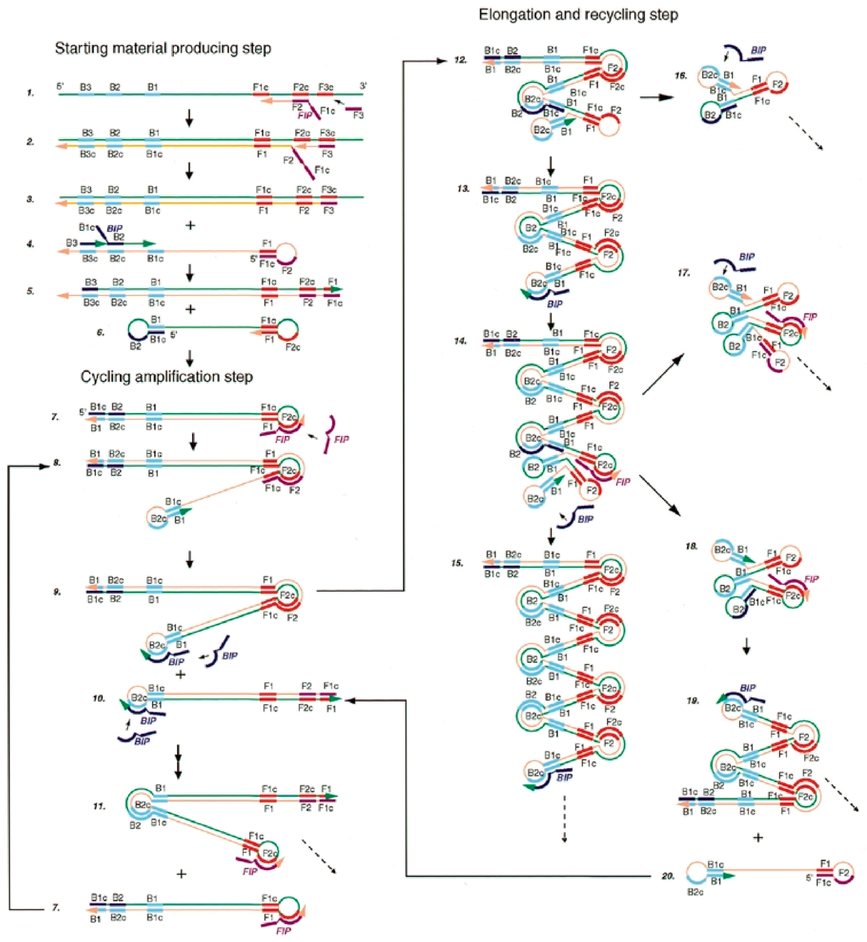
2.3. Loop-Mediated Isothermal Amplification (LAMP)

Loop-mediated isothermal amplification (LAMP) was first proposed in 2000 [21]. It is a very specific one-step reaction that uses four target-specific primers, namely an internal forward primer (FIP), an internal reverse primer (BIP), and two external primers (F3 and B3), to recognize six different sites present at the level of the DNA sequence to be amplified. FIP and BIP primers contain two functional sequences, one for priming extension in the first step and the other for self-priming in the second step of the reaction, corresponding to the sense and antisense sequences of the dsDNA target. As mentioned above, the reaction takes place in two phases and is catalyzed by Bst DNA polymerase: the first phase is the formation of the starting structure in which all four primers are used, whereas the second one is the phase of cyclic amplification, where only the internal primers are used. The complete process of LAMP isothermal amplification is shown in Figure 1.
In summary, FIP hybridizes to a specific region in the target DNA and initiates complementary strand synthesis. External primer F3 hybridizes to another specific area in the target DNA and initiates strand displacement DNA synthesis, releasing a FIP-linked complementary strand, which can form a looped-out structure at one end. The single-stranded DNA serves as a template for the synthesis of DNA by BIP. Subsequent B3-primed strand displacement produces a DNA ring structure. At this point, the cyclic amplification process is initiated by an internal primer that is complementary to the loop formed on the product and is continued by all internal primers in an alternating fashion, eventually resulting in 109 copies of the target sequence in less than 1 h. The products obtained are stem–loop DNA and structures with multiple loops, and these products can be detected by both real-time and end-point assays. The LAMP method has several advantages over PCR and other isothermal techniques. Compared to PCR, it provides easy handling and needs less expensive equipment [22], and it also maintains certain robustness to pH and temperature changes. Finally, untreated samples, such as urine and whole blood, which inhibit PCR, do not affect the sensitivity of the LAMP reaction [23]. Compared to other isothermal amplification procedures, it requires the use of fewer enzymes and does not require a process of pre-amplification ligation.
The LAMP assay is widely used for the detection of bacteria [24], viruses [24,25], and parasites [26,27].

2.4. Helicase-Dependent Amplification (HDA)

Helicase-dependent amplification, first described in 2004 [28], makes use of a helicase enzyme instead of thermal treatment to denaturate dsDNA. The process begins with the helicase unwinding of dsDNA to which forward and reverse primers can bind, followed by polymerase-mediated elongation. After elongation, helicase can act again on the synthesized dsDNA and the cycle asynchronously repeats. HDA has shown amplification kinetics similar to current PCR, but it is performed at a constant temperature of 60–65 °C for 60–120 min [29]. HDA can detect genomic eukaryotic and prokaryotic DNA and can be performed on the extracted and purified DNA sample or directly on a crude sample containing complex biofluids with a 10-fold lower sensitivity [28]. In summary, HDA is a simple, rapid, one-step isothermal amplification technique.

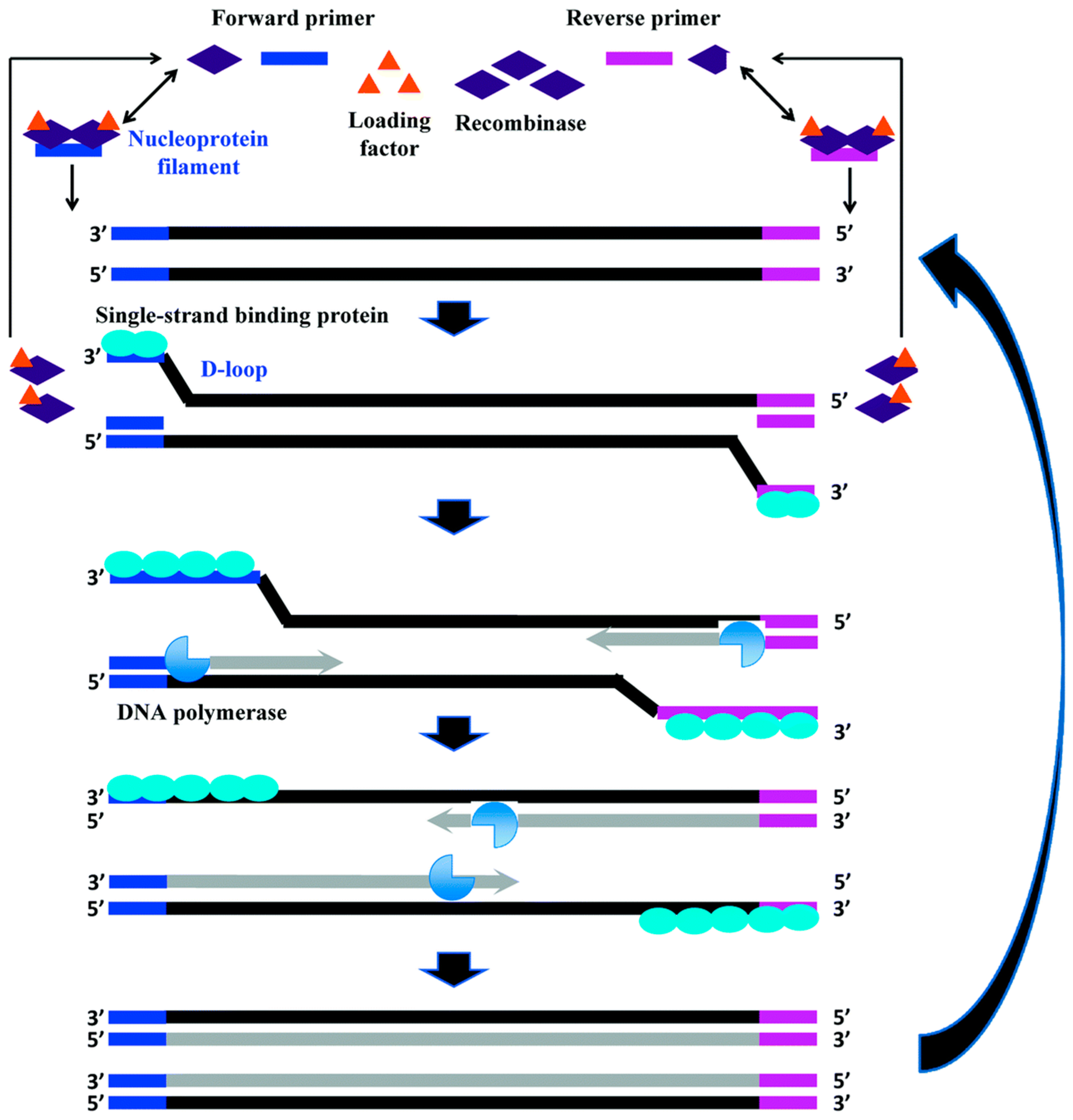
2.5. Recombinase Polymerase Amplification (RPA)

Recombinase polymerase amplification (RPA) was first introduced in 2006 [30], based on a probe amplification approach that exploits the specific action of recombinase and takes advantage of its simplicity, flexibility, low reaction temperature of 37 °C, and speed by obtaining results in 5–20 min [31]. The RPA reaction involves two primers, a recombinase enzyme, a single-stranded binding protein, and a DNA polymerase to displace the DNA strands. The recombinase aided by some proteins binds to oligonucleotide primers and forms recombination strands that scan the target dsDNA for homologous sequences; when these are found, a displacement-loop (D-loop) structure is formed as a product. The D-loop structure is stabilized by the single-stranded DNA-binding protein. At this point, replication is performed by a DNA polymerase that has strand dislocation activity necessary to extend the primer. The new DNA strands produced are used for another round of RPA, thus providing exponential amplification of the target. The entire amplification process is described in Figure 2.
The first application of RPA integration in a microfluidic device was first carried out in 2010 [32] for the detection of the antibiotic resistance gene mecA from S. aureus.
In conclusion, isothermal nucleic acid amplification techniques have innumerable strengths over classical PCR, and therefore they appear to be of considerable interest in the development of lab-on-a-chip systems for the determination of nucleic acids of clinical and environmental interest coupled with electrochemical detection devices.
To better understand the advantages of these systems, a series of examples are given in the following sections.
Table 1. Summary table of isothermal amplification techniques and their main characteristics.
MethodTemp (°C)Reaction Time (min)TargetPros and Cons vs. PCRReference
NASBA4190–120RNAPower-saving (41 °C); ideal for RNA target; not ideal for DNA target[17]
SDA37–4920–120ssDNA
RNA
Power-saving (37–50 °C); suitable for miRNA; sample preparation required[18]
LAMP60–6540–60ssDNA Highly specific; commercial kits available; not suitable for small targets; complex primers design[21]
HDA37–6060–120dsDNAUse of helicase enzyme to melt the dsDNA template; simplicity[28]
RPA25–425–20dsDNA
ssDNA
RNA
Lower temperature, 37 °C; fast amplification (20 min); commercial kits available; stringent reaction conditions[33]
NASBA, nucleic acid sequence–based amplification; SDA, strand displacement amplification; LAMP, loop-mediated isothermal amplification; HDA, helicase-dependent amplification; RPA, recombinase polymerase amplification; ssDNA, single-strand DNA; dsDNA, double-strand DNA.

2.6. Electrochemical Detection of Nucleic Acids

Electrochemical detection strategies are based on measurements of a current signal (amperometric and voltammetric biosensors), a change in the potential at the equilibrium (potentiometric sensors), or a difference in impedance. Several examples of papers in which this kind of detection strategies are discussed in more detail are reported in the literature; we kindly ask the reader to refer to detailed books [34,35] for the theory and setup of electrochemical techniques, as well as review papers for an extensive discussion on electrochemical principles employed in nucleic acid determination [10,11,36,37]. Briefly, voltammetric techniques rely on the monitoring of the current response obtained by scanning the applied potential in a specific range of values. By contrast, the technique based on the constant application of a fixed potential value is known as amperometry. In both of these techniques, the current is correlated to the concentration of the target analyte. Potentiometric sensors rely on the passive measurement of the potential between two electrodes, namely the indicator and the reference electrode, at the thermodynamic equilibrium conditions. The electrochemical impedance spectroscopy (EIS) technique is used to carry out impedance measurements. EIS is based on the observation of the system upon the perturbation of the cell with an alternating signal of small magnitude, overlaid on a constant DC bias potential, commonly as a function of the frequency. EIS is particularly used for monitoring the hybridization reaction in a label-free format [38]. Other electrochemical label-free methods for the detection of nucleic acids rely on the electrochemical reduction or oxidation of bases as a consequence of the hybridization between the capture probe and the target sequence. Both guanine and adenine show an oxidation peak, in a potential range from +1 V to +1.5 V (vs. Ag/AgCl) on most electrode materials, and are frequently used for this purpose. The changes in the electrical properties at the interface [39] can also be investigated for the development of a label-free-format assay.
Label-based methods possess many intriguing features since transduction can be obtained in a number of different ways and can be higher sensitive and selective than a label-free approach. The labels are typically redox-active molecules (such as methylene blue, ferrocene derivatives, Ru-complexes and others) [40,41] that intercalate within the DNA duplex, or bind preferentially to either single-strand (ss)DNA, RNA, or double-strand (ds)DNA, or are capable of monitoring a difference in the strand conformation and flexibility before and after the hybridization reaction.
Enzymes such as Horseradish Peroxidase (HRP) and Alkaline Phosphatase are commonly used as enzymatic labels. The most used electrochemical substrates for alkaline phosphatases are p-aminophenyl phosphate and 1-naphthyl phosphate [42,43]. In the case of HRP, tetramethylbenzidine (TMB) and hydroquinone (HQ), together with H2O2, are frequently used [44]. Enzyme-like catalysts and nanozymes (e.g., metal nanoparticles, metal oxides, G-quadruplets/hemin, and porphyrins) that mimic the activity of natural enzymes, while often maintaining higher stability, are also frequently used.
Recycling of redox-active species, such as ruthenium hexamine (Ru(NH3)63+/2+) and ferrocyanide-ferricyanide (Fe(CN)63−/4−), by implementing additional catalytic redox cycles, are being more frequently proposed as labelling strategies to further improve the assay’s sensitivity. Metal nanoparticles and liposomes have also been used as labels of the hybridization event [43,45].
Different materials can be used as transducers in electrochemical measurements such as metals (Au, Pt), metal oxides (TiO2), and carbon-based materials (e.g., glassy carbon, graphite, graphene, carbon nanotubes etc.) [46]. Nanostructuration of the sensor surface can dramatically improve the sensitivity of the assay [47]. Nanostructured electrode platforms provide an improved mass transport and electrochemically active surface area. Furthermore, nanostructuration could effectively accelerate electron transfers at the interface between electrodes and detection molecules, leading to a rapid and sensitive current response. In this sense, many examples of graphene-based sensors have now demonstrated excellent analytical performances compared to other common carbon electrodes [48].
Functional organic and inorganic conductive polymers have been tested to modify the electrode surface and improve the efficiency of the redox reaction.
Photoelectrochemistry (PEC) can be exploited as an electroanalytical technique for nucleic acid detection. The different strategies for label-free and label-based PEC assay formats are schematically discussed in Reference [49].

3. Isothermal Amplification Techniques Coupled with Electrochemical Detection of Microbial Agents for Medical, Food Safety, and Environmental Applications

Bacterial contamination of food and water is an issue of considerable interest because it is linked to human and animal infectious diseases, some of which may lead to death. About 91% of all foodborne diseases are due to bacterial proliferation mainly of Escherichia coli, Salmonella, and Campylobacter [50]. Such a rapid spread of bacterial agents is mainly due to factors related to globalization and makes it necessary to monitor pathogens more effectively. In addition to bacterial contamination of food and water, of considerable importance are bacterial infections in humans, such as Mycobacterium tuberculosis (Mtb) or the recent Covid-19 infection.
Current methods of microbial determination are based on laboratory techniques, such as cell culture, microscopic analysis, and biochemical assays [51]. These techniques require trained personnel and are time-consuming [52,53,54].
Portable and autonomous systems capable of performing accurate and rapid in situ analyses are highly requested. The combination of isothermal DNA amplification methods with microfluidic and electrochemical systems may address this need [52]. The integration of these techniques has resulted in biosensing platforms with interesting sensitivity and specificity for the detection of different analytes such as viruses, fungi, protozoa, bacteria, and microalgae. Many publications can be found in the literature [55,56,57,58,59,60] regarding bacterial detection by coupling isothermal amplification techniques and electrochemical signal detection. Table 2 shows different examples of isothermal amplification and electrochemical detection. Examples of chip-based systems are also reported.
As shown in Table 2, the coupling of isothermal amplification strategies with an electrochemical readout in a chip format allows a decrease in the complexity and a speed up of the analysis in comparison to what reported for other biosensing platforms developed for nucleic acid analysis.
As an example, Tsaloglou et al. [61] developed a fully-integrated device that combines RPA-based isothermal DNA amplification with the electrochemical detection of Mycobacterium smegmatis, a tuberculosis simulant. In this work, the RPA reagents, the genomic target DNA, and the redox label, [Ru(NH3)6]3+, were all included in a disposable paper-based strip that is then inserted inside the heating module on the device (Figure 3). With this scheme, the amplification reaction takes place for 20 min at 39 °C, automatically followed by the electrochemical readout. Particularly, when the dsDNA binds to the ruthenium complex, it leads to a drop in the cathodic current, proportional to the concentration of DNA in the sample. Square wave voltammetry (SWV), was chosen in this study because it is a sensitive, rapid, and compatible technique for a POC device. The developed device allows electrochemical data to be transferred directly to the “cloud” via USB or Bluetooth communication. With this strategy, the authors achieved a LOD equivalent to 11 CFU/mL of M. tuberculosis.
A different kind of disposable device was studied by Liu’s group [64], focusing on the point-of-care detection of lambda DNA in spiked saliva samples. In particular, they developed an integrated electronic-tube cap that featured a 3D-printed electrode equipped with a membrane capable of extracting, concentrating, and purifying the DNA sequences. The assembled cap with the captured DNA was then transferred to a reaction tube, where the LAMP reaction took place, using an external heating block at 63 °C. To monitor the electrochemical signal in a real-time and label-free method, a pH-sensitive layer (based on IrOx) was introduced on the 3D-printed electrode substrate, which was connected to the working electrode port of a multichannel potentiostat, while the carbon-paste-coating electrode was connected to the reference electrode port. A sensitivity of 100 DNA copies/mL within 30 min was achieved.
A similar strategy was also reported in Reference [62] for the real-time monitoring of the LAMP reaction. In particular, the researchers used methylene blue as a redox probe for the hybridization reaction. Methylene blue is easily intercalated into the double helix of E. coli O157:H7 and Salmonella DNAs. The real-time electrochemical LAMP device introduced in this study consists of a disposable eight-well PET electrochemical chip with three chemically plated gold electrodes on one side that can be fixed on a heating block that allows a constant temperature of 65 °C (for the LAMP amplification reaction). Differential pulse voltammetry (DPV) measurements exhibited LODs of 10 and 1 DNA copies for the two abovementioned species, respectively. Moreover, the platform was tested on turbid food samples, such as milk and juice.
In addition to the detection of bacteria, the detection of viruses is also becoming increasingly important, given the recent SARS-CoV-2 pandemic. COVID-19 is a viral respiratory infection that is part of severe acute respiratory syndromes [65]. The SARS-CoV-2 pandemic started in 2019 when the first case was identified in December in Wuhan, China, and is presumably due to animal-to-human transmission [66]. Therefore, in addition to antiviral treatment and the use of vaccines, early diagnosis is also necessary to reduce the virulence of the disease and prevent transmission [67]. The diagnostic tests that are in use today involve PCR, but, to simplify the analysis, a procedure without the need for thermocycling and based on isothermal amplification could be used. Interestingly, RPA combined with electrochemical devices has enabled the development of wearable devices. Indeed, the temperature used in RPA (i.e., 37 °C) allows these systems to operate by simple skin contact, hence eliminating the need for an external heating device. Several studies have already been reported in the literature focusing on devices that run RPA by exploiting body temperature. Among those, Kim and co-workers [63] designed an electrochemical biosensor based on microelectrode array chips consisting of five individual gold working electrodes for the determination of multiple target genes related to SARS-CoV-2 by DPV. This allows the measurement of different targets or the repeated measurement of the same target for at least three times, avoiding any misdiagnosis. Notably, the RPA isothermal amplification was carried out at human body temperature for 20 min and led to the hybridization of amplicons with thiol-modified primers immobilized on the working electrode. A decrease in current density can be noted as the amplicons accumulate, as displayed in Figure 4.
Similarly, Trinh and Lee [68] developed a flexible wearable microdevice based on polydimethylsiloxane (PDMS) that adheres to human skin. The system takes advantage of body heat to perform the isothermal amplification reaction. In detail, the polymer ratio of PDMS was optimized to improve the flexibility and adhesion of the device. Using body heat and storing the RPA reagents in PDMS molded microchambers, the authors managed to amplify 210 bp from Escherichia coli O157:H7 and 203 bp of SARS-CoV-2 DNA plasmid within 23 min, reaching LODs of approximately 500 pg/reaction and 600 fg/reaction (based on gel electrophoresis), respectively. The overall process is shown in Figure 5.
Indeed, the works presented pave the way for simplified DNA amplification in remote and resource-limited settings.
Environmental monitoring is another interesting field of applications of isothermal amplification and electrochemical detection of nucleic acids. As an example, monitoring harmful algal blooms (HABs) is important to maintain the safety and health of coastal environments. HABs are natural phenomena caused by the proliferation of microalgae, macroalgae, or cyanobacteria that not only have a detrimental impact on ecosystems but also on socio-economic activities, such as tourism and agriculture, and on human health. These HABs phenomena have increased most likely due to globalization and sudden climate change. Therefore, the identification and quantification of HAB species are of great importance. In a recent paper, an electrochemical assay, based on RPA amplification was developed. An eight-electrode array was used for monitoring the cathodic current due to the enzymatic reaction between the HRP-labeled report probe and the enzyme substrate (TMB and H2O2). A limit of detection of 9 pg/μL of Ostreopsis cf. ovata DNA (which corresponds to ~640 cells/L) was reported, with no interferences from two non-target Ostreopsis species (O. cf. siamensis and O. fattorussoi) [44].
In addition to HABs, microbial infections of different species of environmental importance can be easily monitored by coupling isothermal amplification and electrochemical detection. A major threat to shellfish aquaculture is ostreid herpesvirus 1 (OsHV-1), particularly to pacific oyster (Crassostrea gigas) production [69]. The electrochemical biosensor developed here is formed by using gold electrodes by chronoamperometric signal determination. The calibration curve resulted in an LOD of 207 target copies of Os-HV1 [70].
Many publications in the literature address the characterization of such species in a simple, fast, reliable, and sensitive manner, applying isothermal amplification techniques and electrochemical devices. However, unfortunately, no examples of coupling in a single chip are reported.

4. Isothermal Amplification Techniques Coupled to Electrochemical Detection of Cancer Biomarkers

Molecular diagnostics plays an important role in current oncology trials, trying to decrease the number of recurrences and improve cancer treatment.
A number of nucleic acid sequences are studied as cancer biomarkers and biomarker candidates, including DNA mutations, abnormally methylated DNA, the DNA of associated viruses, and circulating tumor DNA, as well as long non-coding RNA (lncRNA) and small RNAs [71]. The continuous discovery of the importance of new biomarkers demands new sensitive and specific technologies for their detection [72].
Lab-on-a-chip technologies present interesting features for point-of-care testing since their main advantages are portability, user-friendly interfaces, and low cost.
Oliveira et al. [73] have developed a new diagnostic system that combines microfluidics with droplet digital LAMP (ddLAMP) on a unique device that allows for the quantification of the target. The multilayer 3D chip reported in this work can be developed in less than 30 min, using manufacturing mechanisms that are cheap and exploit direct laser writing technology in polystyrene “Shrinky-dinks” sheets. Isothermal amplification and target DNA quantification are performed directly on-chip. The microfluidic device has been validated by amplification of targets ranging from 5 to 500,000 copies/reaction; moreover, the amplification was performed in a volume of 10 mL, reaching a detection limit of five copies/mL in 60 min. Specifically, this technique was used to quantify the cancer biomarker c-Myc, but it can be extended to many others. The c-Myc gene encodes for a transcription factor that is believed to regulate the expression of about 15% of all genes by binding to Enhancer Box sequences (also called E-boxes) and using histone acetyltransferase (HAT). Therefore, the c-Myc gene product acts as a protoncogene; its overexpression or mutation in an activating sense can transform Myc into an oncogene. In many malignancies, the Myc gene is constitutively mutated and expressed.
In their work, Moranova et al. [74] developed, optimized, and validated a diagnostic assay for the detection of two prostate cancer RNA markers, namely PSA RNA (as control) and PCA3 lncRNA. The authors monitored LAMP amplicons by using magnetic beads (MBs) which were then deposited on working electrodes by the use of magnets so as to concentrate them. Finally, the target amplicons were determined by chronoamperometry in the presence of the system HRP/H2O2/HQ, measuring the cathodic current corresponding to the HRP-catalyzed reduction of H2O2 mediated by HQ. Specifically, the MBs were deposited on the surface of the screen-printing carbon working electrode due to the presence of a magnetic holder placed under the electrode’s surface. The MBs were then coated with a hydroquinone solution, and the electrochemical current resulting from the enzymatic reaction of hydroquinone oxidation was measured chronoamperometrically at −0.3 V [75]. This diagnostic bioassay has been used not only to determine the presence of biomarkers in a selected group of prostate cancer cell lines, but also in the urine of men affected by primary non-metastatic prostate tumors.
Exosomes, membranous vesicles with a size ranging from 30 to 150 nm, are very important for intracellular communication and molecular exchange, being able to transport mRNA, microRNA, lncRNA, proteins, and portions of DNA [76,77]. Lin et al. [78] developed a “lab in a tube” system for the determination of exosomal nucleic acids. As shown in Figure 6, the system combines three key steps, all performed within the same vial: (i) isolation of exosomes, using immunomagnetic beads; (ii) lysis of exosomes to extract nucleic acids of interest; and (iii) amplification, using the LAMP technique, and determination of exosomal RNA of interest. This system was applied to the detection of two exosomal lncRNAs of gastric cancer (HOTTIP and lncRNA-GC1), with a dynamic detection from 300 to 10 ng/μL and a detection limit of 10 ng/μL. The system has also been shown to be applicable to human serum of lncRNA HOTTIP exosomal RNA.
LAMP was also used by Hashimoto et al. [79] for the development of a multiplex system based on a single-channel liquid-flow chip for the determination of microRNAs isolated from serum samples. The target sequences were initially elongated up to 100 base pairs by reverse transcription (RT) and elongation reactions (EL). Then the primers required for LAMP were adsorbed at five different positions on the surface of a liquid-flow channel. After that, the solution containing polymerase, dNTPs, and the products from RT and EL was entered into the DNA chip, and the primers came off slowly from their binding sites and spread in the liquid-flow channel. Then a LAMP reaction took place only where the primers were stopped. Target determination was performed by using the Ru(NH3)63+/2+ redox reaction. The coprecipitation of Ru(NH3)63+/2+ with the pyrophosphate produced by the LAMP amplification reaction causes an increase in the redox current. Specifically, the electrochemical determination was made by linear scanning voltammetry (LSV). The cathodic peak current and peak potential were recorded.
Izandi et al. [80] demonstrated the relationship between cervical cancer and human Papilloma virus (HPV) by using the LAMP isothermal amplification technique coupled with an electrochemical detection platform. Specifically, they focused on the detection of HPV16 and HPV18, using a protocol that involves (1) lysis of tumor cells, (2) amplification by the LAMP reaction of virus DNA, (3) hybridization of the products on modified magnetic beads with specific capture probes, and (4) monitoring of the amplification reaction by the electrochemical detection. Excellent agreement with classical PCR was achieved in the study.
In addition to the LAMP isothermal amplification technique, also RPA coupled to electrochemical devices can be used for HPV detection. In particular, Ma et al. [5] developed a method combining RPA-induced isothermal amplification, lateral flow dipstick (LFD), and reverse dot blot (RDB) in the rapid identification of HPV genotypes within 1 h by two complementary functional nucleic acid assays. From the conclusive data, the research team showed that LFD had a short run time and could detect viral HPV down to a minimum of 102 copies, and RBD identified genotyping at a level of 103 copies.
RPA was also used in another study by Li et al. [81] for the development of an enhanced electrochemical field (EFE)-based CRISPR biosensor to detect target DNA in solution in a homogenous phase. The EFE–CRISPR biosensor takes advantage of the diffusivity difference between electrochemical oligonucleotide probes and CRISPR-cleaved probes toward a negatively charged working electrode, allowing simple DNA detection. Thus, in the absence of the target DNA, the ssDNA-MB probe is repelled by the negative charge present at the electrode level during the pulsed differential voltammetry measurement (power range −0.6 to 1 V) because the DNA itself is negatively charged; this leads to a low current signal recorded at the working electrode level. By contrast, in the presence of the target DNA, the CRISPR-Cas12a protein is specifically activated and acts by nonspecifically cutting the ssDNA-MB, which releases the MB probes that, having a lower negative charge and higher perfusivity toward the electrode surface, leads to an increase in the electrochemical signal. In contrast to other existing CRISPR biosensors, the one developed by this research group enables the electrochemical detection of DNA on homogeneous samples, which not only facilitates sample adhesion to the electrode but also improves the efficiency of the reaction. The developed biosensor was then applied to the detection of human papillomavirus (HPV16) DNA in clinical samples by coupling it with the RPA isothermal amplification technique.

5. Patents Involving Isothermal Amplification in Electrochemical Chip-Based Platforms

Patent databases contain several inventions that combine the techniques mentioned in the previous sections. Hence, such strategies do not always remain mere proofs-of-concept or theoretical experiments but might find applications in various fields, including the clinical one. Patent no. CN110129188A (2019) [82] by Haiyun and co-workers discloses an integrated device capable of running an extraction of nucleic acids, an isothermal amplification following a LAMP reaction, and finally an electrochemical detection all inside its structure. The sample and all reagents are loaded dropwise inside a hole in the upper layer of the platform. A middle layer controls the continuous introduction of the samples, using a slidable magnetic mechanism. The system’s design provides a chamber for the amplification, which is carried out at 65 °C, for 60 min, on a hot plate. On the bottom layer, an electrode chip is located and is apt to record the DPV measurements.
Likewise, Shouhui and colleagues (patent no. CN104407036A, 2015) [83] invented a microfluidic device built on a printed electrode, enclosed by two layers of polydimethylsiloxane. Sample injection is performed through a network of microchannels and serpentines on the top layer, whereas the bottom layer creates two separate chambers for (i) DNA hybridization and amplification reactions and (ii) electrochemical detection. A micro-device integrated into the platform controls the temperature needed for the isothermal LAMP reaction.
Focusing on the detection of viruses, including SARS-CoV-2, the same inventors (CN213012858U, 2021) [84] have recently built a small microfluidic electrochemical sensor composed of three layers. In particular, the one on the bottom comprises a screen-printed three-electrode system, whereas the middle part encases the microfluidic channels incised on poly(methyl methacrylate), using a laser. Finally, a microfluidic covering layer closes the top. A reaction chamber is used for the LAMP reaction and the electrochemical detection. The latter is achieved by immobilizing viral nucleic acids by using capture probes fixed through S-Au bonds onto the surface of the working electrode.
Such examples highlight the possibility of producing small devices that integrate all the multiple steps required to analyze nucleic acids, but in a portable and simple design that eases POCT applications.

6. Conclusions

The aim of this review is to offer an overlook at alternative technologies for nucleic acid amplification coupled with an electrochemical readout strategy. Different examples of hyphenation of isothermal amplification methods to electrochemical platforms were summarized in this review, focusing the attention on lab-on-a-chip platforms. An insight into the application of isothermal amplification methods to wearable electrochemical sensing platforms was also shown, and a brief overview of examples of the most recent patents was given.
Indeed, as described in the present manuscript, the concept of electrochemical biosensing of nucleic acids coupled with isothermal amplification has already been proved for several applications, leading to the possible development of interesting point-of-care tests. Two decades ago, the World Health Organization Special Programme for Research and Training in Tropical Diseases (WHO/TDR) published a number of criteria that an ideal point-of-care test should meet at all levels of the healthcare system in the developing world to manage infectious diseases. These criteria are known by the acronym ASSURED (affordable, sensitive, specific, user-friendly, rapid, equipment-free, and delivered) and have become widely accepted as the benchmark for an ideal test that can be used at the point of care. Indeed, the analytical performance of any diagnostic test, independently from the final application, should be rigorously validated by the analysis in the selected real matrices and by comparison with standard tests. Validation refers to the limit of detection, the limit of quantification, the selectivity, the stability, and the robustness of the test, with the analysis performed in real matrices. Considering the analytical performances of the examples discussed in this review, we can conclude that, in many cases, the sensitivity reached in standard conditions could be compliant with the requirement of the different applications. The technology level is mature enough to afford the reliability in the real world, and advances in technology have made the ASSURED criteria for lab-on-a-chip platforms somewhat less daunting. However, information regarding the validation of the lab-on-a-chip platforms in real matrices is still needed.
Other criteria that, in the near future, lab-on-a-chip tests should meet are real-time connectivity, ease of specimen collection, and environmental friendliness. In particular, an important issue, as recently demonstrated by the Covid pandemic, is the environmental impact (i.e., environmental friendliness) of these platforms, and more future efforts should be devoted to the environmental impact of these tests. Biodegradable and recyclable materials should be used, when possible, for the support and encasings, as well as reagents used in the tests. Indeed, some of the examples of lab-on-a-chip devices are based on PDMS or other biocompatible materials. Nevertheless, more efforts should be devoted to this purpose.

Author Contributions

Conceptualization, I.P.; writing—original draft preparation, C.P. and P.S.S.; writing—review and editing, I.P., S.L., S.P. and A.B. (Anna Boddi); funding acquisition, I.P, A.B. (Andrea Bernini), S.P. and O.C. All authors have read and agreed to the published version of the manuscript.

Funding

This work was supported by the Tuscany Region within the framework of Bando Salute 2018 (Research project CUP no. D78D20000870002) and by Fondazione CR Firenze ID 2020.1662.

Data Availability Statement

Not applicable.

Conflicts of Interest

The authors declare no conflict of interest.

References

  1. Miranda-Castro, R.; Palchetti, I.; de-Los-Santos-Álvarez, N. The Translational Potential of Electrochemical DNA-Based Liquid Biopsy. Front. Chem. 2020, 8, 143. [Google Scholar] [CrossRef] [PubMed]
  2. Pillozzi, S.; Bernini, A.; Palchetti, I.; Crociani, O.; Antonuzzo, L.; Campanacci, D.; Scoccianti, G. Soft Tissue Sarcoma: An Insight on Biomarkers at Molecular, Metabolic and Cellular Level. Cancers 2021, 13, 3044. [Google Scholar] [CrossRef] [PubMed]
  3. Martinez-Dominguez, M.V.; Zottel, A.; Šamec, N.; Jovčevska, I.; Dincer, C.; Kahlert, U.D.; Nickel, A.-C. Current Technologies for RNA-Directed Liquid Diagnostics. Cancers 2021, 13, 5060. [Google Scholar] [CrossRef] [PubMed]
  4. Doyle, J.; Uthicke, S. Sensitive Environmental DNA Detection via Lateral Flow Assay (Dipstick)—A Case Study on Corallivorous Crown-of-Thorns Sea Star (Acanthaster cf. solaris) Detection. Environ. DNA 2021, 3, 323–342. [Google Scholar] [CrossRef]
  5. Ma, B.; Fang, J.; Lin, W.; Yu, X.; Sun, C.; Zhang, M. A Simple and Efficient Method for Potential Point-of-Care Diagnosis of Human Papillomavirus Genotypes: Combination of Isothermal Recombinase Polymerase Amplification with Lateral Flow Dipstick and Reverse Dot Blot. Anal. Bioanal. Chem. 2019, 411, 7451–7460. [Google Scholar] [CrossRef] [PubMed]
  6. Anik, Ü. 12-Electrochemical Medical Biosensors for POC Applications. In Medical Biosensors for Point of Care (POC) Applications; Narayan, R.J., Ed.; Woodhead Publishing: Sawston, UK, 2017; pp. 275–292. ISBN 978-0-08-100072-4. [Google Scholar]
  7. Wang, J. Electrochemical Biosensors: Towards Point-of-Care Cancer Diagnostics. Biosens. Bioelectron. 2006, 21, 1887–1892. [Google Scholar] [CrossRef]
  8. Schaad, N.W.; Frederick, R.D. Real-Time PCR and Its Application for Rapid Plant Disease Diagnostics. Can. J. Plant Pathol. 2002, 24, 250–258. [Google Scholar] [CrossRef]
  9. Diehl, F.; Li, M.; He, Y.; Kinzler, K.W.; Vogelstein, B.; Dressman, D. BEAMing: Single-Molecule PCR on Microparticles in Water-in-Oil Emulsions. Nat. Methods 2006, 3, 551–559. [Google Scholar] [CrossRef]
  10. Ferapontova, E.E. Basic Concepts and Recent Advances in Electrochemical Analysis of Nucleic Acids. Curr. Opin. Electrochem. 2017, 5, 218–225. [Google Scholar] [CrossRef]
  11. Labuda, J.; Brett, A.M.O.; Evtugyn, G.; Fojta, M.; Mascini, M.; Ozsoz, M.; Palchetti, I.; Paleček, E.; Wang, J. Electrochemical nucleic acid-based biosensors: Concepts, terms, and methodology (IUPAC Technical Report). Pure Appl. Chem. 2010, 82, 1161–1187. [Google Scholar] [CrossRef]
  12. Sfragano, P.S.; Pillozzi, S.; Palchetti, I. Electrochemical and PEC Platforms for MiRNA and Other Epigenetic Markers of Cancer Diseases: Recent Updates. Electrochem. Commun. 2021, 124, 106929. [Google Scholar] [CrossRef]
  13. Zhao, Y.; Chen, F.; Li, Q.; Wang, L.; Fan, C. Isothermal Amplification of Nucleic Acids. Chem. Rev. 2015, 115, 12491–12545. [Google Scholar] [CrossRef] [PubMed]
  14. Li, J.; Macdonald, J. Advances in Isothermal Amplification: Novel Strategies Inspired by Biological Processes. Biosens. Bioelectron. 2015, 64, 196–211. [Google Scholar] [CrossRef] [PubMed]
  15. Giuffrida, M.C.; Spoto, G. Integration of Isothermal Amplification Methods in Microfluidic Devices: Recent Advances. Biosens. Bioelectron. 2017, 90, 174–186. [Google Scholar] [CrossRef] [PubMed]
  16. Trinh, T.N.D.; Lee, N.Y. Advances in Nucleic Acid Amplification-Based Microfluidic Devices for Clinical Microbial Detection. Chemosensors 2022, 10, 123. [Google Scholar] [CrossRef]
  17. Compton, J. Nucleic Acid Sequence-Based Amplification. Nature 1991, 350, 91–92. [Google Scholar] [CrossRef]
  18. Walker, G.T.; Fraiser, M.S.; Schram, J.L.; Little, M.C.; Nadeau, J.G.; Malinowski, D.P. Strand Displacement Amplification—An Isothermal, in Vitro DNA Amplification Technique. Nucleic Acids Res. 1992, 20, 1691–1696. [Google Scholar] [CrossRef] [Green Version]
  19. Spargo, C.; Dean, C.; Nycz, C.; Walker, G. SDA Target Amplification. In Nonradioactive Analysis of Biomolecules; Springer: Berlin/Heidelberg, Germany, 2000; pp. 356–366. ISBN 978-3-540-64601-3. [Google Scholar]
  20. Deiman, B.; van Aarle, P.; Sillekens, P. Characteristics and Applications of Nucleic Acid Sequence-Based Amplification (NASBA). Mol. Biotechnol. 2002, 20, 163–179. [Google Scholar] [CrossRef]
  21. Notomi, T.; Okayama, H.; Masubuchi, H.; Yonekawa, T.; Watanabe, K.; Amino, N.; Hase, T. Loop-Mediated Isothermal Amplification of DNA. Nucleic Acids Res. 2000, 28, E63. [Google Scholar] [CrossRef] [Green Version]
  22. Keikha, M. LAMP Method as One of the Best Candidates for Replacing with PCR Method. Malays. J. Med. Sci. MJMS 2018, 25, 121–123. [Google Scholar] [CrossRef]
  23. Francois, P.; Tangomo, M.; Hibbs, J.; Bonetti, E.-J.; Boehme, C.C.; Notomi, T.; Perkins, M.D.; Schrenzel, J. Robustness of a Loop-Mediated Isothermal Amplification Reaction for Diagnostic Applications. FEMS Immunol. Med. Microbiol. 2011, 62, 41–48. [Google Scholar] [CrossRef] [PubMed] [Green Version]
  24. Seo, J.H.; Park, B.H.; Oh, S.J.; Choi, G.; Kim, D.H.; Lee, E.Y.; Seo, T.S. Development of a High-Throughput Centrifugal Loop-Mediated Isothermal Amplification Microdevice for Multiplex Foodborne Pathogenic Bacteria Detection. Sens. Actuators B Chem. 2017, 246, 146–153. [Google Scholar] [CrossRef]
  25. Ali, Z.; Aman, R.; Mahas, A.; Rao, G.S.; Tehseen, M.; Marsic, T.; Salunke, R.; Subudhi, A.K.; Hala, S.M.; Hamdan, S.M.; et al. ISCAN: An RT-LAMP-Coupled CRISPR-Cas12 Module for Rapid, Sensitive Detection of SARS-CoV-2. Virus Res. 2020, 288, 198129. [Google Scholar] [CrossRef]
  26. Kato, Y.; Yanagisawa, T.; Nakai, M.; Komatsu, K.; Inoue, M.N. Direct and Sensitive Detection of a Microsporidian Parasite of Bumblebees Using Loop-Mediated Isothermal Amplification (LAMP). Sci. Rep. 2020, 10, 1118. [Google Scholar] [CrossRef] [PubMed]
  27. Lannutti, L.; Mira, A.; Basualdo, M.; Rodriguez, G.; Erler, S.; Silva, V.; Gisder, S.; Genersch, E.; Florin-Christensen, M.; Schnittger, L. Development of a Loop-Mediated Isothermal Amplification (LAMP) and a Direct LAMP for the Specific Detection of Nosema Ceranae, a Parasite of Honey Bees. Parasitol. Res. 2020, 119, 3947–3956. [Google Scholar] [CrossRef]
  28. Vincent, M.; Xu, Y.; Kong, H. Helicase-Dependent Isothermal DNA Amplification. EMBO Rep. 2004, 5, 795–800. [Google Scholar] [CrossRef]
  29. Deng, H.; Gao, Z. Bioanalytical Applications of Isothermal Nucleic Acid Amplification Techniques. Anal. Chim. Acta 2015, 853, 30–45. [Google Scholar] [CrossRef]
  30. Li, J.; Macdonald, J.; Stetten, F. von Review: A Comprehensive Summary of a Decade Development of the Recombinase Polymerase Amplification. Analyst 2019, 144, 31–67. [Google Scholar] [CrossRef] [Green Version]
  31. Daher, R.K.; Stewart, G.; Boissinot, M.; Bergeron, M.G. Recombinase Polymerase Amplification for Diagnostic Applications. Clin. Chem. 2016, 62, 947–958. [Google Scholar] [CrossRef]
  32. Lutz, S.; Weber, P.; Focke, M.; Faltin, B.; Hoffmann, J.; Müller, C.; Mark, D.; Roth, G.; Munday, P.; Armes, N.; et al. Microfluidic Lab-on-a-Foil for Nucleic Acid Analysis Based on Isothermal Recombinase Polymerase Amplification (RPA). Lab. Chip 2010, 10, 887–893. [Google Scholar] [CrossRef]
  33. Piepenburg, O.; Williams, C.H.; Stemple, D.L.; Armes, N.A. DNA Detection Using Recombination Proteins. PLoS Biol. 2006, 4, e204. [Google Scholar] [CrossRef]
  34. Brett, C.M.A.; Brett, A.M.O. Electrochemistry: Principles, Methods, and Applications; Oxford Science Publications; Oxford University Press: New York, NY, USA, 1993; ISBN 978-0-19-855389-2. [Google Scholar]
  35. Compton, R.G.; Banks, C.E. Understanding Voltammetry, 2nd ed.; Imperial College Press: London, UK, 2010; ISBN 978-1-84816-585-4. [Google Scholar]
  36. Ronkainen, N.J.; Halsall, H.B.; Heineman, W.R. Electrochemical Biosensors. Chem. Soc. Rev. 2010, 39, 1747–1763. [Google Scholar] [CrossRef] [PubMed]
  37. Palchetti, I.; Laschi, S.; Marrazza, G.; Mascini, M. Electrochemical Imaging of Localized Sandwich DNA Hybridization Using Scanning Electrochemical Microscopy. Anal. Chem. 2007, 79, 7206–7213. [Google Scholar] [CrossRef]
  38. Voccia, D.; Sosnowska, M.; Bettazzi, F.; Roscigno, G.; Fratini, E.; De Franciscis, V.; Condorelli, G.; Chitta, R.; D’Souza, F.; Kutner, W.; et al. Direct Determination of Small RNAs Using a Biotinylated Polythiophene Impedimetric Genosensor. Biosens. Bioelectron. 2017, 87, 1012–1019. [Google Scholar] [CrossRef] [PubMed] [Green Version]
  39. Odenthal, K.J.; Gooding, J.J. An Introduction to Electrochemical DNAbiosensors. Analyst 2007, 132, 603–610. [Google Scholar] [CrossRef] [PubMed]
  40. Meric, B.; Kerman, K.; Marrazza, G.; Palchetti, I.; Mascini, M.; Ozsoz, M. Disposable Genosensor, a New Tool for the Detection of NOS-Terminator, a Genetic Element Present in GMOs. Food Control 2004, 15, 621–626. [Google Scholar] [CrossRef]
  41. Lai, R.Y.; Lagally, E.T.; Lee, S.-H.; Soh, H.T.; Plaxco, K.W.; Heeger, A.J. Rapid, Sequence-Specific Detection of Unpurified PCR Amplicons via a Reusable, Electrochemical Sensor. Proc. Natl. Acad. Sci. USA 2006, 103, 4017–4021. [Google Scholar] [CrossRef] [Green Version]
  42. Laschi, S.; Miranda-Castro, R.; González-Fernández, E.; Palchetti, I.; Reymond, F.; Rossier, J.S.; Marrazza, G. A New Gravity-Driven Microfluidic-Based Electrochemical Assay Coupled to Magnetic Beads for Nucleic Acid Detection. Electrophoresis 2010, 31, 3727–3736. [Google Scholar] [CrossRef]
  43. Voccia, D.; Bettazzi, F.; Fratini, E.; Berti, D.; Palchetti, I. Improving Impedimetric Nucleic Acid Detection by Using Enzyme-Decorated Liposomes and Nanostructured Screen-Printed Electrodes. Anal. Bioanal. Chem. 2016, 408, 7271–7281. [Google Scholar] [CrossRef]
  44. Toldrà, A.; Alcaraz, C.; Diogène, J.; O’Sullivan, C.K.; Campàs, M. Detection of Ostreopsis cf. ovata in Environmental Samples Using an Electrochemical DNA-Based Biosensor. Sci. Total Environ. 2019, 689, 655–661. [Google Scholar] [CrossRef]
  45. Wang, J.; Xu, D.; Kawde, A.-N.; Polsky, R. Metal Nanoparticle-Based Electrochemical Stripping Potentiometric Detection of DNA Hybridization. Anal. Chem. 2001, 73, 5576–5581. [Google Scholar] [CrossRef] [PubMed]
  46. Berti, F.; Lozzi, L.; Palchetti, I.; Santucci, S.; Marrazza, G. Aligned Carbon Nanotube Thin Films for DNA Electrochemical Sensing. Electrochim. Acta 2009, 54, 5035–5041. [Google Scholar] [CrossRef]
  47. Campuzano, S.; Yáñez-Sedeño, P.; Pingarrón, J.M. Tailoring Sensitivity in Electrochemical Nucleic Acid Hybridization Biosensing: Role of Surface Chemistry and Labeling Strategies. ChemElectroChem 2019, 6, 60–72. [Google Scholar] [CrossRef]
  48. Ingrosso, C.; Corricelli, M.; Bettazzi, F.; Konstantinidou, E.; Bianco, G.V.; Depalo, N.; Striccoli, M.; Agostiano, A.; Curri, M.L.; Palchetti, I. Au Nanoparticle in Situ Decorated RGO Nanocomposites for Highly Sensitive Electrochemical Genosensors. J. Mater. Chem. B 2019, 7, 768–777. [Google Scholar] [CrossRef] [PubMed]
  49. Bettazzi, F.; Palchetti, I. Photoelectrochemical Genosensors for the Determination of Nucleic Acid Cancer Biomarkers. Curr. Opin. Electrochem. 2018, 12, 51–59. [Google Scholar] [CrossRef]
  50. Holmberg, S.D.; Blake, P.A. Staphylococcal Food Poisoning in the United States: New Facts and Old Misconceptions. JAMA 1984, 251, 487–489. [Google Scholar] [CrossRef]
  51. Váradi, L.; Luo, J.L.; Hibbs, D.E.; Perry, J.D.; Anderson, R.J.; Orenga, S.; Groundwater, P.W. Methods for the Detection and Identification of Pathogenic Bacteria: Past, Present, and Future. Chem. Soc. Rev. 2017, 46, 4818–4832. [Google Scholar] [CrossRef]
  52. Toldrà, A.; O’Sullivan, C.K.; Campàs, M. Detecting Harmful Algal Blooms with Isothermal Molecular Strategies. Trends Biotechnol. 2019, 37, 1278–1281. [Google Scholar] [CrossRef]
  53. Toldrà, A.; O’Sullivan, C.K.; Diogène, J.; Campàs, M. Detecting Harmful Algal Blooms with Nucleic Acid Amplification-Based Biotechnological Tools. Sci. Total Environ. 2020, 749, 141605. [Google Scholar] [CrossRef]
  54. Reynoso, E.C.; Laschi, S.; Palchetti, I.; Torres, E. Advances in Antimicrobial Resistance Monitoring Using Sensors and Biosensors: A Review. Chemosensors 2021, 9, 232. [Google Scholar] [CrossRef]
  55. Jaroenram, W.; Kampeera, J.; Arunrut, N.; Karuwan, C.; Sappat, A.; Khumwan, P.; Jaitrong, S.; Boonnak, K.; Prammananan, T.; Chaiprasert, A.; et al. Graphene-Based Electrochemical Genosensor Incorporated Loop-Mediated Isothermal Amplification for Rapid on-Site Detection of Mycobacterium Tuberculosis. J. Pharm. Biomed. Anal. 2020, 186, 113333. [Google Scholar] [CrossRef] [PubMed]
  56. Ng, B.Y.C.; Xiao, W.; West, N.; Wee, E.; Wang, Y.; Trau, M. Rapid, Single-Cell Electrochemical Detection of Mycobacterium Tuberculosis Using Colloidal Gold Nanoparticles. Anal. Chem. 2015, 87, 10613–10618. [Google Scholar] [CrossRef] [PubMed]
  57. Torres-Chavolla, E.; Alocilja, E.C. Nanoparticle Based DNA Biosensor for Tuberculosis Detection Using Thermophilic Helicase-Dependent Isothermal Amplification. Biosens. Bioelectron. 2011, 26, 4614–4618. [Google Scholar] [CrossRef]
  58. Yang, A.K.L.; Lu, H.; Wu, S.Y.; Kwok, H.C.; Ho, H.P.; Yu, S.; Cheung, A.K.L.; Kong, S.K. Detection of Panton-Valentine Leukocidin DNA from Methicillin-Resistant Staphylococcus Aureus by Resistive Pulse Sensing and Loop-Mediated Isothermal Amplification with Gold Nanoparticles. Anal. Chim. Acta 2013, 782, 46–53. [Google Scholar] [CrossRef] [PubMed]
  59. Cai, R.; Zhang, Z.; Chen, H.; Tian, Y.; Zhou, N. A Versatile Signal-on Electrochemical Biosensor for Staphylococcus Aureus Based on Triple-Helix Molecular Switch. Sens. Actuators B Chem. 2021, 326, 128842. [Google Scholar] [CrossRef]
  60. Barreda-García, S.; Miranda-Castro, R.; de-Los-Santos-Álvarez, N.; Miranda-Ordieres, A.J.; Lobo-Castañón, M.J. Helicase-Dependent Isothermal Amplification: A Novel Tool in the Development of Molecular-Based Analytical Systems for Rapid Pathogen Detection. Anal. Bioanal. Chem. 2018, 410, 679–693. [Google Scholar] [CrossRef]
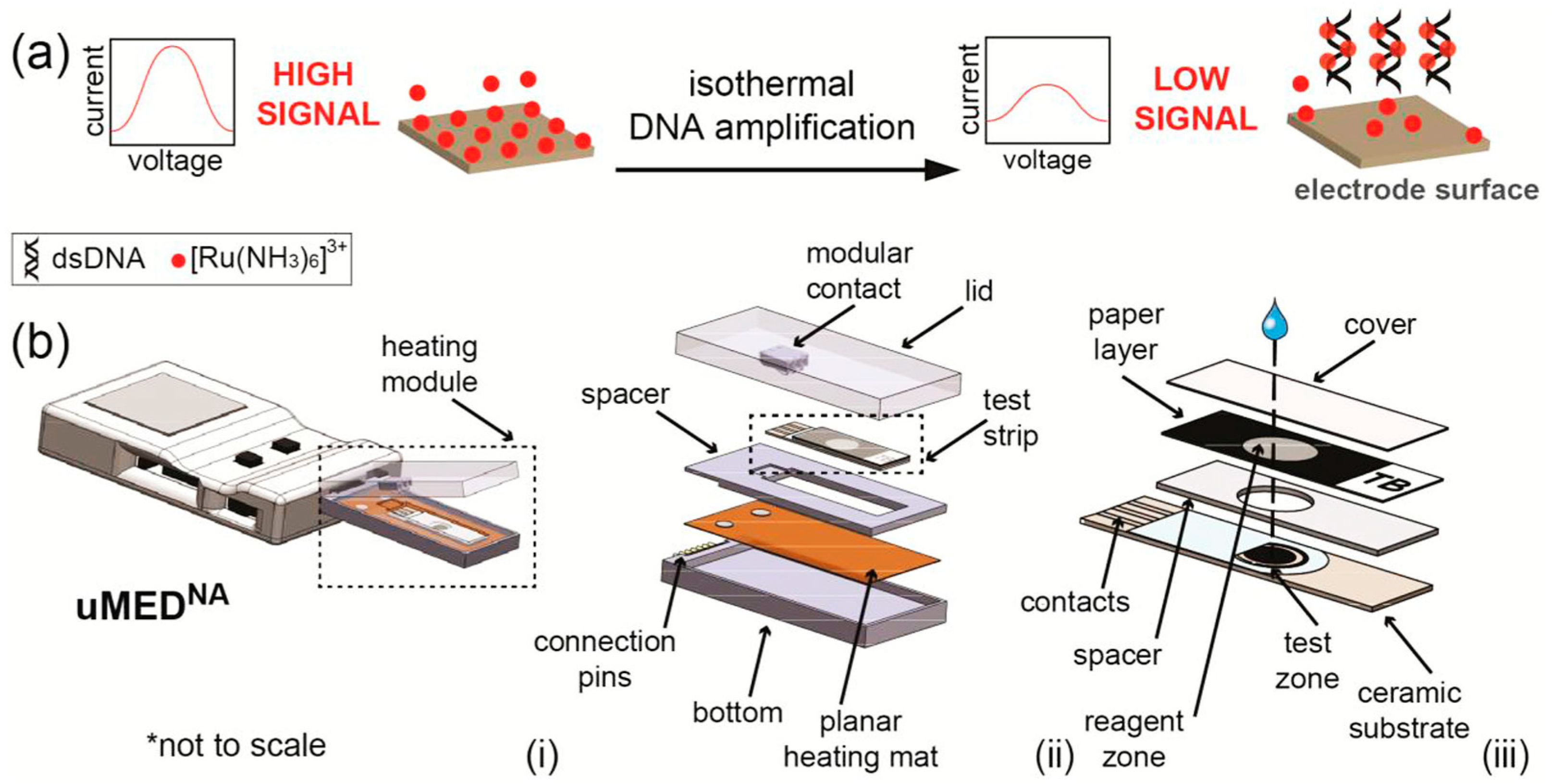
  61. Tsaloglou, M.-N.; Nemiroski, A.; Camci-Unal, G.; Christodouleas, D.C.; Murray, L.P.; Connelly, J.T.; Whitesides, G.M. Handheld Isothermal Amplification and Electrochemical Detection of DNA in Resource-Limited Settings. Anal. Biochem. 2018, 543, 116–121. [Google Scholar] [CrossRef]
  62. Huang, T.-T.; Liu, S.-C.; Huang, C.-H.; Lin, C.-J.; Huang, S.-T. An Integrated Real-Time Electrochemical LAMP Device for Pathogenic Bacteria Detection in Food. Electroanalysis 2018, 30, 2397–2404. [Google Scholar] [CrossRef]
  63. Kim, H.E.; Schuck, A.; Lee, S.H.; Lee, Y.; Kang, M.; Kim, Y.-S. Sensitive Electrochemical Biosensor Combined with Isothermal Amplification for Point-of-Care COVID-19 Tests. Biosens. Bioelectron. 2021, 182, 113168. [Google Scholar] [CrossRef]
  64. Xu, Z.; Yin, K.; Ding, X.; Li, Z.; Sun, X.; Li, B.; Lalla, R.V.; Gross, R.; Liu, C. An Integrated E-Tube Cap for Sample Preparation, Isothermal Amplification and Label-Free Electrochemical Detection of DNA. Biosens. Bioelectron. 2021, 186, 113306. [Google Scholar] [CrossRef]
  65. Gorbalenya, A.E.; Baker, S.C.; Baric, R.S.; de Groot, R.J.; Drosten, C.; Gulyaeva, A.A.; Haagmans, B.L.; Lauber, C.; Leontovich, A.M.; Neuman, B.W.; et al. The Species Severe Acute Respiratory Syndrome-Related Coronavirus: Classifying 2019-NCoV and Naming It SARS-CoV-2. Nat. Microbiol. 2020, 5, 536–544. [Google Scholar] [CrossRef] [Green Version]
  66. Andersen, K.G.; Rambaut, A.; Lipkin, W.I.; Holmes, E.C.; Garry, R.F. The Proximal Origin of SARS-CoV-2. Nat. Med. 2020, 26, 450–452. [Google Scholar] [CrossRef] [PubMed] [Green Version]
  67. Miripour, Z.S.; Sarrami-Forooshani, R.; Sanati, H.; Makarem, J.; Taheri, M.S.; Shojaeian, F.; Eskafi, A.H.; Abbasvandi, F.; Namdar, N.; Ghafari, H.; et al. Real-Time Diagnosis of Reactive Oxygen Species (ROS) in Fresh Sputum by Electrochemical Tracing; Correlation between COVID-19 and Viral-Induced ROS in Lung/Respiratory Epithelium during This Pandemic. Biosens. Bioelectron. 2020, 165, 112435. [Google Scholar] [CrossRef] [PubMed]
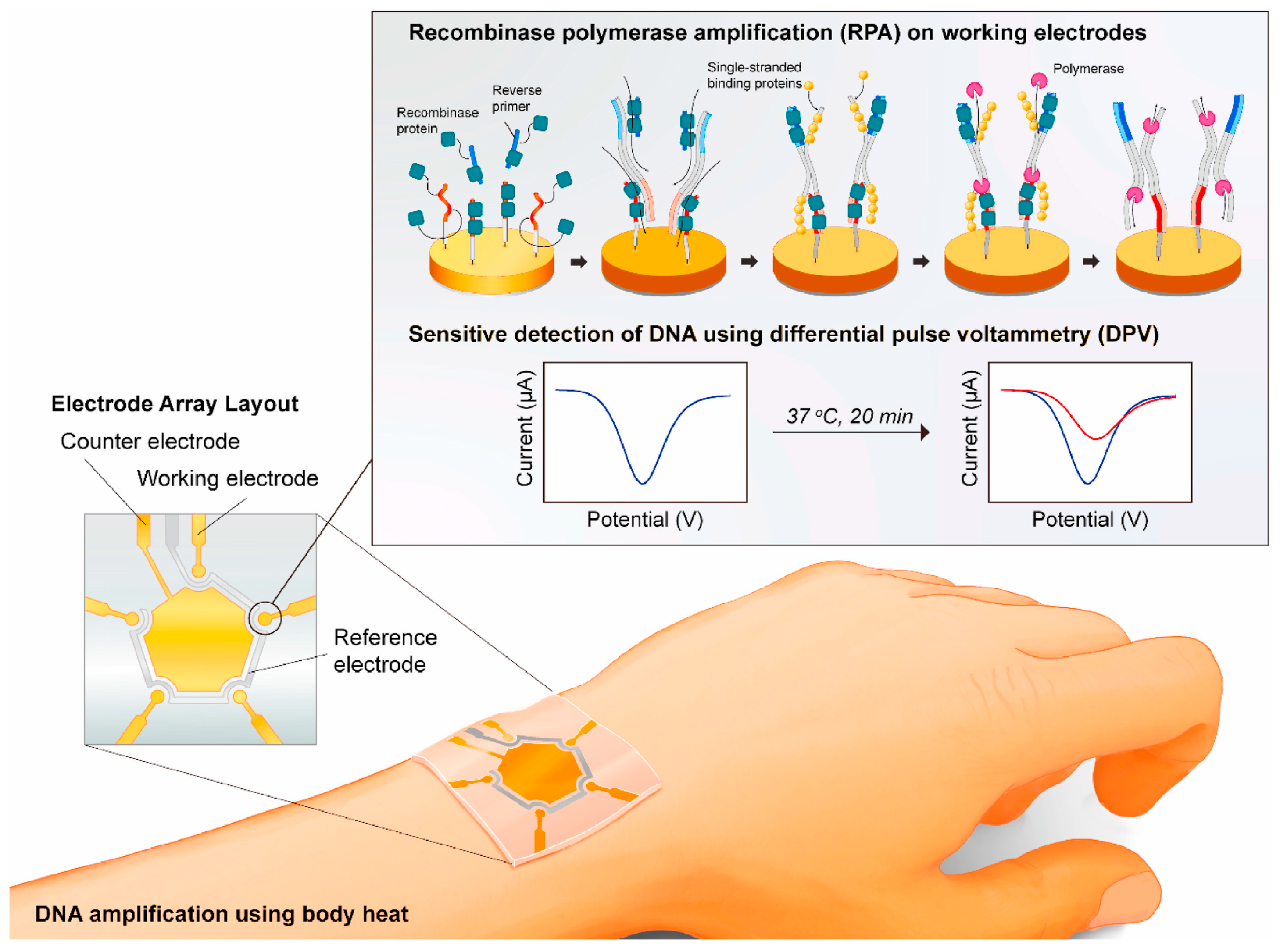
  68. Trinh, K.T.L.; Lee, N.Y. Fabrication of Wearable PDMS Device for Rapid Detection of Nucleic Acids via Recombinase Polymerase Amplification Operated by Human Body Heat. Biosensors 2022, 12, 72. [Google Scholar] [CrossRef] [PubMed]
  69. Rodgers, C.; Arzul, I.; Carrasco, N.; Furones Nozal, D. A Literature Review as an Aid to Identify Strategies for Mitigating Ostreid Herpesvirus 1 in Crassostrea Gigas Hatchery and Nursery Systems. Rev. Aquac. 2019, 11, 565–585. [Google Scholar] [CrossRef] [Green Version]
  70. Toldrà, A.; Furones, M.D.; O’Sullivan, C.K.; Campàs, M. Detection of Isothermally Amplified Ostreid Herpesvirus 1 DNA in Pacific Oyster (Crassostrea gigas) Using a Miniaturised Electrochemical Biosensor. Talanta 2020, 207, 120308. [Google Scholar] [CrossRef]
  71. Martisova, A.; Holcakova, J.; Izadi, N.; Sebuyoya, R.; Hrstka, R.; Bartosik, M. DNA Methylation in Solid Tumors: Functions and Methods of Detection. Int. J. Mol. Sci. 2021, 22, 4247. [Google Scholar] [CrossRef]
  72. Zhang, P.; Zhou, X.; He, M.; Shang, Y.; Tetlow, A.L.; Godwin, A.K.; Zeng, Y. Ultrasensitive Detection of Circulating Exosomes with a 3D-Nanopatterned Microfluidic Chip. Nat. Biomed. Eng. 2019, 3, 438–451. [Google Scholar] [CrossRef]
  73. Oliveira, B.; Veigas, B.; Fernandes, A.R.; Águas, H.; Martins, R.; Fortunato, E.; Baptista, P.V. Fast Prototyping Microfluidics: Integrating Droplet Digital Lamp for Absolute Quantification of Cancer Biomarkers. Sensors 2020, 20, 1624. [Google Scholar] [CrossRef] [Green Version]
  74. Moranova, L.; Stanik, M.; Hrstka, R.; Campuzano, S.; Bartosik, M. Electrochemical LAMP-Based Assay for Detection of RNA Biomarkers in Prostate Cancer. Talanta 2022, 238, 123064. [Google Scholar] [CrossRef]
  75. Camacho, C.; Matías, J.C.; Chico, B.; Cao, R.; Gómez, L.; Simpson, B.K.; Villalonga, R. Amperometric Biosensor for Hydrogen Peroxide, Using Supramolecularly Immobilized Horseradish Peroxidase on the β-Cyclodextrin-Coated Gold Electrode. Electroanalysis 2007, 19, 2538–2542. [Google Scholar] [CrossRef]
  76. Ma, C.; Jiang, F.; Ma, Y.; Wang, J.; Li, H.; Zhang, J. Isolation and Detection Technologies of Extracellular Vesicles and Application on Cancer Diagnostic. Dose-Response Publ. Int. Hormesis Soc. 2019, 17, 1559325819891004. [Google Scholar] [CrossRef] [Green Version]
  77. Dilsiz, N. Role of Exosomes and Exosomal MicroRNAs in Cancer. Future Sci. OA 2020, 6, FSO465. [Google Scholar] [CrossRef] [PubMed] [Green Version]
  78. Lin, Q.; Huang, Z.; Ye, X.; Yang, B.; Fang, X.; Liu, B.; Chen, H.; Kong, J. Lab in a Tube: Isolation, Extraction, and Isothermal Amplification Detection of Exosomal Long Noncoding RNA of Gastric Cancer. Talanta 2021, 225, 122090. [Google Scholar] [CrossRef]
  79. Hashimoto, K.; Inada, M.; Ito, K. Multiplex Real-Time Loop-Mediated Isothermal Amplification Using an Electrochemical DNA Chip Consisting of a Single Liquid-Flow Channel. Anal. Chem. 2019, 91, 3227–3232. [Google Scholar] [CrossRef]
  80. Izadi, N.; Sebuyoya, R.; Moranova, L.; Hrstka, R.; Anton, M.; Bartosik, M. Electrochemical Bioassay Coupled to LAMP Reaction for Determination of High-Risk HPV Infection in Crude Lysates. Anal. Chim. Acta 2021, 1187, 339145. [Google Scholar] [CrossRef]
  81. Li, Z.; Ding, X.; Yin, K.; Xu, Z.; Cooper, K.; Liu, C. Electric Field-Enhanced Electrochemical CRISPR Biosensor for DNA Detection. Biosens. Bioelectron. 2021, 192, 113498. [Google Scholar] [CrossRef]
  82. Liu, H.; He, T.; Gao, Y.; Yu, J. Novel Integrated Type Nucleic Acid Detecting Device. Available online: https://worldwide.espacenet.com/patent/search/family/067580275/publication/CN110129188A?q=CN110129188A (accessed on 13 July 2022).
  83. Chen, S.; Xu, Y.; Yuan, X. Preparation and Application of Electrochemical Micro-Fluidic Device for Nucleic Acid Isothermal Amplification. 2015. Available online: https://worldwide.espacenet.com/patent/search/family/052644683/publication/CN104407036A?q=pn%3DCN104407036A (accessed on 13 July 2022).
  84. Chen, S.; Lai, J. Micro-Fluidic Electrochemical Sensor Capable of Rapidly Detecting Viruses. 2021. Available online: https://worldwide.espacenet.com/patent/search/family/075442249/publication/CN213012858U?q=pn%3DCN213012858U (accessed on 13 July 2022).
Figure 1. Representation of the LAMP reaction. F2c and B2 are the sequences inside both ends of the DNA target region. The inner sequences from the ends of F2c and B2 are designated F1c and B1 The internal forward primer (FIP) contains the sequence F1c, a spacer, and the sequence (F2) complementary to F2c. The internal reverse primer (BIP) contains the sequence (B1c) complementary to B1, a spacer, and the sequence B2. The two outer primers consist of B3 and the sequence (F3) complementary to F3c, respectively. In order to explain the reaction, the DNA sample containing the target sequence and the four primers is heat denatured and rapidly cooled on ice. The LAMP reaction is then initiated by the addition of the Bst DNA polymerase large fragment and carried out at 65 °C for 1 h. The figure shows the process that starts with the primer. However, DNA synthesis can also take place by starting with the BIP primer. Reprinted with permission from Reference [21].
Chemosensors 10 00278 g001
Figure 2. Mechanism of the RPA reaction. The reaction begins with the binding of the recombinase (T4 UvsX) to the primers with the support of the loading factor (T4 UvsY). This produces a nucleoprotein fragment that searches for the homologous sequence in the double-stranded DNA. Once homology is found, the complex invades the duplex DNA, producing a D-loop structure to initiate a strand exchange reaction, while the unwound filament is stabilized by the single-stranded binding proteins (T4 gp32). The recombinase (Bsu or Sau) comes off from the nucleoprotein filament once the strand exchange is performed and will be accessible for the next pair of primers. Then the DNA polymerase elongates from the 3′ end of the primers. As the polymerization continues, the two parental strands begin to separate and form two duplexes, and then the entire process repeats. Reprinted with permission from Reference [30].
Chemosensors 10 00278 g002
Figure 3. (a) Representation of the RPA methods. (b) Representation of the portable assay for DNA amplification (double-strand DNA) and detection: (i) uMEDNA, (ii) view of module, and contacts to test strip, and (iii) view of the disposable paper-based strip. Reprinted with permission from Reference [61].
Chemosensors 10 00278 g003
Figure 4. Electrochemical biosensor coupled with recombinase polymerase amplification (RPA). The RPA reaction takes place on the working electrodes, and the determination of amplicons is accomplished by DPV. Reprinted with permission from Reference [63].
Chemosensors 10 00278 g004
Figure 5. (a) Illustration of the fabrication of a wearable PDMS microdevice. (b) Steps involved in the use of such device to run RPA using body heat. Reprinted from Reference [68].
Chemosensors 10 00278 g005
Figure 6. Schematic view of the integrated “lab on tube” system for the detection of exosomal nucleic acids. Reprinted with permission from Reference [78].
Chemosensors 10 00278 g006
Table 2. Some examples of isothermal amplification-based strategies for the electrochemical detection of microbial contamination.
Isothermal Amplification
Method
Limit of DetectionLab-on-a-Chip/Wearable PlatformReal SamplesElectrochemical DetectionType of ElectrodeAnalysis TimeReferences
Mycobacterium tuberculosis
LAMP40 CFU/equivalence-Sputum(CV)SPGE <65 min[55]
RPA1 CFU/mL-None(DPV)SPCE -[56]
HDA0.5 aM-None(DPV)SPCE From 2 to 8 h[57]
RPA11 CFU/mLYesNone(SWV)SPCE -[61]
Staphylucoccus aureus
LAMP530 copies of target-NoneResistive pulse sensingSPCE-[58]
SDA8 CFU/mL-Lake water and honeyDPVSPCE -[59]
Salmonella spp./E. coli
LAMP 10 and 1 Bacteria DNA copiesYesNoneDPVGold electrodesLess than 30 min[62]
SARS-CoV-2
RPA3.925 fg/μLYesNoneDPVGold ElectrodeLess than 20 min[63]
Lambda DNA
LAMP100 DNA copies/mLYesSaliva(EIS)pH-sensitive electrode30 min[64]
SDA, strand displacement amplification; LAMP, loop-mediated isothermal amplification; HDA, helicase-dependent amplification; RPA, recombinase polymerase amplification; CV, cyclic voltammetry; DPV, differential pulse voltammetry; SWV, square wave voltammetry; EIS, electrochemical impedance spectroscopy; SPCE, screen-printed carbon electrodes; SPGE, screen-printed gold electrode; CFU, colony-forming unit.
Publisher’s Note: MDPI stays neutral with regard to jurisdictional claims in published maps and institutional affiliations.

