Article

A Sensitive Co-MOF/CNTs/SiO2 Composite Based Electrode for Determination of Gallic Acid

Department of Biosystems Engineering, Zhejiang University, 866 Yuhangtang Road, Hangzhou 310058, China
*
Author to whom correspondence should be addressed.
Chemosensors 2022, 10(11), 443; https://doi.org/10.3390/chemosensors10110443
Submission received: 5 September 2022 / Revised: 16 October 2022 / Accepted: 21 October 2022 / Published: 26 October 2022
(This article belongs to the Section Electrochemical Devices and Sensors)

Abstract

:
A novel Co-based organic frameworks/carbon nanotubes/silicon dioxide (Co-MOF/CNTs/SiO2)-modified Au electrode was fabricated and taken as a platform for gallic acid (GA) detection. The composite combined the advantages of Co-MOF, CNTs and SiO2, and higher electrochemical response of Co-MOF/CNTs/SiO2-modified electrode indicated that the composite material exhibited satisfied the catalytic activity towards GA. Moreover, the electrochemical oxidation process of GA was deeply investigated on the surface of electrode based on computational investigations. Hirshfeld charges and condensed Fukui functions of each atom in GA were calculated. Besides, the catalysis of Co-MOF to GA was further investigated based on density functional theory. The quantitative determination of GA was carried out and showed a linear range between 0.05–200 μM, with low limit of detection. The sensitivity value of the self-assembled electrochemical sensor was calculated to be 593.33 μA cm−2 mM−1, and the selectivity, reproducibility and stability of the gallic acid sensor were also confirmed in the study.

1. Introduction

Gallic acid (GA; 3,4,5-triphydroxyl-benzoic acid) is a kind of polyphenolic compound found in nature, which is widespread in plants, fruits and vegetables [1,2,3]. GA has three hydroxyl groups (-OH) and the property of allowing straightforward copolymerization through its carboxylic group (-COOH), which makes it act as a strong antioxidant [4,5]. Besides, GA also exhibits other biochemical merits, such as antibiotic, anti-inflammatory, and antitumor qualities. It helps reduce the risk of development of diseases such as cancers, diabetes and cardiovascular diseases [6], by prevent or slowing down the oxidative stress in human body. The quantitative determination of GA can provide guidance for the diagnosis and treatment of some diseases, and it is of great importance to determine GA by alternative analytical methods.
So far, different traditional methods of GA determination are high-performance liquid chromatography (HPLC), flow injection analysis (FIA), thin-layer chromatography (TLC), chemiluminescence (CL), and spectrophotometry [7,8,9,10,11]. These traditional techniques need expensive instruments, long operating time, skilled operators and complicated sample preparations. By comparison, electrochemically based technology has attracted more attention on account of its high sensitivity, high selectivity, low cost, short detection time and simple operation. It also requires little or no sample pretreatment and provide more information about the course of the reaction. The electrochemical detections are often based on various kinds of electrochemical electrodes in the market, such as a glassy carbon electrode (GCE), carbon paste electrode (CPE), screen printing electrode (SPE) and metal electrode (Au/Pt/Cu) [12,13,14].
Nowadays, in order to improve electroconductivity and selectivity of the electrode [15], a variety of functional materials would be modified on the surface of electrode, such as metallic oxide nanoparticles, conducting polymers, cyclodextrins and imprinted materials [16,17,18,19]. Metal–organic frameworks (MOFs) are a novel class of material with a specific frame structure formed by metal ions and organic ligands. Compared with traditional adsorption materials, MOFs are characterized by a large specific surface area, high porosity and designable structure. These characteristics enable MOFs to have a larger saturation capacity and faster adsorption time, making them widely applicable in fuel storage, gas capture, catalysis, chemical sensing and other fields [20,21,22]. Likewise, MOFs have been regarded as an ideal candidate for electrocatalysts. Despite the recognition of such potential, until now, the intrinsic conductivity of MOFs has been poor because of the low mobility of the charge carriers [23,24,25], hindering its application and development in the field of electrochemical sensing. In order to overcome this shortcoming, CNTs were introduced to and combined with MOFs. The ultrahigh conductivity of carbon, simple synthesis and low costs [26,27] contribute to the popularity of CNTs in electrochemical research. On the other hand, the poor water stability of MOFs also restricts their application. At the early stage, the MOF structures can easily be destroyed in solution, due to the weak ligand–metal bonds in MOFs under the attack of water molecules [28]. With an increase in understanding of the fundamental chemistry of MOFs, plenty of research techniques have been developed to enhance their water stability. The conventional methods of enhancing the water stability of MOFs focus on MOFs themselves, including introducing water-repellent functional groups and water-shielding linkers in the MOF core, or substituting in multivalent metal ions [29]. Nevertheless, in order to improve the water stability of composite materials, hydrophobic materials, together with MOFs as the sensing composite, are proposed. Among them, hydrophobic silica particles exhibited excellent hydrophobicity, which they obtained easily [30,31,32], and had a large potential for widespread application.
In this work, a novel sensor based on a Co-based organic frameworks/carbon nanotubes/silicon dioxide (Co-MOF/CNTs/SiO2) was fabricated and applied for the determination of GA. The morphologies and microstructures of composite materials were characterized, and the electrochemical properties was also investigated by the cyclic voltammetry (CV) method. The aims of this study were as follows: (1) taking the CNTs as substrate, to synthesize Co-MOF/CNTs by diffusion method at room temperature; (2) to prepare the hydrophobic silica particles and combine them with Co-MOF/CNTs successfully; (3) to modify the Au electrode (AuE) with the composite, and characterize the electrochemical properties of electrodes; (4) the electrochemical oxidation process of GA and the catalysis of Co-MOF to GA were to be thoroughly investigated.

2. Materials and Methods

2.1. Chemicals and Reagents

Potassium ferricyanide, potassium hexacyanoferrate (II), carbon nanotubes (CNTs, ≥98%), polyvinyl pyrrolidone (PVP), tetraethyl orthosilicate (TEOS, 99.99%), triethoxy-1H,1H,2H,2H-perfluorooctylsilane (POTS, 97%), gallic acid (GA), glucose, glutamic acid (Glu) and zinc chloride (ZnCl2) were purchased from Aladdin Chemical Co. Ltd., Shanghai, China. Potassium chloride (KCl), aqueous ammonia (NH3, 28%), anhydrous ethanol, methanol, nitric acid, cobalt nitrate hexahydrate (Co(NO3)2·6H2O), sodium chloride (NaCl), citric acid and disodium hydrogen phosphate were purchased from Sinopharm Chemical Reagent Co., Ltd., Shanghai, China. 2-methylimidazole and uric acid (UA) were purchased from Shanghai Macklin Biochemical Co., Ltd., Shanghai, China. All the reagents used in this work were of analytical grade and were used directly without further purification. Deionized water was used throughout all the experiments.

2.2. Instruments

A field emission-scanning electron microscope (FE-SEM; GEMINI300, ZEISS, Jena, Germany) was used to characterize the morphology and energy-dispersive spectroscopy (EDS) of composite materials. X-ray diffraction (XRD) pattern analysis of Co-MOF was carried out by the PANanalytica X’Pert PRO diffractometer system (Cu Kα radiation, λ = 1.5406 Å) within the 2θ range of 5° to 50°. The chemical state was investigated by X-ray photoelectron spectroscopy (XPS, Thermo Scientific ESCALAB 250Xi, USA). The Fourier transform infrared (FTIR) spectra were collected on a Thermo Scientific Nicolet iS50 FTIR spectrometer (Waltham, MA, USA). The thermogravimetric analysis (TGA) was recorded using a TA-Q500 (TA Instruments Co., Milford, MA, USA). Water contact angles (WCA) on the fabricated electrode were measured on an OCA15EC instrument (Dataphysics, Stuttgart, Germany). All the electrochemical experiments were performed with the CHI660E electrochemical workstation (CH Instruments Co., Shanghai, China), including the characterization of the modified electrode and the detection of GA. In the electrochemical process, the platinum wire electrode and Ag/AgCl electrode were employed as counter and reference electrodes, respectively. The frequencies of electrodes were recorded by the frequency counter (QCM 922A, Ametek Inc., Berwyn, PA, USA) The AT-cut 9.00 MHz quartz crystals with Au electrodes (D = 5 mm) were purchased from Ametek Inc., Shanghai, China. This Au electrode was used throughout the whole range of experiments, including the electrochemical experiment and the characterization of the modified Au electrode by QCM equipment.

2.3. Synthesis of Co-MOF/CNTs and Co-MOF

The preparation of Co-MOF/CNTs was carried out by in situ via the growth of Co-MOF on carbon nanotubes. However, differing from other research, the metal ion Co2+ was adsorbed beforehand by physical interaction on the surface of CNTs. The CNT network acted as the support and the Co2+ acted as the nucleation sites, with the result that the Co-MOF and CNTs would be subjected to much stronger coupling. The specific preparation process of Co-MOF/CNTs was as follows: firstly, 200 mg of PVP was dissolved in 50 mL of methanol, and then 10 mg of CNTs were added under sonication. Afterwards, 582 mg of Co(NO3)2·6H2O was added into black suspension under mild stirring for about 1 h. Meanwhile, 660 mg of 2-methylimidazole was dissolved in 50 mL of methanol, and then the transparent solution was mixed with suspension containing CNTs and Co(NO3)2·6H2O. The mixtures were incubated at room temperature for 12 h. Finally, the precipitates were washed with methanol and centrifuged at 6000 rpm three times, and then dried at 30 °C for 12 h. The final purple-black product was the Co-MOF/CNTs compound. Furthermore, we also prepared Co-MOF using a similar procedure without the addition of CNTs.

2.4. Synthesis of Co-MOF/CNTs/SiO2

We have prepared hydrophobic Co-MOF/CNTs/SiO2 using the procedure reported in the literature, with some modifications [32,33]. Firstly, 200 mL of ethanol and 10 mL of aqueous ammonia were well blended, and then 5 mL of TEOS was added. The mixture was stirred at 30 °C for 10 h, then the silica particle suspension was obtained. Meanwhile, 90 mL of ethanol and 2 mL of POTS were also mixed and stirred at 30 °C for 5 h. After that, 3 mL of silica particles suspension was injected, and the mixture was incubated at 30 °C under mild stirring for 10 h. To synthesize the composite Co-MOF/CNTs/SiO2, 0.5 mL of hydrophobic silica particles suspension and 5 mg Co-MOF/CNTs in 4.5 mL ethanol were ultrasonically mixed for 2 h.

2.5. Preparation of Modified AuE

The bare AuEs were pretreated before, being modified with Co-MOF/CNTs/SiO2. They were rinsed in ultrasonic cleaner with ethanol and deionized water successively three times and then dried. Then, 2 μL Co-MOF/CNTs/SiO2 suspension was dropped on one side of the cleaned Au electrode. When the modified electrode was dried in th N2 atmosphere, the procedure of dropping was repeated twice more. Preparation of the modified sensor (Co-MOF/CNTs/SiO2/AuE) was shown in Figure 1.

3. Results and Discussion

3.1. Characterization of Co-MOF/CNTs and Co-MOF

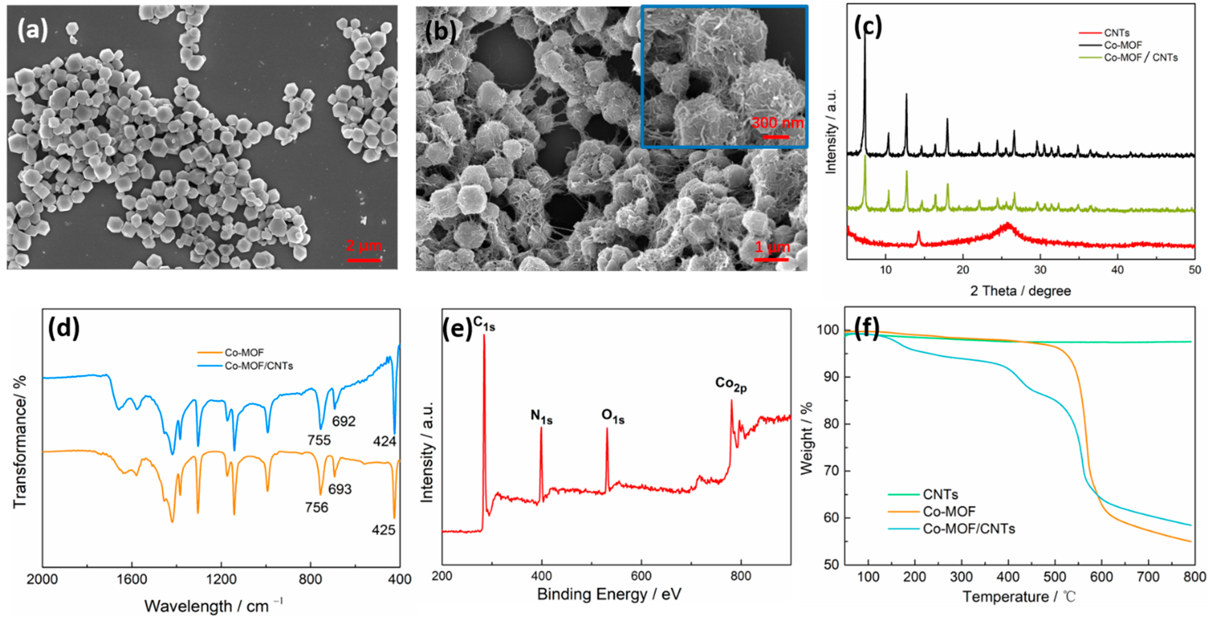
The morphologies and microstructures of the as-synthesized Co-MOF/CNTs were observed by FE-SEM. As shown in Figure 2a, pure Co-MOF exhibited good crystal morphology, with a particle size distribution mostly concentrated around 1 μm. Co-MOF grains were rhombic and dodecahedral, with smooth surfaces. When combined with CNTs, as Figure 2b shows, the size of the Co-MOF matrix was retained, but the morphology of Co-MOF has changed. Numerous CNTs were covered and wrapped around the Co-MOF grains, and the Co-MOF/CNTs composites possessed a rougher surface compared to pure Co-MOF, which had a surface consisting of abundant wrinkles. Besides, CNTs network helped Co-MOF grains establish contact with each other, contributing to the formation of Co-MOF/CNTs hybridization system. The CNTs, Co-MOF and Co-MOF/CNTs crystal structures were assessed by XRD analysis. As shown in Figure 2c, the diffraction peaks of Co-MOF and Co-MOF/CNTs are similar, and both possess highly crystalline features. The FTIR spectra of CNTs, Co-MOF and Co-MOF/CNTs were shown in Figure 2d. For Co-MOF and Co-MOF/CNTs, the ring out-of-plane bending peaks at ~692 cm−1 and ~755 cm−1 belonged to the imidazole, along with the stretch peaks at ~424 cm−1 which corresponded to Co-N and were clearly observed [34]. In order to further probe the element composition and surface chemical state of Co-MOF/CNTs, the XPS survey spectrum of composite was displayed in Figure 2e. The results indicated the presence of C, N, O and Co elements, which was consistent with the theoretical structural composition of Co-MOF/CNTs hybrids. Furthermore, in order to confirm the accurate structural composition of the synthesized Co-MOF/CNTs, thermogravimetric analysis (TGA) method was conducted. The TGA results of CNTs, Co-MOF, and Co-MOF/CNTs were shown in Figure 2f. It can be observed that the weight loss of the CNTs was negligible when the temperature rose from room temperature to 800 °C. For pure Co-MOF, the weight barely changed with a rise of temperature, whereas a sharp weight loss at about 550 °C was observed. This could be ascribed to the decomposition of the ligand (2-methylimidazole) [35], and led to the collapse of frame in Co-MOF. By comparison, the progress of weight loss for Co-MOF/CNTs was divided into three stages. Besides the weight loss in 100–250 °C, which was caused by the evaporation of absorbed water, the weight loss occurred in 350–450 °C and proved that the presence of CNTs has an effect on thermostability of Co-MOF/CNTs. On the whole, the combination of CNTs and Co-MOF barely changed the structure and properties Co-MOF.

3.2. Characterization of Co-MOF/CNTs/SiO2

The morphology of obtained silica particles was studied by FE-SEM. It can be seen from Figure 3a that these SiO2 particles presented a relatively uniform spherical structure, with a diameter of 300 nm. In order to verify the assembly of SiO2 particles on Co-MOF/CNTs and investigate hydrophobicity of Co-MOF/CNTs/SiO2 composites, FE-SEM was applied, and the WCAs of Co-MOF/CNTs and Co-MOF/CNTs/SiO2 were measured. As shown in inset in Figure 3b,c, Co-MOF/CNTs exhibited hydrophilicity with low WCA of 64°, and Co-MOF/CNTs/SiO2 owned hydrophobicity with high WCA of 140°, which was attributed to the participation of hydrophobic SiO2 particles. What is more, in order to observe the element distributions of the Co-MOF/CNTs/SiO2 sample, the element mapping images and EDS analysis were employed (Figure 3d,e,f). It can be seen that C, O, Si, Co elements are distributed in the Co-MOF/CNTs/SiO2 composite, and that Si and O elements show a different distribution with C and Co. The combination of Si and O elements form SiO2, which can be obviously observed in Figure 3d,f. Combined with the analysis above, it was noticeable that the hydrophobic Co-MOF/CNTs/SiO2 composite is successfully synthesized.

3.3. Electrochemical Characterization of Co-MOF/CNTs/SiO2-Modified Au Electrodes

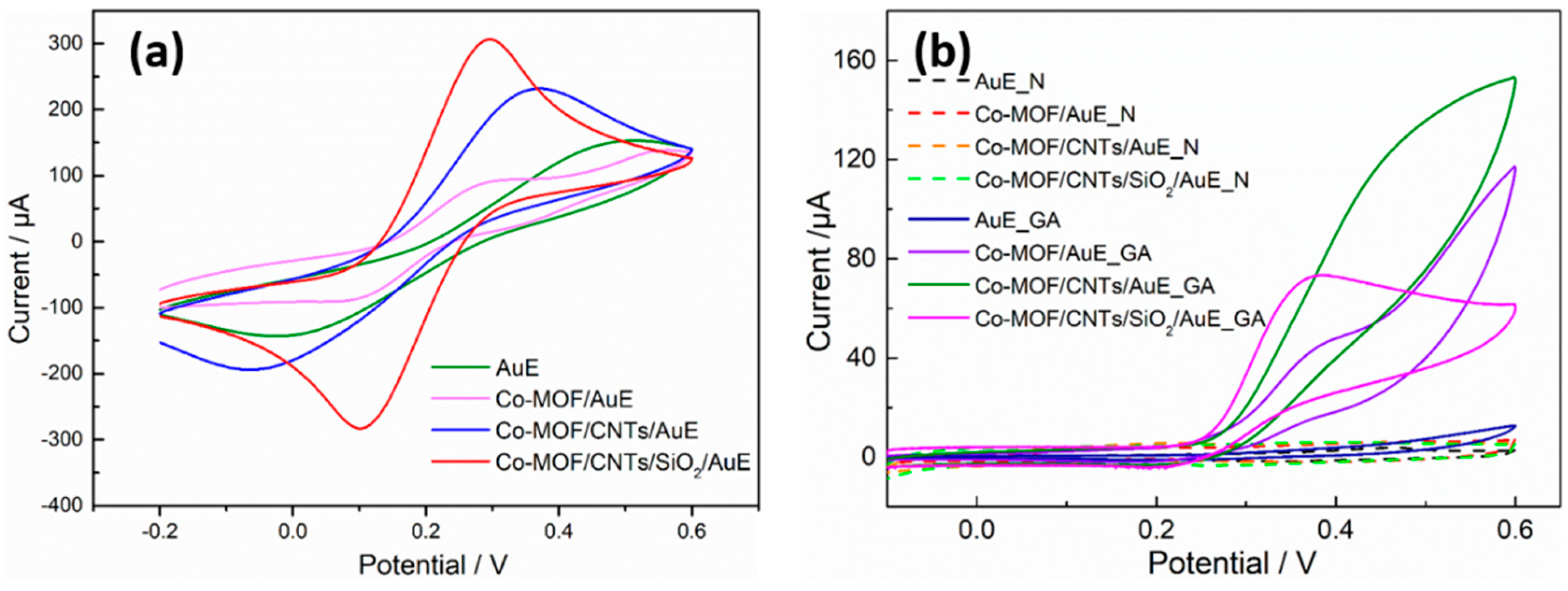
In order to investigate the interface properties of different sensors including the bare AuE, Co-MOF/AuE, Co-MOF/CNTs/AuE and Co-MOF/CNTs/SiO2/AuE, CV was introduced in this part of the study. These four sensors were characterized in a 0.1 M KCl solution containing 5 mM K3[Fe(CN)6]/K4[Fe(CN)6]. Figure 4a shows CV responses of different electrodes. The unmodified AuE presents a pair of redox peaks with large peak-to-peak separation values (ΔEp = 0.437 V) and low peak current on account of a lack of an activated electrode surface. After modification with Co-MOF/CNTs/SiO2, the electrochemical performance of Co-MOF/CNTs/SiO2/AuE was optimized to a large extent, with higher peak current (Ipa = 285.4 μA, Ipc = 301.1 μA) and lower peak-to-peak separation values (ΔEp = 0.196 V) than bare AuE, Co-MOF/AuE, and Co-MOF/CNTs/AuE. The peak currents of Co-MOF/CNTs/SiO2/AuE were about 1.3 times those of Co-MOF/CNTs/AuE and 3 times those of Co-MOF/AuE. This phenomenon benefited from good properties of Co-MOF/CNTs/SiO2 composite.

3.4. Characterization of Surface Quality Change by EQCM

The frequency of uncoated AuE after cleaning and drying was measured by EQCM, and the frequency was 9.00 MHz. When the electrode was modified with Co-MOF/CNTs/SiO2 composite and dried, the frequency was also recorded, and the value was 8.93 MHz. According to the Sauerbrey equation (Equation (1)) [36], the added mass can be calculated by the frequency shifts.
Δ f   = 2 f 0 2 A ρ q μ q   · Δ m
where Δf is the frequency change value of the quartz crystal, f0 is the fundamental frequency of the unloaded piezoelectric quartz crystal, A is the surface area of the electrode, ρq is the density of the quartz crystal, μq is the shear modulus of the quartz crystal, and Δm is the mass change loading on the surface of the crystal. Finally, the changed mass was 84.8 ng on the electrode. It gave a specific reference value of electrode preparation, better than that from dropping the equal volume solution onto the electrode surface to control the experimental parameters. In this experiment, the mass change of each modified electrode was checked, and the amount of Co-MOF/CNTs/SiO2 composite modified on the surface of electrode was controlled at about 85 ng.

3.5. Electrochemical Behavior of Gallic Acid on Modified AuE

CV measurements were employed to investigate the electrochemical behavior of gallic acid on bare AuE, Co-MOF/AuE, Co-MOF/CNTs/AuE and Co-MOF/CNTs/SiO2/AuE. In this part of the experiment, disodium hydrogen phosphate–citric acid buffer (pH = 5) was chosen as the supporting electrolyte. Figure 4b shows the CV responses of four electrodes at a scan rate of 100 mV s−1 and a scan potential range from −0.1 V to +0.6 V, both in the presence of 0.1 mM GA and not. It can be seen that the CV responses of four electrodes in buffer (dash line) are similar, without any visible current peak. Besides, the response of bare AuE sensor towards GA was also like four electrodes in buffer. It had weak response current and there was no significant oxidation peak, which indicated that the bare electrode had scarcely any catalysis toward GA. Compared with bare AuE, Co-MOF/AuE, Co-MOF/CNTs/AuE and Co-MOF/CNTs/SiO2/AuE present the better CV response for GA. There was a peak current observed for Co-MOF/AuE, but it was not measurable. The response current of Co-MOF/CNTs/AuE was larger, but there was no peak current. As for Co-MOF/CNTs/SiO2/AuE, the peak current was observed obviously, and the peak current was 73.4 μA when the peak potential was 0.383 V. Among these, Co-MOF/CNTs/SiO2/AuE tended to perform the best, with obvious oxidation peaks, which may be ascribed to the synergistic amplification effect of Co-MOF and CNTs. Besides, the large surface area and excellent catalysis of Co-MOF and good conductivity of CNTs also contribute to the oxidation of GA. A common denominator which can be seen from CV response curves of different modified AuEs is that the oxidation of GA is almost irreversible. The oxidation of the pyrogallol moiety in GA generates the unstable quinone species, preventing their electrochemical reduction in the subsequent cycle because the unstable quinone species may undergo further chemical reactions [37]. One GA molecule has four hydrogen bond donors and five hydrogen bond receptors, and Co-MOF also had hydrogen bond donors, hydrogen bond receptors and active sites, which may be attributed to the electrochemical reaction on the surface of the modified electrode.

3.6. The Effect of pH and Scan Rate

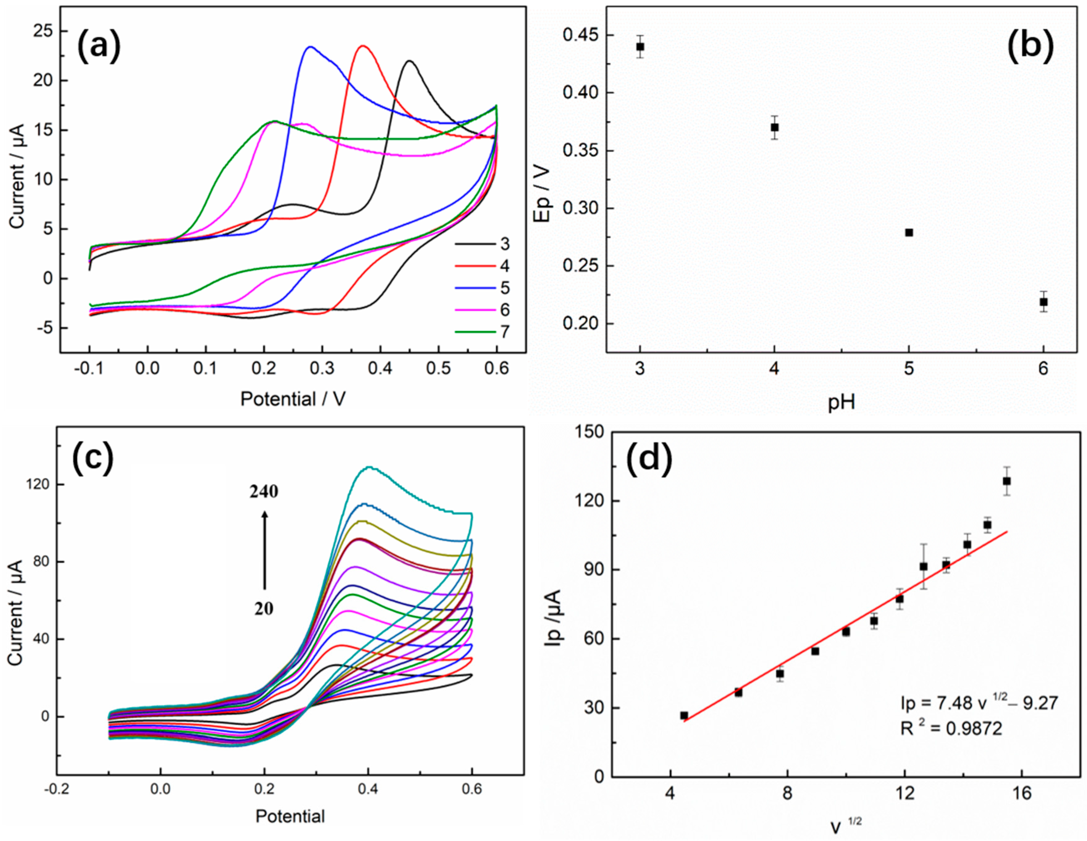
The pH of the buffer has a significant effect on the electrochemical response of GA, and the effect was investigated by the CV method. Figure 5a shows the CV responses of 0.1 mM GA on Co-MOF/CNTs/SiO2-modified electrodes in disodium hydrogen phosphate–citrate buffer at a pH range of 3 to 7 and scan rate of 100 mV s−1. It can be seen that the electrochemical behavior of GA is affected by different pH values. The relationship of peak potential (Ep) and pH values between 3 to 6 was also studied, and the plot was shown in Figure 5b. The results showed that, in the pH range of 2 to 6, the anodic peak potential was negatively shifted by the increase in pH, presenting a continuous dynamic change. This phenomenon demonstrated the participation of protons in the reactions. The presence of protonated hydroxyl groups of GA was conductive to the GA oxidation in the acidic environment. Besides, the GA oxidation potential decreased linearly with the increase in pH values, and the slope Ep/pH was 66 mV/pH, which was close to the theoretical Nernstian value of 59 mV/pH. This result suggested that the oxidation of GA is a transfer reaction, involving equal number of protons and electrons, which is in agreement with other references [37]. Meanwhile, an increase in peak current (Ip) was observed with pH ranging from 3 to 5, followed by a decrease. In combination with the above statements, pH 5.0 was chosen as the optimal pH for subsequent experiments.
The effect of scan rate on the electrochemical behavior of 1 mM GA on the surface of Co-MOF/CNTs/SiO2/AuE was investigated by CV in the range of 20–260 mV s−1 (Figure 5c). Figure 5d shows that the peak current of GA was linear with the square root of the scan rate, and the linear regression equation of the oxidation peak currents and scan rate was Ip (μA) = 7.48 v1/2(mV s−1) − 9.27), with R2 = 0.9872. The results indicated that the oxidation of GA was typical of a diffusion-controlled process on the surface of a Co-MOF/CNTs/SiO2/AuE sensor. Further, the increase in scan rate produced a positive shift in the peak current and a linear increase in the peak current, which suggests a kinetic limitation in the reaction of GA. In conclusion, considering that the faster scan rate would lead to the decline of signal-to-noise ratio and acceleration of electrode consumption, and low scan rate would take longer detection time, 100 mV s−1 was selected for the experiments. We chose 100 mV s−1 throughout the experiment to ensure the systematicity of our research.

3.7. Analysis of Electrooxidation Mechanism of GA

3.7.1. The Oxidation Process of GA

It was calculated that the electrooxidation of GA may be a two-electron and two-proton process, according to the electrochemical results. Meanwhile, some computational analyses were introduced to investigate the oxidation process of GA. First, the structure of GA was optimized by Gaussian 05, and the final structure was shown in Figure 6a. The energies of Highest Occupied Molecular Orbitals (HOMO) and Lowest Unoccupied Molecular Orbitals (LUMO) of a molecule are important quantum chemical descriptors. The HOMO and LUMO of GA are shown in Figure 6b. The band gap of GA was 5.35 eV, which indicated that the GA molecule is stable in general, but our experiment has proved that GA could be oxidized by electrochemical excitation. According to the Natural Bond Orbital (NBO) analysis, the strength of four O-H bonds was obtained, and the bond which would be broken first in electrooxidation can be predicted. Table 1 shows that the bond order of bond O12-H18 was lesser, which indicated that O12-H18 breaks more easily in the reaction. In the same way, in can be inferred that O11-H17 would be broken later. Therefore, according to the NBO computational investigations above, the two bonds O12-H18 and O11-H17 were more likely to be broken in the electrochemical oxidation process. Besides, we calculated Hirshfeld charges for all atoms in GA molecule in its N, N + 1 and N − 1 electrons states, respectively. Based on Hirsheld charges, condensed Fukui functions were investigated (Table 2), and the highest f(+) value for the H18, which represented this area may, be the first reactive site in electrochemical reaction. This can be seen visually from Figure 6c. Synthesizing the above theories and analytical results, the possible electrochemical reaction mechanism of GA on Co-MOF/CNTs/SiO2 modified electrode is shown in Figure 7 as follows:

3.7.2. The Catalysis of Co-MOF to GA

Based on the possible electrochemical reaction process analyzed above, the catalysis effect of Co-MOF to GA was investigated further. The MOF structure was too big to perform the ab initio calculation; thus, the simplified cobalt–methylimidazole cluster was exported from the MOF structure for our calculation. The molecular structures of the gallic acid, the cobalt–methylimidazole cluster and the gallic acid adsorbed on cobalt–methylimidazole cluster were all optimized under the framework of density functional theory, with dispersion corrected to B3LYP functional [38,39,40,41] and Pople basis sets of 6–31 g* [42,43]. In order to simulate the solvation effect of electrolyte, the implicit solvent model of SMD (Solvation Model Based on Density) [44] was applied in all calculations. The vibrational frequency analysis was carried out for the optimized structure with the same calculation method to obtain the zero-point energy and free-energy corrections. In order to obtain the electron energy with higher accuracy which has the major impact on the accuracy of Gibbs free energy, a single point calculation for the optimized structure with M062x functional [45] and def2TZVP basis set [46] was performed. Finally, the single point energy is added to the free energy correction calculated before to obtain the Gibbs free energy. The Gibbs free energies of the electrochemical oxidation reactions of pure gallic acid and gallic acid adsorbed on the cobalt–methylimidazole cluster were calculated from the formular:
ΔG = G(cation) − G(Neutral)
where G(cation) and G(Neutral) were the Gibbs free energy of oxidation state and initial state of the molecule. The oxidation potentials were calculated further from the Nernst equation. The φ value of every step where GA loses an electron was shown in Figure 6d. It can be seen visually that the presence of “Co” reduces the oxidation potential of GA; in other words, Co-MOF has a catalytic effect on the oxidation of GA. Co centers was acted as redox mediators in the process of GA oxidation. The Co2+ ions of Co-MOF were oxidized to Co3+ upon the external potential was applied. The results support that the electrochemically generated Co3+ species can oxidize GA efficiently. The corresponding electrocatalytic mechanism is illustrated by the following equations:
(1)
2Co-MOF—Co(II) → 2Co-MOF—Co(III) + 2e
(2)
C7H6O6 − 2e − 2H+ → C7H4O6
(3)
2Co-MOF—Co(III) + 2e → 2Co-MOF—Co(II)
The presence of “Co” not only speeds up electron transfer, but also catalyzes chemical reactions.

3.8. Electrochemical Determination of GA

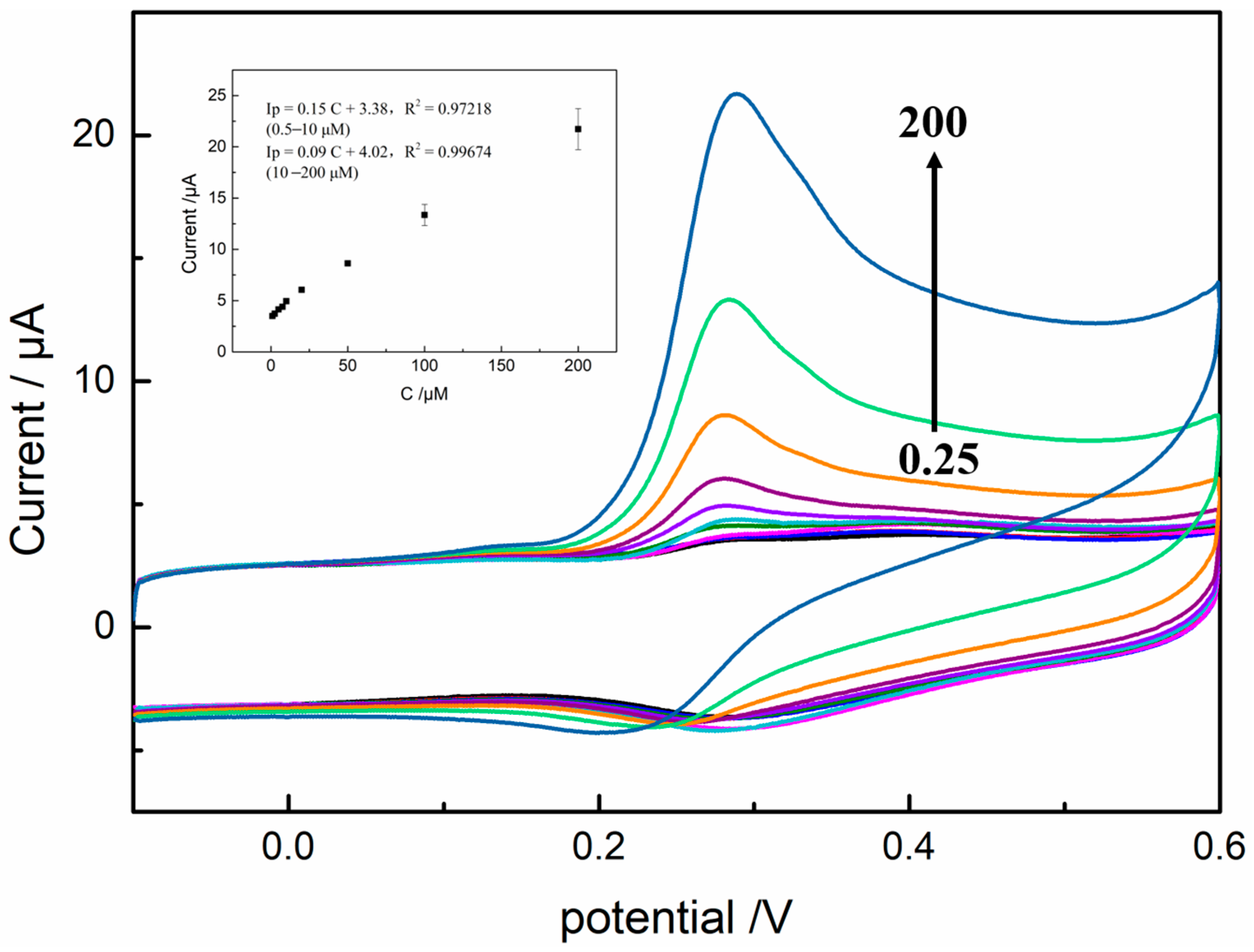
CV was used for the determination of GA on the surface of Co-MOF/CNTs/SiO2/AuE. The CV curves in Figure 8 show the electrochemical responses of different concentrations, ranging from 0.05 to 200 μM, under the optimized experimental parameters. It was obvious that oxidation peak currents show an increasing trend with the increase in GA concentrations. Meanwhile, it can be seen that the reduction peak currents of different concentrations of GA were approximate, which proves the conclusion that the oxidation of GA is almost irreversible further. As shown in inset of Figure 8, the relationship between the oxidation peak currents and GA concentrations was plotted. The modified electrode exhibits two parts of linearity in the concentration ranges of 0.5–10 μM and 10–200 μM. The linear regression equations are Ip (μA) = 0.15c (μM) + 3.38, R2 = 0.97218 (0.5–10 μM) and Ip (μA) = 0.09c (μM) + 4.02, and R2 = 0.99674(10–200 μM). The limit of detection was calculated to be 2 × 10⁻⁷ M (S/N = 3). The sensitivity value of the modified sensor was calculated to be 2.48 μA cm−2 μM−1 and 2.29 μA cm−2 μM−1 for GA. On the other hand, the sensitivity of modified sensor can also be described by EQCM data, which was calculated to be 274.76 μA mg−1. Table 3 displays other recent research about GA electrochemical detection previously reported in the literature, including electrodes, electrochemical methods, linear range and limit of detection (LOD). By comparison, the home-made sensors exhibited better detection performances than other modified electrodes based on different methodology including differential pulse voltammetry (DPV), linear sweep voltammetry (LSV) and square wave voltammetry (SWV).

3.9. The Selectivity, Reproducibility and Stability of the GA Sensor

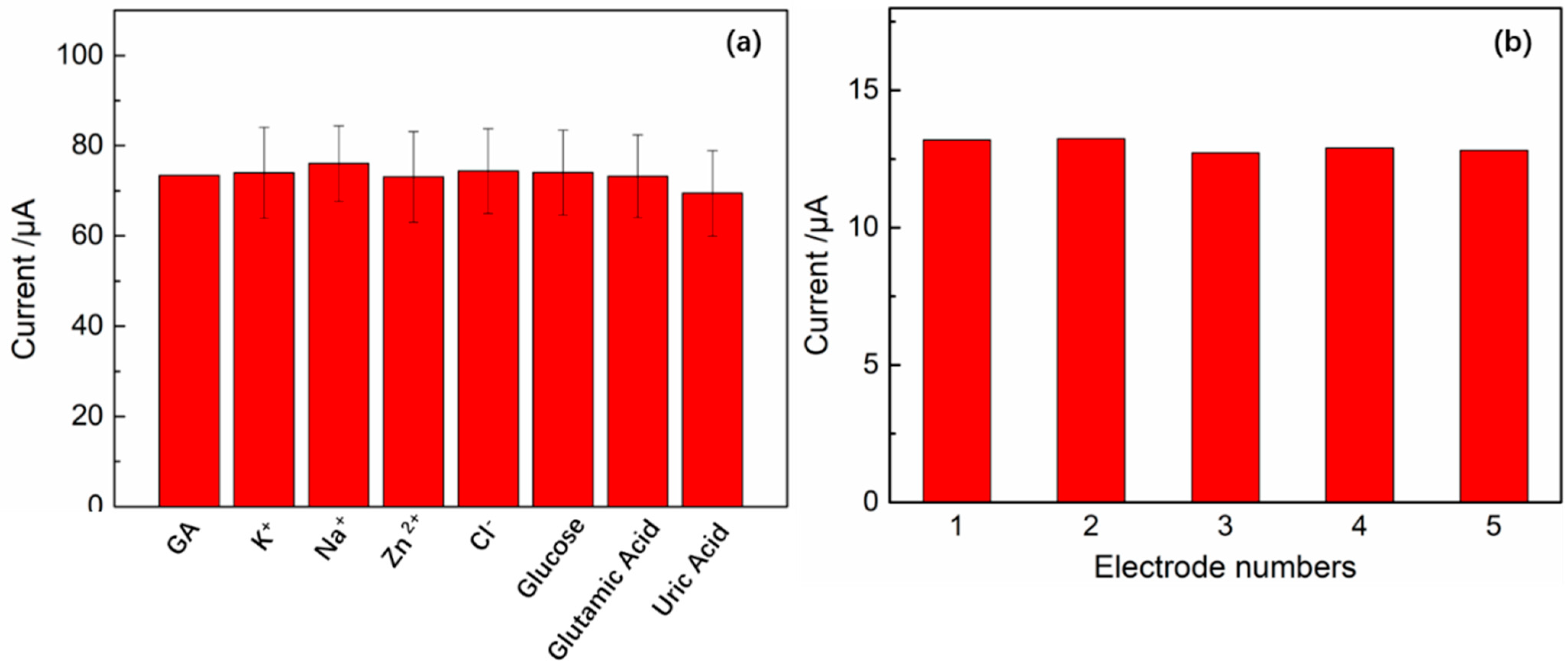
In order to investigate the effects of different interfering substances in the determination of GA on Co-MOF/CNTs/SiO2/AuE, a CV method was introduced. The peak currents of 0.1 mM GA in the presence of a 100-fold concentration of K+, Na+, Zn2+, Cl, and 50-fold concentration of glucose, glutamic acid and uric acid were recorded. Figure 8a shows that these metal ions and organic compounds do not significantly impact on the determination of GA, suggesting the good selectivity of modified sensors. The reproducibility of Co-MOF/CNTs/SiO2/AuE was examined by five individual sensors, and fabricated by the same process, in the presence of 0.1 mM GA. In Figure 9b, these five sensors showed nearly the same responses, and the relative standard deviations (RSD) of the oxidation peak currents was 1.75%. The stability was also one of the most important properties of modified sensors before actual application, and it was evaluated by CV. The modified electrodes were stored at room temperature for 15 days. The responses of electrodes for the same concentration of GA were changed slightly, remaining about 96.2% of their initial currents 15 days ago. The result demonstrated that the GA sensors own good selectivity, reproducibility and stability, indicating that the Co-MOF/CNTs/SiO2/AuE has the feasibility for practical application.

4. Conclusions

In this work, the hydrophobic Co-MOF/CNTs/SiO2 composite was strategically designed and synthesized, and the Co-MOF/CNTs/SiO2-modified AuE was successfully developed for the detection of GA. The characterization of materials was carried by various techniques such as FE-SEM, XRD, FTIR, XPS, TGA. The electrochemical behaviors of Co-MOF/CNTs/SiO2/AuE were investigated by CV. The homemade sensor exhibited good electrocatalysis for GA, on account of the active sites of Co-MOF, as well as the good conductivity of CNTs and their cooperation. The presence of hydrophobic SiO2 particle enhanced water stability of the composite. Under the optimal conditions, the GA sensor presented a satisfying determination performance with wide linear relationship over the range of 0.05–200 μM, and a low detection limit of 0.02 μM (S/N = 3). The GA sensor also showed good selectivity, reproducibility and stability. Besides, the oxidation mechanism of gallic acid was briefly expounded. We anticipate that this work may open up a new approach for the application of hydrophobic MOF-based materials in electrochemical sensors.

Author Contributions

L.Z.: conceptualization, methodology, writing—original draft preparation. Q.Z.: formal analysis, data curation, formal analysis. W.S.: methodology, investigation, supervision. Z.W.: methodology, writing—reviewing and editing. J.W.: writing—reviewing and editing, funding acquisition. All authors have read and agreed to the published version of the manuscript.

Funding

This research was funded by the Chinese National Key Technology R&D Program through Project 2017YFD0400102.

Data Availability Statement

Not applicable.

Conflicts of Interest

All other authors declare no competing financial interest.

References

  1. Vilian, A.T.E.; Song, J.Y.; Lee, Y.S.; Hwang, S.K.; Kim, H.J.; Jun, Y.S.; Huh, Y.S.; Han, Y.K. Salt-templated three-dimensional porous carbon for electrochemical determination of gallic acid. Biosens. Bioelectron. 2018, 117, 597–604. [Google Scholar] [CrossRef] [PubMed]
  2. Sainz-Urruela, C.; Vera-Lopez, S.; San Andres, M.P.; Diez-Pascual, A.M. Graphene-based sensors for the detection of bioactive compounds: A review. Int. J. Mol. Sci. 2021, 22, 3316. [Google Scholar] [CrossRef] [PubMed]
  3. Zhao, H.; Ran, Q.; Li, Y.; Li, B.; Liu, B.; Ma, H.; Zhang, M.; Komarneni, S. Highly sensitive detection of gallic acid based on 3D interconnected porous carbon nanotubes/carbon nanosheets modified glassy carbon electrode. J. Mater. Res. Technol. 2020, 9, 9422–9433. [Google Scholar] [CrossRef]
  4. Das, D.; Biswas, D.; Hazarika, A.K.; Sabhapondit, S.; Roy, R.B.; Tudu, B.; Bandyopadhyay, R. CuO nanoparticles decorated MIP-based electrode for sensitive determination of gallic acid in green tea. IEEE Sens. J. 2021, 21, 5687–5694. [Google Scholar] [CrossRef]
  5. Lin, X.; Deng, Y.; He, Y.; Chen, J.; Hu, S. Construction of hydrophilic N, O-rich carboxylated triazine-covalent organic frameworks for the application in selective simultaneous electrochemical detection. Appl. Surf. Sci. 2021, 545, 149047. [Google Scholar] [CrossRef]
  6. Nazari, F.; Ghoreishi, S.M.; Khoobi, A. Bio-based Fe3O4/chitosan nanocomposite sensor for response surface methodology and sensitive determination of gallic acid. Int. J. Biol. Macromol. 2020, 160, 456–469. [Google Scholar] [CrossRef] [PubMed]
  7. Fernandes, F.H.; Salgado, H.R. Gallic acid: Review of the methods of determination and quantification. Crit. Rev. Anal. Chem. 2016, 46, 257–265. [Google Scholar] [CrossRef] [PubMed]
  8. Zhang, L.L.; Liu, Y.C.; Hu, X.Y.; Xu, M.; Wang, Y.M. Studies on interactions of pentagalloyl glucose, ellagic acid and gallic acid with bovine serum albumin: A spectroscopic analysis. Food Chem. 2020, 324, 6. [Google Scholar] [CrossRef]
  9. Zaripour, M.; Zare-Shahabadi, V.; Jahromi, H.J. Application of ultrasonic-assisted inclusion complex formation with alpha-cyclodextrin for simultaneous spectrophotometric determination of gallic acid and vanillic acids in fruit samples. Spectrochim. Acta A Mol. Biomol. Spectrosc. 2019, 222, 117197. [Google Scholar] [CrossRef]
  10. Sangeetha, N.S.; Narayanan, S.S. A novel bimediator amperometric sensor for electrocatalytic oxidation of gallic acid and reduction of hydrogen peroxide. Anal. Chim. Acta 2014, 828, 34–45. [Google Scholar] [CrossRef]
  11. Kaur, S.; Shiekh, B.A.; Kaur, D.; Kaur, I. Highly sensitive sensing of Fe(III) harnessing schiff based ionophore: An electrochemical approach supported with spectroscopic and dft studies. J. Mol. Liq. 2021, 333, 115954. [Google Scholar] [CrossRef]
  12. Seenivasan, R.; Chang, W.J.; Gunasekaran, S. Highly sensitive detection and removal of lead ions in water using cysteine-functionalized graphene oxide/polypyrrole nanocomposite film electrode. ACS Appl. Mater. Interfaces 2015, 7, 15935–15943. [Google Scholar] [CrossRef] [PubMed]
  13. Angizi, S.; Hatamie, A.; Ghanbari, H.; Simchi, A. Mechanochemical green synthesis of exfoliated edge-functionalized boron nitride quantum dots: Application to vitamin c sensing through hybridization with gold electrodes. ACS Appl. Mater. Interfaces 2018, 10, 28819–28827. [Google Scholar] [CrossRef] [PubMed]
  14. Zhu, L.; Wei, Z.; Wang, J.; Zhong, J. An electrochemical biosensor based on NiO nanoflowers/polymethylene blue composite for non-enzymatic glucose detection. J. Electrochem. Soc. 2020, 167, 146512. [Google Scholar] [CrossRef]
  15. Pwavodi, P.C.; Ozyurt, V.H.; Asir, S.; Ozsoz, M. Electrochemical sensor for determination of various phenolic compounds in wine samples using Fe3O4 nanoparticles modified carbon paste electrode. Micromachines 2021, 12, 312. [Google Scholar] [CrossRef]
  16. Goda, M.A.; El-Moghny, M.G.A.; El-Deab, M.S. Enhanced electrocatalytic oxidation of urea at CuOx-NiOx nanoparticle-based binary catalyst modified polyaniline/GC electrodes. J. Electrochem. Soc. 2020, 167, 064522. [Google Scholar] [CrossRef]
  17. Aryal, K.P.; Jeong, H.K. Electrochemical detection of ascorbic acid with chemically functionalized carbon nanofiber/beta-cyclodextrin composite. Chem. Phys. Lett. 2020, 757, 137881. [Google Scholar] [CrossRef]
  18. Zeng, X.; Li, N.; Wang, J. Electrochemical synthesis of (poly) dimethoxyaniline on glassy carbon electrodes and their applications in the detection of L- and D-glutamic acids. J. Electrochem. Soc. 2019, 166, B3066–B3071. [Google Scholar] [CrossRef]
  19. Phonklam, K.; Wannapob, R.; Sriwimol, W.; Thavarungkul, P.; Phairatana, T. A novel molecularly imprinted polymer PMB/MWCNTs sensor for highly-sensitive cardiac troponin T detection. Sens. Actuators B Chem. 2020, 308, 127630. [Google Scholar] [CrossRef]
  20. Marsh, C.; Shearer, G.C.; Knight, B.T.; Paul-Taylor, J.; Burrows, A.D. Supramolecular aspects of biomolecule interactions in metal–organic frameworks. Coord. Chem. Rev. 2021, 439, 213928. [Google Scholar] [CrossRef]
  21. Wang, B.; Xie, L.-H.; Wang, X.; Liu, X.-M.; Li, J.; Li, J.-R. Applications of metal–organic frameworks for green energy and environment: New advances in adsorptive gas separation, storage and removal. Green Energy Env. 2018, 3, 191–228. [Google Scholar] [CrossRef]
  22. Liu, J.; Thallapally, P.K.; McGrail, B.P.; Brown, D.R.; Liu, J. Progress in adsorption-based CO2 capture by metal-organic frameworks. Chem. Soc. Rev. 2012, 41, 2308–2322. [Google Scholar] [CrossRef]
  23. Wang, Q.; Astruc, D. State of the art and prospects in metal-organic framework (MOF)-based and MOF-derived nanocatalysis. Chem. Rev. 2020, 120, 1438–1511. [Google Scholar] [CrossRef]
  24. Liao, X.; Fu, H.; Yan, T.; Lei, J. Electroactive metal-organic framework composites: Design and biosensing application. Biosens. Bioelectron. 2019, 146, 111743. [Google Scholar] [CrossRef]
  25. Karim, M.R.; Hatakeyama, K.; Koinuma, M.; Hayami, S. Proton conductors produced by chemical modifications of carbon allotropes, perovskites and metal organic frameworks. J. Mater. Chem. A 2017, 5, 7243–7256. [Google Scholar] [CrossRef]
  26. Li, Y.; Shen, Y.; Zhang, Y.; Zeng, T.; Wan, Q.; Lai, G.; Yang, N. A UiO-66-NH2/carbon nanotube nanocomposite for simultaneous sensing of dopamine and acetaminophen. Anal. Chim. Acta 2021, 1158, 338419. [Google Scholar] [CrossRef]
  27. Lei, J.; Ju, H. Nanotubes in biosensing. Wiley Interdiscip. Rev. Nanomed. Nanobiotechnol. 2010, 2, 496–509. [Google Scholar] [CrossRef] [PubMed]
  28. Wang, C.; Liu, X.; Keser Demir, N.; Chen, J.P.; Li, K. Applications of water stable metal-organic frameworks. Chem. Soc. Rev. 2016, 45, 5107–5134. [Google Scholar] [CrossRef] [PubMed]
  29. Raza, W.; Kukkar, D.; Saulat, H.; Raza, N.; Azam, M.; Mehmood, A.; Kim, K.-H. Metal-organic frameworks as an emerging tool for sensing various targets in aqueous and biological media. TrAC Trend. Anal. Chem. 2019, 120, 115654. [Google Scholar] [CrossRef]
  30. Chen, W.; Wang, Z.; Gu, S.; Wang, J. Detection of hexanal in humid circumstances using hydrophobic molecularly imprinted polymers composite. Sens. Actuators B Chem. 2019, 291, 141–147. [Google Scholar] [CrossRef]
  31. Li, L.; Li, B.; Dong, J.; Zhang, J. Roles of silanes and silicones in forming superhydrophobic and superoleophobic materials. J. Mater. Chem. A 2016, 4, 13677–13725. [Google Scholar] [CrossRef]
  32. Ren, T.; He, J. Substrate-versatile approach to robust antireflective and superhydrophobic coatings with excellent self-cleaning property in varied environments. ACS Appl. Mater. Interfaces 2017, 9, 34367–34376. [Google Scholar] [CrossRef] [PubMed]
  33. Ren, T.; Yang, M.; Wang, K.; Zhang, Y.; He, J. CuO nanoparticles-containing highly transparent and superhydrophobic coatings with extremely low bacterial adhesion and excellent bactericidal property. ACS Appl. Mater. Interfaces 2018, 10, 25717–25725. [Google Scholar] [CrossRef] [PubMed]
  34. Li, Z.; Zhou, G.; Dai, H.; Yang, M.; Fu, Y.; Ying, Y.; Li, Y. Biomineralization-mimetic preparation of hybrid membranes with ultra-high loading of pristine metal–organic frameworks grown on silk nanofibers for hazard collection in water. J. Mater. Chem. A 2018, 6, 3402–3413. [Google Scholar] [CrossRef]
  35. Lashgari, S.M.; Yari, H.; Mahdavian, M.; Ramezanzadeh, B.; Bahlakeh, G.; Ramezanzadeh, M. Synthesis of graphene oxide nanosheets decorated by nanoporous zeolite-imidazole (ZIF-67) based metal-organic framework with controlled-release corrosion inhibitor performance: Experimental and detailed DFT-D theoretical explorations. J. Hazard. Mater. 2021, 404, 124068. [Google Scholar] [CrossRef]
  36. Sauerbrey, G. Verwendung von schwingquarzen zur wagung dunner schichten und zur mikrowagung. Z. Phys. 1959, 155, 206–222. [Google Scholar] [CrossRef]
  37. Yan, Y.; Bo, X.; Guo, L. MOF-818 Metal-organic framework-reduced graphene oxide/multiwalled carbon nanotubes composite for electrochemical sensitive detection of phenolic acids. Talanta 2020, 218, 121123. [Google Scholar] [CrossRef]
  38. Giannozzi, P.; Baseggio, O.; Bonfa, P.; Brunato, D.; Car, R.; Carnimeo, I.; Cavazzoni, C.; de Gironcoli, S.; Delugas, P.; Ferrari Ruffino, F.; et al. Quantum espresso toward the exascale. J. Chem. Phys. 2020, 152, 154105. [Google Scholar] [CrossRef] [Green Version]
  39. Lee, C.; Yang, W.; Parr, R.G. Development of the colle-salvetti correlation-energy formula into a functional of the electron density. Phys. Rev. B Condens. Matter. 1988, 37, 785–789. [Google Scholar] [CrossRef] [Green Version]
  40. Becke, A.D. Density-functional exchange-energy approximation with correct asymptotic behavior. Phys. Rev. A Gen. Phys. 1988, 38, 3098–3100. [Google Scholar] [CrossRef]
  41. Becke, A.D. Density-functional thermochemistry.3. the role of exact exchange. J. Chem. Phys. 1993, 98, 5648–5652. [Google Scholar] [CrossRef] [Green Version]
  42. Petersson, G.A.; Allaham, M.A. A complete basis set model chemistry.2. open-shell systems and the total energies of the 1st-row atoms. J. Chem. Phys. 1991, 94, 6081–6090. [Google Scholar] [CrossRef]
  43. Petersson, G.A.; Bennett, A.; Tensfeldt, T.G.; Allaham, M.A.; Shirley, W.A.; Mantzaris, J. A complete basis set model chemistry.1. the total energies of closed-shell atoms and hydrides of the 1st-row elements. J. Chem. Phys. 1988, 89, 2193–2218. [Google Scholar] [CrossRef]
  44. Marenich, A.V.; Cramer, C.J.; Truhlar, D.G. Universal solvation model based on solute electron density and on a continuum model of the solvent defined by the bulk dielectric constant and atomic surface tensions. J. Phys. Chem. B 2009, 113, 6378–6396. [Google Scholar] [CrossRef] [PubMed]
  45. Zhao, Y.; Truhlar, D.G. The M06 suite of density functionals for main group thermochemistry, thermochemical kinetics, noncovalent interactions, excited states, and transition elements: Two new functionals and systematic testing of four M06-class functionals and 12 other functionals. Theor. Chem. Acc. 2008, 120, 215–241. [Google Scholar] [CrossRef] [Green Version]
  46. Weigend, F.; Ahlrichs, R. Balanced Basis Sets of Split Valence, Triple zeta valence and quadruple zeta valence quality for H to Rn: Design and assessment of accuracy. Phys. Chem. Chem. Phys. 2005, 7, 3297–3305. [Google Scholar] [CrossRef]
  47. Chikere, C.O.; Hobben, E.; Faisal, N.H.; Kong-Thoo-Lin, P.; Fernandez, C. Electroanalytical determination of gallic acid in red and white wine samples using cobalt oxide nanoparticles-modified carbon-paste electrodes. Microchem. J. 2021, 160, 105668. [Google Scholar] [CrossRef]
  48. Chikere, C.O.; Faisal, N.H.; Kong Thoo Lin, P.; Fernandez, C. The synergistic effect between graphene oxide nanocolloids and silicon dioxide nanoparticles for gallic acid sensing. J. Solid State Electr. 2019, 23, 1795–1809. [Google Scholar] [CrossRef] [Green Version]
  49. Tashkhourian, J.; Nami-Ana, S.F. A sensitive electrochemical sensor for determination of gallic acid based on SiO2 nanoparticle modified carbon paste electrode. Mater. Sci. Eng. C Mater. Biol. Appl. 2015, 52, 103–110. [Google Scholar] [CrossRef]
  50. Liang, Z.; Zhai, H.; Chen, Z.; Wang, H.; Wang, S.; Zhou, Q.; Huang, X. A simple, ultrasensitive sensor for gallic acid and uric acid based on gold microclusters/sulfonate functionalized graphene modified glassy carbon electrode. Sen. Actuators B Chem. 2016, 224, 915–925. [Google Scholar] [CrossRef]
  51. Wang, H.; Feng, X.; Bo, X.; Zhou, M.; Guo, L. Nickel-based metal-organic framework/crosslinked tubular poly(3,4-ethylenedioxythiophene) composite as an electrocatalyst for the detection of gallic acid and tinidazole. ChemElectroChem 2020, 7, 4031–4037. [Google Scholar] [CrossRef]
  52. Datta, S.; Kanjilal, B.; Sarkar, P. Electrochemical sensor for detection of polyphenols in tea and wine with differential pulse voltammetry and electrochemical impedance spectroscopy utilizing tyrosinase and gold nanoparticles decorated biomembrane. J. Electrochem. Soc. 2017, 164, B118–B126. [Google Scholar] [CrossRef]
  53. Luo, J.H.; Li, B.L.; Li, N.B.; Luo, H.Q. Sensitive detection of gallic acid based on polyethyleneimine-functionalized graphene modified glassy carbon electrode. Sens. Actuators B Chem. 2013, 186, 84–89. [Google Scholar] [CrossRef]
  54. Ziyatdinova, G.; Guss, E.; Morozova, E.; Budnikov, H.; Davletshin, R.; Vorobev, V.; Osin, Y. Simultaneous voltammetric determination of gallic and ellagic acids in cognac and brandy using electrode modified with functionalized SWNT and poly(pyrocatechol violet). Food Anal. Method. 2019, 12, 2250–2261. [Google Scholar] [CrossRef]
  55. Madhusudhana; Manasa, G.; Bhakta, A.K.; Mekhalif, Z.; Mascarenhas, R.J. Bismuth-nanoparticles decorated multi-wall-carbon-nanotubes cast-coated on carbon paste electrode; an electrochemical sensor for sensitive determination of gallic acid at neutral pH. Mater. Sci. Energy Tech. 2020, 3, 174–182. [Google Scholar] [CrossRef]
  56. Refat, A.H.; Ahmed, B.; Emad, F.N.; François, G. Simultaneous voltammetric determination of gallic and protocatechuic acids in mango juice using a reduced graphene oxide-based electrochemical sensor. Beverages 2019, 5, 17. [Google Scholar] [CrossRef]
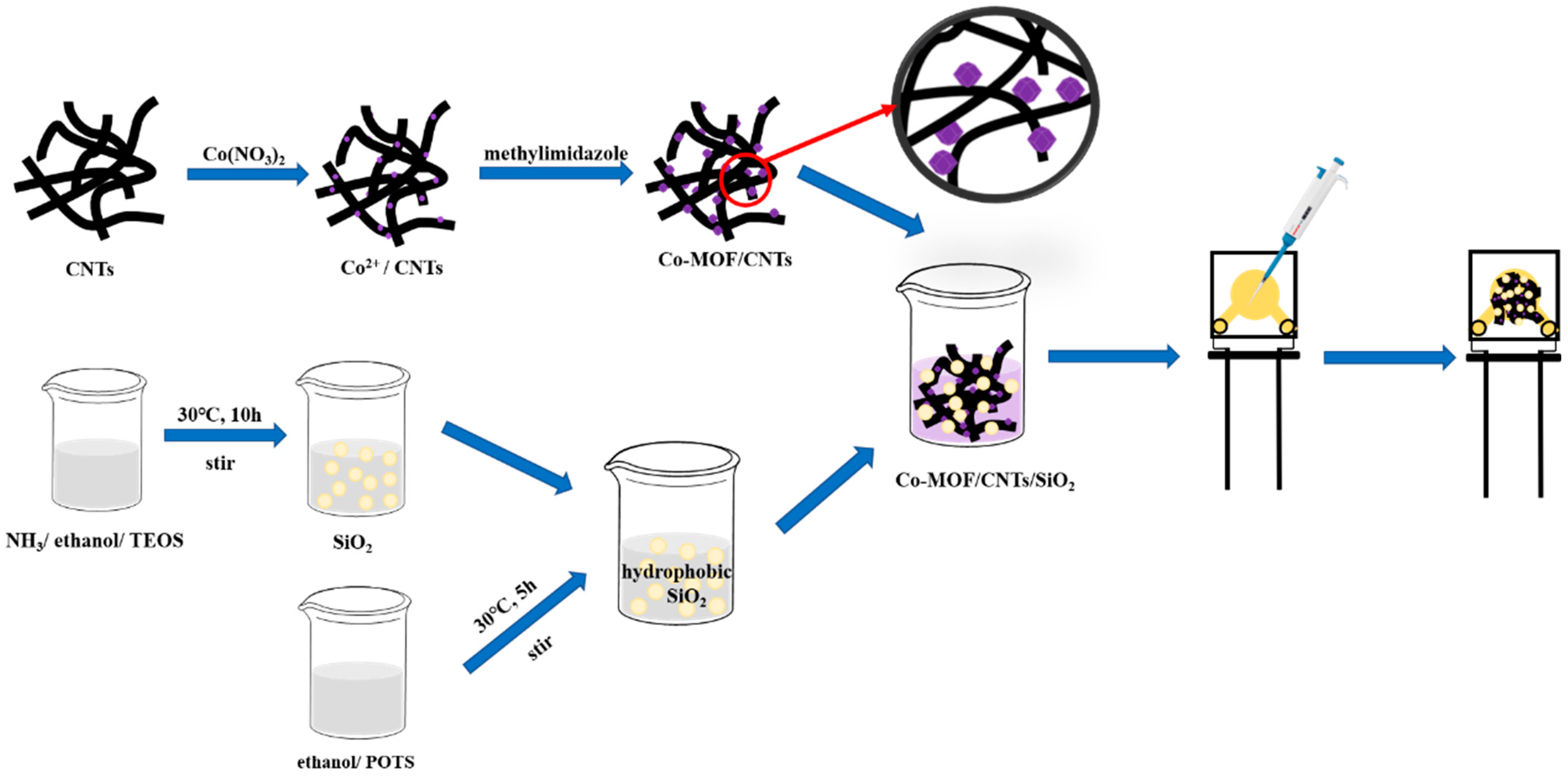
Figure 1. Schematic diagram of the fabrication of the Co-MOF/CNTs/SiO2/AuE.
Chemosensors 10 00443 g001
Figure 2. FE-SEM images of Co-MOF (a) and Co-MOF/CNTs (b); XRD pattern of CNTs, Co-MOF and Co-MOF/CNTs (c); FTIR spectrum of Co-MOF and Co-MOF/CNTs (d); XPS spectrum of Co-MOF/CNTs (e); TGA results of CNTs, Co-MOF and Co-MOF/CNTs (f).
Chemosensors 10 00443 g002
Figure 3. FE-SEM image of the silica particles (a); FE-SEM images and WCA photographs (inset) of Co-MOF/CNTs (b) and Co-MOF/CNTs/SiO2 (c); element mapping images of Co-MOF/CNTs/SiO2 (df).
Chemosensors 10 00443 g003
Figure 4. CV curves of the bare AuE, Co-MOF/AuE, Co-MOF/CNTs/AuE and Co-MOF/CNTs/SiO2/AuE in 0.1 M KCl solution containing 5 mM K3[Fe(CN)6]/K4[Fe(CN)6] (a); CV curves of bare AuE, Co-MOF/AuE, Co-MOF/CNTs/AuE and Co-MOF/CNTs/SiO2/AuE for 1 mM GA or without GA (pH = 5) (b).
Chemosensors 10 00443 g004
Figure 5. (a) CV curves of Co-MOF/CNTs/SiO2/AuE in solutions containing 0.1 mM GA at different pH values; (b) the relationship of peak potentials and pH values; (c) CV curves of Co-MOF/CNTs/SiO2/AuE at different scan rates ranging from 20 to 260 mV s−1; (d) the relationship of peak current and scan rates.
Chemosensors 10 00443 g005
Figure 6. Optimized structure of GA and atomic label (a); HOMO and LUMO of GA (b); f (+) of N−1 (c); the oxidation potential of each step with or without Co-MOF (d).
Chemosensors 10 00443 g006
Figure 7. The possible electrochemical reaction mechanism of GA on modified electrode.
Chemosensors 10 00443 g007
Figure 8. CV curves of various concentrations of GA on Co-MOF/CNTs/SiO2/AuE (Inset: the linear relationship of Ip and GA concentrations).
Chemosensors 10 00443 g008
Figure 9. Selectivity of Co-MOF/CNTs/SiO2/AuE (a); Reproducibility of Co-MOF/CNTs/SiO2/AuE for GA (b).
Chemosensors 10 00443 g009
Table 1. Bond order values of GA.
BondBond Order
O1-H130.9261
O7-H150.9256
O11-H170.9196
O12-H180.8975
Table 2. Hirshfeld charges, condensed Fukui functions of each atom in GA.
Atomq(N)q(N + 1)q(N − 1)ff+f0
1(O)−0.1753−0.2358−0.14930.02600.06050.0433
2(C)0.20680.09750.22840.02160.10930.0655
3(O)−0.2833−0.4062−0.21750.06580.12280.0943
4(C)−0.0337−0.10390.05630.09000.07020.0801
5(C)−0.0793−0.1476−0.01750.06180.06830.0651
6(C)0.05080.00760.10140.05060.04330.0469
7(O)−0.1867−0.2223−0.13440.05230.03560.0440
8(C)0.0443−0.04020.14980.10560.08450.0950
9(C)0.06660.02080.14030.07370.04580.0597
10(C)−0.0564−0.1276−0.01880.03760.07120.0544
11(O)−0.1839−0.2248−0.08040.10350.04090.0722
12(O)−0.1832−0.2414−0.06920.11410.05820.0861
13(H)0.18440.14320.20990.02560.04120.0334
14(H)0.03960.00180.07670.03710.03780.0374
15(H)0.18860.16550.21930.03070.02310.0269
16(H)0.05280.01450.08550.03280.03830.0355
17(H)0.17130.14880.20310.03180.02240.0271
18(H)0.17660.15010.21630.03960.02660.0331
Table 3. Compared with other recent reported electrochemical detection of GA.
ElectrodesElectrochemical MethodLinear Range (μM)LOD (μM)Sensitivities (μA μM1 cm2)Ref.
CoONPs/CPEDPV100–10,0001.52[47]
Nano-GO/SiO2/GCEDPV6.25–10002.09[48]
Nano-SiO2/CPEDPV0.8–1000.2547.12[49]
AuMCs/SF-GR/GCEDPV0.05–80.01[50]
MOF818@RGO/MWCNTs/GCEDPV4–150
150–500
0.187.50
2.83
[37]
Ni-MOF/PEDOT-2/GCEDPV0.8–25.5
25.5–150
0.2512.38[51]
ESM/AuNPs/Tyr/GCEDPV5–651.710.08[52]
PEI-rGO/GCELSV0.59–58.780.41[53]
polyPCV/f-SWNT/GCEDPV0.75–10
10–100
0.12[54]
Bi-MWCNT/MCPEDPV1–1000.16[55]
rGO/GCESWV20–14430.80[56]
Co-MOF/CNTs/SiO2/AuECV0.05–10
10–200
0.22.48
2.29
This work
Publisher’s Note: MDPI stays neutral with regard to jurisdictional claims in published maps and institutional affiliations.