Article

A Flexible Capacitive Paper-Based Pressure Sensor Fabricated Using 3D Printing

1
Shanxi Key Laboratory of Micro Nano Sensors & Artificial Intelligence Perception, College of Information and Computer, Taiyuan University of Technology, Taiyuan 030024, China
2
Key Lab of Advanced Transducers and Intelligent Control System of the Ministry of Education, Taiyuan University of Technology, Taiyuan 030024, China
*
Author to whom correspondence should be addressed.
These authors contributed equally to this work.
Chemosensors 2022, 10(10), 432; https://doi.org/10.3390/chemosensors10100432
Submission received: 8 September 2022 / Revised: 14 October 2022 / Accepted: 16 October 2022 / Published: 19 October 2022
(This article belongs to the Section Materials for Chemical Sensing)

Abstract

:
Flexible pressure sensors usually exhibit high sensitivity, excellent resolution, and can be mass-produced. Herein, a high-resolution, capacitive, paper-based, 3D-printed pressure sensor with a simple, low-cost preparation method is proposed. The sensor has a wide detection range (300–44,000 Pa), a short response time (<50 ms), and high mechanical stability during repeated loading/unloading (3750 Pa). It can measure the weight of an object precisely, from which the shape of the object can be predicted. The sensor can also perform gait detection. The advantages presented by low-cost, high sensitivity, wide detection range, and the ability to be mass-produced make these sensors potential candidates for applications in contact detection and wearable medical devices.

1. Introduction

Three-dimensional (3D) printing, also known as multilayer manufacturing or additive manufacturing, is a rapid manufacturing technology in which certain metals or plastics are printed layer by layer through a digital model file [1,2,3]. Three-dimensional printing can be used to manufacture prototypes, functional parts, and even entire equipment. This emerging technology can completely overturn traditional manufacturing methods and transform raw materials into complex devices in a customized, efficient, and flexible manner [1]. Three-dimensional printing has been widely used in microfluidics, electronics, chemical reaction vessels, tissue engineering, and aerospace applications [4,5,6,7]. Several 3D printing technologies, including digital light processing (DLP), laser cladding deposition (LCD), fused deposition models (FDM), three-dimensional printing and gluing (3DP), patternless casting manufacturing (PCM), and stereolithography equipment (SL), have been developed to meet different needs [8,9,10,11]. It has also found use in the fabrication of precise and flexible sensors [12]. At present, flexible polymeric materials such as polydimethylsiloxane (PDMS), thermoplastic polyurethanes (TPU), and photosensitive resin (UV resin) are used to fabricate 3D-printed sensors [13,14,15,16,17,18]. For instance, Wang et al. reported a stretchable temperature sensor in the form of a grid, triangle, or hexagonal structure prepared by 3D printing a composite of graphene/PDMS. This study shows that 3D printing can be used for the rapid preparation of sensors with custom structures [13]. In another research, Joung et al. developed 3D-printed load cells (PLC) and nanocarbon composite strain sensors (NCSS). The miniature PLC was fabricated using a low-cost LCD-based 3D printer with UV resin [14]. The current methods for the fabrication of the microstructure dielectric layer of a capacitive pressure sensor are the traditional photolithography and molding methods. Photolithography is disadvantageous owing to its high cost and complex fabrication method, while the fabrication success rate of new sensors, inspired by nature to mimic biological surface microstructures, is very low [19,20,21,22]. In contrast, because of its convenience and large-scale production capability, 3D printing has become a promising technology for the development of flexible electronic products [23]. Therefore, 3D printing technology is chosen to fabricate the structural dielectric layer in this manuscript, which has the characteristics of low cost, a simple preparation process and feasibility for mass production.
Cellulose is the most abundant biopolymer in nature and is also paper’s main ingredient. It is extracted from plants such as cotton, wood, bamboo, and grass [24]. With the development of papermaking machines, the requirements for different types of paper products can be met by technical adjustments, such as within the structure of the paper, including its fiber orientation and surface topography [24]. In addition to being an essential product, paper has some advantageous properties, such as being lightweight, cheap, flexible, and renewable. These properties make it particularly suitable for use as a substrate for electronic devices. Moreover, it is compatible with other sensitive materials. Based on these advantages, many paper-based devices, such as photodiodes, displays, thin film transistors, radios, and NCY recognition devices, have been reported [25]. Paper-based materials are also widely used in flexible tactile sensors [26,27,28,29,30,31]. Li et al. reported a highly flexible resistive strain sensor composed of PDMS elastomer and carbon paper (CP) prepared using a simple high-temperature pyrolytic paper technology. The sensitivity of the fabricated CP/PDMS strain sensor was much higher than that of traditional metallic strain gauges. It has proven practical value in wearable electronics for breath monitoring and robotic control. Due to their excellent performance, low cost, and simple manufacturing process, CP/PDMS strain sensors have good application prospects in flexible, stretchable, and wearable electronics [26]. Long et al. reported a paper-based strain/pressure sensor that uses a laser to write directly to a paper substrate. The sensor detects weak sounds and can be used to detect and recognize human movements and sounds of seven piano notes [27]. Liao et al. fabricated a crack-based, waterproof, bending strain sensor, which combined commercial abrasive paper and a micro-cracked Au nanofilm. The sensor has fast response and recovery times of 20 ms, ultra-high stability of greater than 18,000 loading/unloading cycles with flexibility, and a wide range of application prospects in wearable electronic devices and intelligent health monitoring systems [28].
Based on the above discussion, it is clear that paper-based materials are low-cost, commonly available, and have a microporous layered structure, high flexibility, and a strong adsorption capacity for nanomaterials. In this study, four kinds of capacitive flexible pressure sensors were assembled with 3D-printed UV resin, 3D-printed TPU, flat PDMS, and air as the dielectric, respectively, using qualitative filter paper as the substrate and silver nanowires (AgNWs) as the electrode material. To verify that the filter paper possessed a microporous, multilayer structure and can be tightly combined with sensitive materials, it was characterized by scanning electron microscopy (SEM). Sensing performance tests were conducted on the four sensors, and the results showed that using grid-structured flexible UV resin as the dielectric layer of the dielectric material can help to improve the sensor’s sensitivity. A series of experimental tests and functional analyses of the sensor showed that it has stable and excellent performance. Finally, practical application measurements of the sensor were conducted, indicating its enormous potential in wearable detectors. For example, the sensor could be used to fabricate a highly sensitive pressure-sensing array. As another example, the sensor could be installed in shoe soles to detect the amount of force experienced by the foot, providing real-time information about the state of the body during various activities, which could be applied to the training of athletes and the physical therapy of patients.

2. Experimental Section

2.1. Materials

AgNWs were purchased from Shanghai Buwei Applied Materials Technology Co., Ltd. (Shanghai, China, Diameter: 60 nm, L: 20 μm, purity: 99.9%, solvent: anhydrous ethanol, concentration: 10 mg/mL, volume: 100 mL). The qualitative filter paper was purchased from Beijing Norblad Technology Co., Ltd. (Beijing, China, No. 1004 4, particle retention in liquid: 20–25 μm, thickness: 205 μm). The UV resin was purchased from Guangdong Boxing New Materials Technology Co., Ltd. (Guangzhou, China). The TPU was purchased from Weihai Tongge Technology Co., Ltd. (Weihai, China). The PDMS used in the comparison experiment was purchased from Microflu Microfluidics Technology (Changzhou) Co., Ltd. (Changzhou, China, 184 sets).

2.2. Methods

A detailed preparation method for the capacitive paper-based pressure sensor is shown in Figure 1a. The first is the preparation of paper-based electrodes. An appropriate amount of AgNWs was poured on a 1 × 1 cm2 qualitative filter paper, which was then heated on a heating plate at 40 °C. (The resistance of the qualitative filter paper after adding AgNWs was about 1 Ω.) The second is the preparation of the dielectric. The grid-like TPU and the grid-like UV resin dielectric were obtained by 3D printing. (The grid-like TPU was prepared by an extruded 3D printer, and the grid-like UV resin was prepared by a DLP bioprinter). As shown in Figure 1a, the nozzle selected for extruded 3D printing was 400 μm, and the 3D printer was set to scan horizontally and then vertically, with a scanning interval of 2 mm. The printing temperature was 240 °C, the height of the printing needle head for each layer was set to 350 μm, and the printing speed was 12 mm/s. For DLP printing, import the pre-designed grid shape into the printer; set the slice thickness to 105 μm, the peel distance of each layer to 3 mm, the peeling speed to 25 mm/min, the lifting speed to 100 mm/min, the ultraviolet light (20 mW/cm2) to a duration of 2 s to cure the UV resin; and then print ten layers in total. The size of the 3D-printed grid is 4.84 cm × 2.44 cm. Flat PDMS dielectric was obtained through a rectangular-shaped mold with a length, width, and height of 4.84 cm × 2.44 cm × 0.105 cm. Finally, the paper-based electrodes were installed on the upper and lower sides of the dielectric, and then the wires were laid to complete the preparation of the capacitive sensor.

2.3. Characterization and Measurement

The grid-like dielectric layer was prepared by 3D printers (Extruded 3D printer (German envision TEC company, Gladbeck, Germany); DLP bioprinter (Suzhou Yongqinquan Intelligent Equipment Co., Ltd., Suzhou, China)). A field emission SEM identified the sample’s microstructure (Scanning Electron Microscope, JEOL-7100F, Hitachi Limited, Tokyo, Japan). The composition of the AgNWs in the paper substrate was analyzed using EDS (Energy Dispersive Spectrometer, SU8100, Hitachi Limited, Tokyo, Japan). A PDMS substrate was heated and molded at 40 °C with a constant temperature heating platform (XG-2020). A pressure tester (ZHIQU) was used to apply pressure to the pressure sensor. (A ZQ-DS2-50N digital pressure gauge was used for the constant pressure test, and a ZQ-990 electric tension and pressure testing machine was used for the repetitive pressure test.) The sensor was fixed to the pressure testing platform, and the upper and lower substrates, respectively, led out copper wires to connect to the impedance analyzer (Keysight Technologies E4990A, Shanxi Chaoyuan Science and Trade Co. Ltd., Taiyuan, China, accessory: 16047E, measurement: Cs-D, constant frequency: 2 kHz, points: 1500, point averaging factor: 500), which was used to measure the change in the capacitance of the sensor with the application of pressure (Figure 1c).

3. Results and Discussion

SEM was used to characterize the surface structure and cross-section of the sensor’s paper substrate to verify that it had a rough structure and was firmly bound to the AgNWs. EDS subsequently analyzed the distribution of AgNWs on the filter paper.
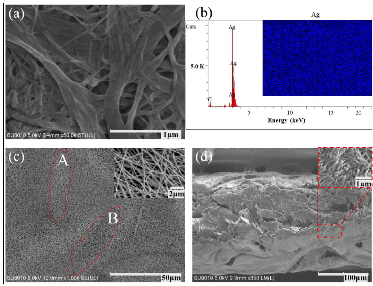
Analyzing the untreated filter paper using SEM, it was confirmed that its surface has fibrous micro-concave–convex structures. As shown in Figure 2a, the filter paper is composed of a multi-layered, microporous structure, and its surface is visibly rough. EDS and SEM then characterized the AgNWs, and their distribution and surface structure on the qualitative filter paper were investigated. Based on the EDS analysis (Figure 2b), it was observed that the surface contained carbon (which was due to the paper itself) and a large amount of Ag. The lower carbon content is because the surface is largely covered by AgNWs, reducing the amount of carbon that can be detected. The inset in Figure 2b shows the distribution of Ag on the paper substrate, wherein it can be observed that the AgNWs were uniformly distributed. Figure 2c shows the SEM image of the paper-based electrode surface wherein two regions of differing contrasts, as demarcated by the letters A and B, can be clearly distinguished. This indicates that the surface of the electrode is rough. By comparing this with Figure 2a, it can be observed that the surface roughness of the AgNW-treated paper is similar to that of the untreated filter paper. Additionally, the inset shows the micromorphology of the AgNWs, wherein it can be observed that they are straight and of similar width. Based on the cross-sectional SEM image of the AgNW-treated filter paper shown in Figure 2d, it can be observed that the AgNWs were partially adsorbed in the filter paper. Additionally, the inset shows that the AgNWs were uniformly distributed across the qualitative filter paper. Thus, due to the good permeability of AgNWs and the superior adsorption capacity of the filter paper, the filter paper is fully compatible with the sensitive AgNWs.
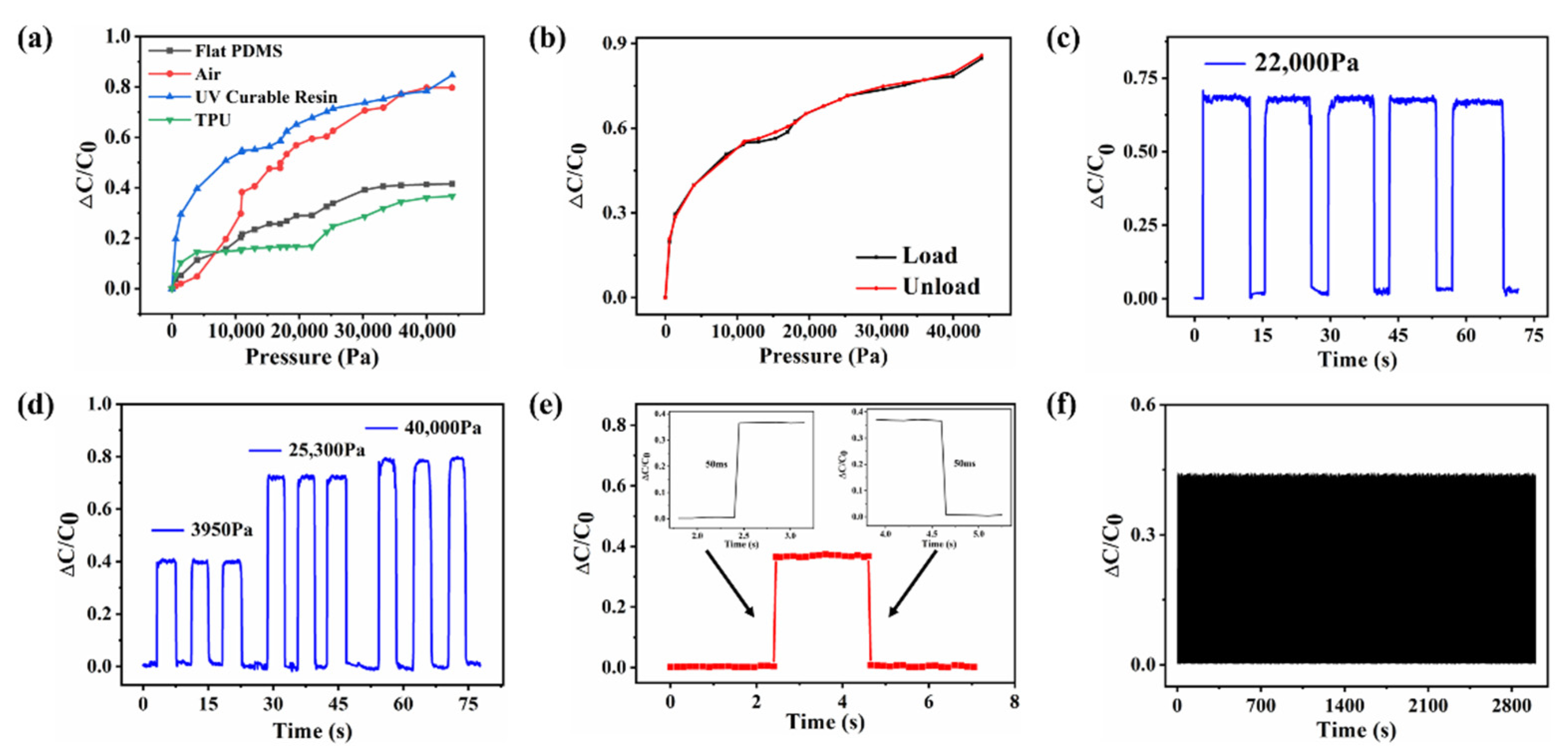
The measured characteristic pressure curves of the fabricated capacitive pressure sensor are shown in Figure 3. The change in the relative capacitance is expressed as ΔC/C0, where ΔC is C−C0 (C is the capacitance value obtained by the impedance analyzer, and C0 represents the initial capacitance of the sensor) [32]. The slope of the ΔC/C0 vs. the pressure curve is the sensitivity of the sensor, i.e., S = δ ( Δ C / C 0 ) / δ p [32]. The measured pressure curves of different dielectrics with the same thickness are shown in Figure 3a, wherein the sensor’s sensitivity is significantly higher when 3D-printed UV resin is used as the dielectric. Therefore, subsequent measurements are mainly performed on sensors using UV resins as the dielectric. From the trend indicated by the blue line in Figure 3a, it can be observed that the pressure sensor’s relative capacitance increases as the external pressure increases, and it is composed of two, roughly linear parts. When the pressure is within 0–3950 Pa, ΔC/C0 is about 0–0.39721, and the calculated sensitivity is about 0.101 kPa−1. In contrast, when the pressure is within 3950–44,000 Pa, ΔC/C0 varies from 0.39721 to 0.84682, and the calculated sensitivity is about 0.01123 kPa−1. Moreover, it is observed that when the applied pressure exceeds 3950 Pa, the sensitivity of the sensor is significantly reduced. This is because the initial application of lower pressure to the grid structure of the UV curable resin results in greater deformation, and the thickness of the grid becomes narrower. Thus, the distance between the upper and lower substrates is drastically reduced, which changes the output capacitance. However, when the applied pressure exceeded 3950 Pa, the elasticity of the dielectric reached its extremum and its thickness could no longer change. Thus, the distance between the upper and lower substrates is slightly reduced, which causes a slight change in the output capacitance, thereby greatly reducing the sensitivity of the sensor. Figure 3b shows the sensor’s behavior during loading and unloading. From this, it can be observed that, upon application of pressure, the relative capacitance increases linearly in two parts. Moreover, it can also be observed that the change in the relative capacitance upon releasing the applied pressure is similar to that when the pressure was applied. Upon complete removal of external pressure, the change in the relative capacitance returns to zero. Thus, the loading and unloading behaviors are similar, indicating that the sensor is stable during pressurization and depressurization, with smaller hysteresis and higher reliability. Figure 3c shows the repeatability measurements of the pressure sensor at an external pressure of 22,000 Pa. Upon application of pressure, the relative capacitance of the sensor increased from 0 to around 0.68 and remained at that value during the pressurized state. Upon releasing the pressure, the relative capacitance of the sensor is reduced to approximately zero. Moreover, upon repeating the loading and unloading cycle five times, the relative capacitance in each cycle is observed to be almost the same, which indicates that it has high stability and repeatability. Even after repeated loading and unloading, the sensor at different pressures (3950 Pa, 25,300 Pa, and 40,000 Pa for three cycles; see Figure 3d) is observed to have excellent repeatability, and the relative capacitance is also observed to increase with a corresponding increase in the pressure. The stable value of the capacitance during loading (even at different external pressures) illustrates that the output of the fabricated pressure sensor has high reliability, and hence, the magnitude of the applied pressure could be confidently determined from the output of the sensor. Furthermore, based on the enlarged graph of the relative capacitance change curve of one-cycle pressure (see the inset of Figure 3e), it is concluded that the response time of the sensor does not exceed 50 ms. The flexible pressure sensor should have high mechanical durability because it needs to maintain a stable input–output relationship under long-term or cyclic loading. Therefore, the sensor is measured under repetitive cyclic loads of 3.75 kPa and 0.5 Hz, and the results are shown in Figure 3f. Based on this, it can be observed that the sensor maintains reliable and consistent pressure-sensing ability even after 1500 cycles.
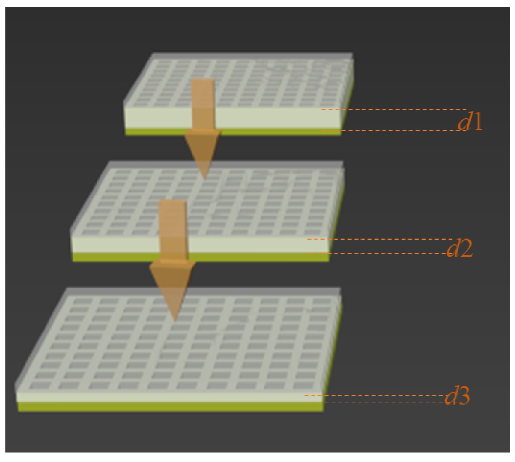
A capacitor consists of two parallel plates and a dielectric sandwiched between them. The capacitance (C) of the capacitor is given by C = ε r ε 0 A / d , where ε0 is the dielectric constant of vacuum, εr is the relative permittivity of the dielectric layer (its change is related to the characteristics of the dielectric), A is the overlapping area of two parallel plates, and d is the distance between the two parallel plates (which depends on the normal force, shear force, and strain). The capacitance of a capacitor is sensitive to εr, A, and d [32]. The pressure-sensing mechanism of the sensor is shown in Figure 4, which depicts a change in the shape of the sensor when pressure is applied. The dielectric layer of the sensor has a regular grid structure when no external pressure is applied. During this state, the distance between the two substrates is d1, and the initial capacitance is C0. Since the dielectric layer has a grid structure, the degree of structural change in the dielectric layer is much higher than that of the no grid structure upon the application of external force, and, hence, its sensitivity is much higher than that of the no-grid electrode. When external pressure is applied to the sensor, the upper substrate of the sensor bends inward and both sides of the dielectric layer are squeezed and deformed. This results in changes to the contact area (A) on both sides of the electrode and the distance between the two substrates (which now becomes d2), which ultimately results in a change in the output capacitance of the sensor. In addition, as the pressure increases, the gap between the grid structures decreases and the grid lines become wider. Therefore, εr increases. These are conducive to the increase in the capacitance of the sensor, thereby improving its sensitivity. When the pressure increases to a certain level, the deformation of the dielectric layer is small due to excessive extrusion of the dielectric layer, and the capacitance first decreases and then saturates, which determines the maximum detection limit of the sensor. The sensitivity of the sensor is compared with related work, as shown in Table 1.
The capacitive pressure sensor was prepared as a sensor array to observe the spatial distribution of different weights. For this, the AgNW-adsorbed filter paper was first cut into rectangles of appropriate size (the width of the rectangle is related to the size of the sensor array and the pixels). The preparation process of the sensor array is shown in Figure 5a. Figure 5b–e shows that two pressure sensor arrays with different pixels were fabricated. The size of the entire pressure sensor array was 30 mm × 30 mm, and the size of the capacitor unit of the pressure sensor with 3 × 3 pixels was 4 mm × 4 mm. For this, six 30 mm × 4 mm filter paper blocks had to be cut out. The size of the capacitor unit of the 5 × 5-pixel pressure sensor was 3 mm × 3 mm, which implies that ten 30 mm × 3 mm pieces from the filter paper were cut out. Two weights with masses of 2 g and 10 g were placed on a 3 × 3-pixel pressure sensor array to measure the spatial resolution and plot the intensity distribution map. As shown in the right of Figure 5b, the magnitudes of the increases in relative capacitance at coordinates (1,3) and (3,1) corresponding to 10 g and 2 g, respectively, are different. In addition, the changes in the relative capacitance at other positions are small. This shows that the capacitance increases only at the location of the load. Similarly, two weights with masses 2 g and 20 g were placed diagonally on a 5 × 5-pixel sensor array, as shown in Figure 5d. The area of the sensing unit of each 5 × 5-pixel pressure sensor array was smaller than that of the 3 × 3-pixel pressure sensor array, and the corresponding two-dimensional intensity distribution graph also shows that the sensor array had an excellent spatial resolution. Next, a weight with a mass of 50 g was placed on the 5 × 5-pixel pressure sensor array at different locations, as shown in Figure 5c,e. Accordingly, different contact areas showed different two-dimensional intensity distributions. This further proves that the sensor has good spatial resolution and that the morphological characteristics of the target can be roughly estimated through the two-dimensional intensity distribution map. To summarize, the above results show that the fabricated flexible paper-based pressure sensor can be used to identify the pressure distribution and has potential application prospects in wearable sensing devices or electronic skins.
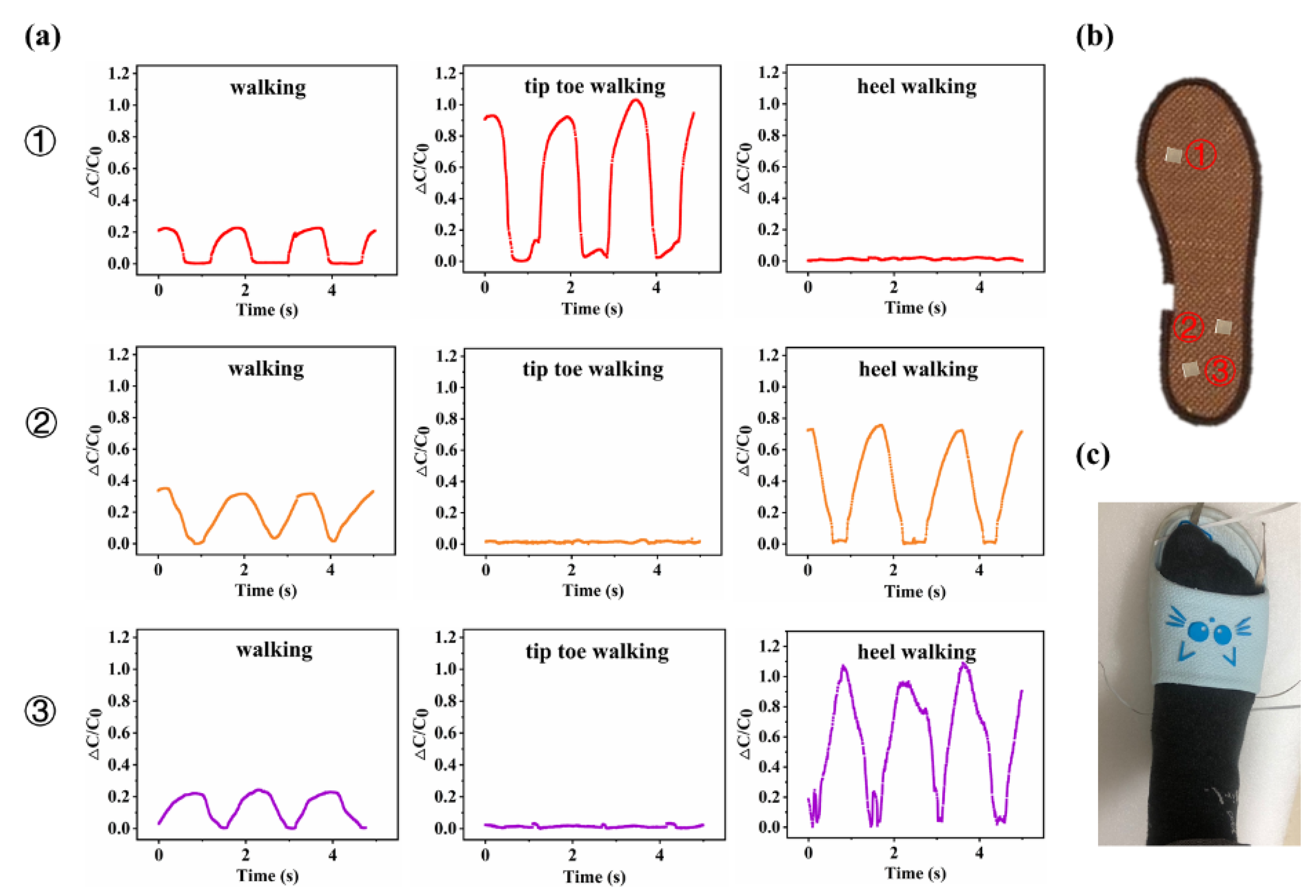
In addition, as shown in Figure 6b,c, three such sensors were installed at different positions on the sole of a shoe for gait measurement. The corresponding results are shown in Figure 6a. When walking normally, it was observed that the change in the relative capacitances of the three sensors was similar, and the pressure at position ② was highest. When tiptoeing, it was observed that the relative capacitance at position ① showed the greatest change, while those at positions ② and ③ remained unchanged. When walking on heels, it was observed that the relative capacitance changed only at positions ② and ③, wherein the change at position ③ was significantly higher than that at position ②, and no pressure change at position ① was observed. This shows that the sensor can provide real-time information about the human body’s activity and, hence, can be used to monitor various activities, such as those undertaken during patient and athlete training.

4. Conclusions

In summary, we report a capacitive paper-based pressure sensor with a dielectric layer that possesses a grid structure. The grid structure of the dielectric layer was prepared by 3D printing, which is simple, easy to implement, and conducive to mass production. In addition, the materials used in the fabrication process of the sensor are low-cost and readily available. The sensor was able to achieve high sensitivity, a wide detection range, and was stable. Tests have proven that an array of such sensors can be used to monitor pressure and analyze gait and, hence, can be applied to wearable sensing devices or electronic skins to monitor various activities of patients and athletes. The above analysis shows that 3D printing technology has broad application prospects for the fabrication of sensors in general.

Author Contributions

Methodology, W.J. and Q.Z.; validation, Y.C. and H.Z., writing—original draft preparation, W.J.; writing—review and editing, Q.Z. and J.J.; funding acquisition, S.S., Y.C., J.W. and J.J. All authors have read and agreed to the published version of the manuscript.

Funding

This research was financially supported by the State Key Program of National Natural Science of China (No. 62031022, S.S.), the National Natural Science Foundation of China (NO. 51975400, S.S.), the Shanxi Provincial Key Research and Development Project (201903D421044, Y.C.), the Natural Science Foundation of Shanxi Province (201901D111097, J.W.), and the Natural Science Foundation Project of Shanxi Province (No.20210302123136, J.J.).

Institutional Review Board Statement

Not applicable.

Informed Consent Statement

Not applicable.

Data Availability Statement

Data available on request.

Conflicts of Interest

The authors declare that they have no known competing financial interest or personal relationships that could have appeared to influence the work reported in this paper.

References

  1. Guo, Y.; Ji, Z.; Zhang, Y.; Wang, X.; Zhou, F. Solvent-free and photocurable polyimide inks for 3D printing. J. Mater. Chem. A 2017, 5, 16307–16314. [Google Scholar] [CrossRef]
  2. Fan, Z.; Feng, H. Study on selective laser melting and heat treatment of Ti-6Al-4V alloy. Results Phys. 2018, 10, 660–664. [Google Scholar] [CrossRef]
  3. Rastogi, P.; Kandasubramanian, B. Breakthrough in the printing tactics for stimuli-responsive materials: 4D printing. Chem. Eng. J. 2019, 366, 264–304. [Google Scholar] [CrossRef]
  4. Zeng, X.; Chen, H.; Chen, L.; Zheng, B. Insights into the relationship between structure and rheological properties of starch gels in hot-extrusion 3D printing. Food Chem. 2021, 342, 128362. [Google Scholar] [CrossRef]
  5. Yang, Y.; Song, X.; Li, X.; Chen, Z.; Zhou, C.; Zhou, Q.; Chen, Y. Recent Progress in Biomimetic Additive Manufacturing Technology: From Materials to Functional Structures. Adv. Mater. 2018, 30, 1706539. [Google Scholar] [CrossRef]
  6. Yang, Y.; Li, X.; Zheng, X.; Chen, Z.; Zhou, Q.; Chen, Y. 3D-Printed Biomimetic Super-Hydrophobic Structure for Microdroplet Manipulation and Oil/Water Separation. Adv. Mater. 2018, 30, 1704912. [Google Scholar] [CrossRef]
  7. Jian, Z.; Zhuang, T.; Qinyu, T.; Liqing, P.; Kun, L.; Xujiang, L.; Diaodiao, W.; Zhen, Y.; Shuangpeng, J.; Xiang, S. 3D bioprinting of a biomimetic meniscal scaffold for application in tissue engineering. Bioact. Mater. 2021, 6, 1711–1726. [Google Scholar] [CrossRef]
  8. Li, Y.; Zhong, J.; Wu, L.; Weng, Z.; Zheng, L.; Peng, S.; Zhang, X. High performance POSS filled nanocomposites prepared via UV-curing based on 3D stereolithography printing. Compos. Part A Appl. Sci. Manuf. 2019, 117, 276–286. [Google Scholar] [CrossRef]
  9. Ji, X.F.; Zhu, H.; Zhao, L.M.; Xiao, J. Recent advances in 3D bioprinting for the regeneration of functional cartilage. Regen. Med. 2018, 13, 73–87. [Google Scholar]
  10. Chin, S.Y.; Dikshit, V.; Meera Priyadarshini, B.; Zhang, Y. Powder-based 3D printing for the fabrication of device with micro and mesoscale features. Micromachines 2020, 11, 658. [Google Scholar] [CrossRef]
  11. Karakurt, I.; Lin, L. 3D printing technologies: Techniques, materials, and post-processing. Curr. Opin. Chem. Eng. 2020, 28, 134–143. [Google Scholar] [CrossRef]
  12. Lu, B.; Lan, H.; Liu, H. Additive manufacturing frontier: 3D printing electronics. Opto-Electron. Adv. 2018, 1, 170004. [Google Scholar] [CrossRef]
  13. Wang, Z.; Gao, W.; Zhang, Q.; Zheng, K.; Xu, J.; Xu, W.; Shang, E.; Jiang, J.; Zhang, J.; Liu, Y. 3D-printed graphene/polydimethylsiloxane composites for stretchable and strain-insensitive temperature sensors. ACS Appl. Mater. Interfaces 2018, 11, 1344–1352. [Google Scholar] [CrossRef] [PubMed]
  14. Joung, K.Y.; Kim, S.Y.; Kang, I.; Cho, S.H. 3D-Printed load cell using nanocarbon composite strain sensor. Sensors 2021, 21, 3675. [Google Scholar] [CrossRef]
  15. Seo, M.; Hwang, S.; Hwang, T.; Yeo, J. Fabrication of soft sensor using laser processing techniques: For the alternative 3D printing process. Materials 2019, 12, 2955. [Google Scholar] [CrossRef] [Green Version]
  16. Eijking, B.; Sanders, R.; Krijnen, G. Development of whisker inspired 3D multi-material printed flexible tactile sensors. In Proceedings of the 2017 IEEE Sensors, Glasgow, UK, 29 October–1 November 2017; pp. 1–3. [Google Scholar]
  17. Li, B.; Zhang, S.; Zhang, L.; Gao, Y.; Xuan, F. Strain sensing behavior of FDM 3D printed carbon black filled TPU with periodic configurations and flexible substrates. J. Manuf. Process. 2022, 74, 283–295. [Google Scholar] [CrossRef]
  18. Xiang, D.; Zhang, Z.; Han, Z.; Zhang, X.; Zhou, Z.; Zhang, J.; Luo, X.; Wang, P.; Zhao, C.; Li, Y. Effects of non-covalent interactions on the properties of 3D printed flexible piezoresistive strain sensors of conductive polymer composites. Compos. Interfaces 2021, 28, 577–591. [Google Scholar] [CrossRef]
  19. Wan, Y.; Wang, Y.; Guo, C.F. Recent progresses on flexible tactile sensors. Mater. Today Phys. 2017, 1, 61–73. [Google Scholar] [CrossRef]
  20. Huang, Y.; Fan, X.; Chen, S.C.; Zhao, N. Emerging technologies of flexible pressure sensors: Materials, modeling, devices, and manufacturing. Adv. Funct. Mater. 2019, 29, 1808509. [Google Scholar] [CrossRef]
  21. Luo, Y.; Shao, J.; Chen, S.; Chen, X.; Tian, H.; Li, X.; Wang, L.; Wang, D.; Lu, B. Flexible capacitive pressure sensor enhanced by tilted micropillar arrays. ACS Appl. Mater. Interfaces 2019, 11, 17796–17803. [Google Scholar] [CrossRef]
  22. Li, T.; Luo, H.; Qin, L.; Wang, X.; Xiong, Z.; Ding, H.; Gu, Y.; Liu, Z.; Zhang, T. Flexible capacitive tactile sensor based on micropatterned dielectric layer. Small 2016, 12, 5042–5048. [Google Scholar] [CrossRef] [PubMed]
  23. Gross, B.C.; Erkal, J.L.; Lockwood, S.Y.; Chen, C.; Spence, D.M. Evaluation of 3D printing and its potential impact on biotechnology and the chemical sciences. Anal. Chem. 2014, 86, 3240–3253. [Google Scholar] [CrossRef] [PubMed]
  24. Chen, S.; Song, Y.; Ding, D.; Ling, Z.; Xu, F. Flexible and anisotropic strain sensor based on carbonized crepe paper with aligned cellulose fibers. Adv. Funct. Mater. 2018, 28, 1802547. [Google Scholar] [CrossRef]
  25. Hua, D.; Zhang, X.; Ji, Z.; Yan, C.; Yu, B.; Li, Y.; Wang, X.; Zhou, F. 3D printing of shape changing composites for constructing flexible paper-based photothermal bilayer actuators. J. Mater. Chem. C 2018, 6, 2123–2131. [Google Scholar] [CrossRef]
  26. Li, Y.; Samad, Y.A.; Taha, T.; Cai, G.; Fu, S.Y.; Liao, K. Highly flexible strain sensor from tissue paper for wearable electronics. ACS Sustain. Chem. Eng. 2016, 4, 4288–4295. [Google Scholar] [CrossRef]
  27. Long, Y.; He, P.; Xu, R.; Hayasaka, T.; Shao, Z.; Zhong, J.; Lin, L. Molybdenum-carbide-graphene composites for paper-based strain and acoustic pressure sensors. Carbon 2020, 157, 594–601. [Google Scholar] [CrossRef]
  28. Liao, X.; Zhang, Z.; Liang, Q.; Liao, Q.; Zhang, Y. Flexible, cuttable, and self-waterproof bending strain sensors using microcracked gold nanofilms@ paper substrate. ACS Appl. Mater. Interfaces 2017, 9, 4151–4158. [Google Scholar] [CrossRef]
  29. Guo, H.; Yin, Z.; Namkoong, M.; Li, Y.; Nguyen, T.; Salcedo, E.; Arizpe, I.; Tian, L. Printed Ultrastable Bioplasmonic Microarrays for Point-of-Need Biosensing. ACS Appl. Mater. Interfaces 2022, 14, 10729–10737. [Google Scholar] [CrossRef]
  30. Santhiago, M.; Corrêa, C.C.; Bernardes, J.S.; Pereira, M.P.; Oliveira, L.J.; Strauss, M.; Bufon, C.C. Flexible and foldable fully-printed carbon black conductive nanostructures on paper for high-performance electronic, electrochemical, and wearable devices. ACS Appl. Mater. Interfaces 2017, 9, 24365–24372. [Google Scholar] [CrossRef]
  31. Li, Y.Q.; Liang, F. Progress in paper-based colorimetric sensor array. Chin. J. Anal. Chem. 2020, 48, 1448–1457. [Google Scholar] [CrossRef]
  32. Zhang, Q.; Jia, W.; Ji, C.; Pei, Z.; Jing, Z.; Cheng, Y.; Zhang, W.; Zhuo, K.; Ji, J.; Yuan, Z. Flexible wide-range capacitive pressure sensor using micropore PE tape as template. Smart Mater. Struct. 2019, 28, 115040. [Google Scholar] [CrossRef]
  33. He, Z.; Chen, W.; Liang, B.; Liu, C.; Yang, L.; Lu, D.; Mo, Z.; Zhu, H.; Tang, Z.; Gui, X. Capacitive pressure sensor with high sensitivity and fast response to dynamic interaction based on graphene and porous nylon networks. ACS Appl. Mater. Interfaces 2018, 10, 12816–12823. [Google Scholar] [CrossRef] [PubMed]
  34. Ke, K.; McMaster, M.; Christopherson, W.; Singer, K.D.; Manas-Zloczower, I. Highly sensitive capacitive pressure sensors based on elastomer composites with carbon filler hybrids. Compos. Part A Appl. Sci. Manuf. 2019, 126, 105614. [Google Scholar] [CrossRef]
  35. Metzger, C.; Fleisch, E.; Meyer, J.; Dansachmüller, M.; Graz, I.; Kaltenbrunner, M.; Keplinger, C.; Schwödiauer, R.; Bauer, S. Flexible-foam-based capacitive sensor arrays for object detection at low cost. Appl. Phys. Lett. 2008, 92, 013506. [Google Scholar] [CrossRef]
  36. Lei, K.F.; Lee, K.F.; Lee, M.Y. Development of a flexible PDMS capacitive pressure sensor for plantar pressure measurement. Microelectron. Eng. 2012, 99, 1–5. [Google Scholar] [CrossRef]
  37. Wang, X.; Li, T.; Adams, J.; Yang, J. Transparent, stretchable, carbon-nanotube-inlaid conductors enabled by standard replication technology for capacitive pressure, strain and touch sensors. J. Mater. Chem. A 2013, 1, 3580–3586. [Google Scholar] [CrossRef]
  38. Dobrzynska, J.A.; Gijs, M. Polymer-based flexible capacitive sensor for three-axial force measurements. J. Micromech. Microeng. 2012, 23, 015009. [Google Scholar] [CrossRef]
  39. Lei, K.F.; Lee, K.F.; Lee, M.Y. A flexible PDMS capacitive tactile sensor with adjustable measurement range for plantar pressure measurement. Microsyst. Technol. 2014, 20, 1351–1358. [Google Scholar] [CrossRef]
  40. Yao, S.; Zhu, Y. Wearable multifunctional sensors using printed stretchable conductors made of silver nanowires. Nanoscale 2014, 6, 2345–2352. [Google Scholar] [CrossRef]
  41. Atalay, O.; Atalay, A.; Gafford, J.; Walsh, C. A highly sensitive capacitive-based soft pressure sensor based on a conductive fabric and a microporous dielectric layer. Adv. Mater. Technol. 2018, 3, 1700237. [Google Scholar] [CrossRef]
  42. Zhang, B.; Xiang, Z.; Zhu, S.; Hu, Q.; Cao, Y.; Zhong, J.; Zhong, Q.; Wang, B.; Fang, Y.; Hu, B. Dual functional transparent film for proximity and pressure sensing. Nano Res. 2014, 7, 1488–1496. [Google Scholar] [CrossRef]
  43. Subramaniam, S.; Majumder, S.; Faisal, A.I.; Deen, M.J. Insole-based systems for health monitoring: Current solutions and research challenges. Sensors 2022, 22, 438. [Google Scholar] [CrossRef] [PubMed]
  44. Baek, S.; Jang, H.; Kim, S.Y.; Jeong, H.; Han, S.; Jang, Y.; Kim, D.H.; Lee, H.S. Flexible piezocapacitive sensors based on wrinkled microstructures: Toward low-cost fabrication of pressure sensors over large areas. RSC Adv. 2017, 7, 39420–39426. [Google Scholar] [CrossRef]
  45. Maddipatla, D.; Narakathu, B.B.; Ali, M.M.; Chlaihawi, A.A.; Atashbar, M.Z. Development of a novel carbon nanotube based printed and flexible pressure sensor. In Proceedings of the 2017 IEEE Sensors Applications Symposium (SAS), Glassboro, NJ, USA, 13–15 March 2017; pp. 1–4. [Google Scholar]
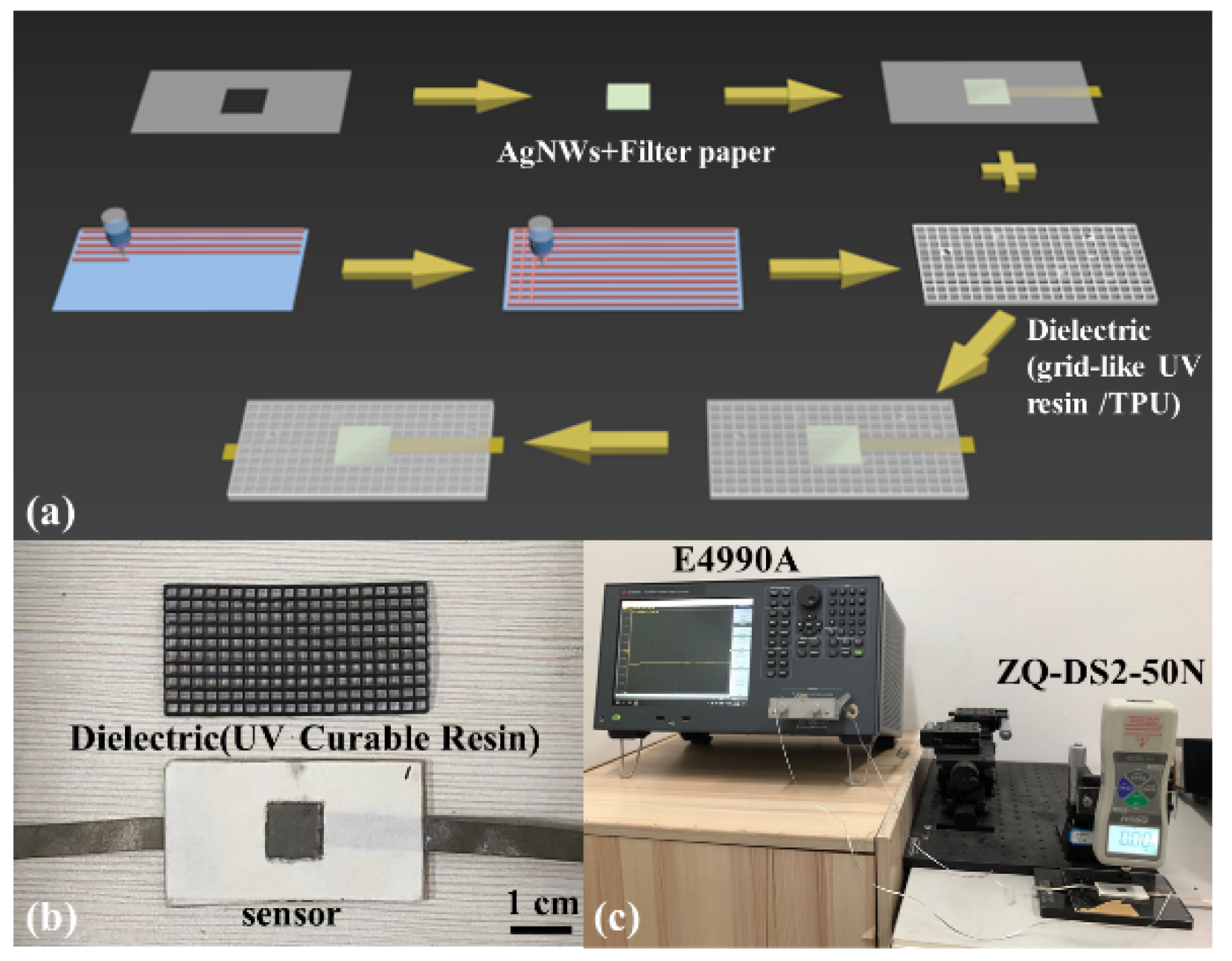
Figure 1. (a) The fabrication process of a capacitive paper-based pressure sensor using 3D printing; (b) Optical photo of UV-curable resin and sensor (4.84 cm × 2.44 cm); (c) Test platform.
Chemosensors 10 00432 g001
Figure 2. (a) SEM image of the filter paper surface; (b) EDS data and distribution of Ag across the electrode; (c) SEM image of the electrode, an enlarged image of the electrode is shown in the inset (Area A is the bright part of the figure, and area B is the shaded part); and (d) cross-sectional SEM image of the electrode.
Chemosensors 10 00432 g002
Figure 3. (a) Change in the relative capacitance of the paper-based pressure sensor with different dielectric; (b) Change in the relative capacitance during loading and unloading; (c) Repeatability measurement of the sensor under an external pressure of 22,000 Pa; (d) Repeatability measurement of the sensor at three different pressures; (e) Sensor response time; (f) Sensor durability measurement.
Chemosensors 10 00432 g003
Figure 4. Sensing mechanism of the paper-based capacitive pressure sensor.
Chemosensors 10 00432 g004
Figure 5. Application measurements of the pressure sensor: changes in the relative capacitance due to different weights and contact areas placed on the sensor array.
Chemosensors 10 00432 g005
Figure 6. Application measurement curve of the sensor: changes in the relative capacitances of sensors attached to the sole of a shoe during walking. (a) Variation of the relative capacitance output under different gaits when the paper-based pressure sensor is installed at the sole position ①, ② and ③, respectively; (b) Position distribution of paper-based pressure sensors installed at the sole; (c) Optical photograph of the gait analysis test.
Chemosensors 10 00432 g006
Table 1. Comparison of sensors’ sensitivity.
Dielectric MaterialPressure Range (kPa)Sensitivity (kPa−1)Ref.
Graphene and porous nylon n/w0–1
1–5
0.33
0.007
[33]
Branched-CNTs and GNPs (3:1)0–12000.00205[34]
Polyolefin foam0–1.0298.25 fF[35]
PDMS0–9450.0019[36]
Ecoflex10–1500.00059[37]
PDMS/Parylene-C0–10
10–140
0.024
0.00066
[38]
PDMS240–10000.000022[39]
Ecoflex0–500
500–1400
0.0016
0.00057
[40]
Silicone0–1000.0121[41]
PDMS0–500.0004[42]
PDMS0–15.601
15.601–45
0.0486
0.0025
[32]
Ecoflex silicone elastomer0–2000.012[43]
Wrinkled μ-structures Ecoflex<1
>8
0.0012
0.0000042
[44]
PDMS1.52–3370.021[45]
Grid-like UV resin0.3–3.95
3.95–44
0.101
0.01123
This work
Publisher’s Note: MDPI stays neutral with regard to jurisdictional claims in published maps and institutional affiliations.