Melanoma of the Hand: Current Practice and New Frontiers

Abstract
:1. Introduction
2. General Principles of Hand Melanoma (Non-Nail Unit Lesions)
2.1. What Is Different about Hand Melanoma?
2.2. Diagnosis
2.3. Staging and Management
2.3.1. Margins of Excision

2.3.2. Sentinel Lymph Node Biopsy
2.4. Role of Mohs Micrographic Surgery
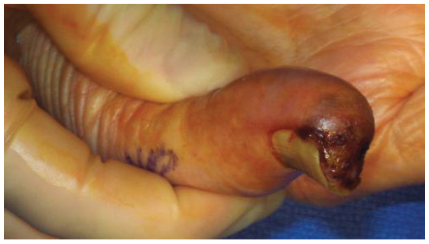
3. Subungual Melanoma
3.1. Diagnosis
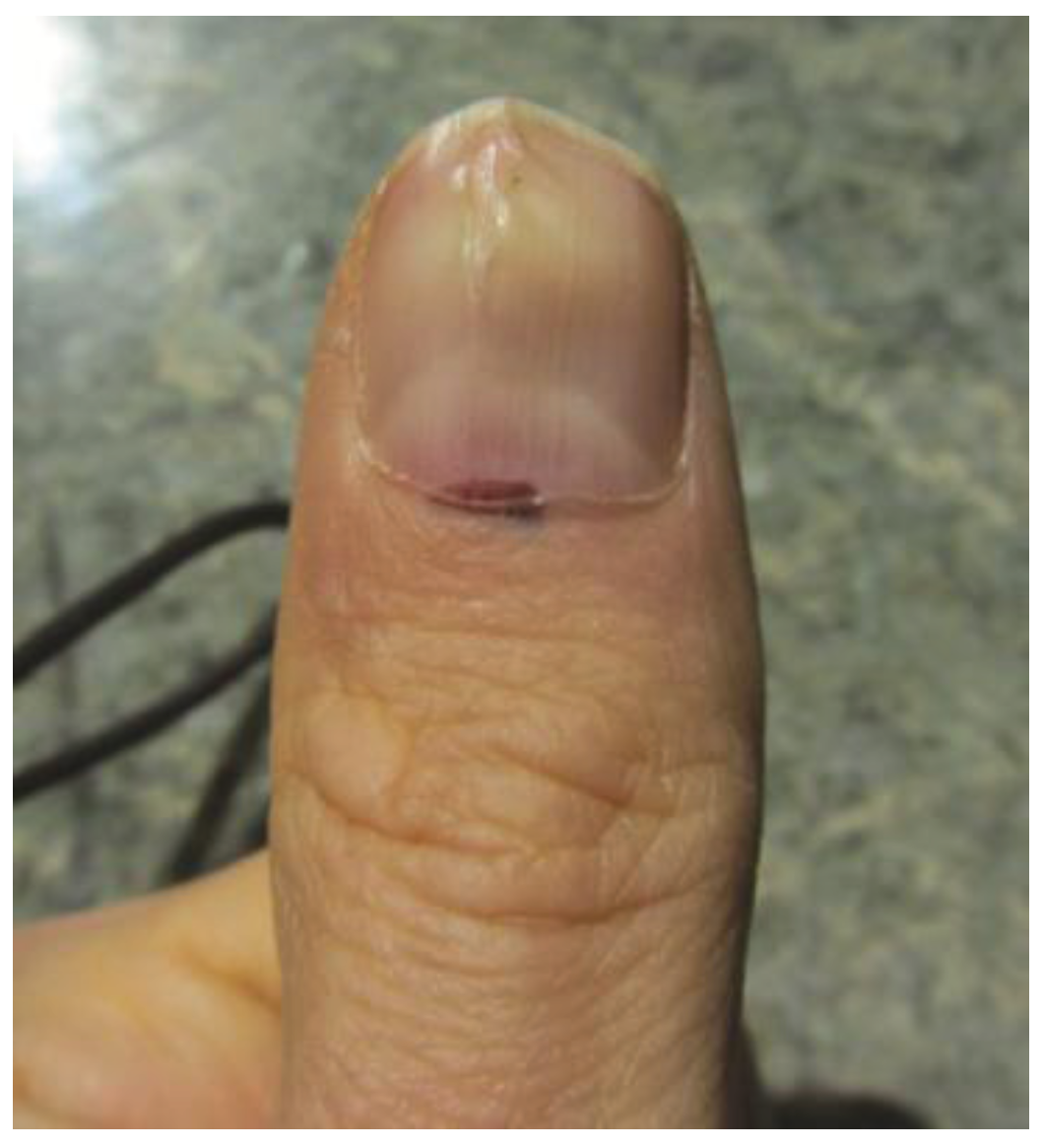
3.2. Staging

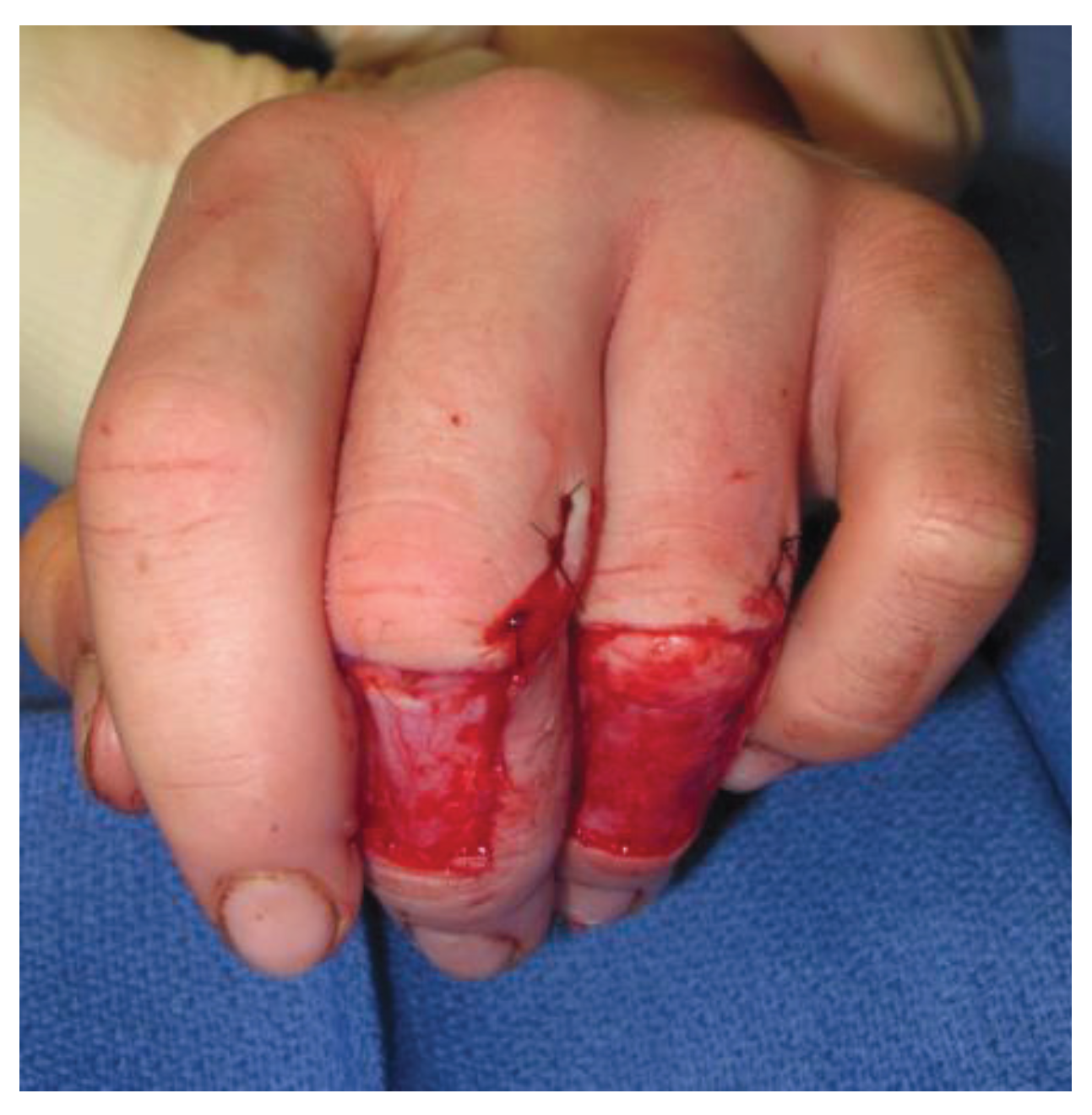
3.3. Staging and Management
3.4. Prognostic Data
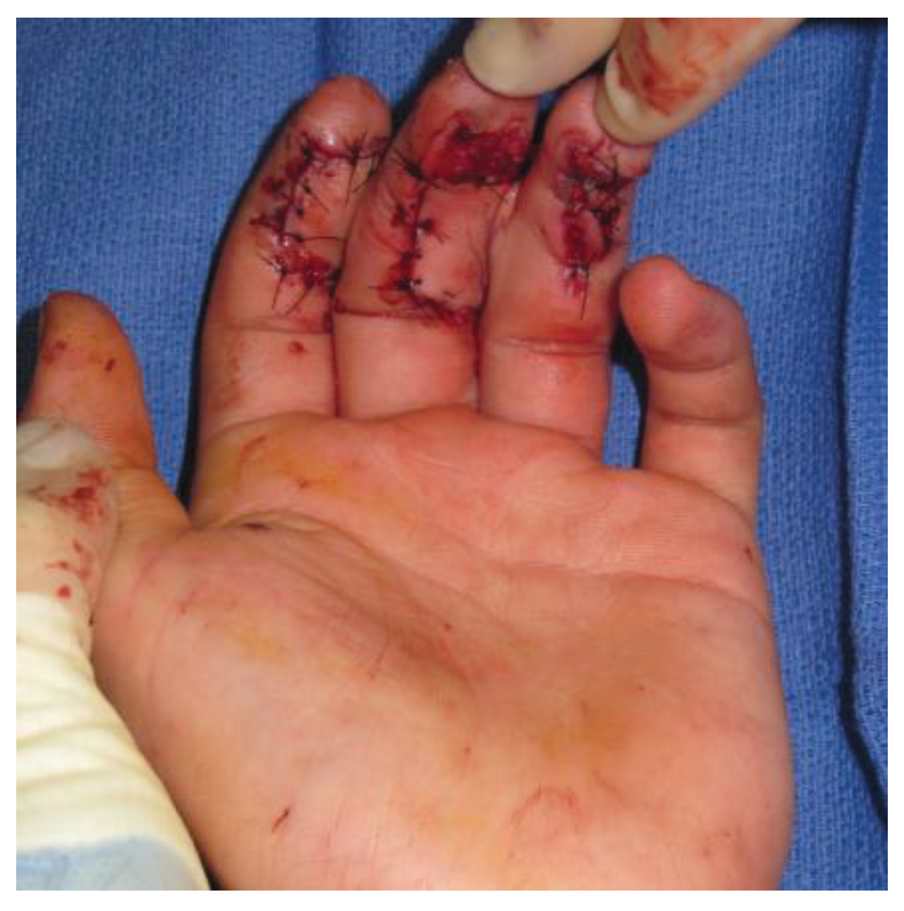
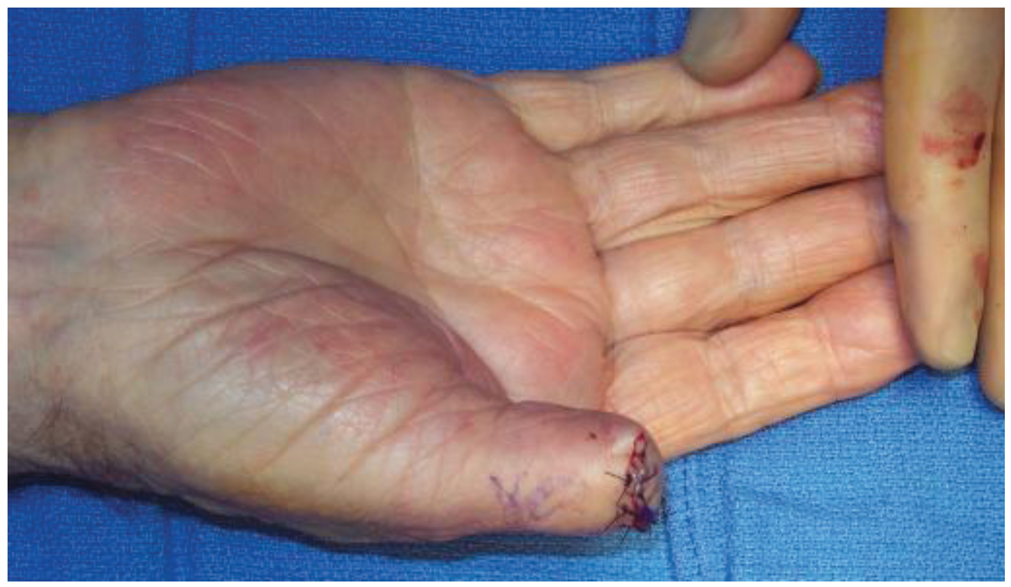
4. Surgical Management
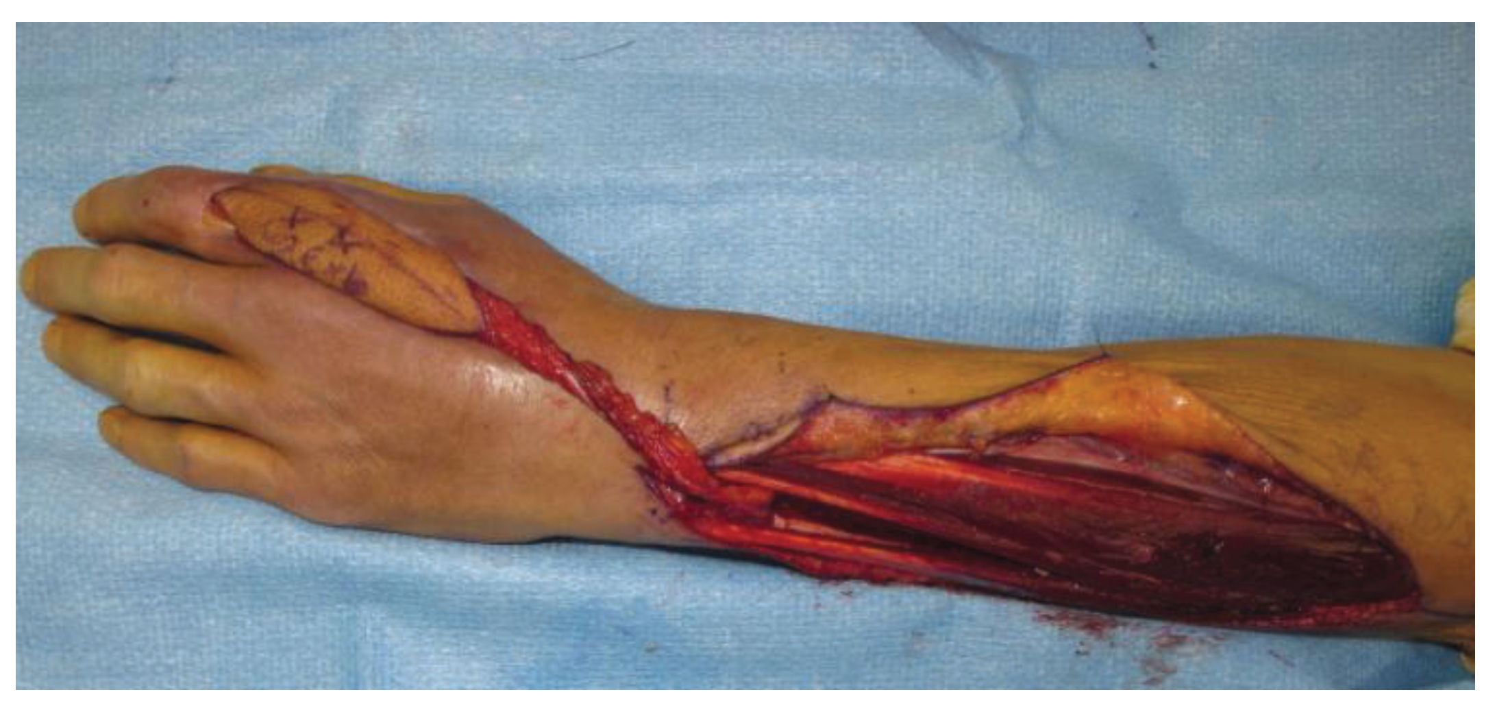
4.1. Reconstructive Goals in Hand Surgery
4.2. Soft Tissue Reconstruction

5. Adjunct Therapy
5.1. General Considerations
5.2. Hyperthermic Isolated Limb Perfusion and Isolated Limb Infusion
6. Conclusions
Acknowledgments
Author Contributions
Conflicts of Interest
References
- American Cancer Society: Cancer Facts and Figures 2013. Available online: http://www.cancer.org/acs/groups/content/@epidemiologysurveilance/documents/document/acspc-036845/ (accessed on 28 September 2013).
- Mason, M.L. Carcinoma of the hand. Arch. Surg. 1929, 18, 2107–2158. [Google Scholar] [CrossRef]
- English, C.; Hammert, W.C. Cutaneous malignancies of the upper extremity. J. Hand Surg. Am. 2012, 37, 367–377. [Google Scholar] [CrossRef]
- Yu, H.L.; Chase, R.A.; Strauch, B. Atlas of Hand Anatomy and Clinical Implications; Mosby: St. Louis, MO, USA, 2004. [Google Scholar]
- Maciburko, S.J.; Townley, W.A.; Hollowood, K.; Giele, H.P. Skin cancers of the hand: Series of 541 malignancies. Plast. Reconstr. Surg. 2012, 129, 1329–1336. [Google Scholar] [CrossRef]
- Rex, J.; Paradelo, C.; Mangas, C.; Hilari, J.M.; Fernández-Figueras, M.T.; Ferrándiz, C. Management of primary cutaneous melanoma of the hands and feet: A clinicoprognostic study. Dermatol. Surg. 2009, 35, 1505–1513. [Google Scholar] [CrossRef]
- Durbec, F.; Martin, L.; Derancourt, C.; Grange, F. Melanoma of the hand and foot: Epidemiological, prognostic and genetic features. A systematic review. Br. J. Dermatol. 2012, 166, 727–739. [Google Scholar] [CrossRef]
- Neila, J.; Soyer, H. Key points in dermoscopy for diagnosis of melanomas, including difficult to diagnose melanomas, on the trunk and extremities. J. Dermatol. 2011, 38, 3–9. [Google Scholar] [CrossRef]
- Brehmer, F.; Ulrich, M.; Haenssle, H. Strategies for early recognition of cutaneous melanoma-present and future. Dermatol. Pract. Concept. 2012, 2, 29–37. [Google Scholar]
- Breslow, A. Thickness, cross-sectional areas and depth of invasion in the prognosis of cutaneous melanoma. Ann. Surg. 1970, 172, 902–908. [Google Scholar] [CrossRef]
- Corona, R.; Mele, A.; Amini, M.; de Rosa, G.; Coppola, G.; Piccardi, P.; Fucci, M.; Pasquini, P.; Faraggiana, T. Interobserver variability on the histopathologic diagnosis of cutaneous melanoma and other pigmented skin lesions. J. Clin. Oncol. 1996, 14, 1218–1223. [Google Scholar]
- Morton, D.L.; Cochran, A.J.; Thompson, J.F. The rationale for sentinel-node biopsy in primary melanoma. Nat. Clin. Pract. Oncol. 2008, 5, 510–511. [Google Scholar] [CrossRef]
- Handley, W.S. The pathology of melanotic growths in relation to their operative treatment. Lancet 1907, i, 996–1003. [Google Scholar]
- McKenna, D.B.; Lee, R.J.; Prescott, R.J.; Doherty, V.R. The time from diagnostic excision biopsy to wide local excision for primary cutaneous malignant melanoma may not affect patient survival. Br. J. Dermatol. 2002, 147, 48–54. [Google Scholar] [CrossRef]
- Ross, M.I.; Balch, C.M. Surgical treatment of primary melanoma. In Cutaneous Melanoma, 3rd ed.; Balch, C.M., Houghton, A.N., Sober, A.J., Soong, S., Eds.; Quality Medical Publishing Inc.: St. Louis, MO, USA, 1998; pp. 142–152. [Google Scholar]
- Clinical Practice Guidelines for the Management of Cutaneous Melanoma in Australia and New Zealand. Available online: http://www.cancer.org.au/content/pdf/HealthProfessionals/ClinicalGuidelines/ClinicalPracticeGuidelines-ManagementofMelanoma.pdf (accessed on 28 September 2013).
- Van Everdingen, J.J.; van der Rhee, H.J.; Koning, C.C.; Nieweg, O.E.; Kruit, W.H.; Coebergh, J.W.; Ruiter, D.J. Guideline “Melanoma” (3rd revision). Ned. Tijdschr. Geneeskd. 2005, 149, 1839–1843. [Google Scholar]
- Bishop, J.A.; Corrie, P.G.; Evans, J.; Gore, M.E.; Hall, P.N.; Kirkham, N.; Roberts, D.L.; Anstey, A.V.; Barlow, R.J.; Cox, N.H. UK guidelines for the management of cutaneous melanoma. Br. J. Plast. Surg. 2002, 55, 46–54. [Google Scholar] [CrossRef]
- Namikawa, K.; Yamazaki, N. Sentinel lymph node biopsy guided by indocyanine green fluorescence for cutaneous melanoma. Eur. J. Dermatol. 2011, 21, 184–190. [Google Scholar]
- Suver, D.W.; Friedrich, J.B. Sentinel lymph node biopsy for tumors of the hand and wrist. J. Hand Surg. Am. 2010, 35, 1209–1210. [Google Scholar] [CrossRef]
- Ling, A.; Dawkins, R.; Bailey, M.; Leung, M.; Cleland, H.; Serpell, J.; Kelly, J. Short-termmorbidity associated with sentinel lymph node biopsy in cutaneous malignant melanoma. Australas. J. Dermatol. 2010, 51, 13–17. [Google Scholar] [CrossRef]
- Connolly, S.M.; Baker, D.R.; Coldiron, B.M.; Fazio, M.J.; Storrs, P.A.; Vidimos, A.T.; Zalla, M.J.; Brewer, J.D.; Smith-Begolka, W.; Berger, T.G.; et al. AAD/ACMS/ASDSA/ASMS 2012 appropriate use criteria for Mohs micrographic surgery: A report of the American Academy of Dermatology, American College of Mohs Surgery, American Society for Dermatologic Surgery Association, and the American Society for Mohs Surgery. J. Am. Acad. Dermatol. 2012, 67, 531–550. [Google Scholar] [CrossRef]
- Brodland, D.G. The treatment of nail apparatus melanoma with Mohs micrographic surgery. Dermatol. Surg. 2001, 27, 269–273. [Google Scholar] [CrossRef]
- Do, A.N.; Goleno, K.; Geisse, J.K. Mohs micrographic surgery and partial amputation preserving function and aesthetics in digits: Case reports of invasive melanoma and digital dermatofibrosarcoma protuberans. Dermatol. Surg. 2006, 32, 1516–1521. [Google Scholar] [CrossRef]
- Kottschade, L.A.; Grotz, T.E.; Dronca, R.S.; Salomao, D.R.; Pulido, J.S.; Wasif, N.; Jakub, J.W.; Bagaria, S.P.; Kumar, R.; Kaur, J.S.; et al. Rare Presentations of Primary Melanoma and Special Populations: A Systematic Review. Am. J. Clin. Oncol. 2013. [Google Scholar] [CrossRef]
- O’Connor, E.A.; Dzwierzynski, W. Longitudinal melonychia: Clinical evaluation and biopsy technique. J. Hand Surg. Am. 2011, 36, 1852–1854. [Google Scholar] [CrossRef]
- Baek, H.J.; Lee, S.J.; Cho, K.H.; Choo, H.J.; Lee, S.M.; Lee, Y.H.; Suh, K.J.; Moon, T.Y.; Cha, J.G.; Yi, J.H.; et al. Subungual tumors: Clinicopathologic correlation with US and MR imaging findings. Radiographics 2010, 30, 1621–1636. [Google Scholar] [CrossRef]
- Levit, E.K.; Kagem, M.H.; Scher, R.K.; Grossman, M.; Altman, E. The ABC rule for clinical detection of subungual melanoma. J. Am. Acad. Dermatol. 2000, 42, 269–274. [Google Scholar] [CrossRef]
- Puhaindran, M.E.; Rothrock, C.P.; Athanasian, E.A. Surgical management for malignant tumors of the thumb. Hand 2011, 6, 373–377. [Google Scholar] [CrossRef]
- Moehrle, M.; Metzger, S.; Shippert, W.; Garbe, C.; Rassner, G.; Breuninger, H. “Functional” surgery in subungual melanoma. Dermatol. Surg. 2003, 29, 366–374. [Google Scholar] [CrossRef]
- Yun, M.J.; Park, J.U.; Kwon, S.T. Surgical options for malignant skin tumors of the hand. Arch. Plast. Surg. 2013, 40, 238–243. [Google Scholar] [CrossRef]
- Friedrich, J.B.; Katolik, L.I.; Vedder, N.B. Soft tissue reconstruction of the hand. J. Hand Surg. Am. 2009, 34A, 1148–1155. [Google Scholar] [CrossRef]
- Eberlin, K.R.; Chang, J.; Curtin, C.M.; Sammer, D.M.; Saint-Cyr, M.; Taghinia, A.H. Soft tissue coverage of the hand: A case-based approach. Plast. Reconstr. Surg. 2014, 133, 91–101. [Google Scholar] [CrossRef]
- Kudchadkar, R.R.; Smalley, K.S.; Glass, L.F.; Trimble, J.S.; Sondak, V.K. Targeted therapy in melanoma. Clin. Dermatol. 2013, 31, 200–208. [Google Scholar] [CrossRef]
- Beasley, G.M.; Petersen, R.P.; Yoo, J.; McMahon, N.; Aloia, T.; Petros, W.; Sanders, G.; Cheng, T.Y.; Pruitt, S.K.; Seigler, H.; et al. Isolated limb infusion for in-transit malignant melanoma of the extremity: A well-tolerated but less effective alternative to hyperthermic isolated limb perfusion. Ann. Surg. Oncol. 2008, 15, 2195–2205. [Google Scholar] [CrossRef]
© 2014 by the authors; licensee MDPI, Basel, Switzerland. This article is an open access article distributed under the terms and conditions of the Creative Commons Attribution license (http://creativecommons.org/licenses/by/3.0/).
Share and Cite
Turner, J.B.; Rinker, B. Melanoma of the Hand: Current Practice and New Frontiers. Healthcare 2014, 2, 125-138. https://doi.org/10.3390/healthcare2010125
Turner JB, Rinker B. Melanoma of the Hand: Current Practice and New Frontiers. Healthcare. 2014; 2(1):125-138. https://doi.org/10.3390/healthcare2010125
Chicago/Turabian StyleTurner, John Brad, and Brian Rinker. 2014. "Melanoma of the Hand: Current Practice and New Frontiers" Healthcare 2, no. 1: 125-138. https://doi.org/10.3390/healthcare2010125
APA StyleTurner, J. B., & Rinker, B. (2014). Melanoma of the Hand: Current Practice and New Frontiers. Healthcare, 2(1), 125-138. https://doi.org/10.3390/healthcare2010125