Screening for Left Ventricular Hypertrophy Using Artificial Intelligence Algorithms Based on 12 Leads of the Electrocardiogram—Applicable in Clinical Practice?—Critical Literature Review with Meta-Analysis
Abstract
1. Introduction
2. Materials and Methods
- -
- Population: adult patients (out- and inpatients) with written electrocardiogram
- -
- Intervention: analysis of electrocardiogram
- -
- Comparator: human interpretation with imaging modalities
- -
- Outcome: sensitivity and specificity of AI algorithms
- Inclusion criteria:
- -
- Studies that utilize ML or DL algorithms with ECG data for diagnosing LVH
- -
- Research conducted in the last few years
- -
- Studies where LVH diagnosis is confirmed through echocardiography or cardiac MRI.
- -
- Research that directly compares AI results with the imaging-based LVH diagnosis.
- Exclusion criteria:
- -
- Studies that do not involve ML or DL techniques
- -
- Research performed before 2000
- -
- Studies lacking standardized datasets for evaluation
- -
- Models with limited interpretability
- -
- Inconsistency in performance when using new datasets
- -
- Concerns related to security, privacy of health data, and collaboration with physicians
- -
- Studies using other ECG configurations (e.g., single-lead or 3-lead)
- -
- Research unrelated to LVH
- -
- Studies with inadequate ECG data quality
- -
- Studies using other imaging modalities not specified (e.g., thoracic computed tomography)
- -
- Research without a direct comparison between AI and imaging-based LVH diagnosis
- -
- Investigations with inadequate sample sizes or methodological flaws.
- -
- Definition of title and objective of the review
- -
- Searching the diagnostic studies
- -
- Selection of included studies and data extraction
- -
- Assessment of study quality
- -
- Statistical analysis
- -
- Interpretation of results and development of recommendations
3. Results
4. Discussion
4.1. Performance Variability and Model Selection
4.2. Impact of Clinical Data Integration
4.3. Complexity of LVH Diagnosis
4.4. Imaging Modality Considerations
4.5. Challenges in Machine Learning Application
5. Conclusions
Author Contributions
Funding
Institutional Review Board Statement
Informed Consent Statement
Data Availability Statement
Conflicts of Interest
References
- Weir, M.R.; Townsend, R.R. What is left ventricular hypertrophy and is there a reason to regress left ventricular hypertrophy? J. Clin. Hypertens. 2009, 11, 407–410. [Google Scholar] [CrossRef] [PubMed] [PubMed Central]
- Levy, D.; Garrison, R.J.; Savage, D.D.; Kannel, W.B.; Castelli, W.P. Prognostic implications of echocardiographically determined left ventricular mass in the Framingham Heart Study. N. Engl. J. Med. 1990, 322, 1561–1566. [Google Scholar] [CrossRef]
- Bacharova, L.; Chevalier, P.; Gorenek, B.; Jons, C.; Li, Y.-G.; Locati, E.T.; Maanja, M.; Pérez-Riera, A.R.; Platonov, P.G.; Ribeiro, A.L.P.; et al. ISE/ISHNE expert consensus statement on the ECG diagnosis of left ventricular hypertrophy: The change of the paradigm. Ann. Noninvasive Electrocardiol. 2024, 29, e13097. [Google Scholar] [CrossRef] [PubMed]
- Siranart, N.; Deepan, N.; Techasatian, W.; Phutinart, S.; Sowalertrat, W.; Kaewkanha, P.; Pajareya, P.; Tokavanich, N.; Prasitlumkum, N.; Chokesuwattanaskul, R. Diagnostic accuracy of artificial intelligence in detecting left ventricular hypertrophy by electrocardiograph: A systematic review and meta-analysis. Sci. Rep. 2024, 10, 15882. [Google Scholar] [CrossRef] [PubMed] [PubMed Central]
- Petmezas, G.; Stefanopoulos, L.; Kilintzis, V.; Tzavelis, A.; Rogers, J.A.; Katsaggelos, A.K.; Maglaveras, N. State-of-the-Art Deep Learning Methods on Electrocardiogram Data: Systematic Review. JMIR Med. Inform. 2022, 10, e38454. [Google Scholar] [CrossRef] [PubMed]
- Ansari, Y.; Moura, O.; Qaraqe, K.; Serpedin, E. Deep learning for ECG Arrhythmia detection and classification: An overview of progress for period 2017–2023. Front. Physiol. 2023, 14, 1246746. [Google Scholar] [CrossRef]
- Ding, C.; Yao, T.; Wu, C.; Ni, J. Advances in deep learning for personalized ECG diagnostics: A systematic review addressing inter-patient variability and generalization constraints. Biosens. Bioelectron. 2025, 271, 117073. [Google Scholar] [CrossRef] [PubMed]
- Akbilgic, O.; Butler, L.; Karabayir, I.; Chang, P.P.; Kitzman, D.W.; Alonso, A.; Chen, L.Y.; Soliman, E.Z. ECG-AI: Electrocardiographic artificial intelligence model for prediction of heart failure. Eur. Heart J. Digit. Health 2021, 2, 626–634. [Google Scholar] [CrossRef] [PubMed] [PubMed Central]
- Ko, W.Y.; Siontis, K.C.; Attia, Z.I.; Carter, R.E.; Kapa, S.; Ommen, S.R.; Demuth, S.J.; Ackerman, M.J.; Gersh, B.J.; Arruda-Olson, A.M.; et al. Detection of Hypertrophic Cardiomyopathy Using a Convolutional Neural Network-Enabled Electrocardiogram. J. Am. Coll. Cardiol. 2020, 25, 722–733. [Google Scholar] [CrossRef] [PubMed]
- Jabbour, G.; Nolin-Lapalme, A.; Tastet, O.; Corbin, D.; Jordà, P.; Sowa, A.; Delfrate, J.; Busseuil, D.; Hussin, J.; Dubé, M.P.; et al. Prediction of incident atrial fibrillation using deep learning, clinical models and polygenic scores. Eur. Heart J. 2024; Epub ahead of print. [Google Scholar] [CrossRef] [PubMed]
- Choi, S.; Choi, K.; Yun, H.K.; Kim, S.H.; Choi, H.H.; Park, Y.S.; Joo, S. Diagnosis of atrial fibrillation based on AI-detected anomalies of ECG segments. Heliyon 2023, 10, e23597. [Google Scholar] [CrossRef] [PubMed] [PubMed Central]
- Zhao, X.; Huang, G.; Wu, L.; Wang, M.; He, X.; Wang, J.-R.; Zhou, B.; Liu, Y.; Lin, Y.; Liu, D.; et al. Deep learning assessment of left ventricular hypertrophy based on electrocardiogram. Front. Cardiovasc. Med. 2022, 9, 952089. [Google Scholar] [CrossRef]
- Revathi, J.; Anitha, J. Hybrid LSTM models-based detection of left ventricular hypertrophy in electrocardiogram signals. Intell. Decis. Technol. 2024, 18, 2621–2641. [Google Scholar] [CrossRef]
- Katsushika, S.; Kodera, S.; Sawano, S.; Shinohara, H.; Setoguchi, N.; Tanabe, K.; Higashikuni, Y.; Takeda, N.; Fujiu, K.; Daimon, M.; et al. An explainable artificial intelligence-enabled electrocardiogram analysis model for the classification of reduced left ventricular function. Eur. Heart J. Digit. Health 2023, 4, 254–264. [Google Scholar] [CrossRef] [PubMed] [PubMed Central]
- Emmert-Streib, F.; Moutari, S.; Dehmer, M. Elements of Data Science, Machine Learning, and Artificial Intelligence Using R; Springer Nature: Berlin/Heidelberg, Germany, 2023. [Google Scholar]
- Taconne, M.; Corino, V.D.A.; Mainardi, L. An ECG-Based Model for Left Ventricular Hypertrophy Detection: A Machine Learning Approach. IEEE Open J. Eng. Med. Biol. 2024, 6, 219–226. [Google Scholar] [CrossRef] [PubMed]
- Emmert-Streib, F.; Yang, Z.; Feng, H.; Tripathi, S.; Dehmer, M. An introductory review of deep learning for prediction models with big data. Front. Artif. Intell. 2020, 3, 4. [Google Scholar] [CrossRef]
- PMcardio Review—Features, Pricing and Alternatives. (2 February 2024). Retrieved from The Largest AI Tools Marketplace Ditectory Website. Available online: https://wavel.io/ai-tools/pmcardio/ (accessed on 13 December 2024).
- Herman, R.; Meyers, H.P.; Smith, S.W.; Bertolone, D.T.; Leone, A.; Bermpeis, K.; Viscusi, M.M.; Belmonte, M.; Demolder, A.; Boza, V.; et al. International evaluation of an artificial intelligence-powered ecg model detecting acute coronary occlusion myocardial infarction. Eur. Heart J. 2023, 5, 123–133. [Google Scholar] [CrossRef]
- ECG App|EKG/ECG Data Analysis App|Online ECG Reader|ADI. Available online: https://www.adinstruments.com/products/ecg-analysis (accessed on 12 December 2024).
- A Review Of SonoHealth’s EKGraph Portable ECG Monitor: Comparison To Apple Watch ECG And AliveCor’s Kardia ECG. (8 September 2019). Retrieved from The Skeptical Cardiologist Website. Available online: https://theskepticalcardiologist.com/2019/09/08/a-review-of-sonohealths-ekgraph-portable-ecg-monitor-comparison-to-apple-watch-ecg-and-alivecors-kardia-ecg/ (accessed on 13 December 2024).
- Reed, M.J.; Grubb, N.R.; Lang, C.C.; O’Brien, R.; Simpson, K.; Padarenga, M.; Grant, A.; Tuck, S.; Keating, L.; Coffey, F.; et al. Multi-centre Randomised Controlled Trial of a Smartphone-based Event Recorder Alongside Standard Care Versus Standard Care for Patients Presenting to the Emergency Department with Palpitations and Pre-syncope: The IPED (Investigation of Palpitations in the ED) study. EClinicalMedicine 2019, 8, 37–46. [Google Scholar] [CrossRef]
- María Mónica Marín, O.; Ángel Alberto García, P.; Oscar Mauricio Muñoz, V.; Julio César Castellanos, R.; Edward Cáceres, M.; David Santacruz, P. Portable single-lead electrocardiogram device is accurate for QTc evaluation in hospitalized patients. Heart Rhythm O2 2021, 2, 382–387. [Google Scholar] [CrossRef] [PubMed]
- Cacciamani, G.E.; Chu, T.N.; Sanford, D.I.; Abreu, A.; Duddalwar, V.; Oberai, A.; Kuo, C.-C.J.; Liu, X.; Denniston, A.K.; Vasey, B.; et al. PRISMA AI reporting guidelines for systematic reviews and meta-analyses on AI in healthcare. Nat. Med. 2023, 29, 14–15. [Google Scholar] [CrossRef] [PubMed]
- Haimovich, J.S.; Diamant, N.; Khurshid, S.; Di Achille, P.; Reeder, C.; Friedman, S.; Singh, P.; Spurlock, W.; Ellinor, P.T.; Philippakis, A.; et al. Artificial intelligence-enabled classification of hypertrophic heart diseases using electrocardiograms. Cardiovasc. Digit. Health J. 2023, 7, 48–59. [Google Scholar] [CrossRef] [PubMed] [PubMed Central]
- Pan, H.-Y.; Hsu, W.-Y.B.; Chou, C.-T.; Lee, C.; Lee, W.-J.; Ko, T.M.; Wang, T.D.; Tseng, V.S. Automated Estimation of Computed Tomography-Derived Left Ventricular Mass Using Sex-specific 12-Lead ECG-Based Temporal Convolutional Network. Circulation 2023, 148, 24303061. [Google Scholar] [CrossRef]
- Khurshid, S.; Friedman, S.-F.; Pirruccello, J.P.; Di Achille, P.; Diamant, N.; Anderson, C.D.; Ellinor, P.T.; Batra, P.; Ho, J.E.; Philippakis, A.A.; et al. Deep learning to estimate cardiac magnetic resonance–derived left ventricular mass. Cardiovasc. Digit. Health J. 2021, 2, 109–117. [Google Scholar] [CrossRef]
- Naderi, H.; Ramirez, J.; Van Duijvenboden, S.; Ruiz Pujadas, E.; Aung, N.; Wang, L.; Chahal, C.A.A.; Lekadir, K.; Petersen, S.E.; Munroe, P.B. Classifying hypertension mediated left ventricular hypertrophy phenotypes from the 12-lead electrocardiogram using machine learning. Eur. Heart J.-Cardiovasc. Imaging 2023, 24, 38. [Google Scholar] [CrossRef]
- Pantelidis, P.; Oikonomou, E.; Souvaliotis, N.; Spartalis, M.; Lampsas, S.; Bampa, M.; Bakogiannis, C.; Antonopoulos, A.; Siasos, G.; Vavuranakis, M.; et al. Deep learning to diagnose left ventricular hypertrophy from standard, 12-lead ECG signals: A proof-of-concept study. Europace 2023, 25, 870–871. [Google Scholar] [CrossRef]
- Shimizu, M.; Misu, Y.; Tsunoda, T.; Miyazaki, H.; Tateishi, R.; Yamaguchi, M.; Yamakami, Y.; Kato, N.; Shimada, H.; Isshiki, A.; et al. Comparison of historical criterion and artificial intelligence in patients with left ventricular hypertrophy. Eur. Hear. J. 2023, 44, ehad655-292. [Google Scholar] [CrossRef]
- Soto, J.T.; Hughes, J.W.; Sanchez, P.A.; Perez, M.; Ouyang, D.; Ashley, E.A. Multimodal deep learning enhances diagnostic precision in left ventricular hypertrophy. Eur. Heart J.—Digit. Health 2022, 3, 380–389. [Google Scholar] [CrossRef]
- Wu, J.M.-T.; Tsai, M.-H.; Xiao, S.-H.; Liaw, Y.-P. A deep neural network electrocardiogram analysis framework for left ventricular hypertrophy prediction. A deep neural network electrocardiogram analysis framework for left ventricular hypertrophy prediction. J. Ambient Intell. Humaniz. Comput. 2020, 1, 17. [Google Scholar]
- Kokubo, T.; Kodera, S.; Sawano, S.; Katsushika, S.; Nakamoto, M.; Takeuchi, H.; Kimura, N.; Shinohara, H.; Matsuoka, R.; Nakanishi, K.; et al. Automatic Detection of Left Ventricular Dilatation and Hypertrophy from Electrocardiograms Using Deep Learning. Int. Heart J. 2022, 63, 939–947. [Google Scholar] [CrossRef]
- Kwon, J.-M.; Jeon, K.-H.; Kim, H.M.; Kim, M.J.; Lim, S.M.; Kim, K.-H.; Song, P.S.; Park, J.; Choi, R.K.; Oh, B.-H. Comparing the performance of artificial intelligence and conventional diagnosis criteria for detecting left ventricular hypertrophy using electrocardiography. Europace 2019, 22, 412–419. [Google Scholar] [CrossRef]
- Liu, C.-M.; Hsieh, M.-E.; Hu, Y.-F.; Wei, T.-Y.; Wu, I.-C.; Chen, P.-F.; Lin, Y.-J.; Higa, S.; Yagi, N.; Chen, S.-A.; et al. Artificial Intelligence-Enabled Model for Early Detection of Left Ventricular Hypertrophy and Mortality Prediction in Young to Middle-Aged Adults. Circ. Cardiovasc. Qual. Outcomes 2022, 15, e008360. [Google Scholar] [CrossRef] [PubMed]
- Cai, C.; Imai, T.; Hasumi, E.; Fujiu, K. One-shot Screening: Utilization of a two-dimensional convolutional neural network for automatic detection of left ventricular hypertrophy using electrocardiograms. Comput. Methods Programs Biomed. 2024, 247, 108097. [Google Scholar] [CrossRef] [PubMed]
- Ryu, J.S.; Lee, S.; Chu, Y.; Ahn, M.-S.; Jun Park, Y.; Yang, S. CoAt-Mixer: Self-attention deep learning framework for left ventricular hypertrophy using electrocardiography. PLoS ONE 2023, 18, e0286916. [Google Scholar] [CrossRef]
- De la Garza Salazar, F.; Romero Ibarguengoitia, M.E.; Azpiri López, J.R.; González Cantú, A. Optimizing ECG to detect echocardiographic left ventricular hypertrophy with computer-based ECG data and machine learning. PLoS ONE 2021, 16, e0260661. [Google Scholar] [CrossRef] [PubMed] [PubMed Central]
- Sammani, A.; Jansen, M.; de Vries, N.M.; de Jonge, N.; Baas, A.F.; Te Riele, A.S.J.M.; Asselbergs, F.W.; Oerlemans, M.I.F.J. Automatic Identification of Patients with Unexplained Left Ventricular Hypertrophy in Electronic Health Record Data to Improve Targeted Treatment and Family Screening. Front. Cardiovasc. Med. 2022, 9, 768847. [Google Scholar] [CrossRef] [PubMed]
- Romhilt, D.W.; Bove, K.E.; Norris, R.J.; Conyers, E.; Conradi, S.; Rowlands, D.T.; Scott, R. A critical appraisal of the electrocardiographic criteria for the diagnosis of left ventricular hypertrophy. Circ. Am. Heart Assoc. 1969, 40, 185–196. [Google Scholar] [CrossRef]
- Burrage, M.K.; Ferreira, V.M. Cardiovascular Magnetic Resonance for the Differentiation of Left Ventricular Hypertrophy. Curr. Heart Fail. Rep. 2020, 17, 192–204. [Google Scholar] [CrossRef]
- Cabitza, F.; Campagner, A.; Soares, F.; García de Guadiana-Romualdo, L.; Challa, F.; Sulejmani, A.; Seghezzi, M.; Carobene, A. The importance of being external. methodological insights for the external validation of machine learning models in medicine. Comput. Methods Programs Biomed. 2021, 208, 106288. [Google Scholar] [CrossRef] [PubMed]
- Eche, T.; Schwartz, L.H.; Mokrane, F.Z.; Dercle, L. Toward Generalizability in the Deployment of Artificial Intelligence in Radiology: Role of Computation Stress Testing to Overcome Underspecification. Radiol. Artif. Intell. 2021, 3, e210097. [Google Scholar] [CrossRef] [PubMed]
- Barbierato, E.; Gatti, A. The Challenges of Machine Learning: A Critical Review. Electronics 2024, 13, 416. [Google Scholar] [CrossRef]
- Duffy, G.; Cheng, P.P.; Yuan, N.; He, B.; Kwan, A.C.; Shun-Shin, M.J.; Alexander, K.M.; Ebinger, J.; Lungren, M.P.; Rader, F.; et al. High-Throughput Precision Phenotyping of Left Ventricular Hypertrophy With Cardiovascular Deep Learning. JAMA Cardiol. 2022, 7, 386–395. [Google Scholar] [CrossRef] [PubMed]
- Rabkin, S.W. Searching for the Best Machine Learning Algorithm for the Detection of Left Ventricular Hypertrophy from the ECG: A Review. Bioengineering 2024, 11, 489. [Google Scholar] [CrossRef] [PubMed]
- Wang, Y.C.; Chen, T.C.T.; Chiu, M.C. An improved explainable artificial intelligence tool in healthcare for hospital recommendation. Healthc. Anal. 2023, 3, 100147. [Google Scholar] [CrossRef]
- Chopannejad, S.; Roshanpoor, A.; Sadoughi, F. Attention-assisted hybrid CNN-BILSTM-BiGRU model with SMOTE–Tomek method to detect cardiac arrhythmia based on 12-lead electrocardiogram signals. Digit. Health 2024, 10. [Google Scholar] [CrossRef] [PubMed]
- Lim, D.Y.; Sng, G.; Ho, W.H.; Hankun, W.; Sia, C.-H.; Lee, J.S.; Shen, X.; Tan, B.Y.; Lee, E.C.; Dalakoti, M.; et al. Machine learning versus classical electrocardiographic criteria for echocardiographic left ventricular hypertrophy in a pre-participation cohort. Kardiol. Pol. 2021, 79, 654–661. [Google Scholar] [PubMed]
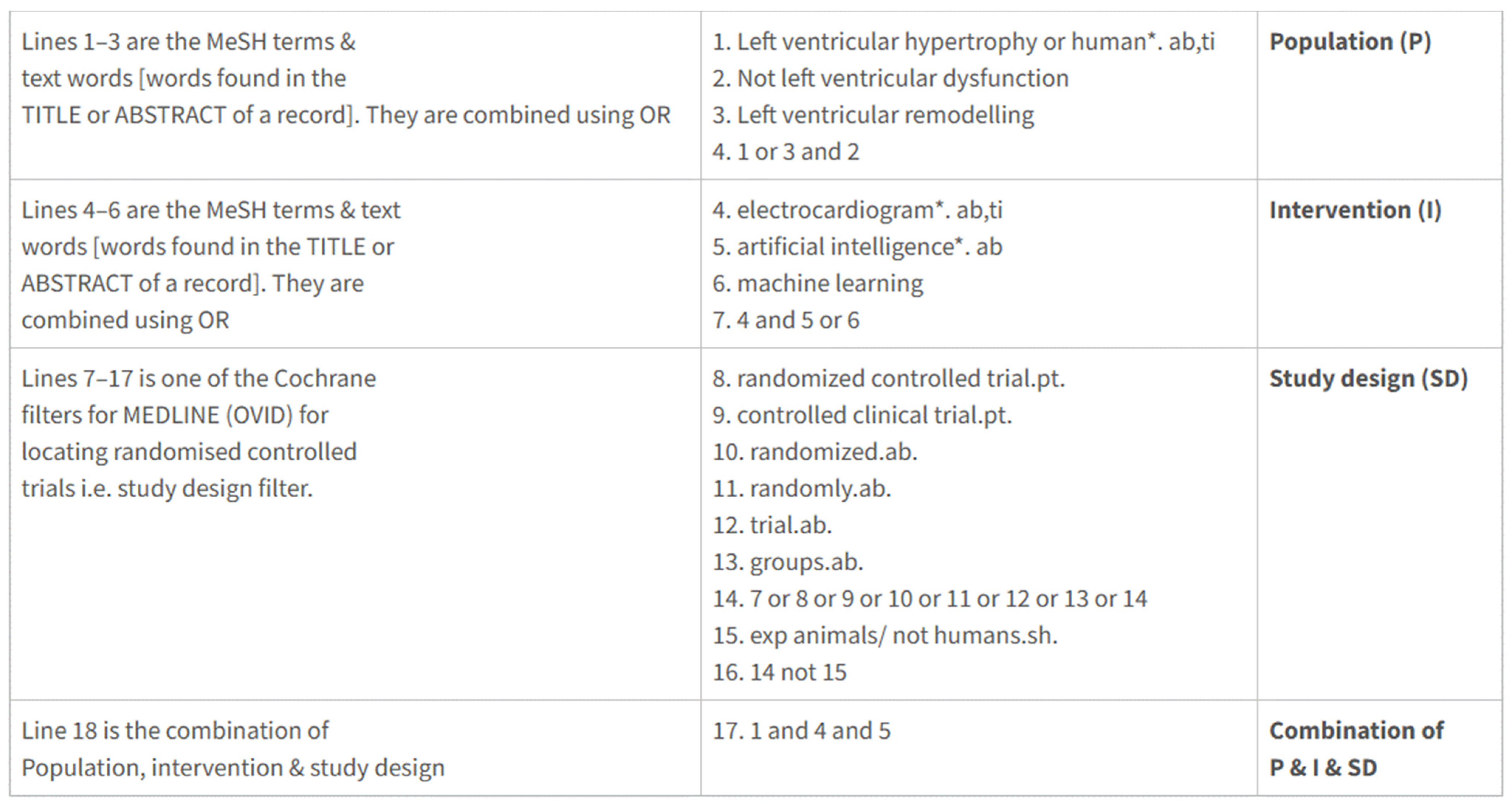
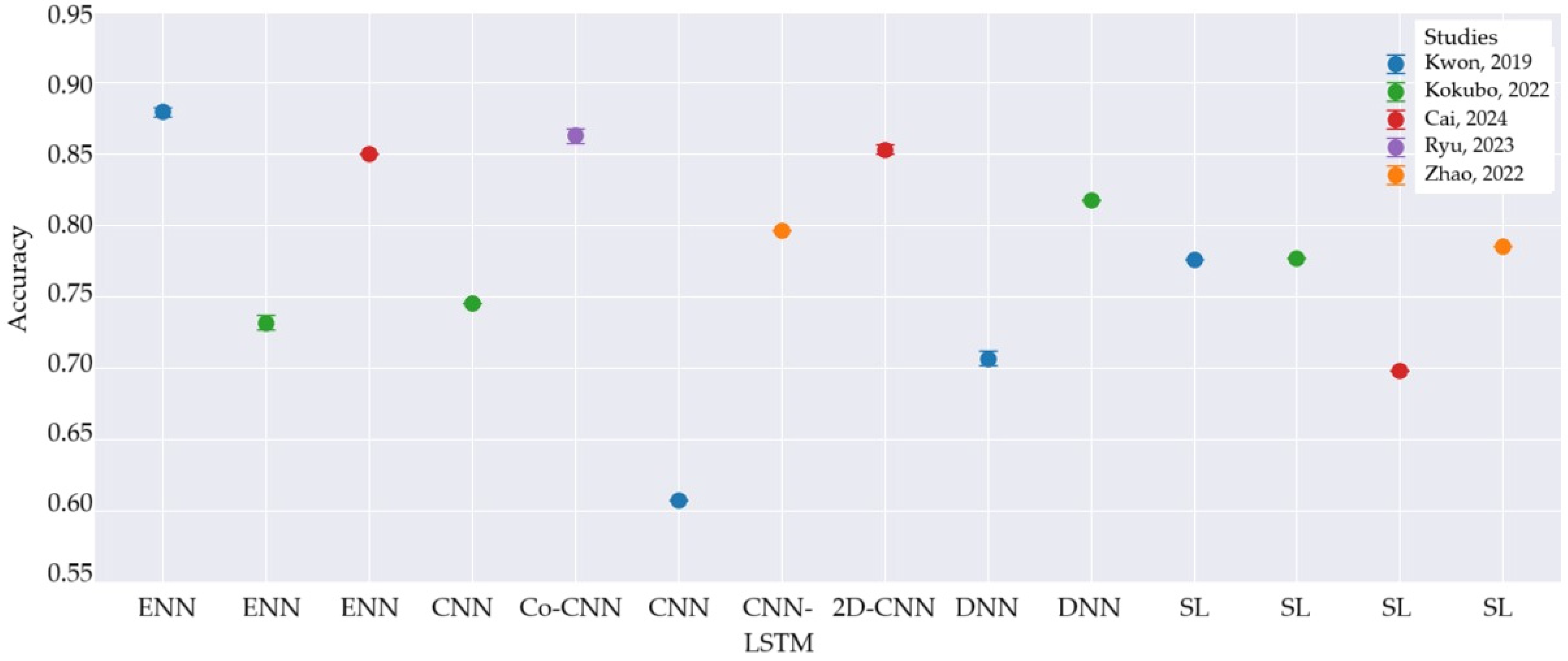
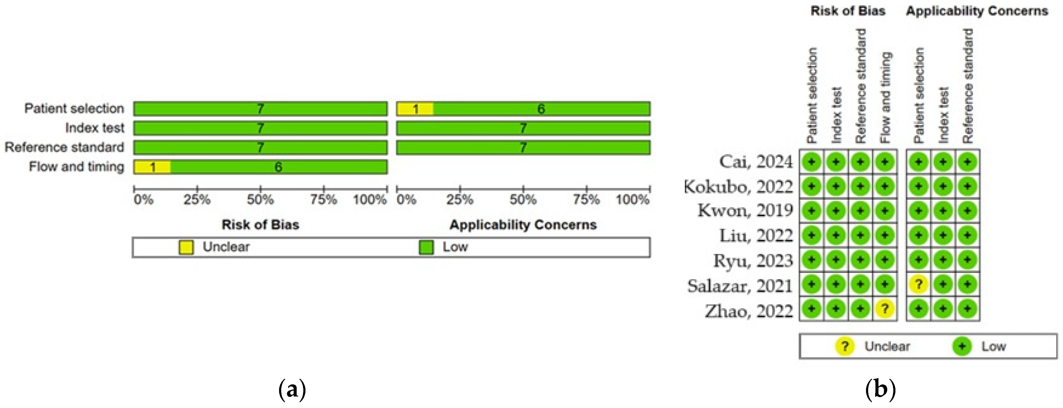
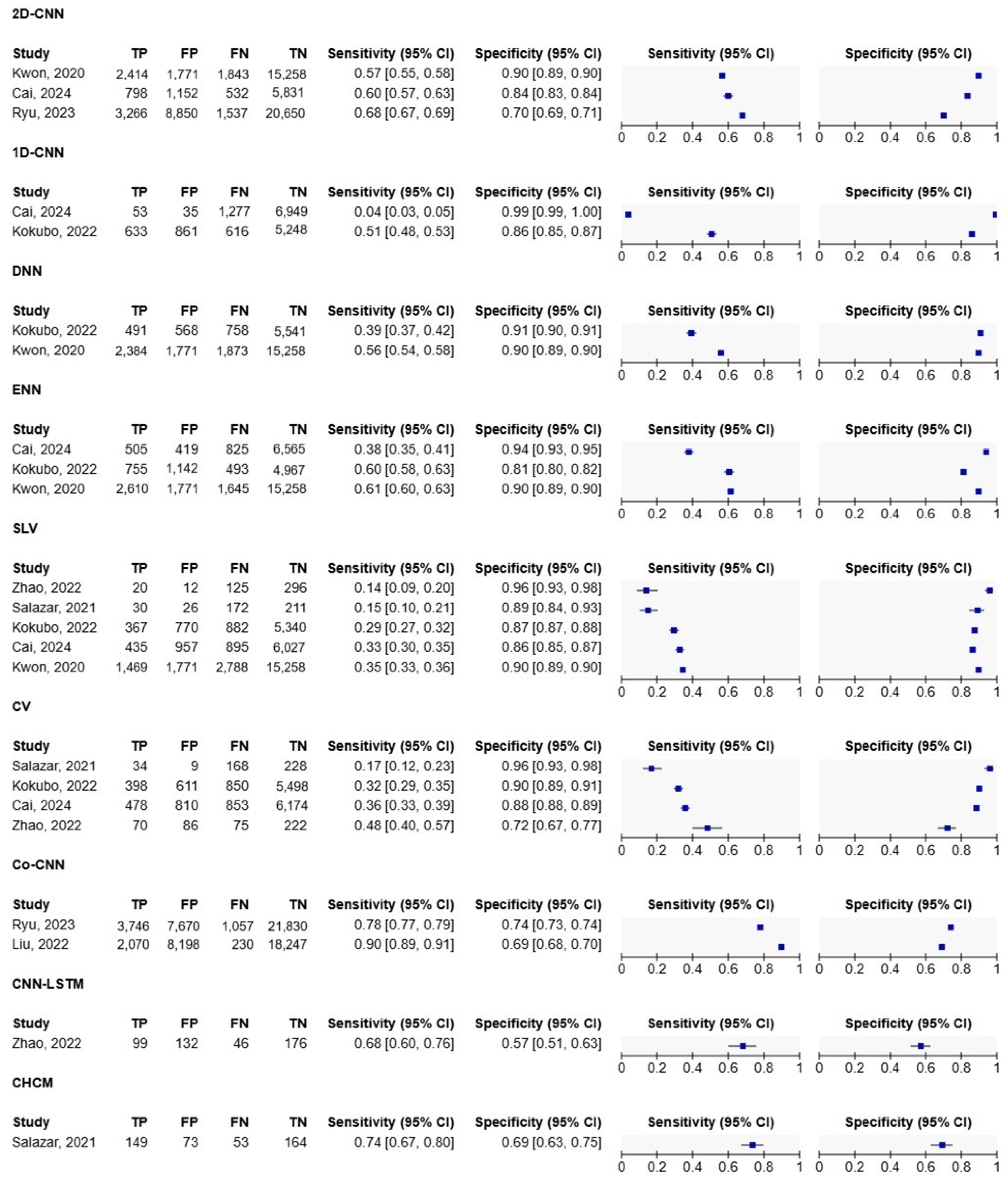
| Application | Advantages | Disadvantages |
|---|---|---|
| PMcardio [18] |
|
|
| LabChart ECG Analysis Module [20] |
|
|
| SonoHealth [21] |
|
|
| KardiaMobile [22] |
|
|
| Excluded Study | Reason for Exclusion |
|---|---|
| Haimovich, 2023 [25] | Discrimination of echocardiographic LVH and multiple cardiac diseases associated with LVH. LVH detection by ECG was not examined. |
| Pan, 2024 [26] | CT-derived LVM, not echocardiographic estimation |
| Khurshid, 2021 [27] | The deep learning algorithm estimates CMR-derived LV mass with fair accuracy using 12-lead ECG |
| Naderi, 2023 [28] | Case-to-case comparisons with echocardiography and ECG-AI analysis are missed |
| Pandelitis, 2023 [29] | Only proof-of-concept study |
| Shimizu, 2023 [30] | Only the random forest method was used. Sensitivity and specificity not known |
| Soto, 2022 [31] | Multimodal training models (the electrocardiogram and echocardiogram data were paired according to unique patient identifiers) |
| Wu, 2020 [32] | The use of six-layer deep neural networks, the electrocardiographic left ventricular hypertrophy classifier (ELVHC), has not been confirmed directly with echocardiography or cardiac MRI. |
| Study | Sample Size | Training Sample | Validation Sample | LVMI (g/m2) | LVH (Number of Patients) | Prevalence (LVH Sample/Sample Size) | Used AI Algorithm/ECG Criteria | Predictive Performance Variables |
|---|---|---|---|---|---|---|---|---|
| Kokubo, 2022 [33] | 7358 | 3881 | 943 | 92.66 ± 96.91 (all patients in the test sample) | 1249 | 0.17 | CNN, DNN, ENN, Logistic Regression, Random Forest/ Sokolow-Lyon voltage, Cornell voltage | Sensitivity Specificity Accuracy PPV NPV |
| Kwon, 2019 [34] | 21,286 | 35,694 | 3162 (internal) 5476 (external) | 67.61 ± 12.46 | 4353 | 0.2 | CNN, DNN, ENN/ Sokolow-Lyon voltage, included clinical variables | Sensitivity Specificity Accuracy PPV NPV |
| Liu, 2022 [35] | 5749 | 23,996 | 3280 (internal) 225 (external) | - | 2435 | 0.08 | CNN and a multimodality module that combines the demographic features | Sensitivity Specificity |
| Cai, 2024 [36] | 8314 | 28,855 | 3370 | 111.96 ± 104.09 | 1363 | 0.16 | CNN, ENN/ Sokolow-Lyon voltage Cornell voltage | Accuracy Sensitivity Specificity |
| Ryu, 2023 [37] | 34,302 | 24,008 | - | 146.86 ± 12.23 (men) 126.96 ± 15.54 (women) | 4873 | 0.14 | Model with the Entire dataset (with demographic features) | Sensitivity Specificity PPV NPV |
| Salazar, 2021 [38] | 132 | 307 | 156 | 134.2 ± 29 | 203 | 0.46 | Cardiac Hypertrophy Computer-based model/ Sokolow- Lyon voltage, Cornell voltage | Sensitivity Specificity Accuracy PPV NPV |
| Zhao, 2022 [12] | 453 | 1120 | 371 | 129.28 ± 28.93 | 144 | 0.32 | CNN-LSTM/ Sokolow-Lyon voltage, Cornell voltage, with demographic features | Accuracy Sensitivity Specificity |
| Study | AI Model | Input | Structure | Optimization Techniques/Parameters |
|---|---|---|---|---|
| Kokubo, 2022 [33] | CNN | raw ECG data (1-ECG lead) |
|
|
| DNN | 19 parameters comprising ECG features and demographic information |
| ||
| ENN | CNN + DNN are combined directly after the convolution layer |
| ||
| Kwon, 2019 [34] | CNN | raw ECG data (12-ECG lead, recording 10 s); 12 × 5000 numbers; 500 Hz
|
|
|
| DNN | 11 variables (age, sex, weight, height, heart rate, presence of atrial fibrillation or atrial flutter, QT interval, QRS duration, QTc, R axis, and T axis) |
| ||
| ENN | The last hidden layer of the DNN and the last layer after the flattened layer of CNN |
| ||
| Liu, 2022 [35] | CNN with multimodality module | full-length ECG record divided into separate heartbeats (one feature extraction layer; recording 10 s; 500 Hz for 12-ECG leads |
|
|
| Cai, 2024 [36] | 2D-CNN | 1-lead ECG data 240 × 240 pixels), recording 10 s; rate of 500 Hz; 12-lead ECG data converted to 2D images (224 × 224 pixels) |
|
|
| Ryu, 2023 [37] | CoAt-Mixer (CNN variant) | 12-lead ECG, duration of 10 s, 500 Hz, 12 × 5000 numbers; demographic features |
|
|
| Salazar, 2021 [38] | Computer-based ECG model | 458 ECG standard and non-standard parameters; 25 mm/s velocity and 10 mm/mV sensitivity |
|
|
| Zhao, 2022 [12] | CNN-LSTM hybrid | sliding window technique for ECG signal processing (window size: 250 samples), 1000 Hz, duration of 5 s; 12 × 5000 numbers |
|
|
| AI Model | Advantages | Disadvantages |
|---|---|---|
| CNN |
|
|
| LSTM Networks |
|
|
| Co-CNN |
|
|
| ENN |
|
|
| DNN |
|
|
Disclaimer/Publisher’s Note: The statements, opinions and data contained in all publications are solely those of the individual author(s) and contributor(s) and not of MDPI and/or the editor(s). MDPI and/or the editor(s) disclaim responsibility for any injury to people or property resulting from any ideas, methods, instructions or products referred to in the content. |
© 2025 by the authors. Licensee MDPI, Basel, Switzerland. This article is an open access article distributed under the terms and conditions of the Creative Commons Attribution (CC BY) license (https://creativecommons.org/licenses/by/4.0/).
Share and Cite
Makowska, A.; Ananthakrishnan, G.; Christ, M.; Dehmer, M. Screening for Left Ventricular Hypertrophy Using Artificial Intelligence Algorithms Based on 12 Leads of the Electrocardiogram—Applicable in Clinical Practice?—Critical Literature Review with Meta-Analysis. Healthcare 2025, 13, 408. https://doi.org/10.3390/healthcare13040408
Makowska A, Ananthakrishnan G, Christ M, Dehmer M. Screening for Left Ventricular Hypertrophy Using Artificial Intelligence Algorithms Based on 12 Leads of the Electrocardiogram—Applicable in Clinical Practice?—Critical Literature Review with Meta-Analysis. Healthcare. 2025; 13(4):408. https://doi.org/10.3390/healthcare13040408
Chicago/Turabian StyleMakowska, Agata, Gayathri Ananthakrishnan, Michael Christ, and Matthias Dehmer. 2025. "Screening for Left Ventricular Hypertrophy Using Artificial Intelligence Algorithms Based on 12 Leads of the Electrocardiogram—Applicable in Clinical Practice?—Critical Literature Review with Meta-Analysis" Healthcare 13, no. 4: 408. https://doi.org/10.3390/healthcare13040408
APA StyleMakowska, A., Ananthakrishnan, G., Christ, M., & Dehmer, M. (2025). Screening for Left Ventricular Hypertrophy Using Artificial Intelligence Algorithms Based on 12 Leads of the Electrocardiogram—Applicable in Clinical Practice?—Critical Literature Review with Meta-Analysis. Healthcare, 13(4), 408. https://doi.org/10.3390/healthcare13040408


