
Association Between Euthyroidism and Muscular Parameters in Adults with an Excess of Fat Mass: A Preliminary Study

 , , ,
, , ,  ,
,  ,
, 
Abstract
1. Introduction
2. Material and Methods
2.1. Study Design
2.2. Participants
2.3. Anthropometric and Body Composition Evaluations
- Height and body mass were measured using a stadiometer with a weighting station to the nearest 0.1 cm and 0.1 kg, respectively (SECA, Intermed S.r.l., Milano, Italy). Body mass index (BMI) was calculated by dividing body mass (kg) by the square of height (m2).
- Body composition was measured using a bioelectrical impedance method (BIA 101 BIVA® PRO AKERN s.r.l., Pisa, Italy) in the morning. Whole-body impedance resistance and reactance were obtained and recorded when stable. Variables of interest were percentage of fat mass (pFM), fat-free mass (FFM) and skeletal muscle mass (SMM).
2.4. Muscular Fitness Assessments
2.5. Physical Activity Levels
2.6. Thyroid Hormone Assays
2.7. Statistical Analysis
3. Results
4. Discussion
5. Limitations of the Study
6. Conclusions
Supplementary Materials
Author Contributions
Funding
Institutional Review Board Statement
Informed Consent Statement
Data Availability Statement
Conflicts of Interest
References
- Mullur, R.; Liu, Y.Y.; Brent, G.A. Thyroid hormone regulation of metabolism. Physiol. Rev. 2014, 94, 355–382. [Google Scholar] [CrossRef]
- Sawicka-Gutaj, N.; Erampamoorthy, A.; Zybek-Kocik, A.; Kyriacou, A.; Zgorzalewicz-Stachowiak, M.; Czarnywojtek, A.; Ruchała, M. The Role of Thyroid Hormones on Skeletal Muscle Thermogenesis. Metabolites 2022, 12, 336. [Google Scholar] [CrossRef] [PubMed]
- Ruiz, S.; Vázquez, F.; Pellitero, S.; Puig-Domingo, M. ENDOCRINE OBESITY: Pituitary dysfunction in obesity. Eur. J. Endocrinol. 2022, 186, R79–R92. [Google Scholar] [CrossRef] [PubMed]
- Fontenelle, L.C.; Feitosa, M.M.; Severo, J.S.; Freitas, T.E.C.; Morais, J.B.S.; Torres-Leal, F.L.; Henriques, G.S.; Marreiro, D.D.N. Thyroid Function in Human Obesity: Underlying Mechanisms. Horm. Metab. Res. 2016, 48, 787–794. [Google Scholar] [CrossRef]
- De Pergola, G.; Ciampolillo, A.; Paolotti, S.; Trerotoli, P.; Giorgino, R. Free triiodothyronine and thyroid stimulating hormone are directly associated with waist circumference, independently of insulin resistance, metabolic parameters and blood pressure in overweight and obese women. Clin. Endocrinol. 2007, 67, 265–269. [Google Scholar] [CrossRef]
- Walczak, K.; Sieminska, L. Obesity and Thyroid Axis. Int. J. Environ. Res. Public Health 2021, 18, 9434. [Google Scholar] [CrossRef] [PubMed]
- Tomlinson, D.J.; Erskine, R.M.; Morse, C.I.; Winwood, K.; Onambélé-Pearson, G. The impact of obesity on skeletal muscle strength and structure through adolescence to old age. Biogerontology 2016, 17, 467–483. [Google Scholar] [CrossRef]
- Valenzuela, P.L.; Maffiuletti, N.A.; Tringali, G.; De Col, A.; Sartorio, A. Obesity-associated poor muscle quality: Prevalence and association with age, sex, and body mass index. BMC Musculoskelet. Disord. 2020, 21, 200. [Google Scholar] [CrossRef]
- Frontera, W.R.; Ochala, J. Skeletal muscle: A brief review of structure and function. Calcif. Tissue Int. 2015, 96, 183–195. [Google Scholar] [CrossRef]
- Donini, L.M.; Busetto, L.; Bischoff, S.C.; Cederholm, T.; Ballesteros-Pomar, M.D.; Batsis, J.A.; Bauer, J.M.; Boirie, Y.; Cruz-Jentoft, A.J.; Dicker, D.; et al. Definition and diagnostic criteria for sarcopenic obesity: ESPEN and EASO consensus statement. Clin. Nutr. 2022, 41, 990–1000. [Google Scholar] [CrossRef]
- Salvatore, D.; Simonides, W.S.; Dentice, M.; Zavacki, A.M.; Larsen, P.R. Thyroid hormones and skeletal muscle: New insights and potential implications. Nat. Rev. Endocrinol. 2014, 10, 206–214. [Google Scholar] [CrossRef] [PubMed]
- Bloise, F.F.; Cordeiro, A.; Ortiga-Carvalho, T.M. Role of thyroid hormone in skeletal muscle physiology. J. Endocrinol. 2018, 236, R57–R68. [Google Scholar] [CrossRef]
- Greco, F.; Moulton, C.; Antinozzi, C.; Lista, M.; Di Luigi, L.; Dimauro, I.; Sgrò, P. Relationship between Euthyroidism and Muscle Mass and Strength: A Systematic Review. Int. J. Sports Med. 2023, 44, 704–710. [Google Scholar] [CrossRef] [PubMed]
- Zupo, R.; Castellana, F.; Sardone, R.; Lampignano, L.; Paradiso, S.; Giagulli, V.A.; Triggiani, V.; Di Lorenzo, L.; Giannelli, G.; De Pergola, G. Higher Muscle Mass Implies Increased Free-Thyroxine to Free-Triiodothyronine Ratio in Subjects with Overweight and Obesity. Front. Endocrinol. 2020, 11, 565065. [Google Scholar] [CrossRef] [PubMed]
- Lee, J.; Jo, K.; Ha, J.; Lim, D.-J.; Lee, J.M.; Chang, S.-A.; Kang, M.I.; Kim, M.-H. Significant Association of Upper Limb Muscle Strength with Thyroid Function in Overweight and Obese Population: A Study of the Sixth Korea National Health and Nutrition Examination Survey (KNHANES 2014-2015). Int. J. Endocrinol. 2020, 2020, 7195846. [Google Scholar] [CrossRef]
- Roberts, H.C.; Denison, H.J.; Martin, H.J.; Patel, H.P.; Syddall, H.; Cooper, C.; Sayer, A.A. A review of the measurement of grip strength in clinical and epidemiological studies: Towards a standardised approach. Age Ageing 2011, 40, 423–429. [Google Scholar] [CrossRef]
- Armstrong, T.; Bull, F. Development of the world health organization global physical activity questionnaire (GPAQ). J. Public Health 2006, 14, 66–70. [Google Scholar] [CrossRef]
- Di Bonito, P.; Corica, D.; Licenziati, M.R.; Di Sessa, A.; del Giudice, E.M.; Faienza, M.F.; Calcaterra, V.; Franco, F.; Maltoni, G.; Valerio, G.; et al. Central sensitivity to thyroid hormones is reduced in youths with overweight or obesity and impaired glucose tolerance. Front. Endocrinol. 2023, 14, 1159407. [Google Scholar] [CrossRef]
- Hosoi, Y.; Murakami, M.; Mizuma, H.; Ogiwara, T.; Imamura, M.; Mori, M. Expression and regulation of type II iodothyronine deiodinase in cultured human skeletal muscle cells. J. Clin. Endocrinol. Metab. 1999, 84, 3293–3300. [Google Scholar] [CrossRef]
- Schorr, M.; Dichtel, L.E.; Gerweck, A.V.; Valera, R.D.; Torriani, M.; Miller, K.K.; Bredella, M.A. Sex differences in body composition and association with cardiometabolic risk. Biol. Sex Differ. 2018, 9, 28. [Google Scholar] [CrossRef]
- Sheng, Y.; Ma, D.; Zhou, Q.; Wang, L.; Sun, M.; Wang, S.; Qi, H.; Liu, J.; Ding, G.; Duan, Y. Association of thyroid function with sarcopenia in elderly Chinese euthyroid subjects. Aging Clin. Exp. Res. 2019, 31, 1113–1120. [Google Scholar] [CrossRef] [PubMed]
- Xiu, S.; Mu, Z.; Zhao, L.; Sun, L. Low free triiodothyronine levels are associated with risk of frailty in older adults with type 2 diabetes mellitus. Exp. Gerontol. 2020, 138, 111013. [Google Scholar] [CrossRef] [PubMed]
- Gu, Y.; Meng, G.; Wu, H.; Zhang, Q.; Liu, L.; Bao, X.; Wang, Y.; Zhang, S.; Sun, S.; Wang, X.; et al. Thyroid Function as a Predictor of Handgrip Strength Among Middle-Aged and Older Euthyroid Adults: The TCLSIH Cohort Study. J. Am. Med. Dir. Assoc. 2019, 20, 1236–1241. [Google Scholar] [CrossRef] [PubMed]


| Variable | N = 40 M | N = 65 F | M vs. F p-Value * |
|---|---|---|---|
| Age (years) | 44.5 (27.8) | 41.0 (29.5) | 0.550 |
| Body mass (kg) | 95.3 (24.3) | 84.0 (16.2) | <0.01 |
| Body Mass Index (kg/m2) | 33.0 (10.6) | 34.0 (7.5) | 0.611 |
| Fat Free Mass (kg) | 65.4 ± 9.2 | 48.8 ± 5.4 | <0.01 |
| Skeletal Muscle Mass (kg) | 32.5 ± 4.8 | 24.0 ± 3.3 | <0.01 |
| Fat Mass (%) | 34.2 ± 8.4 | 43.6 ± 6.4 | <0.01 |
| TSH (ulU/mL) | 1.82 (1.69) | 2.19 (1.68) | 0.145 |
| FT3 (pg/mL) | 3.78 ± 0.41 | 3.40 ± 0.43 | <0.01 |
| FT4 (ng/dL) | 1.19 ± 0.14 | 1.16 ± 0.17 | 0.235 |
| FT3/FT4 | 3.20 ± 0.44 | 2.97 ± 0.43 | <0.01 |
| Handgrip strength (kg) | 44.0 ± 11.5 | 28.1 ± 6.2 | <0.01 |
| Chair Stand Test (seconds) | 18.7 ± 3.5 | 21.9 ± 5.4 | <0.01 |
| Variable | FT3 (pg/mL) | FT4 (ng/dL) | FT3/FT4 | |||
|---|---|---|---|---|---|---|
| R | p-Value | R | p-Value | R | p-Value | |
| Body mass (kg) | 0.265 | 0.099 | 0.287 | 0.073 | −0.040 | 0.805 |
| Body Mass Index (kg/m2) | 0.216 | 0.181 | 0.235 | 0.144 | −0.033 | 0.842 |
| Fat Free Mass(kg) | 0.245 | 0.128 | 0.180 | 0.267 | 0.022 | 0.893 |
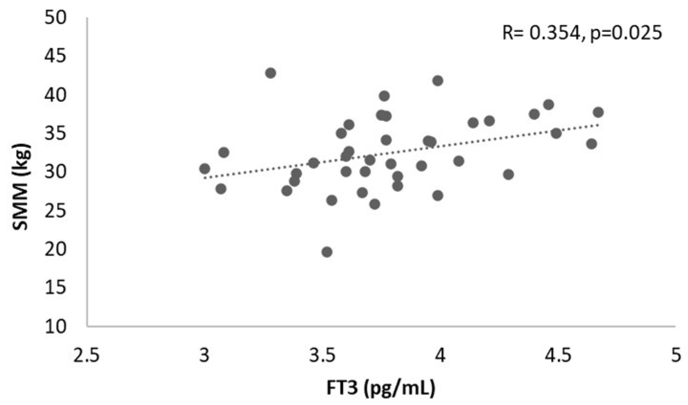
| Skeletal Muscle Mass (kg) | 0.354 | 0.025 * | 0.154 | 0.343 | 0.108 | 0.509 |
| Fat Mass (%) | 0.185 | 0.253 | 0.271 | 0.091 | −0.077 | 0.637 |
| TSH (ulU/mL) | −0.013 | 0.935 | 0.020 | 0.904 | −0.039 | 0.810 |
| Handgrip strength (kg) | 0.005 | 0.973 | 0.178 | 0.273 | −0.160 | 0.325 |
| Chair Stand Test (s) | −0.073 | 0.656 | −0.172 | 0.289 | 0.085 | 0.601 |
| Variable | FT3 (pg/mL) | FT4 (ng/dL) | FT3/FT4 | |||
|---|---|---|---|---|---|---|
| R | p-Value | R | p-Value | R | p-Value | |
| Body mass (kg) | −0.042 | 0.738 | −0.028 | 0.824 | −0.013 | 0.915 |
| Body Mass Index (kg/m2) | −0.119 | 0.346 | 0.014 | 0.912 | −0.126 | 0.318 |
| Fat Free Mass(kg) | 0.030 | 0.815 | −0.008 | 0.949 | 0.038 | 0.763 |
| Skeletal Muscle Mass (kg) | 0.018 | 0.889 | −0.002 | 0.990 | −0.010 | 0.938 |
| Fat Mass (%) | −0.006 | 0.959 | −0.027 | 0.830 | 0.027 | 0.831 |
| TSH (ulU/mL) | 0.049 | 0.700 | −0.198 | 0.115 | 0.238 | 0.056 |
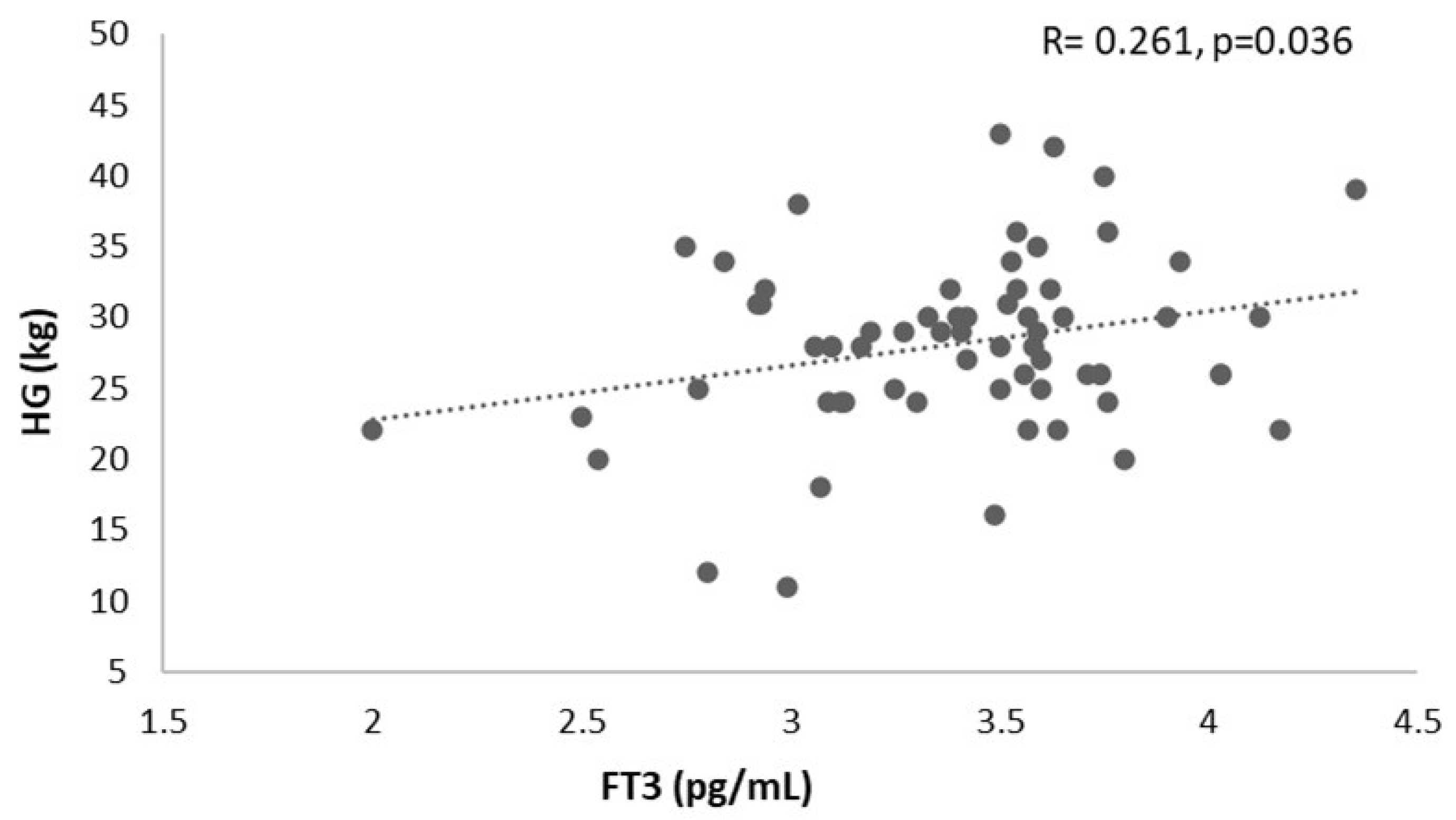
| Handgrip strength (kg) | 0.261 | 0.036 * | 0.195 | 0.120 | 0.026 | 0.838 |
| Chair Stand Test (s) | −0.047 | 0.709 | 0.243 | 0.051 | −0.266 | 0.032 * |
Disclaimer/Publisher’s Note: The statements, opinions and data contained in all publications are solely those of the individual author(s) and contributor(s) and not of MDPI and/or the editor(s). MDPI and/or the editor(s) disclaim responsibility for any injury to people or property resulting from any ideas, methods, instructions or products referred to in the content. |
© 2025 by the authors. Licensee MDPI, Basel, Switzerland. This article is an open access article distributed under the terms and conditions of the Creative Commons Attribution (CC BY) license (https://creativecommons.org/licenses/by/4.0/).
Share and Cite
Greco, F.; Sicilia, L.; Seminara, G.; Iuliano, S.; Tocci, V.; Brunetti, A.; Aversa, A.; Di Luigi, L.; Sgrò, P. Association Between Euthyroidism and Muscular Parameters in Adults with an Excess of Fat Mass: A Preliminary Study. Healthcare 2025, 13, 241. https://doi.org/10.3390/healthcare13030241
Greco F, Sicilia L, Seminara G, Iuliano S, Tocci V, Brunetti A, Aversa A, Di Luigi L, Sgrò P. Association Between Euthyroidism and Muscular Parameters in Adults with an Excess of Fat Mass: A Preliminary Study. Healthcare. 2025; 13(3):241. https://doi.org/10.3390/healthcare13030241
Chicago/Turabian StyleGreco, Francesca, Luciana Sicilia, Giuseppe Seminara, Stefano Iuliano, Vera Tocci, Antonio Brunetti, Antonio Aversa, Luigi Di Luigi, and Paolo Sgrò. 2025. "Association Between Euthyroidism and Muscular Parameters in Adults with an Excess of Fat Mass: A Preliminary Study" Healthcare 13, no. 3: 241. https://doi.org/10.3390/healthcare13030241
APA StyleGreco, F., Sicilia, L., Seminara, G., Iuliano, S., Tocci, V., Brunetti, A., Aversa, A., Di Luigi, L., & Sgrò, P. (2025). Association Between Euthyroidism and Muscular Parameters in Adults with an Excess of Fat Mass: A Preliminary Study. Healthcare, 13(3), 241. https://doi.org/10.3390/healthcare13030241