Systematic Review

Deep Learning Applications in Dental Image-Based Diagnostics: A Systematic Review

by
Osama Khattak
1,
Ahmed Shawkat Hashem
2,
Mohammed Saad Alqarni
3,
Raha Ahmed Shamikh Almufarrij
4,
Amna Yusuf Siddiqui
5,
Rabia Anis
6,
Shahzad Ahmad
7,
Muhammad Amber Fareed
8,9,*,
Osama Shujaa Alothmani
5,
Lama Habis Samah Alkhershawy
4,
Wesam Waleed Zain Alabidin
4,
Rakhi Issrani
10,* and
Anshoo Agarwal
11
1
Department of Restorative Dentistry, College of Dentistry, Jouf University, Sakaka 72311, Saudi Arabia
2
Oral Medicine and Periodontology, Faculty of Dentistry, Damanhour University, Damanhur 22522, Egypt
3
Department of Oral & Maxillofacial Surgery and Diagnostic Sciences, College of Dentistry, Jouf University, Sakaka 72311, Saudi Arabia
4
College of Dentistry, Jouf University, Sakaka 72311, Saudi Arabia
5
Department of Endodontics, Faculty of Dentistry, King Abdulaziz University, Jeddah 22230, Saudi Arabia
6
Department of Health Professions Education, Isra Dental College, Isra University, Hydrabad 73000, Pakistan
7
Faculty of Medicine and Health Science, The University of Buckingham, Buckingham MK18 1EG, UK
8
Clinical Sciences Department, College of Dentistry, Ajman University, Ajman 346, United Arab Emirates
9
Centre of Medical and Bio-Allied Health Sciences Research, Ajman University, Ajman 346, United Arab Emirates
10
Department of Preventive Dentistry, College of Dentistry, Jouf University, Sakaka 72311, Saudi Arabia
11
Department of Pathology, Northern Border University, Arar 91431, Saudi Arabia
*
Authors to whom correspondence should be addressed.
Healthcare 2025, 13(12), 1466; https://doi.org/10.3390/healthcare13121466
Submission received: 5 May 2025 / Revised: 6 June 2025 / Accepted: 9 June 2025 / Published: 18 June 2025
(This article belongs to the Special Issue Artificial Intelligence in Healthcare: Opportunities and Challenges)

Abstract

:
Background: AI has been adopted in dentistry for diagnosis, decision making, and therapy prognosis prediction. This systematic review aimed to identify AI models in dentistry, assess their performance, identify their shortcomings, and discuss their potential for adoption and integration in dental practice in the future. Methodology: The sources of the papers were the following electronic databases: PubMed, Scopus, and Cochrane Library. A total of 20 out of 947 needed further studies, and this was encompassed in the present meta-analysis. It identified diagnostic accuracy, predictive performance, and potential biases. Results: AI models demonstrated an overall diagnostic accuracy of 82%, primarily leveraging artificial neural networks (ANNs) and convolutional neural networks (CNNs). These models have significantly improved the diagnostic precision for dental caries compared with traditional methods. Moreover, they have shown potential in detecting and managing conditions such as bone loss, malignant lesions, vertical root fractures, apical lesions, salivary gland disorders, and maxillofacial cysts, as well as in performing orthodontic assessments. However, the integration of AI systems into dentistry poses challenges, including potential data biases, cost implications, technical requirements, and ethical concerns such as patient data security and informed consent. AI models may also underperform when faced with limited or skewed datasets, thus underscoring the importance of robust training and validation procedures. Conclusions: AI has the potential to revolutionize dentistry by significantly improving diagnostic accuracy and treatment planning. However, before integrating this tool into clinical practice, a critical assessment of its advantages, disadvantages, and utility or ethical issues must be established. Future studies should aim to eradicate existing barriers and enhance the model’s ease of understanding and challenges regarding expense and data protection, to ensure the effective utilization of AI in dental healthcare.

1. Introduction

Artificial intelligence (AI) is transforming dentistry in that early diagnosis is more accurate with advanced treatment planning, treatment coordination, and patient monitoring. Envisaged as the science of creating systems capable of emulating intelligent human actions, AI has undergone extensive deployment across the sphere of healthcare, and dentistry is no exception. With the help of AI technologies, dentists are able to easily analyze various forms of imaging, make better patient-specific treatment decisions, and enhance patient care [1,2].
AI holds a large potential in dentistry, and one of the best areas where it could be applied is medical image analysis. For instance, a sub-type of AI known as convolutional neural networks (CNNs) is used in the diagnosis of dental caries, apical lesions, and bone loss from periapical radiographic images as well as panoramic images [3]. Such models can independently assess affected teeth and separate various structures in the mouth to identify regions that require treatment to minimize diagnostic mistakes and time consumption [4,5]. Prior studies have shown that CNNs have a higher diagnostic accuracy than conventional approaches for detecting carious lesions, and periapical radiographs offer proper image clarity for caries [6].
Other than diagnostics, there is the application of AI in treatment procedures. Current dental operating systems are now fully automated using robotic systems that have incorporated AI, and these systems can now perform 3D tooth preparations without destroying the surrounding tissues [7,8]. In the context of restorative dentistry, ANNs assess material characteristics to avoid the debonding process of composite restorations [9]. Similarly, orthodontic treatments have been on the receiving end, with applications such as cephalometric analyses and planning adjustments to harmonize facial aesthetics [10].
It is concluded that there are great opportunities in the application of AI, but there are also several problems with AI in the field of dentistry. Some of these issues include patient data confidentiality, the ability to obtain patient consent, and potential algorithmic bias, which must be addressed to minimize or eliminate bias and unfair treatment in the equitable sharing of these services [11]. Moreover, the reliability of AI systems depends on the quality and diversity of the training datasets, which hinders the systems’ applicability outside researched settings. The high cost of integrating AI may be a challenge for small firms, particularly in explaining why the implementation of advanced dental technologies may lead to unequal distribution [12].
AI application in dentistry is fundamental because it changes the dental field in a way that makes care data-driven, accurate, and timely. It is proficient in the study of big data and displaces arduous jobs that burden clinicians while enhancing the lives of patients [2,13]. Nevertheless, to unlock the full potential of AI as a resource, much attention has to be paid to AI’s limitations, both ethical and technical, as well as to its availability for a wider range of use. Future advancement in modern dentistry will be premised on findings derived from focused studies exploring the applicability of AI in dental practice [1].

2. Materials and Methods

This review has been registered in the INPLASY database with the registration number INPLASY202530022, and the associated DOI is 10.37766/inplasy2025.3.0022.

2.1. Methodological Framework

This review adhered to the PRISMA guidelines, formulated to address the research question:
“Which types of AI approaches are applied in dentistry and to what extent and in what way is AI improving the diagnosis in dentistry, the quality of decisions made and the outcomes of dental procedures?”
PICOS, which represents population, intervention, comparison, outcome, and study design, was utilized in the formulation of this investigation.
CAD/CAM, computerized representations of the clinic, apical, bitewing, orthopantomographic, or CBCT radiographs, and 2D and 3D patient and simulator facial images were all part of the package. The primary focus was on the application of robotics, natural language processing and deep learning, in dental care evaluation, therapy, and prognosis. The outcomes measured included accuracy, efficiency, support for the critical conclusion, the area under the curve and applications of AI in various specialties of dentistry.

2.2. Search Strategy

To assess machine learning dental applications, as well as neural networks and their role in oral care, we searched databases such as PubMed, Scopus and the Cochrane Library for studies published between 2014 and 2023. The search terms used were (“Artificial Intelligence in dentistry” OR “AI in dentistry”) AND (“Machine Learning”) AND (“Neural Networks”) AND (“Dental”) AND (“Oral Health”).
As part of the search process, Boolean operators such as AND and OR were used to remove any inconsequential results. For example, when entering the search query “AI in dentistry” and “machine learning”, this helped to narrow down the results to show only the studies related to neural networks used in dentistry.
Inter-rater agreement was assessed using Cohen’s kappa coefficient to ensure consistency during study selection and data extraction. The kappa value was calculated at 0.82, indicating strong agreement. Disagreements between the two primary reviewers (1st and 3rd author) were resolved through discussion, and unresolved discrepancies were referred to a third reviewer (12th author). The review hence included 20 papers that passed the inclusion criteria as depicted in [Figure 1], a PRISMA flow diagram.

2.3. Criteria of Exclusion and Inclusion

2.3.1. Criteria for Inclusion

  • Studies along with specified keywords;
  • Articles (research) available from 2014 to 2023;
  • English-language studies; and
  • Original research articles.

2.3.2. Criteria for Exclusion

  • Studies not focusing on AI-based diagnostics in pediatric dentistry;
  • Articles not including full-text access; and
  • Ongoing research projects.
Pediatric dentistry focuses on children who have unique dental conditions and diagnostic needs. AI diagnostics in pediatric dentistry uses specific tools and techniques for children that may not apply to other groups. Including pediatric research could increase dataset diversity, complicating synthesis. The diagnostic challenges in children’s dentistry, including behavior management and limited data, differ significantly from those in adult dentistry, making comparisons less meaningful.
For precise results, two reviewers independently performed the data extraction. The reviewers obtained details regarding the studies and their outcomes. After the extraction, notes were compared to check for any discrepancies, and a consensus was reached in cases where the notes differed.

2.4. Quality Assessment

The appraisal of the studies that were selected was conducted using The Cochrane Handbook for Systematic Reviews of Interventions (v5.1.0) criteria [11]. In addition to this, the AMSTAR 2 checklist was used as a complementary tool to enhance the methodological rigor and transparency of this systematic review.
The QUADAS-2 framework was adapted to assess risk of bias for diagnostic AI models. Assessment criteria included randomization, blinding, dropout rates, the accuracy of outcome variables, sample size estimation, and examiner reliability.

3. Results

Thus, the search was conducted using pre-coordinated terms and Boolean connectors such as AND, resulting in 50 articles. After removing 23 articles that were similar, 27 articles needed further scrutiny. Following the inclusion and exclusion criteria, 20 relevant studies were selected for this systematic review.
Data extraction focused on capturing critical details from each study, including the authors’ names and publication year, input data sources, study objectives, AI models employed, and resulting outcomes. A detailed summary of these studies is presented in Table 1, which encapsulates essential information such as the study design, AI methodologies applied, their practical applications, evaluation metrics, and significant findings. The present tabulation provides a detailed outlook of AI practices in dentistry and their impact on diagnostic efficiency and decision making in therapeutic care.
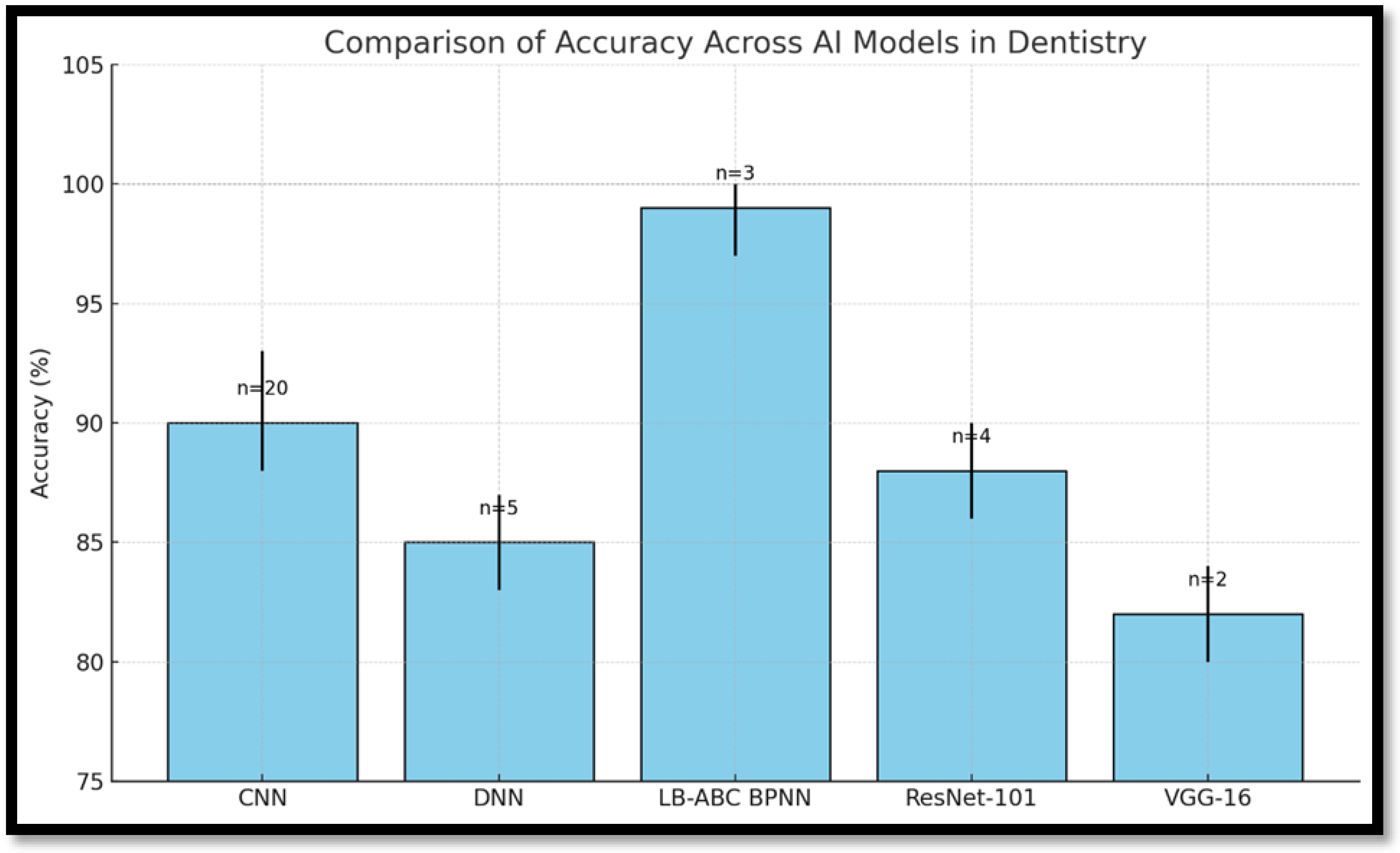
Figure 2 reveals that CNNs perform consistently across diagnostic tasks with an accuracy range of 85–93%, making them the most reliable for image-intensive applications. In contrast, the LB-ABC BPNN model, though evaluated in fewer studies, exhibited exceptional performance with near-perfect classification accuracy in certain tasks, suggesting suitability for complex, non-image-based diagnostic decisions. Figure 3 confirms these observations but also highlights that models like VGG-16 and ResNet-101, while promising, lack broad external validation, limiting current clinical applications.
Figure 2. Comparison of accuracy rates across different AI models in dentistry. (Cantu et al. [14] (2020), Setzer et al. [15] (2020), Sornam et al. [16] (2019), Li et al. [8] (2020), Patcas et al. [12] (2019), Takahashi et al. [7] (2020), Yamaguchi et al. [11] (2019), Arisu et al. [6] (2018), Kuwada et al. [10] (2020), Bouchahma et al. [4] (2019)).
Healthcare 13 01466 g002
Figure 3. A comparison of accuracy rates across different AI models in dentistry. (Error bars represent confidence intervals where available. The sample size (n), indicated above each bar, reflects the number of studies or datasets contributing to each model’s performance assessment).
Healthcare 13 01466 g003
Table 1. A comprehensive report of the studies that were included.
Study
(Author, Year)
Study DesignAI ModelDataset Type and Size (N)Preprocessing TechniquesValidation MethodApplicationExperimental MethodOutcomes
Bouchahma et al., [4] (2019)Clinical studyCNN Panoramic X-rays, N = 700Grayscale conversion, normalizationNot reportedOperative dentistry and EndodontiaDeep Learning → Supervised Learning → CNNAccuracy: 87%; RCT detection: 88%; Fluoride: 98%
Arisu et al., [6] 2018Clinical studyANNIntraoral images, N = 2400Not reportedInternal testing onlyRestorative dentistryDeep Learning → Supervised Learning → ANNComposite curing prediction; No metric reported
Takahashi et al., [7] 2020Experimental studyCNNPanoramic X-rays, N = 1498Image enhancement, segmentationTrain/test split Prosthodontics Deep Learning → Supervised Learning → CNNQualitative classification; No metric reported
Li et al., [8] 2020Clinical studyAutomated photo analysisFacial and intraoral photos, N = 1050Alignment of landmarksManual comparisonEsthetic dentistryImage Analysis → Automated Integration → Not specifiedSubjective cosmetic application; No metric reported
Kuwada et al., [10] 2020Clinical studyDetect Net, Alex Net, and VGG-16Panoramic X-rays, N = 550Grayscale conversion, normalizationTrain/validation/test splitOrthodonticsDeep Learning → Supervised Learning → CNN (DetectNet, etc.)Precision, recall, F1-score reported
Yamaguchi et al., [11] 2019Clinical studyCNNCephalometric radiographs, N = 1146Image resizing, grayscale conversionHoldout validationRestorative dentistryDeep Learning → Supervised Learning → CNNPrediction of crown debonding; No metric reported
Patcas et al., [12] 2019Study of Cohort CNN Orthodontic photos, N = 1200Image cropping, standardizationInternal datasetOrthodonticsDeep Learning → Supervised Learning → CNNSubjective age/attractiveness estimation; No metric reported
Li et al., [13] (2015)Experimental researchGA and BPNNOrthodontic measurements, N = 1000Feature normalizationInternal validationAesthetic dentistryDeep Learning → Supervised Learning → GA + BPNNObjective tooth color matching; No metric reported
Kositbowornchai et al. [17] (2016)Clinical studyLVQ Neural NetworkPanoramic X-rays, N = 600Image normalizationHoldout validationRestorative dentistry Deep Learning → Supervised Learning → LVQ-NNCaries detection; No metric reported
Patcas et al., [18] (2019)Clinical studyCNN Orthodontic photographs, N = 1200Image cropping, standardizationInternal dataset; no k-foldOrthodonticsDeep Learning → Supervised Learning → CNNAssessment of cleft patient profiles and frontal aesthetics; No metric reported
Vranckx et al., [19] (2020)Clinical studyCNN and ResNet-101Dental radiographs, N = 3000+Rescaling, augmentationCross-validation not specifiedOperative dentistry Deep Learning → Supervised Learning → CNN (ResNet-101)Molar segmentation; No metric reported
Lee et al., [20] (2020)Clinical studyML (e.g., Decision Trees, SVM)TMJ radiographs, N = 2100Grayscale conversion, filteringTrain/test splitOral and Maxillofacial Surgery ML → Supervised Learning → SVM, Decision TreesTMJOA classification; Accuracy: 95%
Cui et al., [21] (2020)Cohort studyCDS ML model Dataset from 5 hospitals, N = ~4000 casesData normalization cleaningSeparate test set Oral and Maxillofacial Surgery ML → Supervised Learning → CDS modelAccuracy: 99.16%
Sornam and Prabhakaran [16] (2019)Clinical studyBPNN with LB-ABCDental records, N = 750Data normalization10-fold cross-validation Restorative dentistryDeep Learning → Supervised Learning → Hybrid BPNNAccuracy: 99.16%
Setzer et al., [15] (2020)Clinical studyDL for CBCT CBCT scans, N = 1000Noise removal, segmentationExternal validationEndodonticsDeep Learning → Supervised Learning → CNNLesion detection; No metric reported
Cantu et al., [14] (2020)Clinical studyCNNCBCT images, N = 800Voxel normalization, filteringStratified train/test splitOperative dentistry and Oral Radiology Deep Learning → Supervised Learning → CNNPerformance exceeds clinicians; No metric reported
Aliaga et al., [22] (2020)Experimental studyAutomatic segmentation Facial scans, N = 350Mesh alignment, surface smoothingManual comparisonOperative dentistry and Oral and Maxillofacial Surgery Image Analysis → Automated → Not specifiedOsteoporosis detection; No metric reported
Kim et al., [23] (2018)Case–control studyML classifiers
(SVM, RF, etc.)
Dental images, N = 2500Feature scalingTrain/test splitOral and Maxillofacial Surgery and Oral MedicineML → Supervised Learning → SVM, RFBRONJ prediction; Accuracy: 91.8%
Dumast et al., [24] (2018)Case–control studyCNN and Shape variationRadiographs, N = 1820Histogram equalizationTest set validation Oral and Maxillofacial SurgeryDeep Learning → Supervised Learning → CNN
Bone classification; Accuracy reported
Morphological classification; Accuracy: 86.5%
Sorkhabi and Khajeh [25] (2019)Clinical study3D CNN Intraoral scans, N = 12003D normalization6-month clinical validation Implant dentistryDeep Learning → Supervised Learning → 3D CNNAlveolar bone density classification; Accuracy: 91.2%
DL → SL → CNN: Deep learning through supervised learning using a convolutional neural network. ANN: artificial neural network; BPNN: backpropagation neural network; CBCT: Cone Beam Computed Tomography; GA: Genetic Algorithm; LVQ-NN: Learning Vector Quantization Neural Network; BRONJ: Bisphosphonate-Related Osteonecrosis of the Jaw. “No metric reported” indicates that the study described an outcome without specifying performance statistics like accuracy, precision, or recall.
For caries detection, CNNs have been very accurate with accuracy ranging to 93%. These outcomes have far exceeded the performance of typical diagnostic strategies pointing toward the higher capacity of CNNs to analyze intricate dental images and detect carious lesions effectively and accurately. Likewise, when it comes to orthodontic treatment planning, CNN models also showed very good results across three databases, with accuracy over 85%. This accuracy reaffirms the applicability of radioactivity to well-documented work, such as treatment planning and prognosis. The results reveal the ability of CNNs to revolutionize the dental practice and place CNNs as ideal candidates for implementation into clinical environments to improve diagnostic capability and decision-making rates.
Deep learning, in general, and AI in its various forms hold high degrees of promise for tackling an entire range of pathologies, and even subjects, especially in the medical field of dentistry and its branches. Such applications include threshold caries detection and diagnosis, VR identification, and apical lesion assessment, so that appropriate management can be performed on time. The ability of air systems to diagnose diseases includes salivary gland disorders, maxillofacial cysts, osteoporosis, and detection and monitoring proficiency. They are capable of detecting cancerous lesions, evaluating the degree of alveolar bone loss, and providing information necessary for cephalometric analysis, which is an important facet of orthodontic diagnosis and planning.
Moreover, AI tools have been used to diagnose potential orthodontic extractions, analysis treatment plans, and measurements important for age and sex estimations. These models apply advanced computational techniques to improve diagnostic accuracy and patient outcomes. Because of their ability to work with large volumes of information and provide impartial conclusions, specialists consider them essential for the development of dental and medical practices.
The efficacy of CNNs in such outcomes points out and depicts their roles in enhancing diagnostic accuracy and the decision-making process in dentistry in enhancing its role in contemporary dentistry. The dominance of CNNs in dental research can be attributed primarily to their strong performance in imaging-related applications, where spatial localization and edge detection are crucial. However, CNNs are less suited to tasks involving tabular clinical data, temporal trends, or natural language processing, where other AI models may be more appropriate. For example, alternative models such as ResNet-101 and VGG-16 have demonstrated competitive performance in certain scenarios. For example, ResNet-101 was used successfully for predicting molar angulations and creating segmentation maps in operative dentistry, while VGG-16 showed usefulness in identifying impacted supernumerary teeth in panoramic radiographs. Backpropagation neural networks (BPNNs), though less common in image analysis, showed high performance in non-image-based tasks such as material classification, prediction of crown debonding, and treatment outcome forecasting. A hybrid LB-ABC BPNN model has shown to outperform CNNs in classifying oral cancer lesions, indicating the potential of hybrid models in tasks involving more complex clinical and histopathological data. Moreover, DNNs, when provided with structured input features and sufficient training data, offer high versatility and depth of learning. DNNs are also found to outperform CNNs in treatment planning scenarios where spatial image features were less critical than temporal or multi-factorial data inputs. These findings suggest that while CNNs are generally superior for image-processing tasks, other architectures may be preferable depending on the specific application and the nature of the data. Therefore, AI model selection in dentistry should be tailored to the diagnostic objective, data structure, and required accuracy level to ensure optimal outcomes.
The reviewed studies, from 2014 to 2023, encompassed four primary research designs: randomized controlled trials, clinical trials, analytical cross-sectional studies, and case–control and cohort studies. The utility of AI has been identified and categorized across various subfields of dentistry, proving its effectiveness. This widespread application reflects how deeply AI is rooted across dental specialties, how it affects the existing paradigm of dentistry, and the diverse roles they play in shaping the future of this field.
The key findings from the quality assessment are summarized below:
  • Blinding: In the context of AI model development, blinding is relevant when human annotators are involved in labeling training datasets, as knowledge of patient identity or outcome can introduce bias into the ground truth, potentially compromising model validity. Among the included studies, only Bouchahma et al. [4] explicitly reported the use of blinding during the annotation of training data, underscoring a general lack of attention to this potential bias across the reviewed literature.
  • Randomization: Randomization was implemented in two trials [10,19].
  • Dropout Rates: Nineteen studies addressed and reported dropout rates [4,6,7,8,10,12,13,14,15,16,17,18,19,20,21,22,23,24,25].
  • Accuracy of Research Variables: Research variables were explicitly checked for accuracy in 20 studies [4,6,7,8,10,11,12,13,14,15,16,17,18,19,20,21,22,23,24,25].
  • Sample Size Reporting: Details of all 20 studies provided for sample size details [4,6,7,8,10,11,12,13,14,15,16,17,18,19,20,21,22,23,24,25].
  • Inclusion and Exclusion Criteria: Eighteen studies specified their presence and exclusion standards [4,6,7,8,10,11,12,13,14,15,16,17,18,19,22,23,24,25].
  • Examiner Dependability: The reliability of the examiner was reported in 19 studies [4,6,7,8,10,11,12,14,15,16,17,18,19,20,21,22,23,24,25].
  • Pre-existing Results: The findings of 16 studies were already published or well-documented [4,7,8,10,12,13,14,15,16,17,18,19,22,23,24,25].
  • Bias Risk: Bias risk was assessed using the QUADAS-2 framework, adapted for AI diagnostic performance studies. A risk of bias at a moderate level was found in 5 studies [6,11,13,19,20], while 15 were identified as having low-quality evidence [4,7,8,10,12,14,15,16,17,18,21,22,23,24,25].
Table 2 presents an overview of the delivered trials, described in terms of methodological characteristics and outcomes.
The risk of bias using the JBI checklist was as follows: The risk of bias among analytical cross-sectional studies was 80%. The diagnostic test accuracy studies had scores of 88% suggesting a low risk of bias. Therefore, all 20 papers were included in this systematic review because they met the laid down criteria [Table 3].
As shown in Table 4, CNNs demonstrated consistently high diagnostic accuracy (85–93%) across multiple dental imaging tasks [4,10]. However, they remain less interpretable and computationally demanding. The hybrid BPNN (LB-ABC) reported the highest accuracy (~99.16%) in caries detection [16]. DNN-based models, such as the CDS system evaluated by Cui et al., also showed strong accuracy in clinical decision-making tasks involving structured datasets [21].

4. Discussion

AI has introduced transformative changes across multiple domains of healthcare, including dentistry, by virtue of its capacity to analyze large volumes of data and generate reliable insights. Its integration into dental practice has accelerated in recent years, significantly enhancing diagnostic accuracy, treatment planning, patient care, and administrative efficiency. Beyond clinical diagnostics, AI has contributed to improvements in patient management through tools such as appointment-scheduling systems, virtual assistants, and AI-powered chatbots that assist with common patient queries and oral health education. These technologies also show promise in mitigating dental anxiety. As AI continues to evolve, it is expected to further streamline clinical workflows, reduce human error, and contribute to more efficient and effective dental care delivery.
The studies reviewed in this paper collectively support the high diagnostic performance of AI in various dental applications. For instance, Caliskan et al. [26] demonstrated that CNNs can match expert-level accuracy in identifying submerged primary teeth from orthopantomograms. Similarly, Kilic et al. [27] illustrated how AI can expedite forensic dental identification. Zheng et al. [28] found that among various CNN architectures for pulpitis and deep caries detection, the multimodal CNN performed best. In the area of cephalometric landmark identification, Bulatova et al. [29] noted superior accuracy and efficiency using AI models compared to manual tracing. Zhao et al. [30] reported accurate assessments of adenoid hypertrophy on lateral cephalograms using AI, while Seo et al. [31] and Kim et al. [32] found that AI models could estimate cervical vertebral maturation with accuracy ranging from 90% to 93%. Likewise, Karhade et al. [33] demonstrated high precision in detecting and classifying early childhood caries. While CNNs dominate image-based tasks, models like BPNN demonstrated better classification performance in oral lesion classification [16].
Despite these advancements, several critical challenges must be addressed for AI to achieve widespread clinical adoption in dentistry. One of the foremost considerations is the trade-off between model interpretability, computational requirements, and deployment feasibility. Interpretable models like decision trees and logistic regression offer transparency and are generally more feasible for use in resource-limited settings, but they often lack the predictive power of complex models. Conversely, black-box models such as CNNs and DNNs provide superior diagnostic accuracy, especially in image-based applications, but are difficult to interpret and require substantial computational resources. Their complexity also raises concerns about accountability and patient safety. To address these concerns, interpretability tools like SHAP (Shapley Additive Explanations) and LIME (Local Interpretable Model-Agnostic Explanations) are being explored, although their integration into clinical practice remains limited [34].
Another major limitation is the lack of standardized evaluation metrics and open-access benchmark datasets in dental AI. Unlike general medical imaging, which benefits from widely recognized datasets such as MURA and CheXpert, dentistry lacks large-scale, annotated datasets to validate and compare model performance [35]. This absence hinders reproducibility, comparability, and regulatory approval. Standardized benchmarks are essential for evaluating diagnostic performance and guiding the development of reliable, generalizable AI systems.
The applicability of AI models to real-world clinical environments remains a significant concern. Most reviewed models were validated in controlled research settings using limited or demographically narrow datasets. For example, Kuwada et al. [10] reported performance metrics such as precision, recall, and F1-score for impacted tooth detection using panoramic radiographs; however, their model was only internally validated, limiting its generalizability to broader clinical settings. Similarly, Arisu et al. [6] used an ANN to predict composite curing outcomes but did not report external validation or integration into a clinical workflow. In contrast, Sornam and Prabhakaran [16] achieved a high accuracy of 99.16% using a hybrid LB-ABC BPNN model with 10-fold cross-validation, yet no evidence of clinical deployment was provided. Setzer et al. [15] among the few studies employing external validation with CBCT data, enhancing the reliability of its AI tool for lesion detection, although its real-time clinical performance remains undocumented. These examples emphasize the gap between in-lab model performance and actual clinical implementation, underscoring the need for future research to prioritize external validation, workflow integration, and long-term outcome assessment. Real-world clinical scenarios vary in complexity due to diverse patient populations, differing healthcare infrastructures, and unpredictable case presentations. Therefore, real-world usability studies are crucial to determine how AI tools integrate with existing clinical workflows, influence decision making, and affect patient outcomes. These studies also reveal adoption barriers such as clinician resistance, interoperability issues, and cost-related challenges.
Furthermore, for AI systems to be safely and legally deployed in dental practice, they must comply with stringent regulatory standards. In the United States, the Food and Drug Administration (FDA) mandates rigorous validation through clinical trials for approval of AI-based medical tools [36]. Similarly, in the European Union, AI technologies must conform to the Medical Device Regulation (MDR) and obtain CE marking [37]. To date, only a limited number of dental AI tools have received such regulatory clearance, underscoring the need for stronger governance and transparency in AI development. Long-term, multicenter trials are necessary to assess the sustained performance, safety, and ethical implications of AI systems in diverse dental care settings.
Although initial implementation costs—including software acquisition, hardware infrastructure, staff training, and data security compliance—pose challenges, AI can still be a cost-effective solution in the long term. Scalable and subscription-based AI models, particularly those requiring minimal hardware, are becoming increasingly available. These innovations may make AI adoption feasible even for small dental practices, provided that barriers such as clinician training and workflow compatibility are adequately addressed.

5. Limitations

Despite the promising outcomes identified in this systematic review, several limitations must be acknowledged. First, many of the included studies relied on datasets from limited or homogenous populations, potentially affecting the generalizability of their findings. For example, studies such as those by Patcas et al. [12,18] and Yamaguchi et al. [11] focused on specific geographic regions or institutional datasets without external validation, limiting their broader applicability.
Second, while the diagnostic accuracy of AI models like CNNs and hybrid neural networks (e.g., LB-ABC BPNNs) was consistently high in controlled settings [4,14,16], few studies validated their performance in real-world clinical environments. The absence of external validation in several studies, such as Arisu et al. [6] and Kuwada et al. [10], raises concerns about the robustness and reproducibility of the models when applied across diverse clinical contexts.
Third, reporting inconsistencies were noted across studies, particularly regarding key methodological elements like data preprocessing, sample size estimation, and validation strategies. For instance, validation methods were either not clearly stated or limited to internal testing in studies by Li et al. [8], Lee et al. [20], and others, potentially impacting the reliability of the reported outcomes.
Finally, most reviewed studies did not assess long-term performance or model adaptability over time. Real-world deployment demands continuous learning and validation of AI systems across varying datasets, clinical workflows, and patient demographics. This calls for future longitudinal, multicenter investigations to evaluate the sustained efficacy, ethical implications, and integration feasibility of AI tools in diverse dental practice settings.

6. Conclusions

AI represents a potent and efficacious tool in the field of dentistry, characterized by high diagnostic specificity, sensitivity, and accuracy. These attributes render AI valuable in augmenting diagnostic processes and, more broadly, enhancing patient care. Nevertheless, to substantiate these findings, it is imperative to incorporate large sample sizes. Future research should focus on evaluating the effectiveness and therapeutic interventions facilitated by AI models in dental care.
In conclusion, the prospective integration of artificial intelligence into clinical practice necessitates the implementation of long-term, large-scale multicenter randomized controlled trials to comprehensively evaluate its efficacy, particularly across diverse patient demographics. Establishing minimum benchmarks for comparing AI performance is a crucial step towards building a robust foundation for AI applications in dentistry.
Artificial intelligence and machine learning hold the potential to become significant assets in clinical dentistry, enhancing the efficacy of diagnostic processes and potentially increasing the accuracy of primary diagnoses. However, their widespread applicability and optimal utilization can only be realized through substantial investment in further research and technological advancements. As these technologies evolve, they may facilitate the prompt diagnosis and treatment of young patients within new dental practices, thereby improving clinical outcomes.

Author Contributions

Conceptualization, O.K., A.S.H. and M.S.A.; methodology, O.K., R.A.S.A. and A.Y.S.; software, R.A. and S.A.; validation, M.A.F. and O.S.A.; formal analysis, L.H.S.A. and W.W.Z.A.; investigation, R.I. and A.A.; resources, O.K. and A.S.H.; data curation, R.I. and A.A.; writing—original draft preparation, O.K. and M.A.F.; writing—review and editing, O.K. and M.A.F.; visualization, R.I. and A.A.; supervision, M.A.F.; project administration, R.A.S.A.; funding acquisition, M.A.F. All authors have read and agreed to the published version of the manuscript.

Funding

The authors thank Ajman University, Ajman, UAE, for supporting the APC of this article. The funders had no role in study design, data collection and analysis, decision to publish, or preparation of the manuscript.

Data Availability Statement

The dataset used in this paper will be made available upon request to the corresponding author.

Conflicts of Interest

The authors declare no conflicts of interest.

References

  1. Shapiro, S.C. Artificial intelligence (AI). In Encyclopedia of Computer Science, 2nd ed.; Wiley: Hoboken, NJ, USA, 2003; pp. 89–93. [Google Scholar]
  2. Ramesh, A.N.; Kambhampati, C.; Monson, J.R.; Drew, P.J. Artificial intelligence in medicine. Ann. R. Coll. Surg. Engl. 2004, 86, 334–338. [Google Scholar] [CrossRef] [PubMed]
  3. A Primer: Artificial Intelligence Versus Neural Networks. The Scientist Magazine®. Available online: https://www.the-scientist.com/magazine-issue/artificial-intelligence-versus-neural-networks-65802 (accessed on 15 December 2024).
  4. Bouchahma, M.; Ben Hammouda, S.; Kouki, S.; Alshemaili, M.; Samara, K. An automatic dental decay treatment prediction using a deep convolutional neural network on X-ray images. In Proceedings of the 2019 IEEE/ACS 16th International Conference on Computer Systems and Applications (AICCSA), Abu Dhabi, United Arab Emirates, 1–4 October 2019. [Google Scholar]
  5. Ekert, T.; Krois, J.; Meinhold, L.; Elhennawy, K.; Emara, R.; Golla, T.; Schwendicke, F. Deep Learning for the Radiographic Detection of Apical Lesions. J. Endod. 2019, 45, 917–922.e5. [Google Scholar] [CrossRef]
  6. Deniz Arısu, H.; Eligüzeloglu Dalkilic, E.; Alkan, F.; Erol, S.; Uctasli, M.B.; Cebi, A. Use of artificial neural network in determination of shade, light curing unit, and composite parameters’ effect on bottom/top Vickers hardness ratio of composites. Biomed. Res. Int. 2018, 2018, 4856707. [Google Scholar] [CrossRef] [PubMed]
  7. Takahashi, T.; Nozaki, K.; Gonda, T.; Ikebe, K. A system for designing removable partial dentures using artificial intelligence. Part 1. Classification of partially edentulous arches using a convolutional neural network. J. Prosthodont. Res. 2021, 65, 115–118. [Google Scholar] [CrossRef]
  8. Li, M.; Xu, X.; Punithakumar, K.; Le, L.H.; Kaipatur, N.; Shi, B. Automated integration of facial and intra-oral images of anterior teeth. Comput. Biol. Med. 2020, 122, 103794. [Google Scholar] [CrossRef]
  9. Poplin, R.; Varadarajan, A.V.; Blumer, K.; Liu, Y.; McConnell, M.V.; Corrado, G.S.; Peng, L.; Webster, D.R. Prediction of cardiovascular risk factors from retinal fundus photographs via deep learning. Nat. Biomed. Eng. 2018, 2, 158–164. [Google Scholar] [CrossRef] [PubMed]
  10. Kuwada, C.; Ariji, Y.; Fukuda, M.; Kise, Y.; Fujita, H.; Katsumata, A.; Ariji, E. Deep learning systems for detecting and classifying the presence of impacted supernumerary teeth in the maxillary incisor region on panoramic radiographs. Oral Surg. Oral Med. Oral Pathol. Oral Radiol. 2020, 130, 464–469. [Google Scholar] [CrossRef]
  11. Yamaguchi, S.; Lee, C.; Karaer, O.; Ban, S.; Mine, A.; Imazato, S. Predicting the Debonding of CAD/CAM Composite Resin Crowns with AI. J. Dent. Res. 2019, 98, 1234–1238. [Google Scholar] [CrossRef]
  12. Patcas, R.; Bernini, D.A.J.; Volokitin, A.; Agustsson, E.; Rothe, R.; Timofte, R. Applying artificial intelligence to assess the impact of orthognathic treatment on facial attractiveness and estimated age. Int. J. Oral Maxillofac. Surg. 2019, 48, 77–83. [Google Scholar] [CrossRef]
  13. Li, H.; Lai, L.; Chen, L.; Lu, C.; Cai, Q. The prediction in computer color matching of dentistry based on GA+BP neural network. Comput. Math. Methods Med. 2015, 2015, 816719. [Google Scholar] [CrossRef]
  14. Cantu, A.G.; Gehrung, S.; Krois, J.; Chaurasia, A.; Rossi, J.G.; Gaudin, R.; Elhennawy, K.; Schwendicke, F. Detecting caries lesions of different radiographic extension on bitewings using deep learning. J. Dent. 2020, 100, 103425. [Google Scholar] [CrossRef] [PubMed]
  15. Setzer, F.C.; Shi, K.J.; Zhang, Z.; Yan, H.; Yoon, H.; Mupparapu, M.; Li, J. Artificial intelligence for the computer-aided detection of periapical lesions in cone-beam computed tomographic images. J. Endod. 2020, 46, 987–993. [Google Scholar] [CrossRef]
  16. Sornam, M.; Prabhakaran, M. Logit-based artificial bee colony optimization (LB-ABC) approach for dental caries classification using a back propagation neural network. Integr. Intell. Comput. Commun. Secur. 2019, 771, 79–91. [Google Scholar]
  17. Kositbowornchai, S.; Siriteptawee, S.; Plermkamon, S.; Bureerat, S.; Chetchotsak, D. An artificial neural network for detection of simulated dental caries. Int. J. Comput. Assist. Radiol. Surg. 2016, 1, 91–96. [Google Scholar] [CrossRef]
  18. Patcas, R.; Timofte, R.; Volokitin, A.; Agustsson, E.; Eliades, T.; Eichenberger, M.; Bornstein, M.M. Facial attractiveness of cleft patients: A direct comparison between artificial-intelligence-based scoring and conventional rater groups. Eur. J. Orthod. 2019, 41, 428–433. [Google Scholar] [CrossRef]
  19. Vranckx, M.; Van Gerven, A.; Willems, H.; Vandemeulebroucke, A.; Ferreira Leite, A.; Politis, C.; Jacobs, R. Artificial Intelligence (AI)-Driven Molar Angulation Measurements to Predict Third Molar Eruption on Panoramic Radiographs. Int. J. Environ. Res. Public Health 2020, 17, 3716. [Google Scholar] [CrossRef] [PubMed]
  20. Lee, K.S.; Kwak, H.J.; Oh, J.M.; Jha, N.; Kim, Y.J.; Kim, W.; Baik, U.B.; Ryu, J.J. Automated detection of TMJ osteoarthritis based on artificial intelligence. J. Dent. Res. 2020, 99, 1363–1367. [Google Scholar] [CrossRef]
  21. Cui, Q.; Chen, Q.; Liu, P.; Liu, D.; Wen, Z. Clinical decision support model for tooth extraction therapy derived from electronic dental records. J. Prosthet. Dent. 2021, 126, 83–90. [Google Scholar] [CrossRef]
  22. Aliaga, I.; Vera, V.; Vera, M.; García, E.; Pedrera, M.; Pajares, G. Automatic computation of mandibular indices in dental panoramic radiographs for early osteoporosis detection. Artif. Intell. Med. 2020, 103, 101816. [Google Scholar] [CrossRef]
  23. Kim, D.W.; Kim, H.; Nam, W.; Kim, H.J.; Cha, I.H. Machine learning to predict the occurrence of bisphosphonate-related osteonecrosis of the jaw associated with dental extraction: A preliminary report. Bone 2018, 116, 207–214. [Google Scholar] [CrossRef]
  24. de Dumast, P.; Mirabel, C.; Cevidanes, L.; Ruellas, A.; Yatabe, M.; Ioshida, M.; Ribera, N.T.; Michoud, L.; Gomes, L.; Huang, C.; et al. A web-based system for neural network based classification in temporomandibular joint osteoarthritis. Comput. Med. Imaging Graph. 2018, 67, 45–54. [Google Scholar] [CrossRef] [PubMed]
  25. Sorkhabi, M.M.; Saadat Khajeh, M. Classification of alveolar bone density using 3-D deep convolutional neural network in the cone-beam CT images: A 6-month clinical study. Measurement 2019, 148, 106945. [Google Scholar] [CrossRef]
  26. Caliskan, S.; Tuloglu, N.; Celik, O.; Ozdemir, C.; Kizilaslan, S.; Bayrak, S. A pilot study of a deep learning approach to submerged primary tooth classification and detection. Int. J. Comput. Dent. 2021, 24, 1–9. [Google Scholar] [CrossRef]
  27. Kılıc, M.C.; Bayrakdar, I.S.; Celik, O.; Bilgir, E.; Orhan, K.; Aydın, O.B.; Kaplan, F.A.; Sağlam, H.; Aslan, A.F.; Yılmaz , A.B. Artificial intelligence system for automatic deciduous tooth detection and numbering in panoramic radiographs. Dentomaxillofac. Radiol. 2021, 50, 20200172. [Google Scholar] [CrossRef] [PubMed]
  28. Zheng, L.; Wang, H.; Mei, L.; Chen, Q.; Zhang, Y.; Zhang, H. Artificial intelligence in digital cariology: A new tool for the diagnosis of deep caries and pulpitis using convolutional neural networks. Ann. Transl. Med. 2021, 9, 763. [Google Scholar] [CrossRef]
  29. Bulatova, G.; Kusnoto, B.; Grace, V.; Tsay, T.P.; Avenetti, D.M.; Sanchez, F.J.C. Assessment of automatic cephalometric landmark identification using artificial intelligence. Orthod. Craniofac. Res. 2021, 24 (Suppl. S2), 37–42. [Google Scholar] [CrossRef]
  30. Zhao, T.; Zhou, J.; Yan, J.; Cao, L.; Cao, Y.; Hua, F.; He, H. Automated adenoid hypertrophy assessment with lateral cephalometry in children based on artificial intelligence. Diagnostics 2021, 11, 1386. [Google Scholar] [CrossRef] [PubMed]
  31. Seo, H.; Hwang, J.; Jeong, T.; Shin, J. Comparison of deep learning models for cervical vertebral maturation stage classification on lateral cephalometric radiographs. J. Clin. Med. 2021, 10, 3591. [Google Scholar] [CrossRef]
  32. Kim, E.G.; Oh, I.S.; So, J.E.; Kang, J.; Le, V.N.T.; Tak, M.K.; Lee, D.-W. Estimating cervical vertebral maturation with a lateral cephalogram using the convolutional neural network. J. Clin. Med. 2021, 10, 5400. [Google Scholar] [CrossRef]
  33. Karhade, D.S.; Roach, J.; Shrestha, P.; Simancas-Pallares, M.A.; Ginnis, J.; Burk, Z.J.S.; Ribeiro, A.A.; Cho, H.; Wu, D.; Divaris, K. An automated machine learning classifier for early childhood caries. Pediatr. Dent. 2021, 43, 191–197. [Google Scholar] [PubMed]
  34. Rudin, C. Stop Explaining Black Box Machine Learning Models for High Stakes Decisions and Use Interpretable Models Instead. Nat. Mach. Intell. 2019, 1, 206–215. [Google Scholar] [CrossRef] [PubMed]
  35. Rajpurkar, P.; Irvin, J.; Ball, R.L.; Zhu, K.; Yang, B.; Mehta, H.; Duan, T.; Ding, D.; Bagul, A.; Langlotz, C.P.; et al. Deep learning for chest radiograph diagnosis: A retrospective comparison of the CheXNeXt algorithm to practicing radiologists. PLoS Med. 2018, 15, e1002686. [Google Scholar] [CrossRef] [PubMed]
  36. Available online: https://www.fda.gov/medical-devices/software-medical-device-samd/artificial-intelligence-and-machine-learning-software-medical-device (accessed on 15 November 2024).
  37. Aboy, M.; Minssen, T.; Vayena, E. Navigating the EU AI Act: Implications for regulated digital medical products. NPJ Digit. Med. 2024, 7, 237. [Google Scholar] [CrossRef] [PubMed]
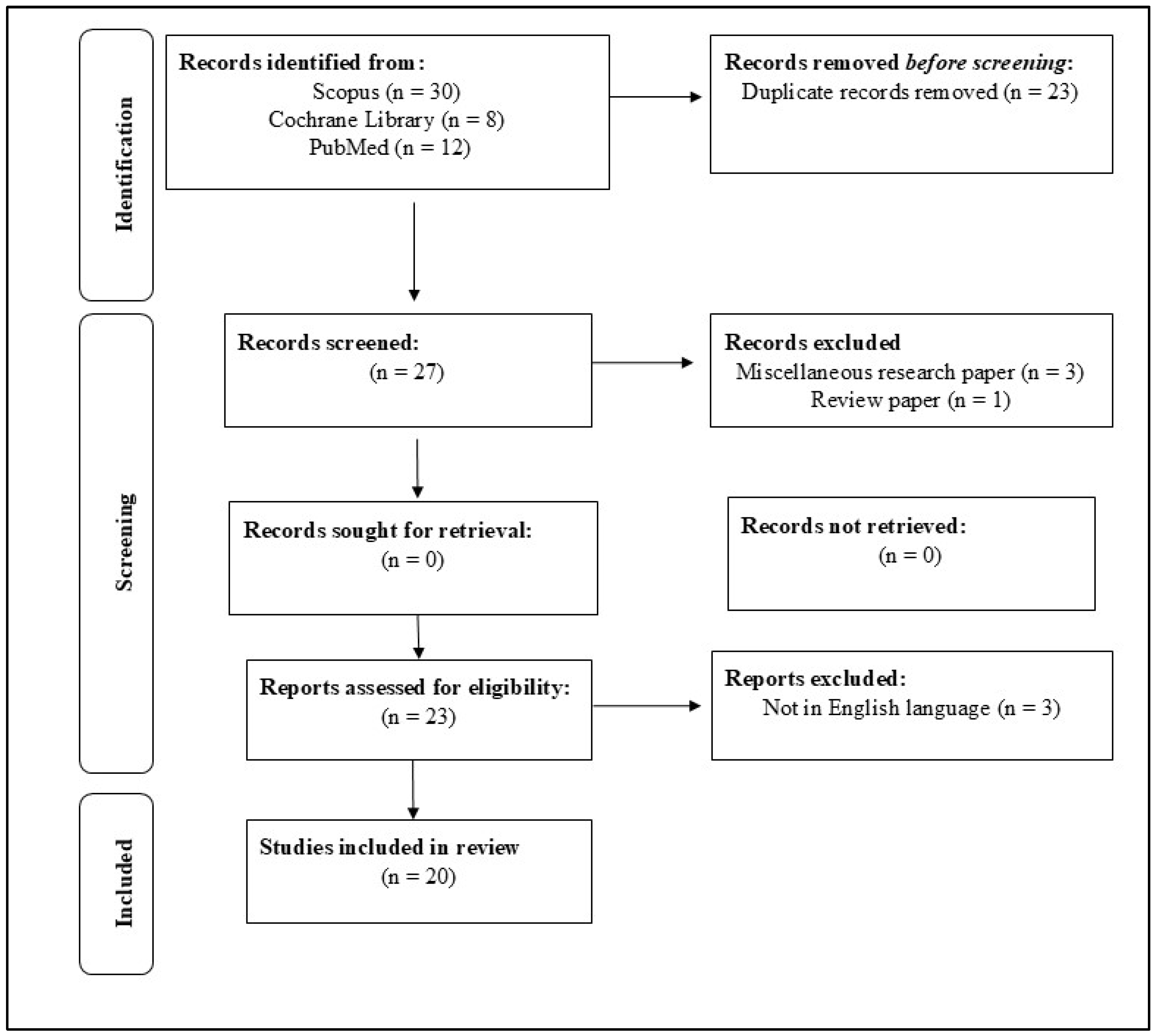
Figure 1. The PRISMA methodology was employed for identifying pertinent studies for this study.
Healthcare 13 01466 g001
Table 2. Evaluations of methodological quality included studies.
Study (Author, Year)RandomizationBlindingWithdrawal/Dropout
Mentioned
Multiple Variables MeasurementEstimation of Sample SizeClear
Exclusion/Inclusion Criteria
Reliability of ExaminerPrespecified OutcomesStudy Quality/Bias Risk
Bouchahma et al., [4] (2019)Not conductedImplementedMentionedMeasured RepeatedlyEstimatedClearly DefinedTestedPredeterminedLow
Arisu et al., [6] 2018Not ConductedNot AppliedMentionedMeasured RepeatedlyEstimatedClearly DefinedTestedNot SpecifiedModerate
Takahashi et al., [7] 2020Not ConductedNot AppliedMentionedMeasured RepeatedlyEstimatedClearly DefinedTestedPredeterminedLow
Li et al., [8] 2020Not ConductedNot AppliedMentionedMeasured RepeatedlyEstimatedClearly DefinedTestedPredeterminedLow
Kuwada et al., [10] 2020ConductedNot AppliedMentionedMeasured RepeatedlyEstimatedClearly DefinedTestedPredeterminedLow
Yamaguchi et al., [11] 2019Not conductedNot AppliedUnclearMeasured repeatedlyEstimatedClearly DefinedTestedNot SpecifiedModerate
Patcas et al., [12] 2019Not ConductedNot AppliedMentionedMeasured RepeatedlyEstimatedClearly DefinedTestedPredeterminedLow
Li et al., [13] (2015)UnclearUnclearMentionedMeasured RepeatedlyEstimatedClearly DefinedNot TestedPredeterminedModerate
Kositbowornchai et al., [17] (2016)Not ConductedNot AppliedMentionedMeasured RepeatedlyEstimatedClearly DefinedTestedPredeterminedLow
Patcas et al., [18] (2019)Not ConductedNot AppliedMentionedMeasured RepeatedlyEstimatedClearly DefinedTestedPredeterminedLow
Vranckx et al., [19] (2020)ConductedNot AppliedMentionedMeasured RepeatedlyEstimatedClearly DefinedTestedPredeterminedModerate
Lee et al., [20] (2020)Not ConductedNot AppliedMentionedNot Measured RepeatedlyEstimatedNot clearly DefinedTestedNot specifiedModerate
Cui et al., [21] (2020)Not ConductedNot AppliedMentionedMeasured RepeatedlyEstimatedNot Clearly DefinedTestedNot SpecifiedLow
Sornam and Prabhakaran [16] (2019)Not ConductedNot AppliedMentionedMeasured RepeatedlyEstimatedClearly DefinedTestedPredeterminedLow
Setzer et al., [15] (2020)Not ConductedNot AppliedMentionedMeasured RepeatedlyEstimatedClearly DefinedTestedPredeterminedLow
Cantu et al., [14] (2020)Not ConductedNot AppliedMentionedMeasured RepeatedlyEstimatedClearly DefinedTestedPredeterminedLow
Aliaga et al., [22] (2020)Not ConductedNot AppliedMentionedMeasured RepeatedlyEstimatedClearly DefinedTestedPredeterminedLow
Kim et al., [23] (2018)Not ConductedNot AppliedMentionedMeasured RepeatedlyEstimatedClearly DefinedTestedPredeterminedLow
Dumast et al., [24] (2018)Not ConductedNot AppliedMentionedMeasured RepeatedlyEstimatedClearly DefinedTestedPredeterminedLow
Sorkhabi and Khajeh [25] (2019)Not ConductedNot AppliedMentionedMeasured RepeatedlyEstimatedClearly DefinedTestedPredeterminedLow
Table 3. Quality assessment results.
Design of StudyNo. of StudiesPercentageBias Risks
Randomized276Moderate
Cross-sectional980Low
Diagnostic test accuracy988Low
Low = low risk/concern; Moderate = some concerns.
Table 4. Comparative overview of AI models used in dental diagnostics.
AI ModelDiagnostic AccuracyInterpretabilityDeployment FeasibilitySuitable Applications
CNNHigh (85–93%)LowRequires GPU resourcesImage-based diagnostics (caries, lesions)
Hybrid BPNN (LB-ABC)Very High (~99%)ModerateModerate complexityOral cancer, caries classification
DNNHigh to Very HighLowModerate to HighTreatment planning, structured data tasks
Disclaimer/Publisher’s Note: The statements, opinions and data contained in all publications are solely those of the individual author(s) and contributor(s) and not of MDPI and/or the editor(s). MDPI and/or the editor(s) disclaim responsibility for any injury to people or property resulting from any ideas, methods, instructions or products referred to in the content.