Article

Serum OxLDL Levels Are Positively Associated with the Number of Ischemic Events and Damaged Blood Vessels in Patients with Coronary Artery Disease

by
Mariana Perez-Robles
1,
Wendy Campos-Perez
1,
Sissi Godinez-Mora
1,
Victor Lopez-Alvarez
1,2,
Liliana Estefanía Ramos-Villalobos
3,
Felisardo Corona-Ruiz
4,
Juan Armendariz-Borunda
5 and
Erika Martinez-Lopez
1,*
1
Instituto de Nutrigenética y Nutrigenómica Traslacional, Departamento de Biología Molecular y Genómica, Centro Universitario de Ciencias de la Salud, Universidad de Guadalajara, Guadalajara 44340, Jalisco, Mexico
2
Doctorado en Ciencias de la Nutrición Traslacional, Departamento de la Reproducción Humana, Crecimiento y Desarrollo Infantil, Centro Universitario de Ciencias de la Salud, Universidad de Guadalajara, Guadalajara 44340, Jalisco, Mexico
3
Servicio de Cardiología del Hospital Civil de Guadalajara “Dr. Juan I. Menchaca”, Guadalajara 44340, Jalisco, Mexico
4
Clínica Medicina Familiar 1 del ISSSTE “Dr. Arturo González Guzmán”, Guadalajara 44340, Jalisco, Mexico
5
Instituto de Biología Molecular en Medicina y Terapia Génica, Centro Universitario de Ciencias de la Salud, Universidad de Guadalajara, Sierra Mojada 950, Independencia Oriente, Guadalajara 44340, Jalisco, Mexico
*
Author to whom correspondence should be addressed.
Healthcare 2025, 13(12), 1426; https://doi.org/10.3390/healthcare13121426
Submission received: 6 May 2025 / Revised: 4 June 2025 / Accepted: 11 June 2025 / Published: 14 June 2025
(This article belongs to the Section Medics)

Abstract

:
Background/Objectives: The severity of coronary artery disease (CAD) depends on several factors. Oxidative stress contributes to the oxidation of lipoproteins, thereby promoting ischemic events (IEs) and damaged blood vessels (DBVs). This study aimed to compare biochemical variables and levels of oxidized lipoproteins in CAD patients stratified by the number of IEs and DBVs. Methods: A cross-sectional study was performed, including 51 patients diagnosed with CAD. Serum oxidized lipoproteins were measured by ELISA. Results: CAD patients with ≥2 IEs and ≥2 DBVs showed higher oxLDL levels than those with one IE and one DBV (8293.35 ng/mL [7131.32–9287.39] vs. 6474.26 ng/mL [5374.52–7574.01], p = 0.029). OxLDL levels were positively associated with the number of IEs (r2 = 15.2, B = 0.142 [0.046–0.239], p = 0.005) and DBVs (r2 = 19.2, B = 0.196 [0.018–0.374], p = 0.020). Conclusions: This study identified a positive association between elevated serum oxLDL levels and the presence of ≥2 IEs and ≥2 DBVs in CAD patients.

Graphical Abstract

1. Introduction

Coronary artery disease (CAD) refers to a group of disorders in which the myocardium receives insufficient blood flow and oxygen due to arterial obstruction, most commonly caused by atheroma [1]. CAD is the most prevalent cardiovascular disease, responsible for 17.9 million deaths worldwide each year, and is the leading cause of mortality [2,3].
Clinically, CAD may present as chronic heart disease (stable angina) or acute coronary syndrome, which includes ST-segment-elevation myocardial infarction (STEMI), non-ST-segment-elevation myocardial infarction (NSTEMI), unstable angina, and mixed CAD [4,5]. Regardless of the type, patients with CAD are commonly treated with pharmacological and surgical interventions; however, most remain at risk of experiencing a second ischemic event (IE), which may affect additional blood vessels [6].
The main risk factors for a second IE and damage to two or more blood vessels are related to the development of atherosclerosis. These include an age >45 years in men and >55 years in women, male sex, and the presence of metabolic conditions such as obesity, arterial hypertension, type 2 diabetes mellitus (T2DM), and dyslipidemias—including hypercholesterolemia, hypertriglyceridemia, elevated low-density lipoprotein cholesterol (LDL), and hypoalphalipoproteinemia [7,8].
In addition, oxidative stress plays an important role in CAD [9,10]. Specifically, peroxyl radicals, fatty hydroperoxides, epoxides, isoprostanes, and aldehydes modify the lipid components of lipoproteins—particularly their polyunsaturated fatty acids (PUFAs)—and alter the structure of cysteine, methionine, and tyrosine residues in apolipoproteins found in native LDL and HDL. This leads to the formation of oxidized low-density and high-density lipoproteins (oxLDLs and oxHDLs, respectively) in the intima of blood vessels, where they are phagocytosed by macrophages forming foam cells. These oxidized lipoproteins exhibit alterations in charge, activity, function, and metabolism, and also promote the secretion of adhesion molecules and proinflammatory cytokines. This process culminates in the formation of atherosclerotic plaques in the intima, which may erode and lead to thrombus formation, resulting in vascular occlusion, CAD progression, or even a new IE in patients already diagnosed with the disease [11,12,13].
Therefore, it has been proposed to evaluate biomarkers associated with oxidative stress in patients with CAD, such as oxLDL and oxHDL, and to analyze their relationship with the number of IEs and damaged blood vessels (DBVs), as this has not yet been explored in the Mexican population. The aim of this study was to compare biochemical variables, including oxidized lipoproteins, in CAD patients stratified by the number of IEs and DBVs.

2. Materials and Methods
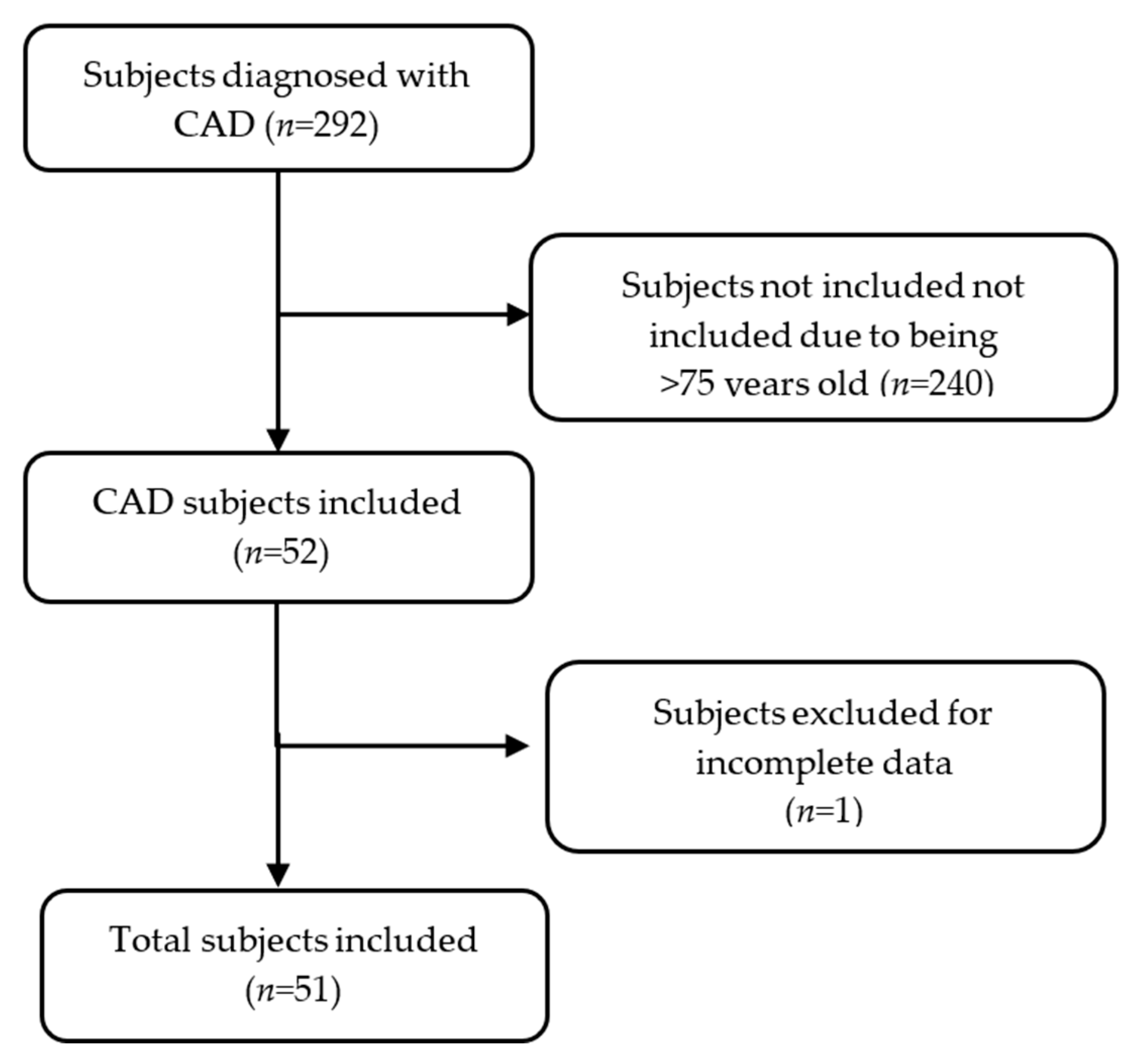
2.1. Study Subjects

A total of 51 individuals from western Mexico with a confirmed diagnosis of CAD participated in this cross-sectional study. These participants were enrolled between 2022 and 2023 through two medical centers: the Cardiology Service of the “Hospital Civil de Guadalajara, Dr. Juan I. Menchaca”, and the Family Medicine Clinic “Instituto de Seguridad y Servicios Sociales de los Trabajadores del Estado (ISSSTE), Dr. Arturo González Guzmán.” The inclusion criteria were an age between 30 and 75 years, a body mass index (BMI) ≥ 18.5 kg/m2, and a confirmed CAD diagnosis. Patients with autoimmune diseases, cancer, or chronic kidney disease were excluded. In addition, individuals with incomplete data or insufficient biological samples were excluded. Study evaluations were conducted at the “Instituto de Nutrigenética y Nutrigenómica Traslacional” affiliated with the “Universidad de Guadalajara”.

2.2. CAD Diagnosis and Cardiological Evaluations

CAD diagnosis and the number of IEs were established by a cardiologist through the assessment of clinical symptoms of ischemia. In men, oppressive chest pain radiating to the left arm, retrosternal pain, and dyspnea at rest or during exertion were evaluated. In women, atypical symptoms without chest pain—such as extreme fatigue, cough, palpitations, dizziness, nausea, gastrointestinal discomfort, shortness of breath, and numbness in the arms, back, jaw, or neck—were considered. Biomarkers of myocardial damage were also considered, specifically, elevated serum cardiac enzymes: creatine kinase ≥12 IU/L, hs-TnI >34 ng/L and hs-TnT >22 ng/L for men, and hs-TnI >15 ng/L and hs-TnT >14 ng/L for women. Electrocardiography was used to assess ST-segment abnormalities. In addition, coronary angiography was performed by a cardiologist to confirm arterial obstruction and determine the number of DBVs [14].
CAD diagnosis was based on the guidelines of the American College of Cardiology (ACC), the American Heart Association (AHA), and the European Society of Cardiology. Patients were categorized as having either chronic CAD (e.g., stable angina) or acute forms (STEMI, NSTEMI, or unstable angina). The mixed CAD type was considered when patients did not meet the criteria for one of the previous diagnoses [4,5].

2.3. Blood Pressure Evaluation

Blood pressure was measured using an automated upper-arm digital device (HEM-7130, Omron Healthcare Co., Ltd., Kyoto, Japan), following the recommendations outlined in the ACC/AHA guidelines [15].

2.4. Pharmacological and Interventional Treatment

Pharmacological treatments were categorized by function into the following groups: anticoagulants and antiplatelets (acetylsalicylic acid, clopidogrel), vasodilators (isosorbide, nifedipine), antihypertensives (metoprolol, losartan, enalapril), lipid-lowering agents (atorvastatin, pravastatin, ciprofibrate, bezafibrate), and glucose-control medication (metformin). Additionally, coronary stent placement was considered an interventional treatment.

2.5. Biochemical Analysis

A peripheral blood sample was collected after a fasting period of 8–10 h. The samples were then centrifuged for 15 min at 3500 rpm (revolutions per minute) to separate the serum. Total cholesterol (TC), high-density lipoprotein cholesterol (HDL), and triglycerides (TG) were measured using dry chemistry with a Vitros 350 Analyzer (Ortho-Clinical Diagnostics, Johnson & Johnson Services Inc., Rochester, NY, USA). Low-density lipoprotein cholesterol (LDL) was calculated using the Friedewald formula when TG concentrations were <400 mg/dL [5]. In addition, the TG/HDL and TC/HDL ratios were calculated by dividing TG by HDL, and TC by HDL, respectively.

2.6. Metabolic Comorbidities and Dyslipidemias

T2DM and arterial hypertension diagnoses were established by an internist according to the guidelines of the American Diabetes Association (ADA) and the American College of Cardiology/American Heart Association (ACC/AHA), respectively [16,17]. Dyslipidemia classifications were based on the thresholds defined by the ACC/AHA. Individuals were categorized as having hypertriglyceridemia when TG levels were ≥150 mg/dL, hypercholesterolemia when TC was ≥ 200 mg/dL, and elevated LDL-C when these levels were ≥70 mg/dL. Low HDL (hypoalphalipoproteinemia) was defined as levels <50 mg/dL for women and <40 mg/dL for men [17].
Thresholds for elevated lipid ratios were defined as a TG/HDL ratio >3.75 in men and >3.0 in women, and a TC/HDL ratio >5.0 in men and >4.0 in women [18,19].

2.7. Quantification of Serum oxLDL and oxHDL

Serum levels of oxLDL and oxHDL were measured using a sandwich-type enzyme-linked immunosorbent assay (ELISA), following the manufacturer’s protocol (Cell Biolabs, Inc., San Diego, CA, USA). Quantification was performed using a ThermoFisher Scientific Multiskan SkyHigh Microplate spectrophotometer (Waltham, MA, USA) at a wavelength of 450 nm. oxLDL/LDL and oxHDL/HDL ratios were calculated converting oxLDL and oxHDL values to mg/dL, and dividing oxLDL by LDL and oxHDL by HDL, respectively. For data analysis, ratio values were log-transformed using the natural logarithm (ln).

2.8. Anthropometric Measurements

Anthropometric measurements were taken after an 8-10 h fast, with participants barefoot and wearing light clothing. Body weight was measured with a SECA 803 scale, while height was measured using an SECA stadiometer (GMBH & Co., Hamburg, Germany). Waist circumference (WC) was assessed using a Lufkin Executive® Thinline measuring tape (2 mm, New Brighton, MN, USA). Body fat percentage (BF%) was determined using tetrapolar electrical bioimpedance (Quantum V, RJL Systems, Clinton Township, MI, USA). The BMI was calculated as the weight in kilograms divided by the height squared in meters (kg/m2).
A waist circumference ≥88 cm in women and ≥102 cm in men was considered indicative of abdominal obesity [20]. The BF% was considered to be excessive when it was ≥25% in men and ≥35% in women [21]. A BMI of 25–29.9 kg/m2 was classified as overweight, and ≥30 kg/m2 as obesity [22].

2.9. Alcohol Consumption

Alcohol intake was assessed through patient self-report, including details on the volume, frequency, and duration of consumption. Significant alcohol intake was defined as >14 g/day for women and >28 g/day for men over the past year, or consumption of ≥5 alcoholic drinks for men or ≥4 for women on the same occasion on at least one day in the past month [23,24]. Additionally, the Alcohol Use Disorders Identification Test (AUDIT) was administered to evaluate risky alcohol intake. A score ≥8 in men and ≥5 in women was considered indicative of risk [25].

2.10. Tobacco Index and Risk Exposure

The tobacco index was determined according to pack-years, obtained by multiplying the daily number of cigarettes by the total number of years of smoking, and dividing the result by 20. The cut-off values were defined as follows: low (<5), moderate (5–15), and high (>15) [26]. Tobacco exposure risk was also assessed using the Tobacco Scale for Primary Care (Escala de Tabaquismo en Atención Primaria, ETAP). A value ≥ 20 years on the ETAP was considered indicative of high-risk exposure [27].

2.11. Statistical Analysis

The sample size was calculated using a formula for studies assessing the correlation between two quantitative variables, based on data obtained from a study conducted in Augsburg that evaluated the relationship between serum oxLDL levels and the risk of an IE [28]. A significance level of p = 0.05, a statistical power of 80%, and an estimated 20% attrition rate were considered, resulting in a required sample of 44 participants.
The Shapiro–Wilk test was used to assess the normal distribution of variables. Descriptive statistics for qualitative variables were reported as frequencies and percentages. Comparative analyses of quantitative variables, adjusted for covariates (age, sex, alcohol consumption, smoking, and pharmacological treatment), were performed using a general univariate model. To analyze the association between two quantitative variables and to determine the correlation coefficient, a linear regression model was used. To determine the risk, binary logistic regression models were used. A 95% confidence interval and a p value <0.05 were considered statistically significant. Statistical analyses were performed using SPSS version 20.0 software (IBM Corp., Armonk, NY, USA).

3. Results

3.1. Characteristics of the Study Subjects

In this study, 51 patients with CAD were included (Figure 1). Of the study participants, 67% (n = 34) were men and 33% (n = 17) were women, with a median age of 60 years (IQR: 55–68).
For clinical variables, the median systolic blood pressure (SBP) was 129 mmHg (IQR: 111–140), and the median diastolic blood pressure (DBP) was 78 mmHg (IQR: 71–86). Regarding cardiological variables, the median number of DBVs was 2 (IQR: 1–2), and the median number of IEs was also 2 (IQR: 1–2). For anthropometric variables, the median body mass index (BMI) was 28.4 kg/m2 (IQR: 26.6–30.9).
The numerical values of the remaining clinical and anthropometric variables are presented in Table 1.
With respect to biochemical variables, the average oxLDL levels were 6500 ng/mL (IQR: 5560–8320). The average oxHDL levels were 3.2 ng/mL (IQR: 2.7–3.6) in men and 3.4 ng/mL (IQR: 3.3- 4.3) in women. The oxLDL/LDL and oxHDL/HDL ratios are presented in Table 2.

3.2. Cardiological, Anthropometric, and Metabolic Variables

Regarding the types of CAD, 66.6% (n = 34) of patients presented with mixed CAD, 15.7% (n = 8) had STEMI, 15.7% (n = 8) had stable angina, and 2% (n = 1) had NSTEMI. Additionally, 51% (n = 26) of patients had two or more DBVs; likewise, 51% (n = 26) had two or more IEs. Based on this distribution, patients were categorized into two groups: those with one DBV and one IE, and those with more than two DBVs and more than two IEs. This grouping allowed for the exploration of whether these variables reflected increased clinical severity.
Regarding qualitative anthropometric analysis, 46% (n = 23) of patients were classified as overweight, and 40% (n = 20) as obese. In addition, 77.1% (n = 27) had an excessive body fat percentage (BF%), and 49% (n = 25) presented with abdominal obesity. The most common metabolic comorbidity was hypertension, present in 37 subjects (73%). Hypoalphalipoproteinemia was present in 70.6% of subjects (n = 36), hypertriglyceridemia in 53% (n = 27), and an elevated TC/HDL ratio in 62.7% (n = 32). Information on other comorbidities and lipid profile abnormalities is provided in Appendix A (Table A1). Additionally, data on alcohol and tobacco consumption are presented in Table A2.

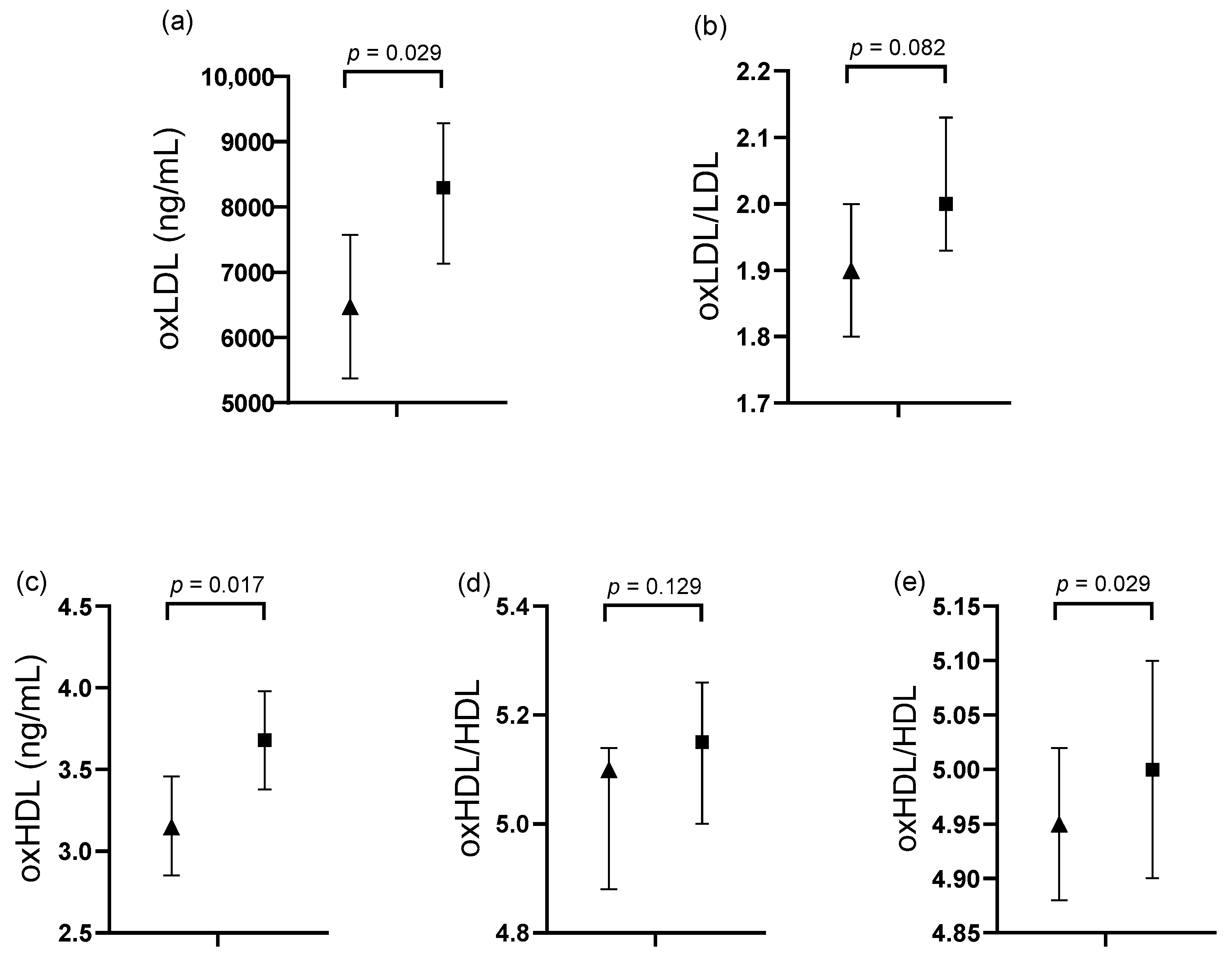
3.3. Comparative Analysis in Subjects Classified by Cardiological Variables

Quantitative variables were compared among study subjects stratified by the number of ischemic events (IEs) and damaged blood vessels (DBVs), as shown in Table 3.
Subjects with ≥2 IEs and ≥2 DBVs had significantly higher oxLDL concentrations compared to those with 1 IE and 1 DBV (8293.35 ng/mL [7131.32–9287.39] vs. 6474.26 ng/mL [5374.52–7574.01], p = 0.029). Similarly, oxHDL levels were significantly higher in the ≥2 IEs and ≥2 DBVs group (3.68 ng/mL [3.38–3.98] vs. 3.15 ng/mL [2.85–3.46], p = 0.017). The oxHDL/HDL ratio was also significantly higher in the ≥2 IEs and ≥2 DBVs group compared to the other group (5.00 [4.9–5.1] vs. 4.95 [4.88–5.02], p = 0.029). A trend toward higher oxLDL/LDL ratios was observed in the ≥2 IEs and ≥2 DBVs group (p = 0.082) (Figure 2).

3.4. Association of oxLDL Levels with Cardiological Variables

A multivariable linear regression analysis was performed to assess the association between serum oxLDL levels and cardiological variables. All biochemical variables that showed a trend or statistical significance in Figure 2 were included in the multivariable model. A positive association was observed between serum oxLDL levels and the number of IEs (R2 = 0.152, p = 0.005) and DBVs (R2 = 0.192, p = 0.020), respectively (Table 4).
Additionally, an odds ratio (OR) analysis was performed, using the population mean serum oxLDL value (7358.82 ng/mL) as the cut-off point. An oxLDL value ≥7358.82 ng/mL was significantly associated with an increased risk of presenting with ≥2 IEs and ≥2 DBVs (OR = 4.667, 95% CI: 1.341–16.239, p = 0.012).

4. Discussion

It is well established that both genetic and environmental factors contribute to the development of chronic diseases, including coronary artery disease (CAD), which remains the most prevalent chronic condition and the leading cause of mortality in Mexico [29]. Although multiple studies have identified diverse risk factors for CAD, limited data are available on the recurrence and severity of ischemic events (IEs) in patients with existing cardiovascular damage [29]. Accordingly, this study aimed to analyze the clinical characteristics and risk factors associated with CAD development and recurrence in a Mexican population.
In terms of sex distribution, a higher percentage of male participants was observed, consistent with prior studies indicating that CAD disproportionately affects men. A descriptive study conducted in Mexico City involving 586 CAD patients reported that 70% were male [30]. This sex disparity is often attributed to hormonal factors—particularly estrogens—which promote vasodilation, reduce fibrosis, enhance mitochondrial function, and mitigate oxidative stress. As a result, premenopausal women exhibit lower susceptibility to CAD [31,32].
Mixed CAD was the most frequent subtype in this study, likely reflecting the coexistence of both stable and unstable atherosclerotic plaques [33]. This finding aligns with data from the 2016 National Registry of Acute Coronary Syndrome, which reported a 50% prevalence of mixed CAD [34]. However, updated prevalence data for CAD subtypes in the Mexican population are lacking.
Comorbidity analysis revealed a high prevalence of hypertension and type 2 diabetes mellitus (T2DM), both of which are strongly implicated in CAD pathogenesis through mechanisms such as endothelial dysfunction, oxidative stress, and chronic inflammation [35,36]. According to the 2023 ENSANUT survey, the prevalence of T2DM and hypertension prevalence in the general Mexican population was 18.4% and 29.9%, respectively [37]—notably lower than the rates observed in this study’s cohort, which likely reflects its clinical profile. These findings reinforce the contribution of these comorbidities as major risk factors for CAD progression.
Despite ongoing pharmacological treatment (Table A3), hypoalphalipoproteinemia and hypertriglyceridemia were highly prevalent. This may be partially attributable to the frequent use of atorvastatin, a lipid-lowering agent that is effective in reducing LDL-C, but has a limited impact on triglycerides and HDL-C levels [38]. Nevertheless, atorvastatin exerts antioxidant effects by enhancing paraoxonase-1 (PON1) activity, which inhibits LDL oxidation and foam cell formation, reinforcing its relevance in secondary prevention [39]. However, pharmacological strategies should be accompanied by lifestyle interventions, including dietary adjustments targeting an omega-6-to-omega-3 PUFA ratio ≤ 5:1, supplementation with 1.8 g/day of marine omega-3s, and personalized physical activity plans [40,41].
Anthropometric indicators, including overweight, an excessive body fat percentage, and abdominal obesity, were also highly prevalent. These conditions are closely linked to CAD, particularly abdominal fat, which has been shown to impair endothelial function, increase oxidative stress, and promote systemic inflammation. In this context, Xue et al. (2021) identified a dose-dependent relationship between abdominal fat and cardiovascular risk, emphasizing its impact on lipid dysregulation and the pathogenesis of dyslipidemia [42]. These findings underscore the importance of monitoring anthropometric variables in clinical practice and cardiovascular risk assessments.
Beyond metabolic disorders, environmental risk factors such as alcohol and tobacco use were also relevant in this cohort. Approximately 25% of participants reported alcohol consumption, and 7.8% exhibited high-risk intake. Wood et al. suggest that even moderate alcohol intake may exacerbate CAD progression by elevating systemic inflammation [43], partly through increased homocysteine, oxLDL, and oxidative stress levels [44].
Moreover, 38.5% of participants were current moderate or heavy smokers—similar to the AHA-reported rate of 35% among high-risk cardiovascular patients [45]. Passive smoke exposure was also high, potentially compounding cardiovascular risk through mechanisms such as platelet activation and reduced oxygen delivery [46].
In addition to hypertriglyceridemia and hypoalphalipoproteinemia, circulating oxidized lipoproteins—namely oxLDL and oxHDL—may serve as novel biomarkers of ischemic heart disease. In this cohort, significantly higher levels of both oxLDL and oxHDL were observed in individuals with more than two IEs and more than two DBVs, compared to those with a single event.
This finding is consistent with a 2022 case–control study by Yazdandoust et al., which examined 572 Iranian patients with stable angina and reported significantly elevated oxHDL levels in patients compared to controls (3.15 [1.01–1.31] vs. 2.85 [0.62–1.06] ng/mL, p < 0.001) [47]. However, their study did not investigate associations between oxHDL levels and the number of DBVs.
Functionally, HDL exerts antioxidant and anti-inflammatory effects, primarily through paraoxonase-1 (PON1) activity, which protects LDL from oxidative modification. Once oxidized, however, HDL loses its protective properties and can become proinflammatory and pro-atherogenic. Oxidized HDL also exhibits impaired reverse cholesterol transport, a key mechanism in atherosclerosis prevention [48].
In comparison, oxLDL levels in our study differed from those reported in a U.S. cohort of 304 individuals, where the mean serum oxLDL concentrations were 31,900 ± 10,200 ng/mL in patients with prior IEs [49]. Similarly, Wang et al. (2020) found that CAD patients with oxLDL ≥139.6 ng/mL had a significantly increased risk of recurrent IEs at 90 days (HR: 1.57) and at one year (HR: 1.49) [9]. Variability in oxLDL concentrations across studies may be attributable to differences in detection methods and the biomarkers selected. In our study, oxLDL was quantified using malondialdehyde (MDA) as a marker of lipid peroxidation. Nonetheless, despite these methodological differences, evidence consistently supports a link between elevated oxLDL levels and CAD risk.
Our findings suggest that elevated oxHDL concentrations and the strong correlation between oxLDL levels, IEs, and DBVs support the inclusion of these oxidized lipoproteins in standard lipid panels. Notably, an oxLDL value ≥ 7358.82 µg/mL was associated with a 4.6-fold increased risk of presenting with more than two IEs and more than two DBVs. Thus, the routine measurement of oxLDL and oxHDL should be considered both in healthy individuals and in patients with established CAD, given their relevance to oxidative stress, lipid peroxidation, disease severity, and recurrence. Importantly, these biomarkers can be incorporated into standard biochemical evaluations.
Although individuals with a history of cancer, chronic kidney disease, or autoimmune disorders were excluded from this study, residual confounding may persist due to the absence of data on key cardiovascular risk factors, including family history, inflammatory markers such as high-sensitivity C-reactive protein, and detailed measures of renal function. The omission of these variables from the regression models is a limitation that should be considered when interpreting the results. Future studies should aim to incorporate these factors to enhance risk adjustment and minimize confounding.
Additionally, the relatively small sample size limited the statistical power of subgroup analyses, particularly those stratified by sex or clinical severity. Therefore, caution is warranted when interpreting these findings. Future research should include larger cohorts and evaluate other cardiac biomarkers, such as small dense LDL and indicators of endothelial function.
Longitudinal studies are also needed to monitor oxLDL levels over time and across diverse populations in order to further validate their clinical utility. Moreover, targeted nutrigenetic and pharmacological interventions should be explored as strategies to reduce oxLDL concentrations and prevent recurrent vascular damage in CAD patients.

5. Conclusions

This study demonstrated that oxLDL concentrations exceeding 7358.82 ng/mL are associated with a 4.6-fold increased likelihood of experiencing a second ischemic event (IE) and presenting with more than one damaged blood vessel (DBV) in patients with coronary artery disease (CAD). Given the significant association observed between circulating oxLDL levels and cardiological variables, oxLDL emerges as a promising clinical biomarker for identifying individuals at elevated cardiovascular risk. Its inclusion in standard biochemical assessments may support the early implementation of targeted medical and nutritional interventions to reduce disease progression and recurrence.

Author Contributions

Conceptualization, M.P.-R. and W.C.-P.; methodology, M.P.-R. and S.G.-M.; software, V.L.-A.; validation, L.E.R.-V. and F.C.-R.; formal analysis, M.P.-R.; investigation, J.A.-B.; resources, V.L.-A.; data curation, S.G.-M.; writing—original draft preparation, M.P.-R.; writing—review and editing, M.P.-R.; visualization, W.C.-P.; supervision, E.M.-L.; project administration, E.M.-L.; funding acquisition, E.M.-L. All authors have read and agreed to the published version of the manuscript.

Funding

This research was funded by the Programa de Fortalecimiento de Institutos, Centros y Laboratorios de Investigación, in 2022 (Fortalecimiento UDG-2022 REC/CINV/0975/2022).

Institutional Review Board Statement

All procedures were conducted in accordance with the guidelines of the Declaration of Helsinki (2013) [50]. The study was approved by the Ethics, Research and Biosafety Committee (Comité de Investigación, Ética en Investigación y de Bioseguridad) of the Centro Universitario de Ciencias de la Salud, Universidad de Guadalajara, Jalisco, México (registration number CI-08320; approval date: 27 October 2020).

Informed Consent Statement

Informed consent was obtained from all subjects involved in the study.

Data Availability Statement

The data presented in this study are available upon reasonable request from the corresponding author.

Acknowledgments

The authors acknowledge the Universidad de Guadalajara and the Instituto de Nutrigenética y Nutrigenómica Traslacional for supporting the development of this research, as well as all the study participants for their valuable contribution.

Conflicts of Interest

The authors declare no conflicts of interest regarding the publication of this article.

Abbreviations

The following abbreviations are used in this manuscript:
ACCAmerican College of Cardiology
ADAAmerican Diabetes Association
AHAAmerican Heart Association
AUDITAlcohol Use Disorders Identification Test
BF%body fat percentage
BMIbody mass index
CADcoronary artery disease
DBVdamaged blood vessel
ELISAenzyme-linked immunosorbent assay
ETAPEscala de Tabaquismo para Atención Primaria (Tobacco Scale for Primary Care)
HDLhigh-density lipoprotein
IEischemic event
LDLlow-density lipoprotein
MDAmalondialdehyde
NSTEMInon-ST-segment-elevation myocardial infarction
oxLDLoxidized low-density lipoprotein
oxHDLoxidized high-density lipoprotein
PON1paraoxonase 1
PUFAspolyunsaturated fatty acids
STEMIST-segment-elevation myocardial infarction
T2DMtype 2 diabetes mellitus
TCtotal cholesterol
TGtriglycerides
WCwaist circumference

Appendix A

Table A1. Frequencies of metabolic comorbidities and dyslipidemias in the study subjects (n = 51).
VariablesValues
%, (n)
Metabolic comorbidities
Hypertension73.0 (37)
T2DM49.0 (25)
Dyslipidemia
Hypoalphalipoproteinemia70.6 (36)
Hypertriglyceridemia53 (27)
Elevated LDL51 (26)
Hypercholesterolemia12 (6)
Lipid ratios
Elevated TC/HDL62.7 (32)
Elevated TG/HDL45.1 (23)
Note: Data are expressed as percentages and absolute frequencies. T2DM: type 2 diabetes mellitus; LDL-c: low-density lipoprotein cholesterol; TG: triglycerides; TC: total cholesterol.
Table A2. Alcohol and tobacco consumption among the study subjects (n = 51).
VariablesValues
%, (n)
Alcohol consumption
Alcohol consumption24.0 (11)
High-risk alcohol consumption7.8 (4)
Smoking
Light smoking23.0 (6)
Moderate smoking38.5 (10)
Severe smoking38.5 (10)
Suspended smoking33.3 (17)
High-risk passive smoking86.2 (25)
Note: Data are expressed as percentages and absolute frequencies.
Table A3. Pharmacological treatments among the study subjects (n = 51).
Medication TypeMedicationValues
%, (n)
Anticoagulants and antiplateletsAcetylsalicylic acid60.8 (31)
Clopidogrel31.4 (16)
VasodilatorsIsosorbide13.7 (7)
Nifedipine3.9 (2)
AntihypertensivesMetoprolol47.1 (24)
Losartan29.4 (15)
Enalapril21.6 (11)
Lipid-loweringAtorvastatin58.8 (30)
Bezafibrate7.8 (4)
Pravastatin3.9 (2)
Ciprofibrate2 (1)
Glucose-controlMetformin35.3 (18)
Note: Data are expressed as percentages and absolute frequencies.

References

  1. Longo, D.L.; Fauci, A.S.; Kasper, D.L.; Hauser, S.L.; Jameson, J.L.; Loscalzo, J. (Eds.) Harrison. In Principios de Medicina Interna, 18th ed.; McGraw Hill: New York, NY, USA, 2013; pp. 1–3000. [Google Scholar]
  2. Brown, J.C.; Gerhardt, T.E.; Kwon, E. Risk factors for coronary artery disease. In StatPearls; StatPearls Publishing: Treasure Island, FL, USA, 2024. Available online: https://www.ncbi.nlm.nih.gov/books/NBK554410/ (accessed on 14 December 2024).
  3. Mensah, G.A.; Fuster, V.; Murray, C.J.; Roth, G.A.; Abate, Y.H.; Abbasian, M.; Abd-Allah, F.; Abdollahi, A.; Abdollahi, M.; Abdulah, D.M.; et al. Global burden of cardiovascular diseases and risks, 1990–2022. J. Am. Coll. Cardiol. 2023, 82, 2350–2473. [Google Scholar] [CrossRef] [PubMed]
  4. Virani, S.S.; Newby, L.K.; Arnold, S.V.; Bittner, V.; Brewer, L.C.; Demeter, S.H.; Dixon, D.L.; Fearon, W.F.; Hess, B.; Johnson, H.M.; et al. 2023 AHA/ACC/ACCP/ASPC/NLA/PCNA guideline for the management of patients with chronic coronary disease: A report of the American Heart Association/American College of Cardiology Joint Committee on Clinical Practice Guidelines. Circulation 2023, 148, e9–e119. [Google Scholar] [CrossRef] [PubMed]
  5. Byrne, R.A.; Rossello, X.; Coughlan, J.J.; Barbato, E.; Berry, C.; Chieffo, A.; Claeys, M.J.; Dan, G.-A.; Dweck, M.R.; Galbraith, M.; et al. 2023 ESC guidelines for the management of acute coronary syndromes. Eur. Heart J. 2023, 44, 3720–3826. [Google Scholar] [CrossRef] [PubMed]
  6. Vickrey, B.G.; Rector, T.S.; Wickstrom, S.L.; Guzy, P.M.; Sloss, E.M.; Gorelick, P.B.; Garber, S.; McCaffrey, D.F.; Dake, M.D.; Levin, R.A. Occurrence of secondary ischemic events among persons with atherosclerotic vascular disease. Stroke 2002, 33, 901–906. [Google Scholar] [CrossRef]
  7. Uzuner, N.; Uzuner, G.T. Risk factors for multiple recurrent ischemic strokes. Brain Circ. 2023, 9, 21–24. [Google Scholar] [CrossRef]
  8. Shahjehan, R.D.; Sharma, S.; Bhutta, B.S. Coronary artery disease. In StatPearls; StatPearls Publishing: Treasure Island, FL, USA, 2024. Available online: https://www.ncbi.nlm.nih.gov/books/NBK564304/ (accessed on 14 May 2025).
  9. Wang, A.; Dai, L.; Zhang, N.; Lin, J.; Chen, G.; Zuo, Y.; Li, H.; Wang, Y.; Meng, X.; Wang, Y. Oxidized low-density lipoprotein (LDL) and LDL cholesterol are associated with outcomes of minor stroke and TIA. Atherosclerosis 2020, 297, 74–80. [Google Scholar] [CrossRef]
  10. Wang, A.; Zhang, X.; Li, S.; Zhao, X.; Liu, L.; Johnston, S.C.; Meng, X.; Lin, J.; Zuo, Y.; Li, H.; et al. Oxidative lipoprotein markers predict poor functional outcome in patients with minor stroke or transient ischaemic attack. Eur. J. Neurol. 2019, 26, 1082–1090. [Google Scholar] [CrossRef]
  11. Nicholls, S.J.; Zheng, L.; Hazen, S.L. Formation of dysfunctional high-density lipoprotein by myeloperoxidase. Trends Cardiovasc. Med. 2005, 15, 212–219. [Google Scholar] [CrossRef]
  12. Ansell, B.J.; Fonarow, G.C.; Fogelman, A.M. The paradox of dysfunctional high-density lipoprotein. Curr. Opin. Lipidol. 2007, 18, 427–434. [Google Scholar] [CrossRef]
  13. Feng, Y.; Cai, Z.; Tang, Y.; Hu, G.; Lu, J.; He, D.; Wang, S. TLR4/NF-κB signaling pathway-mediated and oxLDL-induced up-regulation of LOX-1, MCP-1, and VCAM-1 expressions in human umbilical vein endothelial cells. Genet. Mol. Res. 2014, 13, 680–695. [Google Scholar] [CrossRef]
  14. Basit, H.; Huecker, M.R. Myocardial infarction serum markers. In StatPearls; StatPearls Publishing: Treasure Island, FL, USA, 2024. Available online: https://www.ncbi.nlm.nih.gov/books/NBK532966/ (accessed on 16 October 2024).
  15. Whelton, P.K.; Carey, R.M.; Aronow, W.S.; Casey, D.E., Jr.; Collins, K.J.; Himmelfarb, C.D.; DePalma, S.M.; Gidding, S.; Jamerson, K.A.; Jones, D.W.; et al. 2017 ACC/AHA/AAPA/ABC/ACPM/AGS/APhA/ASH/ASPC/NMA/PCNA guideline for the prevention, detection, evaluation, and management of high blood pressure in adults: Executive summary: A report of the American College of Cardiology/American Heart Association Task Force on Clinical Practice Guidelines. Hypertension 2018, 71, 1269–1324. [Google Scholar] [PubMed]
  16. ElSayed, N.A.; Aleppo, G.; Aroda, V.R.; Bannuru, R.R.; Brown, F.M.; Bruemmer, D.; Collins, B.S.; Gaglia, J.L.; Hilliard, M.E.; Isaacs, D.; et al. 2. Classification and diagnosis of diabetes: Standards of care in diabetes—2023. Diabetes Care 2022, 46 (Suppl. S1), S19–S29. [Google Scholar] [CrossRef] [PubMed]
  17. Stone, N.J.; Robinson, J.G.; Lichtenstein, A.H.; Merz, C.N.B.; Blum, C.B.; Eckel, R.H.; Goldberg, A.C.; Gordon, D.; Levy, D.; Mcbride, P.; et al. 2013 ACC/AHA guideline on the treatment of blood cholesterol to reduce atherosclerotic cardiovascular risk in adults: A report of the American College of Cardiology/American Heart Association Task Force on Practice Guidelines. J. Am. Coll. Cardiol. 2014, 63 Pt B, 2889–2934. [Google Scholar] [CrossRef]
  18. García Muñoz, A.I.; Melo Buitrago, P.J.; Rodríguez Arcila, M.A.; Silva Zambrano, D.A. Índices aterogénicos y composición corporal en cadetes de una escuela de formación militar colombiana. Sanid. Mil. 2020, 76, 13–18. [Google Scholar]
  19. Castelli, W.P. Epidemiology of coronary heart disease: The Framingham study. Am. J. Med. 1984, 76, 4–12. [Google Scholar] [CrossRef]
  20. National Cholesterol Education Program (NCEP) Expert Panel on Detection, Evaluation, and Treatment of High Blood Cholesterol in Adults (Adult Treatment Panel III). Third report of the National Cholesterol Education Program (NCEP) Expert Panel on Detection, Evaluation, and Treatment of High Blood Cholesterol in Adults (Adult Treatment Panel III) final report. Circulation 2002, 106, 3143–3421. [Google Scholar] [CrossRef]
  21. World Health Organization (WHO). Physical Status: The Use and Interpretation of Anthropometry. Report of a WHO Expert Committee; WHO: Geneva, Switzerland, 1995; Available online: http://whqlibdoc.who.int/trs/WHO_TRS_854.pdf (accessed on 25 November 2024).
  22. Centers for Disease Control and Prevention (CDC). Índice de Masa Corporal|Peso Saludable|DNPAO|CDC. 2022. Available online: https://www.cdc.gov/healthyweight/spanish/assessing/bmi/index.html (accessed on 4 December 2022).
  23. U.S. Department of Agriculture; U.S. Department of Health and Human Services. Dietary Guidelines for Americans, 2020–2025, 9th ed.; U.S. Government Publishing Office: Washington, DC, USA, 2020. Available online: https://www.dietaryguidelines.gov/sites/default/files/2021-03/Dietary_Guidelines_for_Americans-2020-2025.pdf (accessed on 4 March 2025).
  24. National Institute on Alcohol Abuse and Alcoholism (NIAAA). National Institute on Alcohol Abuse and Alcoholism (NIAAA). Available online: https://www.niaaa.nih.gov/ (accessed on 4 March 2025).
  25. Saunders, J.B.; Aasland, O.G. Development of the Alcohol Use Disorders Identification Test (AUDIT): WHO collaborative project on early detection of persons with harmful alcohol consumption. Alcohol Health Res. World 1993. [Google Scholar] [CrossRef]
  26. Gobierno Federal. Guía de Práctica Clínica Para la Prevención, Diagnóstico y Tratamiento del Consumo del Tabaco; Centro Nacional de Excelencia Tecnológica en Salud: México City, México, 2009. Available online: https://www.cenetec.salud.gob.mx/interior/gpc.html (accessed on 4 March 2025).
  27. González Romero, M.P.; Cuevas-Fernández, F.J.; Marcelino-Rodríguez, I.; Covas, V.J.; Rodríguez Pérez, M.C.; Cabrera de León, A.; Aguirre-Jaime, A. Aplicación de la Escala de Tabaquismo para Atención Primaria (ETAP) en la práctica clínica. Aten. Primaria 2018, 50, 414–421. [Google Scholar] [CrossRef]
  28. Meisinger, C.; Baumert, J.; Khuseyinova, N.; Loewel, H.; Koenig, W. Plasma oxidized low-density lipoprotein, a strong predictor for acute coronary heart disease events in apparently healthy, middle-aged men from the general population. Circulation 2005, 112, 651–657. [Google Scholar] [CrossRef]
  29. Velázquez Monroy, Ó.; Aldatz, B.; S, F.; Guerra, R.; Francisco, A.; Verdejo, J.; Bello, M.Á.M.; Violante, R.; Esqueda, A.L. Morbilidad y mortalidad de la enfermedad isquémica del corazón y cerebrovascular en México, 2005. Arch. Cardiol. Méx. 2007, 77, 31–39. [Google Scholar]
  30. Solorio, S.; Hernández-González, M.A.; Rangel Abundis, A.; Murillo-Ortiz, B. Cardiopatía isquémica en mujeres mexicanas. Arch. Cardiol. Méx. 2007, 77, 226–231. [Google Scholar] [PubMed]
  31. Franco, Y.; Mendoza-Fernández, V.; Lemini, C. Mecanismos de acción de los efectos protectores de los estrógenos sobre el sistema cardiovascular. Rev. Fac. Med. UNAM 2003, 46, 101–108. [Google Scholar]
  32. Iorga, A.; Cunningham, C.M.; Moazeni, S.; Ruffenach, G.; Umar, S.; Eghbali, M. The protective role of estrogen and estrogen receptors in cardiovascular disease and the controversial use of estrogen therapy. Biol. Sex Differ. 2017, 8, 33. [Google Scholar] [CrossRef] [PubMed]
  33. Kawai, K.; Kawakami, R.; Finn, A.V.; Virmani, R. Differences in stable and unstable atherosclerotic plaque. Arterioscler. Thromb. Vasc. Biol. 2024, 44, 1474–1484. [Google Scholar] [CrossRef]
  34. Martinez-Sanchez, C.; Borrayo, G.; Carrillo, J.; Juarez, U.; Quintanilla, J.; Jerjes-Sanchez, C. Clinical management and hospital outcomes of acute coronary syndrome patients in Mexico: The Third National Registry of Acute Coronary Syndromes (RENASICA III). Arch. Cardiol. Méx. 2016, 86, 221–232. [Google Scholar] [CrossRef]
  35. Volpe, M.; Gallo, G. Hypertension, coronary artery disease and myocardial ischemic syndromes. Vasc. Pharmacol. 2023, 153, 107230. [Google Scholar] [CrossRef]
  36. Gupta, N.; Elnour, A.A.; Sadeq, A.; Gupta, R. Diabetes and the Heart: Coronary Artery Disease. Available online: https://www.escardio.org/Journals/E-Journal-of-Cardiology-Practice/Volume-22/diabetes-and-the-heart-coronary-artery-disease (accessed on 4 March 2025).
  37. Instituto Nacional de Salud Pública (INSP). Encuesta Nacional de Salud y Nutrición Continua 2023; INSP: Cuernavaca, México, 2024; Available online: https://ensanut.insp.mx/encuestas/ensanutcontinua2023/index.php (accessed on 4 February 2025).
  38. Agarwala, A.; Kulkarni, S.; Maddox, T. The association of statin therapy with incident diabetes: Evidence, mechanisms, and recommendations. Curr. Cardiol. Rep. 2018, 20, 50. [Google Scholar] [CrossRef]
  39. Davignon, J.; Jacob, R.F.; Mason, R.P. The antioxidant effects of statins. Coron. Artery Dis. 2004, 15, 251–258. [Google Scholar] [CrossRef]
  40. Franczyk, B.; Gluba-Brzózka, A.; Ciałkowska-Rysz, A.; Ławiński, J.; Rysz, J. The impact of aerobic exercise on HDL quantity and quality: A narrative review. Int. J. Mol. Sci. 2023, 24, 4653. [Google Scholar] [CrossRef]
  41. Torres-Vanegas, J.; Rodríguez-Echevarría, R.; Campos-Pérez, W.; Rodríguez-Reyes, S.C.; Reyes-Pérez, S.D.; Pérez-Robles, M.; Martínez-López, E. Effect of a diet supplemented with marine omega-3 fatty acids on inflammatory markers in subjects with obesity: A randomized active placebo-controlled trial. Healthcare 2025, 13, 103. [Google Scholar] [CrossRef]
  42. Xue, R.; Li, Q.; Geng, Y.; Wang, H.; Wang, F.; Zhang, S. Abdominal obesity and risk of CVD: A dose–response meta-analysis of thirty-one prospective studies. Br. J. Nutr. 2021, 126, 1420–1430. [Google Scholar] [CrossRef] [PubMed]
  43. Wood, A.M.; Kaptoge, S.; Butterworth, A.S.; Willeit, P.; Warnakula, S.; Bolton, T.; Paige, E.; Paul, D.S.; Sweeting, M.; Burgess, S.; et al. Risk thresholds for alcohol consumption: Combined analysis of individual-participant data for 599,912 current drinkers in 83 prospective studies. Lancet 2018, 391, 1513–1523. [Google Scholar] [CrossRef] [PubMed]
  44. Shirpoor, A.; Salami, S.; Khadem Ansari, M.H.; Ilkhanizadeh, B.; Abdollahzadeh, N. Ethanol promotes rat aortic vascular smooth muscle cell proliferation via increase of homocysteine and oxidized-low-density lipoprotein. J. Cardiol. 2013, 62, 374–378. [Google Scholar] [CrossRef] [PubMed]
  45. Virani, S.S.; Alonso, A.; Aparicio, H.J.; Benjamin, E.J.; Bittencourt, M.S.; Callaway, C.W.; Carson, A.P.; Chamberlain, A.M.; Cheng, S.; Delling, F.N.; et al. Heart disease and stroke statistics—2021 update: A report from the American Heart Association. Circulation 2021, 143, e254–e743. [Google Scholar] [CrossRef]
  46. Wells, A.J. Passive smoking as a cause of heart disease. J. Am. Coll. Cardiol. 1994, 24, 546–554. [Google Scholar] [CrossRef]
  47. Yazdandoust, S.; Parizadeh, S.M.R.; Ghayour-Mobarhan, M.; Yaghmaei, P.; Sahebkar, A.H. High-density lipoprotein lipid peroxidation as a diagnostics biomarker in coronary artery disease. Biofactors 2022, 48, 634–642. [Google Scholar] [CrossRef]
  48. Linton, M.F.; Yancey, P.G.; Tao, H.; Davies, S.S. HDL function and atherosclerosis: Reactive dicarbonyls as promising targets of therapy. Circ. Res. 2023, 132, 1521–1545. [Google Scholar] [CrossRef]
  49. Holvoet, P.; Mertens, A.; Verhamme, P.; Bogaerts, K.; Beyens, G.; Verhaeghe, R.; Collen, D.; Muls, E.; Van de Werf, F. Circulating oxidized LDL is a useful marker for identifying patients with coronary artery disease. Arterioscler. Thromb. Vasc. Biol. 2001, 21, 844–848. [Google Scholar] [CrossRef]
  50. World Medical Association (WMA). Declaration of Helsinki: Ethical Principles for Medical Research Involving Human Subjects; WMA: Fortaleza, Brazil, 2013; Available online: https://www.wma.net/policies-post/wma-declaration-of-helsinki-ethical-principles-for-medical-research-involving-human-subjects/ (accessed on 16 July 2020).
Figure 1. Flow diagram of study subjects.
Healthcare 13 01426 g001
Figure 2. Comparative analysis of oxidized lipoproteins in subjects stratified by ischemic events (IEs) and damaged blood vessels (DBVs). ■ Subjects with 1 IE and 1 DBV. ▲ Subjects with ≥2 IEs and ≥2 DBVs. Panels show the following: (a) serum oxLDL levels, (b) oxLDL/LDL ratio, (c) oxHDL levels, and oxHDL/HDL ratio in (d) men and (e) women. Data are expressed as estimated means with 95% confidence intervals. p values were obtained using a general univariate model adjusted for age, sex, alcohol consumption, smoking, and pharmacological treatment. oxLDL/LDL and oxHDL/HDL ratios are reported as natural logarithms (ln). LDL: low-density lipoprotein cholesterol; HDL: high-density lipoprotein cholesterol; oxLDL: oxidized low-density lipoprotein; oxHDL: oxidized high-density lipoprotein. Units: oxLDL and oxHDL in ng/mL (nanograms per milliliter).
Healthcare 13 01426 g002
Table 1. Clinical and anthropometric characteristics of the study subjects (n = 51).
VariablesValues
Clinical
SBP (mmHg)129 (111–140)
DBP (mmHg)78 (71–86)
DBVs (number)2 (1–2)
IEs (number)2 (1–2)
Anthropometric
Body weight (kg)74 (64.7–88)
BMI (kg/m2)28.4 (26.6–30.9)
Body fat (%)
M (n = 34)28.1 (20.5–32.5)
W (n = 17)42.1 (37.9–47.25)
WC (cm)
M (n = 34)98 (90–107)
W (n = 17)97.5 (93.2–102.7)
Data are reported as median (25th–75th percentile). SBP: systolic blood pressure; DBP: diastolic blood pressure; DBVs: damaged blood vessels; IEs: ischemic events; BMI: body mass index; M: men; W: women; WC: waist circumference. Units: millimeters of mercury (mmHg); kilograms (kg); kilograms per square meter (kg/m2).
Table 2. Biochemical characteristics of the study subjects (n = 51).
Biochemical VariablesValues
TC (mg/dL)139 (109–171)
LDL (mg/dL)72 (45–98)
VLDL (mg/dL)30 (21–41)
HDL (mg/dL)
M (n = 34)33 (25.5–40)
W (n = 17)43 (33.5–51.5)
TG (mg/dL)157 (104–208)
TG/HDL-c ratio
M (n = 34)4.67 (2.7–7.1)
W (n = 17)3.94 (2.4–6.2)
TC/HDL-c ratio
M (n = 34)4 (3–5)
W (n = 17)4 (2–5)
oxLDL (ng/mL)6500 (5560–8320)
oxHDL (ng/mL)
M (n = 34)3.2 (2.7–3.6)
W (n = 17)3.4 (3.3–4.3)
oxLDL/LDL5.04 (4.9–5.12)
oxHDL/HDL
M (n = 34)5.04 (4.98–5.19)
W (n = 17)5.03 (4.87–5.09)
Data are presented as median (25th–75th percentile). oxLDL/LDL and oxHDL/HDL ratios are reported as natural logarithms (ln). M: men; W: women; WC: waist circumference; TC: total cholesterol; LDL: low-density lipoprotein cholesterol; HDL: high-density lipoprotein cholesterol; oxLDL: oxidized low-density lipoprotein; oxHDL: oxidized high-density lipoprotein; TG: triglycerides. Units: TC, LDL, VLDL, HDL, and TG in mg/dL (milligrams per deciliter); oxLDL and oxHDL in ng/mL (nanograms per milliliter).
Table 3. Comparative analysis of biochemical variables in subjects classified by the number of IEs and DBVs.
Variables1 IE and 1 DBV
(n = 25)
≥2 IEs and ≥2 DBVs
(n = 26)
p Value
TC (mg/dL)148.15 (132.73–163.56)138.39 (123.27–153.51)0.371
LDL (mg/dL)78.87 (64.84–92.90)70.08 (53.33–83.83)0.376
VLDL (mg/dL)33.80 (27.08–40.52)32.14 (25.56–38.73)0.727
HDL (mg/dL)
M (n = 34)34.03 (29.55–38.59)32.83 (28.82–36.85)0.679
W (n = 17)49.81 (36–63.58)39.69 (23.10–56.27)0.333
TG (mg/dL)187.88 (147.55–228.21)159.34 (119.80–198.87)0.318
TG/HDL ratio
M (n = 34)5.99 (4.03–7.95)5.18 (3.44–6.92)0.932
W (n = 17)4.23 (2.65–5.81)4.33 (2.43–6.24)0.534
TC/HDL-c ratio
M (n = 34)4.20 (3.43–4.97)4.25 (3.57–4.94)0.416
W (n = 17)3.52 (2.59–4.46)4.10 (2.97–5.22)0.914
Values are presented as estimated means with 95% confidence intervals. p values were obtained using a general univariate model adjusted for age, sex, alcohol consumption, smoking, and pharmacological treatment. p values <0.05 were considered statistically significant. IEs: ischemic events; DBVs: damaged blood vessels; M: men; W: women; WC: waist circumference; TC: total cholesterol; TG: triglycerides; LDL: low-density lipoprotein cholesterol; HDL: high-density lipoprotein cholesterol. Units: TC, LDL, VLDL, HDL, and TG in mg/dL (milligrams per deciliter).
Table 4. Association of oxLDL serum levels with cardiological variables.
ModelDependent VariableR2B (95% CI)p Value
1Number of ischemic events0.1520.142 (0.046–0.239)0.005
2Number of damaged blood vessels0.1920.196 (0.018–0.374)0.020
Note: Linear regression models adjusted for age, sex, alcohol consumption, smoking, and pharmacological treatment.
Disclaimer/Publisher’s Note: The statements, opinions and data contained in all publications are solely those of the individual author(s) and contributor(s) and not of MDPI and/or the editor(s). MDPI and/or the editor(s) disclaim responsibility for any injury to people or property resulting from any ideas, methods, instructions or products referred to in the content.

