
Application of Color Doppler with 3- and 4-Dimensional Ultrasonography in the Prenatal Evaluation of Fetal Extracardiac and Placental Abnormalities


Abstract
:1. Introduction
Methods
2. Extracardiac Abnormalities
2.1. Outflow Tracts
2.2. Brain
2.3. Pulmonary Vessels
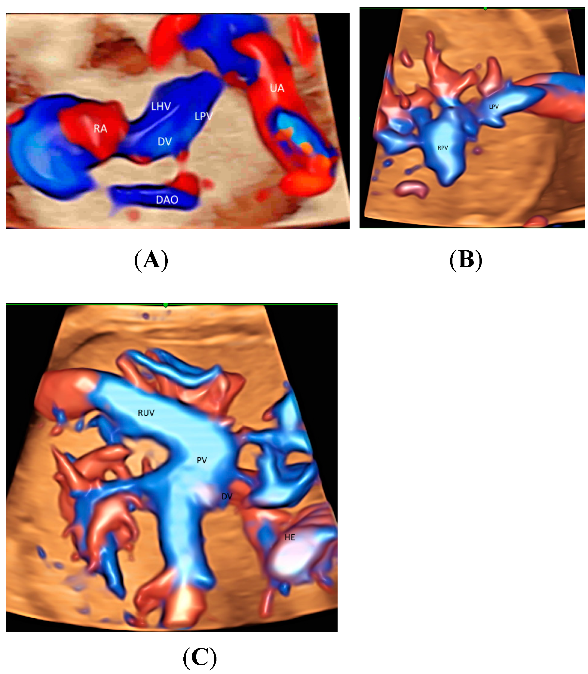
2.4. Abdomen
2.5. Others
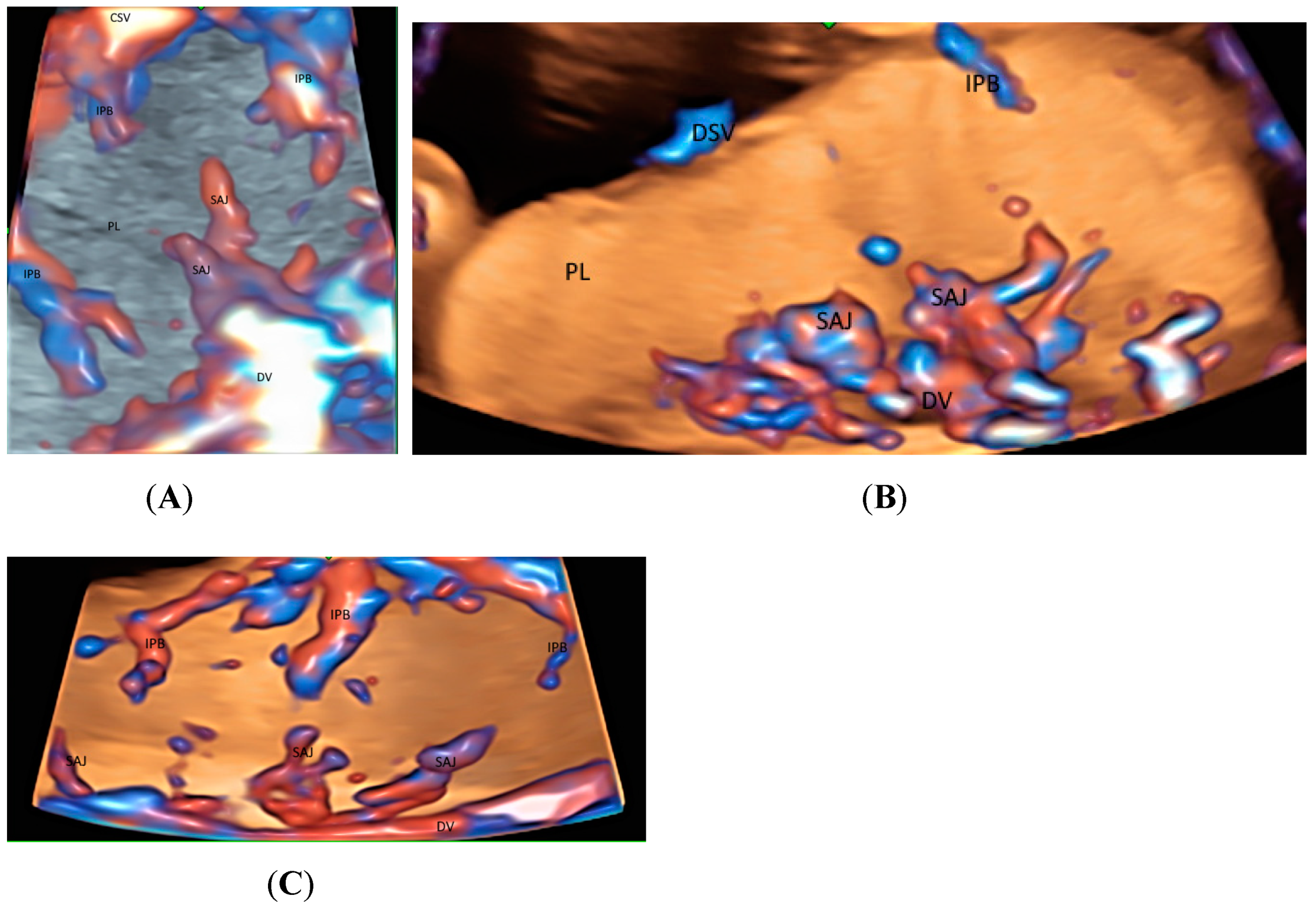
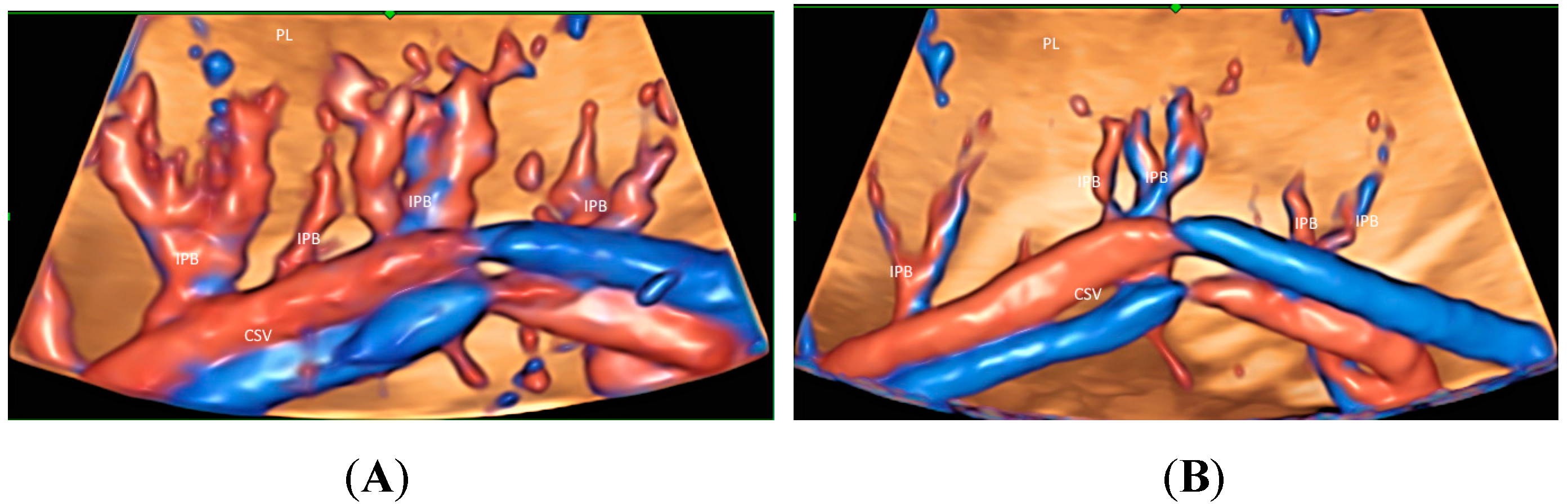
3. Placenta
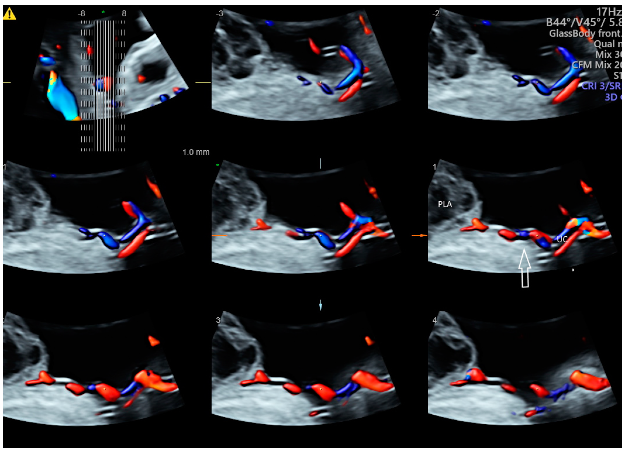
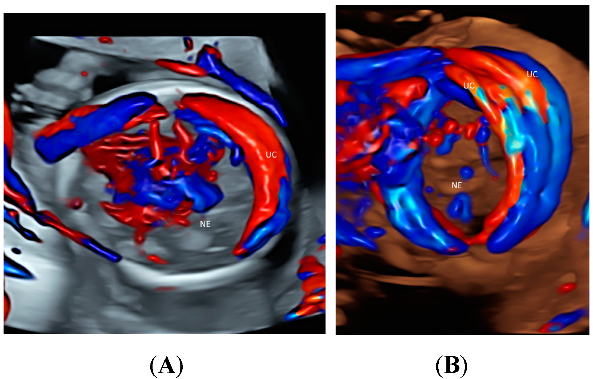
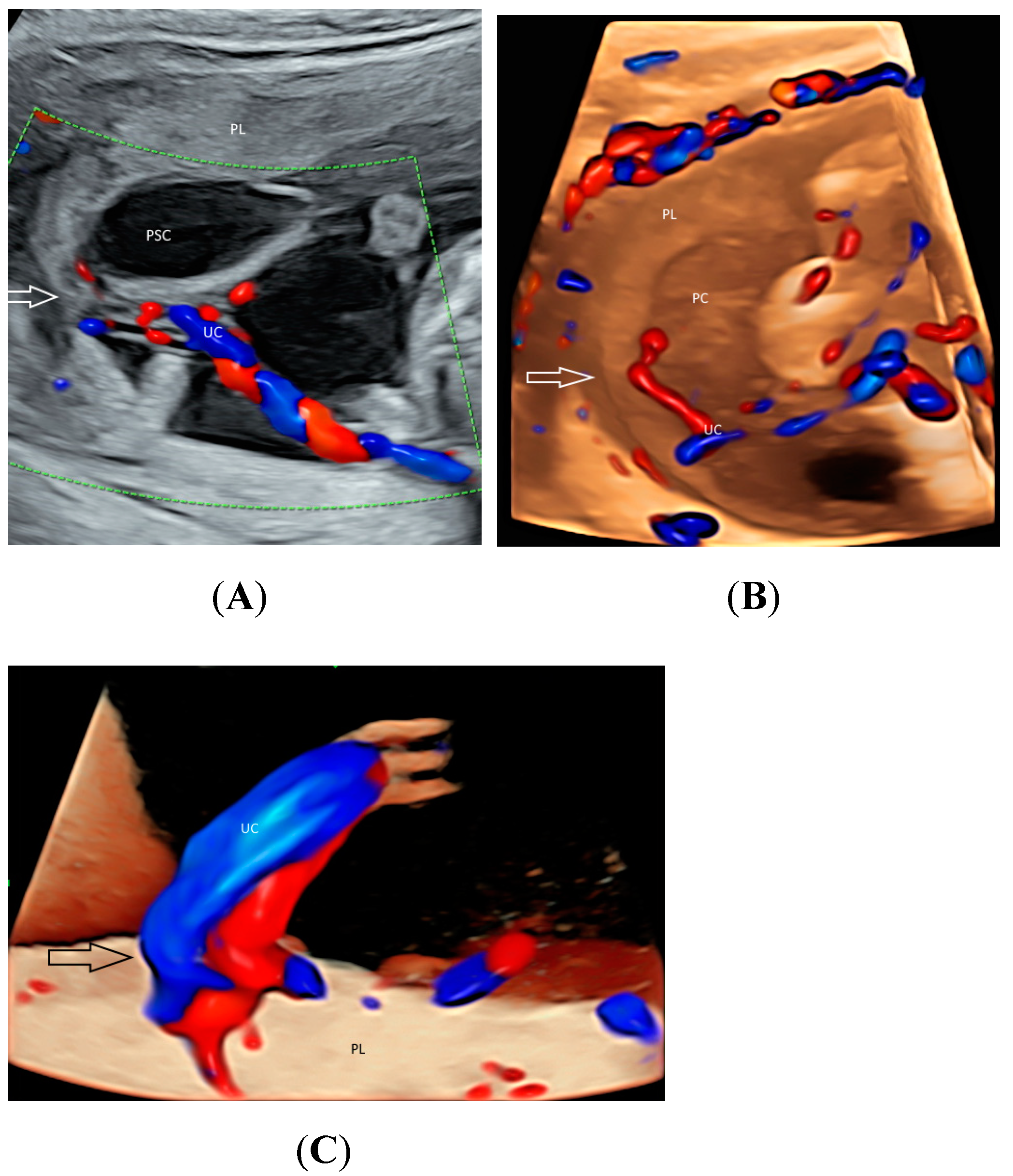
4. Umbilical Cord
5. Twins
6. Pitfalls of Glass-Body Mode
7. Practical Tips and Application
8. Conclusions
Funding
Institutional Review Board Statement
Informed Consent Statement
Data Availability Statement
Conflicts of Interest
References
- Bhide, A.; Acharya, G.; Baschat, A.; Bilardo, C.M.; Brezinka, C.; Cafici, D.; Ebbing, C.; Hernandez-Andrade, E.; Kalache, K.; Kingdom, J.; et al. ISUOG Practice Guidelines (updated): Use of Doppler velocimetry in obstetrics. Ultrasound Obstet. Gynecol. 2021, 58, 331–339. [Google Scholar] [CrossRef] [PubMed]
- Sepulveda, W.; Rojas, I.; Robert, J.A.; Schnapp, C.; Alcalde, J.L. Prenatal detection of velamentous insertion of the umbilical cord: A prospective color Doppler ultrasound study. Ultrasound Obstet. Gynecol. 2003, 21, 564–569. [Google Scholar] [CrossRef] [PubMed]
- Wiechec, M.; Knafel, A.; Nocun, A. Prenatal detection of congenital heart defects at the 11- to 13-week scan using a simple color Doppler protocol including the 4-chamber and 3-vessel and trachea views. J. Ultrasound Med. 2015, 34, 585–594. [Google Scholar] [CrossRef] [PubMed]
- Chaoui, R.; Abuhamad, A.; Martins, J.; Heling, K.S. Recent development in three- and four- dimension fetal echocardiography. Fetal Diagn. Ther. 2020, 47, 345–353. [Google Scholar] [CrossRef]
- Nadel, A.S. Addition of color Doppler to the routine obstetric sonographic survey aids in the detection of pulmonic stenosis. Fetal Diagn. Ther. 2010, 28, 175–179. [Google Scholar] [CrossRef]
- Grosvenor, A.; Silver, R.; Porter, T.F.; Zempolich, K. Optimal management of placenta accreta. Am. J. Obstet. Gynecol. 2006, 195, S82. [Google Scholar] [CrossRef]
- Committee on Obstetric Practice. Committee opinion no. 529. Placenta accreta. Obstet. Gynecol. 2012, 120, 207–211. [Google Scholar] [CrossRef]
- Leung, K.Y.; Wan, Y.L. Update on Color Flow Imaging in Obstetrics. Life 2022, 12, 226. [Google Scholar] [CrossRef]
- Gindes, L.; Pretorius, D.H.; Romine, L.E. Three-Dimensional Ultrasonographic Depiction of Fetal Abdominal Blood Vessels. J. Ultrasound Med. 2009, 28, 977–988. [Google Scholar] [CrossRef]
- Leung, K.Y. Imaging of fetal precordial venous system by four-dimensional ultrasound with spatiotemporal image correlation technology. J. Clin. Ultrasound 2022, 50, 193–197. [Google Scholar] [CrossRef]
- Leung, K.Y. Imaging of placental circulations by 4D sonography with high-definition flow and spatiotemporal image correlation technology. J. Clin. Ultrasound 2023, 51, 91–95. [Google Scholar] [CrossRef]
- Ito, M.; AboEllail, M.A.M.; Yamamoto, K.; Kanenishi, K.; Tanaka, H.; Masaoka, H.; Hata, T. HDlive Flow silhouette mode and spatiotemporal image correlation for diagnosing congenital heart disease. Ultrasound Obstet. Gynecol. 2017, 50, 411–415. [Google Scholar] [CrossRef]
- Wang, Y.; Zhang, Y. Fetal Vascular Rings and Pulmonary Slings: Strategies for Two- and Three-Dimensional Echocardiographic Diagnosis. J. Am. Soc. Echocardiogr. 2021, 34, 336–351. [Google Scholar] [CrossRef]
- Pashaj, S.; Merz, E. Prenatal demonstration of normal variants of the pericallosal artery by 3D Ultrasound. Ultraschall Med. 2014, 35, 129–136. [Google Scholar] [CrossRef]
- Egaña-Ugrinovic, G.; Savchev, S.; Bazán-Arcos, C.; Puerto, B.; Gratacós, E.; Sanz-Cortés, M. Neurosonographic assessment of the corpus callosum as imaging biomarker of abnormal neurodevelopment in late-onset fetal growth restriction. Fetal Diagn. Ther. 2015, 37, 281–288. [Google Scholar] [CrossRef]
- Paules, C.; Miranda, J.; Policiano, C.; Crovetto, F.; Youssef, L.; Hahner, N.; Nakaki, A.; Crispi, F.; Gratacós, E.; Eixarch, E. Fetal neurosonography detects differences in cortical development and corpus callosum in late-onset small fetuses. Ultrasound Obstet. Gynecol. 2021, 58, 42–47. [Google Scholar] [CrossRef]
- Goetzinger, K.R.; Cahill, A.G.; Odibo, L.; Macones, G.A.; Odibo, A.O. Three-Dimensional Power Doppler Evaluation of Cerebral Vascular Blood Flow: A Novel Tool in the Assessment of Fetal Growth Restriction. J. Ultrasound Med. 2018, 37, 139–147. [Google Scholar] [CrossRef]
- Paladini, D.; Deloison, B.; Rossi, A.; Chalouhi, G.E.; Gandolfo, C.; Sonigo, P.; Buratti, S.; Millischer, A.E.; Tuo, G.; Ville, Y.; et al. Vein of Galen aneurysmal malformation (VGAM) in the fetus: Retrospective analysis of perinatal prognostic indicators in a two-center series of 49 cases. Ultrasound Obstet. Gynecol. 2017, 50, 192–199. [Google Scholar] [CrossRef]
- Pooh, R.K. Normal anatomy by three-dimensional ultrasound in the second and third trimesters. Semin. Fetal Neonatal Med. 2012, 17, 269–277. [Google Scholar] [CrossRef]
- Achiron, R.; Gindes, L.; Zalel, Y.; Lipitz, S.; Weisz, B. Three- and four-dimensional ultrasound: New methods for evaluating fetal thoracic anomalies. Ultrasound Obstet. Gynecol. 2008, 32, 36–43. [Google Scholar] [CrossRef]
- Ruano, R.; Aubry, M.C.; Barthe, B.; Mitanchez, D.; Dumez, Y.; Benachi, A. Prediction and probability of neonatal outcome in isolated congenital diaphragmatic hernia using multiple ultrasound parameters. Ultrasound Obstet. Gynecol. 2012, 39, 42–49. [Google Scholar] [CrossRef] [PubMed]
- Cruz-Martinez, R.; Hernandez-Andrade, E.; Moreno-Álvarez, O.; Done, E.; Deprest, J.; Gratacós, E. Prognostic value of pulmonary Doppler to predict response to tracheal occlusion in fetuses with congenital diaphragmatic hernia. Fetal Diagn. Ther. 2011, 29, 18–24. [Google Scholar] [CrossRef]
- Yagel, S.; Cohen, S.M.; Valsky, D.V.; Shen, O.; Lipschuetz, M.; Messing, B. Systematic examination of the fetal abdominal precordial veins: A cohort study. Ultrasound Obstet. Gynecol. 2015, 45, 558–578. [Google Scholar] [CrossRef]
- Yagel, S.; Cohen, S.M.; Valsky, D.V. Simplifying imaging of the abdominal fetal precordial venous system. Ultrasound Obstet. Gynecol. 2019, 53, 571–575. [Google Scholar] [CrossRef]
- Degani, S.; Leibovitz, S.; Shapiro, I.; Ohel, G. Variations of the origin of renal arteries in the fetus identified on power Doppler and 3D sonography. J. Clin. Ultrasound 2010, 38, 59–65. [Google Scholar] [CrossRef]
- Wohlmuth, C.; Bergh, E.; Bell, C.; Johnson, A.; Moise, K.J., Jr.; van Gemert, M.J.C.; van den Wijngaard, J.P.H.M.; Wohlmuth-Wieser, I.; Averiss, I.; Gardiner, H.M. Clinical Monitoring of Sacrococcygeal Teratoma. Fetal Diagn. Ther. 2019, 46, 333–340. [Google Scholar] [CrossRef]
- De Diego, R.; Sabria, J.; Vela, A.; Rodriguez, D.; Gomez, M.D. Role of 3-dimensional power Doppler sonography in differentiating pregnant women with threatened preterm labor from those with an asymptomatic short cervix. J. Ultrasound Med. 2014, 33, 673–679. [Google Scholar] [CrossRef]
- Collins, S.L.; Stevenson, G.N.; Al-Khan, A.; Illsley, N.P.; Impey, L.; Pappas, L.; Zamudio, S. Three-Dimensional Power Doppler Ultrasonography for Diagnosing Abnormally Invasive Placenta and Quantifying the Risk. Obstet. Gynecol. 2015, 126, 645–653. [Google Scholar] [CrossRef]
- Thompson, M.O.; Vines, S.K.; Aquilina, J.; Wathen, N.C.; Harrington, K. Are placental lakes of any clinical significance? Placenta 2002, 23, 685–690. [Google Scholar] [CrossRef]
- Schiffer, V.; Haren, A.v.; Cubber, L.D.; Bons, J.; Coumans, A.; Kuijk, S.M.v.; Spaanderman, M.; Al-Nasiry, S. Ultrasound evaluation of the placenta in healthy and placental syndrome pregnancies: A systematic review. Eur. J. Obstet. Gynecol. Reprod. Biol. 2021, 262, 45–56. [Google Scholar] [CrossRef]
- Colpaert, R.M.; Ramseyer, A.M.; Luu, T.; Quick, C.M.; Frye, L.T.; Magann, E.F. Diagnosis and Management of Placental Mesenchymal Disease. A Review of the Literature. Obstet. Gynecol. Surv. 2019, 74, 611–622. [Google Scholar]
- Pagani, G.; Cali, G.; Acharya, G.; Trisch, I.T.; Palacios-Jaraquemada, J.; Familiari, A.; Buca, D.; Manzoli, L.; Flacco, M.E.; Fanfani, F.; et al. Diagnostic accuracy of ultrasound in detecting the severity of abnormally invasive placentation: A systematic review and meta-analysis. Acta Obstet. Gynecol. Scand. 2018, 97, 25–37. [Google Scholar] [CrossRef]
- Hasegawa, J.; Yamada, H.; Kawasaki, E.; Matsumoto, T.; Takahashi, S.; Suzuki, N. Application of superb micro-vascular imaging (SMI) in obstetrics. J. Matern. Fetal Neonatal Med. 2018, 31, 261–263. [Google Scholar] [CrossRef]
- Sainz, J.A.; Carrera, J.; Borrero, C.; García-Mejido, J.A.; Fernández-Palacín, A.; Robles, A.; Sosa, F.; Arroyo, E. Study of the Development of Placental Microvascularity by Doppler SMI (Superb Microvascular Imaging): A Reality Today. Ultrasound Med. Biol. 2020, 46, 3257–3267. [Google Scholar] [CrossRef]
- Hata, T.; Kanenishi, K.; Yamamoto, K.; AboEllail, M.A.M.; Mashima, M.; Mori, N. Microvascular imaging of thick placenta with fetal growth restriction. Ultrasound Obstet. Gynecol. 2018, 51, 837–839. [Google Scholar] [CrossRef]
- García-Jiménez, R.; Arroyo, E.; Borrero, C.; Garcia-Mejido, J.A.; Sosa, F.; Fernández-Palacín, A.; Sainz, J.A. Evaluation of Placental Micro-vascularization by Superb Micro-vascular Imaging Doppler in Cases of Intra-uterine Growth Restriction: A First Step. Ultrasound Med. Biol. 2021, 47, 1631–1636. [Google Scholar] [CrossRef]
- Santana, E.F.M.; Castello, R.G.; Rizzo, G.; Grisolia, G.; Júnior, E.A.; Werner, H.; Lituania, M.; Tonni, G. Placental and Umbilical Cord Anomalies Diagnosed by Two- and Three-Dimensional Ultrasound. Diagnostics 2022, 12, 2810. [Google Scholar] [CrossRef]
- Eastwood, K.A.; Patterson, C.; Hunter, A.J.; McCance, D.R.; Young, I.S.; Holmes, V.A. Evaluation of the predictive value of placental vascularisation indices derived from 3-Dimensional power Doppler whole placental volume scanning for prediction of pre-eclampsia: A systematic review and meta-analysis. Placenta 2017, 51, 89–97. [Google Scholar] [CrossRef]
- Abdallah, A.; Khairy, M.; Tawfik, M.; Mohamed, S.; Abdel-Rasheed, M.; Salem, S.; Khalifa, E. Role of first-trimester three-dimensional (3D) power Doppler of placental blood flow and 3D placental volume in early prediction of pre-eclampsia. Int. J. Gynaecol. Obstet. 2021, 154, 466–473. [Google Scholar] [CrossRef]
- Farina, A. Placental vascular indices (VI, FI and VFI) in intrauterine growth retardation (IUGR). A pooled analysis of the literature. Prenat. Diagn. 2015, 35, 1065–1072. [Google Scholar] [CrossRef]
- Sun, W.; Chen, L.Z.; Yin, S.W.; Cai, A.L.; Yang, Z.Y. Non-invasive dynamic observation of placental vascular anastomoses in monochorionic twins: Assessment using three-dimensional sonography combined with tomographic ultrasound imaging. Placenta 2020, 95, 84–90. [Google Scholar] [CrossRef]
- Wong, A.E.; Sepulveda, W. Acardiac anomaly: Current issues in prenatal assessment and treatment. Prenat. Diagn. 2005, 25, 796–806. [Google Scholar] [CrossRef]
- Winkler, P.; Helmke, K.; Mahl, M. Major pitfalls in doppler investigations. Pediatr. Radiol. 1990, 20, 304–310. [Google Scholar] [CrossRef]
- Martins, W.P.; Lima, J.C.; Welsh, A.W.; Júnior, E.A.; Miyague, A.H.; Filho, F.M.; Raine-Fenning, N.J. Three-dimensional Doppler evaluation of single spherical samples from the placenta: Intra- and interobserver reliability. Ultrasound Obstet. Gynecol. 2012, 40, 200–206. [Google Scholar] [CrossRef]
- Wataganara, T.; Rekhawasin, T.; Sompagdee, N.; Viboonchart, S.; Phithakwatchara, N.; Nawapun, K. A 10-Year Retrospective Review of Prenatal Applications, Current Challenges and Future Prospects of Three-Dimensional Sonoangiography. Diagnostics 2021, 11, 1511. [Google Scholar] [CrossRef]


| CDFI | PDI | HDFI | MVFI | |
|---|---|---|---|---|
| Sensitive to/ Use with | Rapid flow or large vessels | Medium flow or medium-size vessels | Slow to medium flow or small to medium sized vessels | Slow flow or small vessels |
| Insonation angle | Dependent | Independent | Dependent | Independent |
| Aliasing | Present | Absent | Present | Absent |
| Flash artifacts | Present | Present | Present | Present |
| Motion artifacts | Present | Present | Present | Reduced |
| Directional information | Bi-directional | Uni-directional | Bi-directional | mostly uni-directional |
| Spatial resolution | Low | Low | High | High |
| Radiant flow | Applicable | Applicable | Applicable | Applicable |
| 3D/4D with STIC, glass-body mode | Applicable | Applicable | Applicable | Mostly not applicable |
| Fetus/Placenta | CDFI | PDI/HDFI | MVFI |
|---|---|---|---|
| Heart | Across atrioventricular and semilunar valves | Pulmonary veins and other small/low-flow vessels | First trimester |
| Brain | Circle of Willis | Pericallosal artery Circle of Willis | Medullary veins and other small vessels |
| Abdomen and other parts | Precordial veins | Umbilical arteries, Renal arteries Splenic artery Hepatic arteries | Small vessels in lung, liver, spleen, kidney, adrenal gland and limbs |
| Umbilical cord | Cord vessels and insertion site | Cord vessels and insertion site | |
| Placenta | Placenta accreta spectrum disorders | Chorionic and decidual vessels, Spiral artery jets Placental vascular anastomoses in twin pregnancies | Stem villous vessels |
| Fetus/Placenta | CDFI/HDFI | 3DUS/ STIC | Examples of Abormalities |
|---|---|---|---|
| Heart/chest | High flow (CDFI) Small/low flow (HDFI) | GBM | Abnormal outflow tracts Course of trachea in right aortic arch Pulmonary vessels in lung dysplasia Liver vessels in diaphragmatic hernia Esophageal pouch in esophageal atresia |
| Brain | Pericallosal artery (HDFI) | GBM | Altered callosum development in FGR |
| Abdomen | Precordial veins (HDFI) | GBM TUI | Anomalies of the precoridal venous system Persistent right umbilical vein Abnormal venous returns to the right atrium |
| Umbilical cord | Cord vessels and insertion site (HDFI) | GBM TUI | Velamentous or marginal cord insertion, vasa previa, cord round neck, single umbilical artery, umbilical cord knot, abnormal thickening |
| Placenta | High flow (CDFI) Chorionic and decidual vessels, Spiral artery jets (HDFI) | GBM | Different intraplacental vascularization in PAS, FGR, and PET |
| Twin | Placental vascular anastomoses (HDFI) Intraplacental branches of umbilical artery (HDFI) | GBM TUI | Placental anastomoses in TTTS Different intraplacental vascularization in selective FGR TRAP, Cord entanglement in monoamniotic twin |
Disclaimer/Publisher’s Note: The statements, opinions and data contained in all publications are solely those of the individual author(s) and contributor(s) and not of MDPI and/or the editor(s). MDPI and/or the editor(s) disclaim responsibility for any injury to people or property resulting from any ideas, methods, instructions or products referred to in the content. |
© 2023 by the author. Licensee MDPI, Basel, Switzerland. This article is an open access article distributed under the terms and conditions of the Creative Commons Attribution (CC BY) license (https://creativecommons.org/licenses/by/4.0/).
Share and Cite
Leung, K.-Y. Application of Color Doppler with 3- and 4-Dimensional Ultrasonography in the Prenatal Evaluation of Fetal Extracardiac and Placental Abnormalities. Healthcare 2023, 11, 488. https://doi.org/10.3390/healthcare11040488
Leung K-Y. Application of Color Doppler with 3- and 4-Dimensional Ultrasonography in the Prenatal Evaluation of Fetal Extracardiac and Placental Abnormalities. Healthcare. 2023; 11(4):488. https://doi.org/10.3390/healthcare11040488
Chicago/Turabian StyleLeung, Kwok-Yin. 2023. "Application of Color Doppler with 3- and 4-Dimensional Ultrasonography in the Prenatal Evaluation of Fetal Extracardiac and Placental Abnormalities" Healthcare 11, no. 4: 488. https://doi.org/10.3390/healthcare11040488
APA StyleLeung, K.-Y. (2023). Application of Color Doppler with 3- and 4-Dimensional Ultrasonography in the Prenatal Evaluation of Fetal Extracardiac and Placental Abnormalities. Healthcare, 11(4), 488. https://doi.org/10.3390/healthcare11040488