
Content Analysis of YouTube Videos on Radiographic Anatomy on Dental Panoramic Images


Abstract
:1. Introduction
2. Materials and Methods
3. Results
4. Discussion
5. Conclusions
Supplementary Materials
Funding
Institutional Review Board Statement
Informed Consent Statement
Data Availability Statement
Conflicts of Interest
References
- Yeung, A.W.K.; Tanaka, R.; Jacobs, R.; Bornstein, M.M. Awareness and practice of 2D and 3D diagnostic imaging among dentists in Hong Kong. Br. Dent. J. 2020, 228, 701–709. [Google Scholar] [CrossRef] [PubMed]
- Yeung, A.W.K.; Wong, N.S.M. Reject Rates of Radiographic Images in Dentomaxillofacial Radiology: A Literature Review. Int. J. Environ. Res. Public Health 2021, 18, 8076. [Google Scholar] [CrossRef] [PubMed]
- Hellén-Halme, K.; Johansson, P.-M.; Håkansson, J.; Petersson, A. Image quality of digital and film radiographs in applications sent to the Dental Insurance Office in Sweden for treatment approval. Swed. Dent. J. 2004, 28, 77–84. [Google Scholar] [PubMed]
- Polo, M. Bone resorption under chin implants: The orthodontist’s role in its diagnosis and management. Am. J. Orthod. Dentofacial Orthop. 2017, 151, 201–208. [Google Scholar] [CrossRef] [Green Version]
- Yeung, A.W.K.; Mozos, I. The innovative and sustainable use of dental panoramic radiographs for the detection of osteoporosis. Int. J. Environ. Res. Public Health 2020, 17, 2449. [Google Scholar] [CrossRef] [Green Version]
- Tanaka, R.; Tanaka, T.; Yeung, A.W.K.; Taguchi, A.; Katsumata, A.; Bornstein, M.M. Mandibular radiomorphometric indices and tooth loss as predictors for the risk of osteoporosis using panoramic radiographs. Oral Health Prev. Dent. 2020, 18, 773–782. [Google Scholar]
- Bruno, G.; De Stefani, A.; Balasso, P.; Mazzoleni, S.; Gracco, A. Elongated styloid process: An epidemiological study on digital panoramic radiographs. J. Clin. Exp. Dent. 2017, 9, e1446–e1452. [Google Scholar] [CrossRef]
- Kaval, M.E.; Demirci, G.K.; Atesci, A.A.; Sarsar, F.; Dindaroğlu, F.; Güneri, P.; Caliskan, M.K. YouTube™ as an information source for regenerative endodontic treatment procedures: Quality and content analysis. Int. J. Med. Inform. 2022, 161, 104732. [Google Scholar] [CrossRef]
- Fu, M.W.; Kalaichelvan, A.; Liebman, L.S.; Burns, L.E. Exploring predoctoral dental student use of YouTube as a learning tool for clinical endodontic procedures. J. Dent. Educ. 2021, 86, 726–735. [Google Scholar] [CrossRef]
- Aldallal, S.; Yates, J.; Ajrash, M. Use of YouTube™ as a self-directed learning resource in oral surgery among undergraduate dental students: A cross-sectional descriptive study. Br. J. Oral Maxillofac. Surg. 2019, 57, 1049–1052. [Google Scholar] [CrossRef]
- Jaffar, A.A. YouTube: An emerging tool in anatomy education. Anat. Sci. Educ. 2012, 5, 158–164. [Google Scholar] [CrossRef] [PubMed]
- Azer, S.A. Can “YouTube” help students in learning surface anatomy? Surg. Radiol. Anat. 2012, 34, 465–468. [Google Scholar] [CrossRef] [PubMed]
- Staziaki, P.V.; de Oliveira Santo, I.D.; Skobodzinski, A.A.; Park, L.K.; Bedi, H.S. How to use YouTube for radiology education. Curr. Probl. Diagn. Radiol. 2021, 50, 461–468. [Google Scholar] [CrossRef] [PubMed]
- Kauffman, L.; Weisberg, E.M.; Eng, J.; Fishman, E.K. YouTube and radiology: The viability, pitfalls, and untapped potential of the premier social media video platform for image-based education. Acad. Radiol. 2022, 29, S1–S8. [Google Scholar] [CrossRef] [PubMed]
- Mustafa, A.G.; Taha, N.R.; Alshboul, O.A.; Alsalem, M.; Malki, M.I. Using YouTube to learn anatomy: Perspectives of Jordanian medical students. BioMed Res. Int. 2020, 2020, 6861416. [Google Scholar] [CrossRef] [Green Version]
- Barry, D.S.; Marzouk, F.; Chulak-Oglu, K.; Bennett, D.; Tierney, P.; O’Keeffe, G.W. Anatomy education for the YouTube generation. Anat. Sci. Educ. 2016, 9, 90–96. [Google Scholar] [CrossRef]
- Brown, J.D. What do you meme, professor? An experiment using “memes” in pharmacy education. Pharmacy 2020, 8, 202. [Google Scholar] [CrossRef]
- Wells, D.D. You all made dank memes: Using internet memes to promote critical thinking. J. Political Sci. Educ. 2018, 14, 240–248. [Google Scholar] [CrossRef]
- Miller, G.W.; Lewis, T.L. Anatomy education for the YouTube generation: Technical, ethical, and educational considerations. Anat. Sci. Educ. 2016, 9, 496–497. [Google Scholar] [CrossRef]
- Moxham, B.; Plaisant, O. Perception of medical students towards the clinical relevance of anatomy. Clin. Anat. 2007, 20, 560–564. [Google Scholar] [CrossRef]
- Obrez, A.; Briggs, C.; Buckman, J.; Goldstein, L.; Lamb, C.; Knight, W.G. Teaching clinically relevant dental anatomy in the dental curriculum: Description and assessment of an innovative module. J. Dent. Educ. 2011, 75, 797–804. [Google Scholar] [CrossRef] [PubMed]
- King, A.D.; Bhatia, K.S.S. Magnetic resonance imaging staging of nasopharyngeal carcinoma in the head and neck. World J. Radiol. 2010, 2, 159–165. [Google Scholar] [CrossRef] [PubMed]
- Tekiner, H.; Acer, N.; Kelestimur, F. Sella turcica: An anatomical, endocrinological, and historical perspective. Pituitary 2015, 18, 575–578. [Google Scholar] [CrossRef]
- Devasia, B.; Ranganathan, K.; Umadevi, M.; Shanmugam, S.; Saraswathi, T. Stylocarotid syndrome mimicking dental Pain. Asian J. Oral Maxillofac. Surg. 2004, 16, 47–50. [Google Scholar] [CrossRef]
- Yoshida, K.; Fukuda, M.; Gotoh, K.; Ariji, E. Depression of the maxillary sinus anterior wall and its influence on panoramic radiography appearance. Dentomaxillofac. Radiol. 2017, 46, 20170126. [Google Scholar] [CrossRef]
- Metcalfe, J. Learning from errors. Annu. Rev. Psychol. 2017, 68, 465–489. [Google Scholar] [CrossRef] [Green Version]
- Grillon, M.; Yeung, A.W.K. Content Analysis of YouTube Videos That Demonstrate Panoramic Radiography. Healthcare 2022, 10, 1093. [Google Scholar] [CrossRef]
- Yeung, A.W.K. The Usage of the Terms Mandibular Canal, Inferior Alveolar Canal, and Inferior Dental Canal in the Academia: A Bibliometric Analysis. Int. J. Morphol. 2021, 39, 1058–1062. [Google Scholar] [CrossRef]


| Metric | Mean ± SD | Min; Max |
|---|---|---|
| View count | 7536.2 ± 16,174.1 | 35; 83,549 |
| Like count | 154.5 ± 344.9 | 0; 1800 |
| Comment count | 20.3 ± 56.8 | 0; 314 |
| Duration (s) | 892.2 ± 1138.4 | 60; 6333 |
| Channel subscriber count | 12,507.2 ± 37,211.5 | 3; 214,000 |
| Age of video (y) | 2.0 ± 1.3 | 0.1; 5.0 |
| No. of Videos | % (of 38) | |
|---|---|---|
| Midfacial Landmarks | ||
| Elongated styloid process (or calcification of stylohyoid ligament) may indicate Eagle syndome | 2 | 5.3 |
| Pterygomaxillary fissure may disappear if a maxillary lesion involves the posterior wall of the maxillary bone/sinus | 2 | 5.3 |
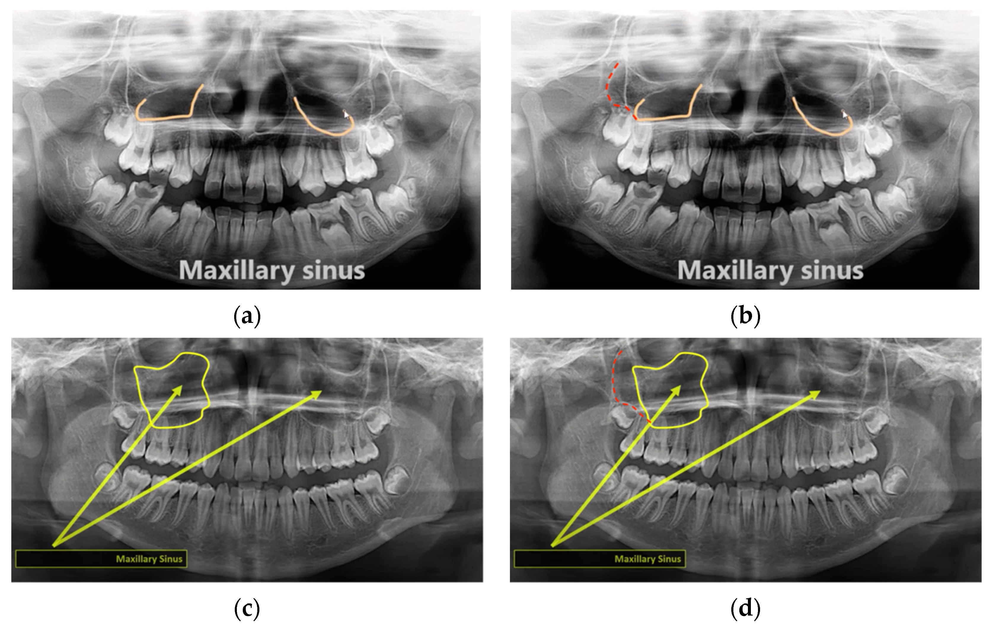
| Maxillary sinus will be radiopaque if pathological | 1 | 2.6 |
| Sinus lift is needed if an implant is placed at maxillary posterior region with a low sinus floor | 1 | 2.6 |
| Zygomaticotemporal suture should not be mistaken as a fracture | 1 | 2.6 |
| Mandibular landmarks | ||
| Mandibular nerve block should be injected at mandibular foramen, and mental nerve block at mental foramen | 2 | 5.3 |
| Condyle may fracture upon severe trauma | 1 | 2.6 |
| Mental foramen should not be mistaken as a periapical lesion | 1 | 2.6 |
| Submandibular gland fossa should not be mistaken as a pathology such as cancer | 1 | 2.6 |
| Soft tissues/air spaces/others | ||
| Hyoid bone should not be mistaken as the clavicle | 1 | 2.6 |
| No. of Videos | % (of 38) | |
|---|---|---|
| Midfacial landmarks | ||
| Named the zygomatic process of the maxilla simply as “zygomatic process” | 2 | 5.3 |
| Wrongly identified the zygomatic process of the maxilla as the posterior wall of maxillary sinus | 2 | 5.3 |
| Wrongly treated the diagonal line due to depression of the maxillary sinus anterior wall as the posterior sinus wall | 2 | 5.3 |
| Wrongly identified the pterygomaxillary fissure | 2 | 5.3 |
| Wrongly identified the zygomatic process of the maxilla | 1 | 2.6 |
| Wrongly identified the lateral pterygoid plate | 1 | 2.6 |
| Wrongly named the zygomatic process of the temporal bone as the zygomatic bone | 1 | 2.6 |
| Mandibular landmarks | ||
| Named the mandibular notch as the coronoid notch | 2 | 5.3 |
| Wrongly named the condyle of the mandible as the coronoid process, and vice versa | 1 | 2.6 |
| Wrongly named the coronoid process as the coronoid notch | 1 | 2.6 |
| Called the shadow of the contralateral angle of the mandible simply as “ghost image” | 1 | 2.6 |
| Soft tissues/air spaces/others | ||
| Grouped or named the palatoglossal air space as the glosso- (or oro-)pharynx | 2 | 5.3 |
| Wrongly identified the palatoglossal air space as the soft palate and uvula | 1 | 2.6 |
| Wrongly identified the shadow of the cervical vertebrae in the midline as the chin rest | 1 | 2.6 |
| Circled a line tracing the inferior border of the nasopharyngeal space and suggested that it was the whole space | 1 | 2.6 |
| Wrongly identified the border of the tongue | 1 | 2.6 |
| Type | Mean ± SD | Min; Max |
|---|---|---|
| Overall | 25.7 ± 11.7 | 4; 48 |
| Midfacial | 12.3 ± 5.7 | 0; 22 |
| Mandibular | 8.2 ± 4.0 | 0; 16 |
| Soft tissue/air space/others | 5.2 ± 3.9 | 0; 15 |
| Like Count | Comment Count | Channel Subscriber Count | Duration (s) | Video Age (y) | Total no. of Landmarks | |
|---|---|---|---|---|---|---|
| View count | 0.940 (p < 0.001) | 0.072 (p = 0.691) | 0.788 (p < 0.001) | −0.121 (p = 0.468) | 0.469 (p = 0.003) | −0.285 (p = 0.082) |
| Like count | 0.128 (p = 0.478) | 0.812 (p < 0.001) | −0.111 (p = 0.508) | 0.245 (p = 0.138) | −0.221 (p = 0.182) | |
| Comment count | 0.018 (p = 0.922) | −0.133 (p = 0.459) | −0.191 (p = 0.287) | −0.199 (p = 0.266) | ||
| Channel subscriber count | −0.078 (p = 0.647) | 0.169 (p = 0.317) | 0.049 (p = 0.775) | |||
| Duration (s) | 0.064 (p = 0.705) | 0.241 (p = 0.144) | ||||
| Video age | −0.153 (p = 0.360) |
Publisher’s Note: MDPI stays neutral with regard to jurisdictional claims in published maps and institutional affiliations. |
© 2022 by the author. Licensee MDPI, Basel, Switzerland. This article is an open access article distributed under the terms and conditions of the Creative Commons Attribution (CC BY) license (https://creativecommons.org/licenses/by/4.0/).
Share and Cite
Yeung, A.W.K. Content Analysis of YouTube Videos on Radiographic Anatomy on Dental Panoramic Images. Healthcare 2022, 10, 1382. https://doi.org/10.3390/healthcare10081382
Yeung AWK. Content Analysis of YouTube Videos on Radiographic Anatomy on Dental Panoramic Images. Healthcare. 2022; 10(8):1382. https://doi.org/10.3390/healthcare10081382
Chicago/Turabian StyleYeung, Andy Wai Kan. 2022. "Content Analysis of YouTube Videos on Radiographic Anatomy on Dental Panoramic Images" Healthcare 10, no. 8: 1382. https://doi.org/10.3390/healthcare10081382
APA StyleYeung, A. W. K. (2022). Content Analysis of YouTube Videos on Radiographic Anatomy on Dental Panoramic Images. Healthcare, 10(8), 1382. https://doi.org/10.3390/healthcare10081382