Article

Usefulness of the Modified Videofluoroscopic Dysphagia Scale in Determining the Allowance of Oral Feeding in Patients with Dysphagia Due to Deconditioning or Frailty

1
Department of Rehabilitation Medicine, Yeungnam University Hospital, Daegu 42415, Korea
2
Department of Neurosurgery, Kyung Hee University Hospital at Gangdong, Kyung Hee University College of Medicine, Seoul 05278, Korea
3
Department of Physical Medicine and Rehabilitation, Ulsan University Hospital, University of Ulsan College of Medicine, Ulsan 44033, Korea
*
Author to whom correspondence should be addressed.
Healthcare 2022, 10(4), 668; https://doi.org/10.3390/healthcare10040668
Submission received: 21 February 2022 / Revised: 28 March 2022 / Accepted: 30 March 2022 / Published: 1 April 2022

Abstract

:
Introduction: In patients with dysphagia due to deconditioning or frailty, as with other disorders that cause swallowing disorders, the videofluoroscopic swallowing study (VFSS) is the gold standard for dysphagia evaluation. However, the interpretation of VFSS results is somewhat complicated and requires considerable experience in the field. Therefore, in this study we evaluated the usefulness of the modified videofluoroscopic dysphagia scale (mVDS) in determining whether to allow oral feeding in patients with dysphagia due to deconditioning or frailty. Methods: Data from the VFSS of 50 patients with dysphagia due to deconditioning or frailty were retrospectively collected. We evaluated the association between mVDS and the selected feeding method based on VFSS findings, and between mVDS and the presence of aspiration pneumonia. Results: Multivariate logistic analysis showed that the mVDS total score had a significant association with oral feeding methods based on VFSS findings in patients with dysphagia due to deconditioning or frailty (p < 0.05). In the receiver operating characteristic (ROC) curve analysis, the area under the ROC curve for the selected feeding method was 0.862 (95% confidence interval, 0.747–0.978; p < 0.0001). Conclusions: mVDS seems a valid scale for determining the allowance of oral feeding, and it can be a useful tool in the clinical setting and in studies that aim to interpret VFSS findings in patients with dysphagia due to deconditioning or frailty. However, studies involving a more general population of patients with dysphagia due to deconditioning or frailty are needed.

1. Introduction

Deconditioning is a complex process of physiological change following a period of inactive, sedentary or bed-rest lifestyle. It causes functional losses in such areas as mental status, and an inability to accomplish activities of daily living. It is frequently associated with hospitalization in the elderly [1]. The most predictable effects of deconditioning are seen in the musculoskeletal system, and include reduced muscle mass and strength. The decline in muscle mass and strength has been linked to falls, functional decline, increased immobility and frailty. This deconditioning also affects the swallowing muscles, which can lead to dysphagia.
Frailty is a complex syndrome that is characterized by the progressive decline in physical, mental, and social functions [2]. Frailty develops because of the age-related decline in several physiological systems [3]. It commonly occurs in older adults and deteriorates physiologic functions and increases the risk of poor health outcomes including disability, hospitalization, and mortality [3]. Frailty also leads to a decline in swallowing function [4].
Although the incidence of dysphagia in deconditioning or frailty has not been accurately reported, deconditioning and frailty have been reported in previous studies as among several causes of dysphagia [5,6,7,8]. When the degree of dysphagia is severe, oral feeding can result in aspiration pneumonia and mortality [9,10]. Therefore, in those with a high risk of aspiration pneumonia, oral feeding should be prohibited. However, because prohibiting oral feeding can lead to malnutrition, and a decline in physical function and general health, decisions regarding oral feeding should be carefully made after detailed ascertainment of its necessity [11].
The videofluoroscopic swallowing study (VFSS) is a standard tool for evaluating the severity of dysphagia, and it provides several details on swallowing function [12,13,14,15]. Based on VFSS results, clinicians can obtain information on dysphagia severity and develop a treatment plan [16]. However, the interpretation of VFSS results is somewhat complicated and requires experience in the field. Accordingly, objective criteria or cutoff values in diagnostic evaluation using quantitative analyses of VFSS should help clinicians decide on oral feeding more easily and precisely.
For the quantitative analysis of VFSS, the videofluoroscopic dysphagia scale (VDS) was developed, but it is limited because of the relatively low inter-rater reliability of some subcategories [17]. To compensate for these limitations, Chang et al. developed a modified version (mVDS) and demonstrated its high validation and inter-rater reliability [18]. mVDS includes nine parameters (lip closure, mastication, oral transit time, trigger pharyngeal swallow, epiglottis inversion, valleculae residue, pyriformis residue, pharyngeal wall coating, and aspiration) [18], and its clinical usefulness in evaluating swallowing function and selecting feeding methods was demonstrated in patients with amyotrophic lateral sclerosis and stroke, respectively [19,20].
Therefore, in the current study, we evaluated whether mVDS could be used to determine the possibility of oral feeding in patients with dysphagia due to deconditioning or frailty. Further, we investigated the mVDS cutoff value for permitting oral feeding.

2. Methods

2.1. Ethics Statements

This retrospective study was approved by the Institutional Review Board (IRB) of Ulsan University Hospital (2021-01-028). This study was performed according to the Declaration of Helsinki for human experiments. Informed consent was waived because of the retrospective nature of the study.

2.2. Study Design and Population

We retrospectively collected the data of patients with dysphagia due to deconditioning or frailty who had any symptoms of difficulty in swallowing and who underwent a VFSS at Ulsan University Hospital between April 2020 and January 2022. We obtained clinical data such as sex, age, presence of tracheal tube or Levin tube, and history of aspiration pneumonia.
The criteria for inclusion (patients with dysphagia due to deconditioning or frailty) were as follows: (1) age at VFSS > 50 years; (2) history of aspiration symptoms, such as coughing or choking; (3) presence of symptoms clinically indicative of dysphagia, such as reduced gag reflex or delayed swallowing reflex; (4) presence of medical records of hospitalization for medical reasons within the last year prior to VFSS (5) presence of chronic disease (hypertension, diabetes, or chronic kidney disease, etc.) (6) presence of oropharyngeal dysphagia due to deconditioning or frailty without a specific diagnosis that could cause dysphagia such as stroke, traumatic brain injury, or other laryngeal pathology [4].
The criteria for exclusion were as follows: (1) inability to sit, or difficulty maintaining consciousness; (2) dysphagia due to known neurologic conditions diagnosed by neurologists including stroke, traumatic brain injury, anoxic brain injury, brain tumor, motor neuron disease, Parkinson disease, or Alzheimer disease; and (3) dysphagia from laryngeal pathology, including laryngeal cancer, stenosis, paralysis, and postoperative head and neck surgery [4].

2.3. Aspiration Pneumonia

A retrospective review of medical records was performed to identify the development of aspiration pneumonia within one month before and after a VFSS in patients with dysphagia due to deconditioning or frailty. The following data were collected: symptoms, such as coughing during feeding; presence of sputum, dyspnea, or fever; chest radiography findings; blood laboratory findings (white blood cell [WBC] counts, C-reactive protein [CRP] level, and erythrocyte sedimentation rate); and use of antibiotics [21,22].
Although a definitive diagnosis of aspiration is difficult, and the diagnostic criteria for aspiration pneumonia are relatively different across studies, patients who met all of the following criteria were considered to have aspiration pneumonia in this study: (1) presence of both objective signs (coarse lung sounds, presence of lung infiltration on chest radiography, and systemic inflammation based on blood laboratory findings, such as increased CRP levels and WBC counts) and subjective symptoms (fever, cough, and increased purulent sputum), (2) clinical suspicion of aspiration (delayed swallowing or coughing during swallowing), and (3) no evidence of micro-organisms, such as Legionella or Mycoplasma, which are common pathogens in atypical pneumonia [21,22]. In addition, the clinical reports from the Department of Internal Medicine were used to diagnose aspiration pneumonia.

2.4. VFSS Protocol

VFSS was conducted with a fluoroscopic device and recorded as a video file. During the VFSS, patients consecutively swallowed the following materials, with a stepwise consistency: water, nectar (51–350 cP), rice porridge (351–1750 cP), and boiled rice (>1750 cP) [23]. The materials were mixed with liquid barium, and the patient swallowed them while in a relaxed sitting position. Dynamic fluoroscopic images were recorded in the anteroposterior and lateral views at 30 frames per second. The VFSS images were analyzed according to the Penetration–Aspiration Scale (PAS) and were considered positive for aspiration if the PAS score was >5 [24].
All studies were reviewed by two physiatrists who had at least eight years of experience in interpreting VFSS results. The conclusion (the allowance of oral feeding) was made by comprehensively considering the findings of VFSS, such as the oral transit time, the swallowing reflex, the VFSS results such as residues and aspiration, the amount of food swallowed, and the viscosity. Patient information, including age, sex, and underlying diseases, was withheld from the interpreters. The interpreters only observed the patients via the movie files on the laptop, described their findings, and chose a feeding method (non-oral feeding versus oral feeding) based on the VFSS results.

2.5. Modification of the VDS

As noted earlier, mVDS was developed based on a study regarding the inter-rater reliability of VDS. Among the VDS categories, the ones with a κ value < 0.2 (bolus formation, mastication, apraxia, tongue-in-palate contact, and pharyngeal transit time) were modified [25]. As mentioned by previous researchers, such categories had somewhat ambiguous guidelines, and three to four multiple selectable choices that led to low reliability [18,19,20]. Therefore, we modified the categories according to a binary scale or deleted the ambiguous categories. The mVDS was drafted as shown in Table 1. We also changed the category of laryngeal elevation to epiglottis inversion, which was reported to be an important factor in the swallowing process in a previous study, because laryngeal elevation and epiglottis inversion are a result of a combination of the contraction and relaxation of the suprahyoid and infrahyoid muscles [18,19,20]. To measure triggering pharyngeal swallow (swallowing reflex), the recorded fluoroscopic images in lateral view at 30 frames per seconds were analyzed by measuring the response time of pharyngeal swallowing reflexes. A study reported that, in the VFSS, the normal value of the response time of the swallowing reflex was 0.53 ± 0.64 s in elderly subjects (≥65 year old) [12]. If the response time of the swallowing reflex was longer than the normal value, we determined that the patient had a delayed swallowing reflex. Residues in the valleculae and pyriformis sinuses were divided into four groups: none, small (<10%), medium (10–50%), and large (>50%) [16].
Originally, to measure these VFSS findings as objective quantitative scores, VDS (with a sum of 100 points) was created based on the odds ratios of various prognostic factors. After modification of the VDS parameters, we rebalanced the score of each mVDS category, which yielded a sum of 100 points (Table 1) [18,19,20]. The inter-rater reliability (Cronbach α value) of the total score of the mVDS was 0.886, which was consistent with very good inter-rater reliability [20].

2.6. Statistical Evaluation

To find differences in parameters between the oral and non-oral feeding groups based on the VFSS findings, and between the aspiration and non-aspiration pneumonia groups, the Pearson chi-square test or Mann–Whitney U-test was used. Using only the parameters that were significant in the Mann–Whitney U-test, multivariate logistic regression analysis with the enter method was performed to identify parameters that had a significant association with the selected feeding method based on VFSS findings. To evaluate the accuracy of predictive factors for oral or non-oral feeding based on the VFSS findings, we performed receiver operating characteristic (ROC) analysis. Statistical analysis was performed using MedCalc (MedCalc Software, Ostend, Belgium) and SPSS version 22.0 (IBM Corp., Armonk, NY, USA).

3. Results

3.1. Patients’ Characteristics

Fifty patients with dysphagia due to deconditioning or frailty were enrolled in this study. Among them, 42 were male and 8 were female (mean age 71.26 ± 11.93 years). The patients’ demographic data are presented in Table 2.

3.2. Difference in Parameters between the Oral and Non-Oral Feeding Groups

In the Mann–Whitney U-test, age, PAS, and mVDS total score were significantly different between the oral and non-oral feeding groups (p < 0.05). In the multivariate logistic analysis, using these three parameters to identify any association with oral feeding methods based on VFSS findings, mVDS total score showed a significant association with oral feeding methods based on VFSS findings in patients with dysphagia due to deconditioning or frailty. (p < 0.05) (Table 3). In the ROC curve analysis, the area under the ROC curve for the selected feeding method was 0.862 (95% confidence interval [CI], 0.747–0.978; p < 0.0001). The optimal cutoff value for allowing oral feeding obtained from the maximal Youden index was ≤48 based on mVDS (sensitivity, 96.15%; specificity, 79.17%) (Figure 1).

3.3. Difference in Parameters between the Aspiration and Non-Aspiration Pneumonia Groups

In the Mann–Whitney U-test, age, PAS, and mVDS score were significantly different between the aspiration and non-aspiration pneumonia groups (p < 0.05). The multivariate logistic analysis using these three parameters to identify any association with the presence of aspiration pneumonia did not show any significant parameter.

4. Discussion

In this study, the mVDS score showed a statistically significant association with the allowance of oral feeding in patients with dysphagia due to deconditioning or frailty. To our knowledge, no previous study has investigated the usefulness of the mVDS score for evaluating dysphagia among patients with dysphagia due to deconditioning or frailty.
In mVDS, as in VDS, a higher score indicates a greater diet limitation and more severe dysphagia [18,19,20]. The results of this study suggest that an mVDS score of ≤48 is significantly associated with the allowance of oral feeding (sensitivity, 96.15%; specificity, 79.17%). Moreover, patients with an mVDS score of ≤12.5 showed 100% specificity for the allowance of oral feeding (sensitivity, 15.38%; specificity, 100.0%).
As mentioned earlier, the interpretation of VFSS results is somewhat complicated and requires considerable experience in the field, although VFSS is the gold standard for dysphagia evaluation because it is able to visualize the entire swallowing process, from the oral phase to the esophageal phase [12,13,14,15]. Therefore, numerous methods have been applied in interpreting VFSS results to quantify the severity of dysphagia and objectively determine the allowance of oral feeding based on the score. One of such methods is PAS, which is an eight-point scale used to describe the depth of food invasion and response to airway infiltration during VFSS [24]. Although it can notably define the extent of airway infiltration, the association between PAS score and feeding method based on VFSS results has been reportedly weak [20]. Another method to quantify dysphagia severity using VFSS video is VDS [25]. Previous studies have shown that VDS is significantly associated with penetration and/or aspiration occurring six months after the initial onset of dysphagia [25]. However, as previously mentioned, because of the ambiguity and complexity of some VDS subcategories, VDS has been reported to have low inter-rater reliability in such subcategories [25]. To improve these disadvantages, some VDS categories have been modified, and the application of these modifications in patients with stroke or other etiologies except dysphagia due to deconditioning or frailty was proven valid in previous studies [18,19,20]. In common with the results of these studies, our findings suggest that mVDS is a useful tool for quantifying the severity of dysphagia and determining the allowance of oral feeding in patients with dysphagia due to deconditioning or frailty.

5. Limitation

There are several limitations to our study. First, the total number of enrolled patients was relatively small. Therefore, generalizing our conclusions may be challenging. Studies with more patients are needed to achieve a more comprehensive conclusion. Second, this was a retrospective study; therefore, some data that might have added more value to the study were unavailable, such as frailty scores which can reflect the severity of disease at the time of VFSS. Lastly, this study was conducted in a single tertiary hospital, which might have also affected the generalizability of the study findings. Further large-scale prospective studies are necessary to achieve a comprehensive understanding of dysphagia due to deconditioning or frailty, excluding specific diseases that cause dysphagia.

6. Conclusions

mVDS seems a useful scale for determining the allowance of oral feeding and appears a relevant tool in the clinical setting, and in studies for interpreting VFSS findings in patients with dysphagia due to deconditioning or frailty. However, further studies involving a more general population of patients with dysphagia due to deconditioning or frailty are needed to elucidate a more accurate cutoff value for allowing oral feeding.

Author Contributions

M.C.C.: Writing—original draft, and Writing—review and editing; H.Y.C.: supervision, data curation; D.P.: Conceptualization, Formal analysis, Writing—original draft, and Writing—review and editing. All authors have read and agreed to the published version of the manuscript.

Funding

This study was supported by the National Research Foundation of Korea Grant funded by the Korean government, No. NRF2021R1A2C1013073.

Institutional Review Board Statement

The study was conducted according to the guidelines of the Declaration of Helsinki, and approved by the Institutional Review Board (or Ethics Committee) of Ulsan University Hospital (protocol code 2021-01-028-001 and date of approval 29 November 2021).

Informed Consent Statement

Informed consent was waived because of the retrospective nature of the study and the analysis used anonymous clinical data.

Data Availability Statement

The data presented in this study are available on request from the corresponding author.

Conflicts of Interest

The authors report no conflict of interest or financial support. No commercial party having a direct financial interest in the results of the research supporting this article has or will confer a benefit on the authors or on any organization with which the authors are associated.

References

  1. Gillis, A.; MacDonald, B. Deconditioning in the hospitalized elderly. Can. Nurse 2005, 101, 16–20. [Google Scholar] [PubMed]
  2. Khezrian, M.; Myint, P.K.; McNeil, C.; Murray, A.D. A Review of Frailty Syndrome and Its Physical, Cognitive and Emotional Domains in the Elderly. Geriatrics 2017, 2, 36. [Google Scholar] [CrossRef] [PubMed] [Green Version]
  3. Chen, X.; Mao, G.; Leng, S.X. Frailty syndrome: An overview. Clin. Interv. Aging 2014, 9, 433–441. [Google Scholar] [CrossRef] [Green Version]
  4. Chang, M.C.; Kwak, S. Videofluoroscopic Swallowing Study Findings Associated with Subsequent Pneumonia in Patients with Dysphagia Due to Frailty. Front. Med. 2021, 8, 690968. [Google Scholar] [CrossRef] [PubMed]
  5. Sella-Weiss, O. Association between swallowing function, malnutrition and frailty in community dwelling older people. Clin. Nutr. ESPEN 2021, 45, 476–485. [Google Scholar] [CrossRef]
  6. Byun, S.E.; Kwon, K.B.; Kim, S.H.; Lim, S.J. The prevalence, risk factors and prognostic implications of dysphagia in elderly patients undergoing hip fracture surgery in Korea. BMC Geriatr. 2019, 19, 356. [Google Scholar] [CrossRef] [Green Version]
  7. Wang, T.; Zhao, Y.; Guo, A. Association of swallowing problems with frailty in Chinese hospitalized older patients. Int. J. Nurs. Sci. 2020, 7, 408–412. [Google Scholar] [CrossRef] [PubMed]
  8. Hathaway, B.; Vaezi, A.; Egloff, A.M.; Smith, L.; Wasserman-Wincko, T.; Johnson, J.T. Frailty measurements and dysphagia in the outpatient setting. Ann. Otol. Rhinol. Laryngol. 2014, 123, 629–635. [Google Scholar] [CrossRef]
  9. Farri, A.; Accornero, A.; Burdese, C. Social importance of dysphagia: Its impact on diagnosis and therapy. Acta Otorhinolaryngol. Ital. 2007, 27, 83–86. [Google Scholar]
  10. Sura, L.; Madhavan, A.; Carnaby, G.; Crary, M.A. Dysphagia in the elderly: Management and nutritional considerations. Clin. Interv. Aging 2012, 7, 287–298. [Google Scholar] [CrossRef] [Green Version]
  11. Volkert, D.; Pauly, L.; Stehle, P.; Sieber, C.C. Prevalence of malnutrition in orally and tube-fed elderly nursing home residents in Germany and its relation to health complaints and dietary intake. Gastroenterol. Res. Pract. 2011, 2011, 247315. [Google Scholar] [CrossRef] [PubMed]
  12. Lee, J.T.; Park, E.; Hwang, J.M.; Jung, T.D.; Park, D. Machine learning analysis to automatically measure response time of pharyngeal swallowing reflex in videofluoroscopic swallowing study. Sci. Rep. 2020, 10, 14735. [Google Scholar] [CrossRef] [PubMed]
  13. Park, D.; Suh, J.H.; Kim, H.; Ryu, J.S. The Effect of Four-Channel Neuromuscular Electrical Stimulation on Swallowing Kinematics and Pressures: A Pilot Study. Am. J. Phys. Med. Rehabil. 2019, 98, 1051–1059. [Google Scholar] [CrossRef] [PubMed]
  14. Yu, K.J.; Moon, H.; Park, D. Different clinical predictors of aspiration pneumonia in dysphagic stroke patients related to stroke lesion: A STROBE-complaint retrospective study. Medicine 2018, 97, e13968. [Google Scholar] [CrossRef] [PubMed]
  15. Park, D.; Lee, H.H.; Lee, S.T.; Oh, Y.; Lee, J.C.; Nam, K.W.; Ryu, J.S. Normal contractile algorithm of swallowing related muscles revealed by needle EMG and its comparison to videofluoroscopic swallowing study and high resolution manometry studies: A preliminary study. J. Electromyogr. Kinesiol. 2017, 36, 81–89. [Google Scholar] [CrossRef]
  16. Park, D.; Oh, Y.; Ryu, J.S. Findings of Abnormal Videofluoroscopic Swallowing Study Identified by High-Resolution Manometry Parameters. Arch. Phys. Med. Rehabil. 2016, 97, 421–428. [Google Scholar] [CrossRef]
  17. Kim, G.E.; Sung, I.Y.; Ko, E.J.; Choi, K.H.; Kim, J.S. Comparison of Videofluoroscopic Swallowing Study and Radionuclide Salivagram for Aspiration Pneumonia in Children with Swallowing Difficulty. Ann. Rehabil. Med. 2018, 42, 52–58. [Google Scholar] [CrossRef] [Green Version]
  18. Chang, M.C.; Lee, C.; Park, D. Validation and Inter-rater Reliability of the Modified Videofluoroscopic Dysphagia Scale (mVDS) in Dysphagic Patients with Multiple Etiologies. J. Clin. Med. 2021, 10, 2990. [Google Scholar] [CrossRef]
  19. Lee, B.J.; Eo, H.; Park, D. Usefulness of the Modified Videofluoroscopic Dysphagia Scale in Evaluating Swallowing Function among Patients with Amyotrophic Lateral Sclerosis and Dysphagia. J. Clin. Med. 2021, 10, 4300. [Google Scholar] [CrossRef]
  20. Lee, B.J.; Eo, H.; Lee, C.; Park, D. Usefulness of the Modified Videofluoroscopic Dysphagia Scale in Choosing the Feeding Method for Stroke Patients with Dysphagia. Healthcare 2021, 9, 632. [Google Scholar] [CrossRef]
  21. Pikus, L.; Levine, M.S.; Yang, Y.X.; Rubesin, S.E.; Katzka, D.A.; Laufer, I.; Gefter, W.B. Videofluoroscopic studies of swallowing dysfunction and the relative risk of pneumonia. AJR Am. J. Roentgenol. 2003, 180, 1613–1616. [Google Scholar] [CrossRef] [PubMed]
  22. Lee, D.H.; Kim, J.M.; Lee, Z.; Park, D. The effect of radionuclide solution volume on the detection rate of salivary aspiration in the radionuclide salivagram: A STROBE-compliant retrospective study. Medicine 2018, 97, e11729. [Google Scholar] [CrossRef] [PubMed]
  23. Yu, K.J.; Park, D. Clinical characteristics of dysphagic stroke patients with salivary aspiration: A STROBE-compliant retrospective study. Medicine 2019, 98, e14977. [Google Scholar] [CrossRef] [PubMed]
  24. Borders, J.C.; Brates, D. Use of the Penetration-Aspiration Scale in Dysphagia Research: A Systematic Review. Dysphagia 2020, 35, 583–597. [Google Scholar] [CrossRef] [PubMed]
  25. Kim, D.H.; Choi, K.H.; Kim, H.M.; Koo, J.H.; Kim, B.R.; Kim, T.W.; Ryu, J.S.; Im, S.; Choi, I.S.; Pyun, S.B.; et al. Inter-rater Reliability of Videofluoroscopic Dysphagia Scale. Ann. Rehabil. Med. 2012, 36, 791–796. [Google Scholar] [CrossRef] [PubMed]
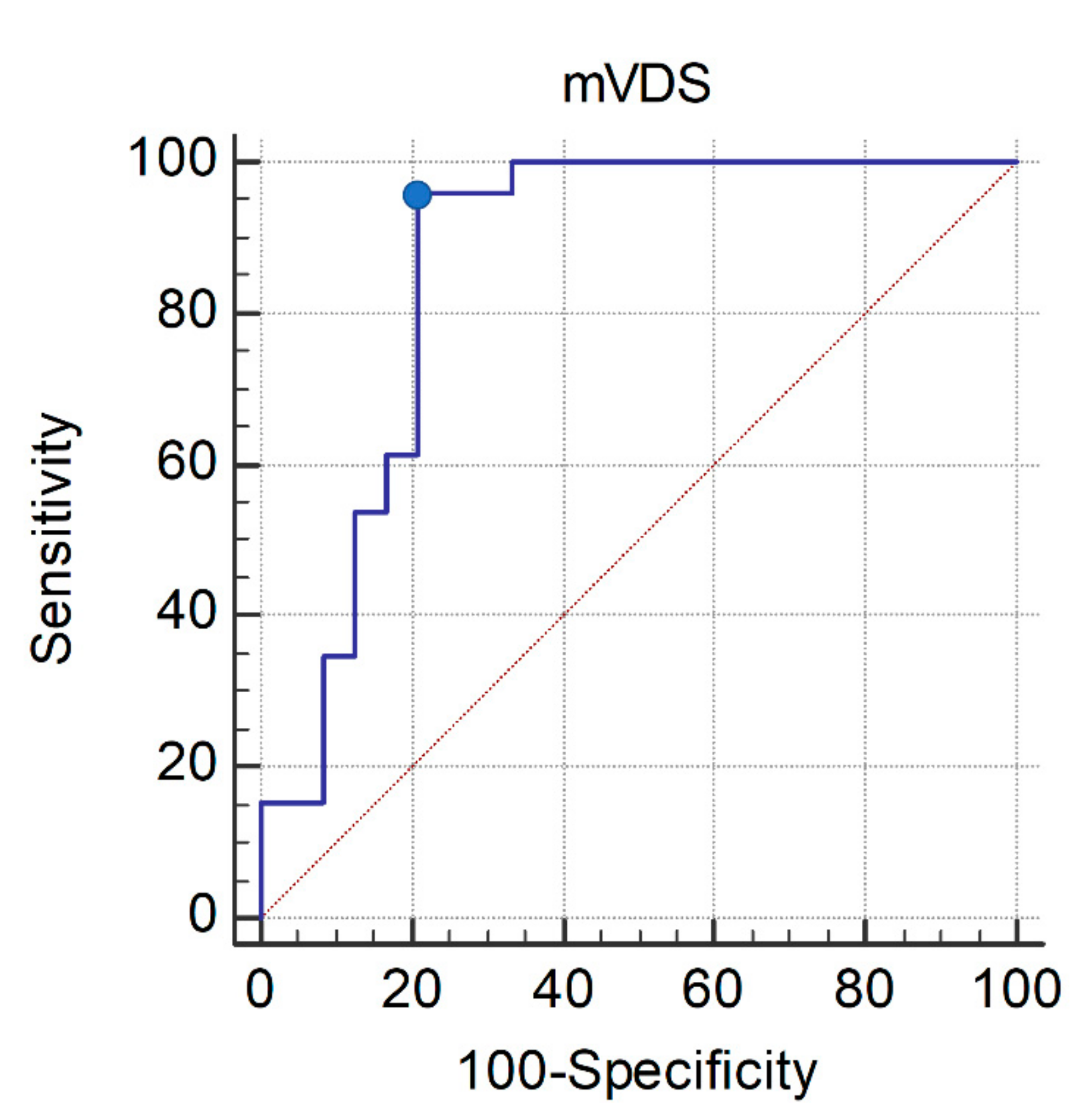
Figure 1. ROC curve of the mVDS score for the allowance of oral feeding in patients with dysphagia due to deconditioning or frailty. The optimal cutoff value (dots on the curves) of the mVDS score, which was obtained from the maximal Youden index, was ≤48 (AUC, 0.862; 95% CI, 0.747–0.978; p < 0.0001; sensitivity, 96.15%; specificity, 79.17%). ROC, receiver operating characteristic; AUC, area under the receiver operating characteristic curve; CI, confidence interval; mVDS, modified version of the videofluoroscopic dysphagia scale.
Healthcare 10 00668 g001
Table 1. Modified version of the videofluoroscopic dysphagia scale.
ParametersScore
lip closureintact/not intact0/6
massificationpossible/not possible0/11.5
oral transit time≤1.5 s/>1.5 s0/4
triggering pharyngeal swallow (swallowing reflex)intact/delayed0/7
epiglottis inversionyes/no0/13
valleculae residue0%/<10%/≥10%, <50%/≥50%0/3/6/9
pyriformis residue0%/<10%/≥10%, <50%/≥50%0/6.5/13/19.5
pharyngeal wall coatingno/yes0/13
aspirationintact/penetration/aspiration0/8.5/17
total score 100
Table 2. Characteristics of patients with dysphagia due to deconditioning or frailty in the present study.
CharacteristicsMean ± Standard Deviation
(Minimum–Maximum)
age (year)71.26 ± 11.93
sex (male:female)42 (84.0%):8 (16.0%)
Tracheal tube (yes:no)11 (22.0%):39 (78.0%)
Oral feeding: Levin-tube feeding29 (58.0%):21 (42.0%)
History of aspiration pneumonia (yes:no)25 (50.0%):25 (50.0%)
PAS grade4.24 ± 3.24 (0–8)
mVDS scores
 lip closure0.36 ± 1.44 (0–6)
 mastification0.92 ± 3.15 (0–11.5)
 oral transit time1.64 ± 2.04 (0–4)
 triggering pharyngeal swallowing3.87 ± 3.53 (0–7)
 epiglottis inversion2.38 ± 5.13 (0–13)
 valleculae residue5.76 ± 3.20 (0–9)
 pyriformis residue11.28 ± 7.08 (0–19.5)
 pharyngeal wall coating7.50 ± 6.73 (0–13)
 aspiration9.18 ± 7.45 (0–17)
 total score42.46 ± 21.70 (0–82.5)
PAS: penetration–aspiration scale, ALSFRS-R: revised amyotrophic lateral sclerosis functional rating scale, mVDS: modified videofluoroscopic dysphagia scale, MMSE: mini-mental status examination, MBI; modified Bathel Index.
Table 3. Multivariate logistic regression analysis (with the enter method) of the association between the modified version of the videofluoroscopic dysphagia scale scores and the allowance of the oral feeding method.
ParameterBeta CoefficientStandard ErrorOR (95% CI)p-Value
Age−0.0880.0490.916
(0.831–1.008)
0.073
PAS−0.2490.4380.780
(0.331–1.839)
0.570
mVDS score−0.0710.0341.081
(1.031–1.132)
0.020
PAS, penetration–aspiration scale; mVDS, modified version of the videofluoroscopic dysphagia scale; OR, odds ratio; CI, confidence interval.
Publisher’s Note: MDPI stays neutral with regard to jurisdictional claims in published maps and institutional affiliations.