
Spontaneous Pneumo-Mediastinum in a Post-COVID-19 Patient with Systemic Sclerosis

 ,
, 
Abstract
1. Introduction
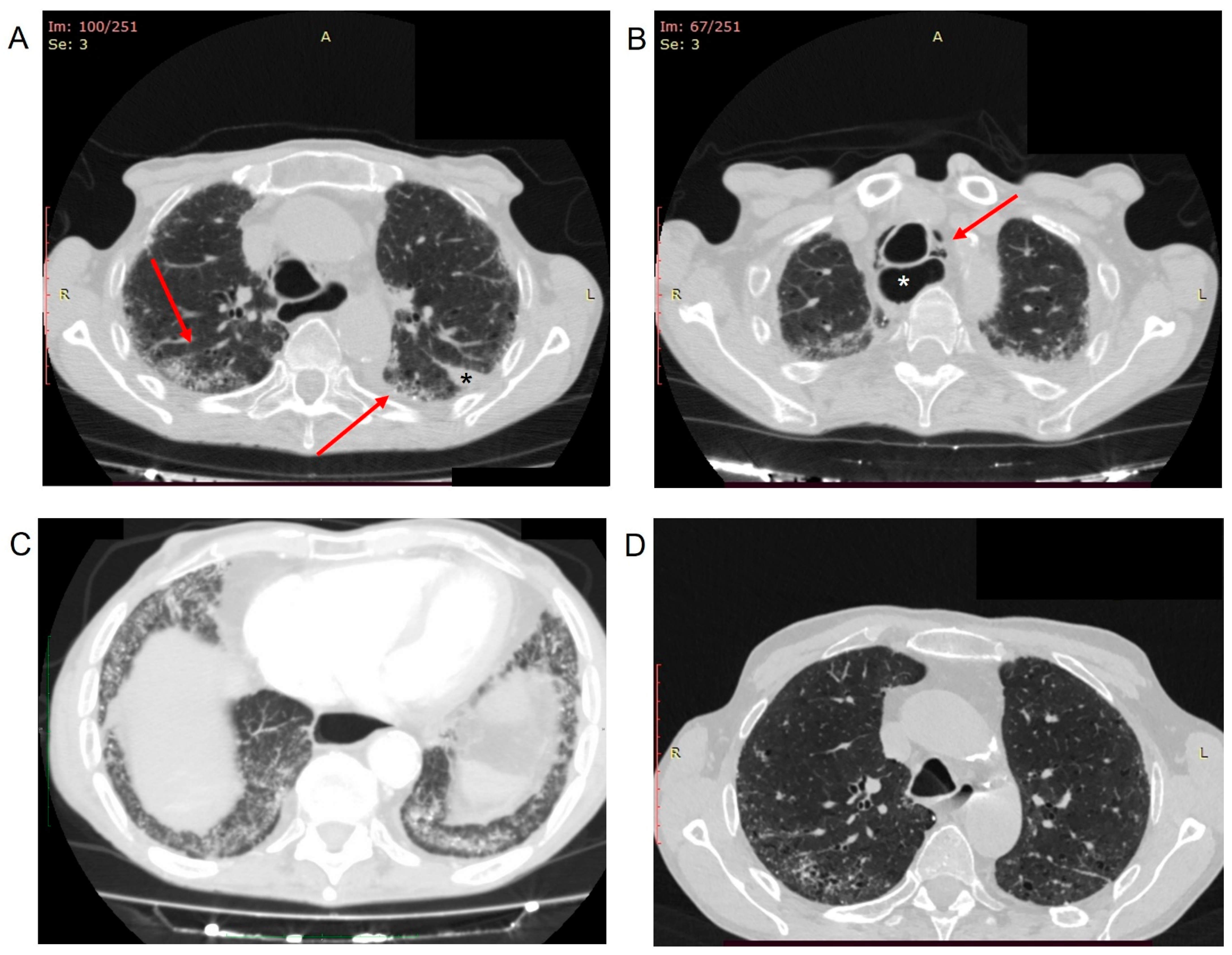
2. Case Description
3. Discussion
4. Conclusions
Author Contributions
Funding
Institutional Review Board Statement
Informed Consent Statement
Data Availability Statement
Acknowledgments
Conflicts of Interest
References
- Doskaliuk, B.; Zaiats, L.; Yatsyshyn, R.; Gerych, P.; Cherniuk, N.; Zimba, O. Pulmonary involvement in systemic sclerosis: Exploring cellular, genetic and epigenetic mechanisms. Rheumatol. Int. 2020, 40, 1555–1569. [Google Scholar] [CrossRef] [PubMed]
- Akter, T.; Silver, R.M.; Bogatkevich, G.S. Recent advances in understanding the pathogenesis of scleroderma-interstitial lung disease. Curr. Rheumatol. Rep. 2014, 16, 411. [Google Scholar] [CrossRef] [PubMed]
- Perelas, A.; Arrossi, A.V.; Highland, K.B. Pulmonary Manifestations of Systemic Sclerosis and Mixed Connective Tissue Disease. Clin. Chest Med. 2019, 40, 501–518. [Google Scholar] [CrossRef]
- Daimon, T.; Johkoh, T.; Honda, O.; Sumikawa, H.; Ichikado, K.; Kondoh, Y.; Taniguchi, H.; Fujimoto, K.; Yanagawa, M.; Inoue, A.; et al. Nonspecific interstitial pneumonia associated with collagen vascular disease: Analysis of CT features to distinguish the various types. Intern. Med. 2009, 48, 753–761. [Google Scholar] [CrossRef] [PubMed][Green Version]
- Okamoto, M.; Fujimoto, K.; Sadohara, J.; Furuya, K.; Kaieda, S.; Miyamura, T.; Suematsu, E.; Kitasato, Y.; Kawayama, T.; Ida, H.; et al. A retrospective cohort study of outcome in systemic sclerosis-associated interstitial lung disease. Respir. Investig. 2016, 54, 445–453. [Google Scholar] [CrossRef] [PubMed]
- Fischer, A.; Swigris, J.J.; Groshong, S.D.; Cool, C.D.; Sahin, H.; Lynch, D.A.; Curran-Everett, D.; Gillis, J.Z.; Meehan, R.T.; Brown, K.K. Clinically significant interstitial lung disease in limited scleroderma: Histopathology, clinical features, and survival. Chest 2008, 134, 601–605. [Google Scholar] [CrossRef] [PubMed]
- Pokeerbux, M.R.; Giovannelli, J.; Dauchet, L.; Mouthon, L.; Agard, C.; Lega, J.C.; Allanore, Y.; Jego, P.; Bienvenu, B.; Berthier, S.; et al. Survival and prognosis factors in systemic sclerosis: Data of a French multicenter cohort, systematic review, and meta-analysis of the literature. Arthritis Res. Ther. 2019, 21, 86. [Google Scholar] [CrossRef]
- Thompson, A.E.; Pope, J.E. A study of the frequency of pericardial and pleural effusions in scleroderma. Br. J. Rheumatol. 1998, 37, 1320–1323. [Google Scholar] [CrossRef]
- Yoon, J.; Finger, D.R.; Pina, J.S. Spontaneous pneumothorax in scleroderma. J. Clin. Rheumatol. 2004, 10, 207–209. [Google Scholar] [CrossRef] [PubMed]
- Dein, E.J.; Lee, K.; Timlin, H.; Hummers, L. Spontaneous pneumomediastinum in limited cutaneous systemic sclerosis and myositis overlap. BMJ Case Rep. 2018, 2018, bcr2018224591. [Google Scholar] [CrossRef]
- Ng, S.C.; Tan, W.C. Bilateral spontaneous pneumothorax in systemic sclerosis--report of two cases. J. Rheumatol. 1990, 17, 689–691. [Google Scholar]
- Matucci-Cerinic, M.; Bruni, C.; Allanore, Y.; Clementi, M.; Dagna, L.; Damjanov, N.S.; de Paulis, A.; Denton, C.P.; Distler, O.; Fox, D.; et al. Systemic sclerosis and the COVID-19 pandemic: World Scleroderma Foundation preliminary advice for patient management. Ann. Rheum. Dis. 2020, 79, 724–726. [Google Scholar] [CrossRef] [PubMed]
- Fang, Y.; Zhang, H.; Xie, J.; Lin, M.; Ying, L.; Pang, P.; Ji, W. Sensitivity of Chest CT for COVID-19: Comparison to RT-PCR. Radiology 2020, 296, E115–E117. [Google Scholar] [CrossRef] [PubMed]
- Ai, T.; Yang, Z.; Hou, H.; Zhan, C.; Chen, C.; Lv, W.; Tao, Q.; Sun, Z.; Xia, L. Correlation of Chest CT and RT-PCR Testing for Coronavirus Disease 2019 (COVID-19) in China: A Report of 1014 Cases. Radiology 2020, 296, E32–E40. [Google Scholar] [CrossRef] [PubMed]
- Prokop, M.; van Everdingen, W.; van Rees Vellinga, T.; Quarles van Ufford, H.; Stoger, L.; Beenen, L.; Geurts, B.; Gietema, H.; Krdzalic, J.; Schaefer-Prokop, C.; et al. CO-RADS: A Categorical CT Assessment Scheme for Patients Suspected of Having COVID-19-Definition and Evaluation. Radiology 2020, 296, E97–E104. [Google Scholar] [CrossRef]
- Fujioka, T.; Takahashi, M.; Mori, M.; Tsuchiya, J.; Yamaga, E.; Horii, T.; Yamada, H.; Kimura, M.; Kimura, K.; Kitazume, Y.; et al. Evaluation of the Usefulness of CO-RADS for Chest CT in Patients Suspected of Having COVID-19. Diagnostics 2020, 10, 608. [Google Scholar] [CrossRef]
- Orlandi, M.; Lepri, G.; Bruni, C.; Wang, Y.; Bartoloni, A.; Zammarchi, L.; Cometi, L.; Guiducci, S.; Matucci-Cerinic, M.; Bellando-Randone, S. The systemic sclerosis patient in the COVID-19 era: The challenging crossroad between immunosuppression, differential diagnosis and long-term psychological distress. Clin. Rheumatol. 2020, 39, 2043–2047. [Google Scholar] [CrossRef] [PubMed]
- Perelas, A.; Silver, R.M.; Arrossi, A.V.; Highland, K.B. Systemic sclerosis-associated interstitial lung disease. Lancet Respir. Med. 2020, 8, 304–320. [Google Scholar] [CrossRef]
- Almeida, M.d.S.T.M.; Dias, L.T.; Fernandes, S.J.; Almeida, J.V. Spontaneous pneumomediastinum and subcutaneous emphysema in systemic sclerosis. Rheumatol. Int. 2007, 27, 675–677. [Google Scholar] [CrossRef] [PubMed]
- Mohammad, A.; Boon Low, T.; O’Dwyer, D.; McElvaney, G.; Kearns, G. Spontaneous pneumo-mediastinum in systemic sclerosis a case report. Rheumatology 2007, 46, 1376–1377. [Google Scholar] [CrossRef] [PubMed]
- Haroon, M.; McLaughlin, P.; Henry, M.; Harney, S. Spontaneous pneumomediastinum in a patient with anti-centromere antibody-positive limited scleroderma. J. Clin. Rheumatol. 2011, 17, 42–43. [Google Scholar] [CrossRef] [PubMed]
- Jun, J.B.; Song, S.Y. The development of pneumomediastinum after pulmonary function testing in a patient with systemic sclerosis. Rheumatol. Int. 2007, 27, 1097–1098. [Google Scholar] [CrossRef] [PubMed]
- Honne, K.; Maruyama, A.; Onishi, S.; Nagashima, T.; Minota, S. Simultaneous pneumatosis cystoides intestinalis and pneumomediastinum in a patient with systemic sclerosis. J. Rheumatol. 2010, 37, 2194–2195. [Google Scholar] [CrossRef]
- Reinig, J.W. Esophagopericardial fistula in a scleroderma patient with peptic esophagitis. Arch. Intern. Med. 1983, 143, 1486–1487. [Google Scholar] [CrossRef] [PubMed]
- Elhakim, T.S.; Abdul, H.S.; Pelaez Romero, C.; Rodriguez-Fuentes, Y. Spontaneous pneumomediastinum, pneumothorax and subcutaneous emphysema in COVID-19 pneumonia: A rare case and literature review. BMJ Case Rep. 2020, 13, e239489. [Google Scholar] [CrossRef] [PubMed]
- Carfi, A.; Bernabei, R.; Landi, F.; Gemelli Against, C.-P.-A.C.S.G. Persistent Symptoms in Patients After Acute COVID-19. JAMA 2020, 324, 603–605. [Google Scholar] [CrossRef]
- Arnold, D.T.; Hamilton, F.W.; Milne, A.; Morley, A.J.; Viner, J.; Attwood, M.; Noel, A.; Gunning, S.; Hatrick, J.; Hamilton, S.; et al. Patient outcomes after hospitalisation with COVID-19 and implications for follow-up: Results from a prospective UK cohort. Thorax 2021, 76, 399–401. [Google Scholar] [CrossRef] [PubMed]
- Munblit, D.; Bobkova, P.; Spiridonova, E.; Shikhaleva, A.; Gamirova, A.; Blyuss, O.; Nekliudov, N.; Bugaeva, P.; Andreeva, M.; DunnGalvin, A.; et al. Incidence and risk factors for persistent symptoms in adults previously hospitalized for COVID-19. Clin. Exp. Allergy 2021, 51, 1107–1120. [Google Scholar] [CrossRef]
- Ejaz, H.; Alsrhani, A.; Zafar, A.; Javed, H.; Junaid, K.; Abdalla, A.E.; Abosalif, K.O.A.; Ahmed, Z.; Younas, S. COVID-19 and comorbidities: Deleterious impact on infected patients. J. Infect. Public Health 2020, 13, 1833–1839. [Google Scholar] [CrossRef] [PubMed]
- Orlandi, M.; Landini, N.; Sambataro, G.; Nardi, C.; Tofani, L.; Bruni, C.; Bellando-Randone, S.; Blagojevic, J.; Melchiorre, D.; Hughes, M.; et al. The Role of Chest Ct in Deciphering Interstitial Lung Involvement: Systemic Sclerosis Versus COVID-19. Rheumatology 2021, keab615. [Google Scholar] [CrossRef] [PubMed]
- Rodriguez-Morales, A.J.; Cardona-Ospina, J.A.; Gutierrez-Ocampo, E.; Villamizar-Pena, R.; Holguin-Rivera, Y.; Escalera-Antezana, J.P.; Alvarado-Arnez, L.E.; Bonilla-Aldana, D.K.; Franco-Paredes, C.; Henao-Martinez, A.F.; et al. Clinical, laboratory and imaging features of COVID-19: A systematic review and meta-analysis. Travel Med. Infect. Dis. 2020, 34, 101623. [Google Scholar] [CrossRef] [PubMed]
- Yang, J.; Zheng, Y.; Gou, X.; Pu, K.; Chen, Z.; Guo, Q.; Ji, R.; Wang, H.; Wang, Y.; Zhou, Y. Prevalence of comorbidities and its effects in patients infected with SARS-CoV-2: A systematic review and meta-analysis. Int. J. Infect. Dis. 2020, 94, 91–95. [Google Scholar] [CrossRef] [PubMed]
- Chen, N.; Zhou, M.; Dong, X.; Qu, J.; Gong, F.; Han, Y.; Qiu, Y.; Wang, J.; Liu, Y.; Wei, Y.; et al. Epidemiological and clinical characteristics of 99 cases of 2019 novel coronavirus pneumonia in Wuhan, China: A descriptive study. Lancet 2020, 395, 507–513. [Google Scholar] [CrossRef]
- Ferri, C.; Giuggioli, D.; Raimondo, V.; L’Andolina, M.; Tavoni, A.; Cecchetti, R.; Guiducci, S.; Ursini, F.; Caminiti, M.; Varcasia, G.; et al. COVID-19 and rheumatic autoimmune systemic diseases: Report of a large Italian patients series. Clin. Rheumatol. 2020, 39, 3195–3204. [Google Scholar] [CrossRef]
- Fineschi, S. Case Report: Systemic Sclerosis After COVID-19 Infection. Front. Immunol. 2021, 12, 686699. [Google Scholar] [CrossRef] [PubMed]
- Bonometti, R.; Sacchi, M.C.; Stobbione, P.; Lauritano, E.C.; Tamiazzo, S.; Marchegiani, A.; Novara, E.; Molinaro, E.; Benedetti, I.; Massone, L.; et al. The first case of systemic lupus erythematosus (SLE) triggered by COVID-19 infection. Eur. Rev. Med. Pharmacol. Sci. 2020, 24, 9695–9697. [Google Scholar] [CrossRef]
- Cardoso, E.M.; Hundal, J.; Feterman, D.; Magaldi, J. Concomitant new diagnosis of systemic lupus erythematosus and COVID-19 with possible antiphospholipid syndrome. Just a coincidence? A case report and review of intertwining pathophysiology. Clin. Rheumatol. 2020, 39, 2811–2815. [Google Scholar] [CrossRef] [PubMed]
- Gracia-Ramos, A.E.; Saavedra-Salinas, M.A. Can the SARS-CoV-2 infection trigger systemic lupus erythematosus? A case-based review. Rheumatol. Int. 2021, 41, 799–809. [Google Scholar] [CrossRef]
- Sedaghat, Z.; Karimi, N. Guillain Barre syndrome associated with COVID-19 infection: A case report. J. Clin. Neurosci. 2020, 76, 233–235. [Google Scholar] [CrossRef] [PubMed]
- Fucikova, J.; Palova-Jelinkova, L.; Bartunkova, J.; Spisek, R. Induction of Tolerance and Immunity by Dendritic Cells: Mechanisms and Clinical Applications. Front. Immunol. 2019, 10, 2393. [Google Scholar] [CrossRef]


| References | Total SSc Patients (n) | Cause | Outcome |
|---|---|---|---|
| [10] | 1 a | Spontaneous | Progressive hypoxia requiring intubation and complicated by Klebsiella pneumonia and renal failure |
| [19] | 1 | Spontaneous | Resolution with conservative treatment strategy |
| [23] | 1 | Spontaneous | Resolution with conservative treatment strategy |
| [22] | 1 | Pulmonary function testing | Resolution with conservative treatment strategy |
| [20] | 1 | Spontaneous | Resolution with conservative treatment strategy |
| [21] | 1 | Spontaneous | Resolution with conservative treatment strategy |
| Present case | 1 | Spontaneous | Resolution with conservative treatment strategy |
Publisher’s Note: MDPI stays neutral with regard to jurisdictional claims in published maps and institutional affiliations. |
© 2022 by the authors. Licensee MDPI, Basel, Switzerland. This article is an open access article distributed under the terms and conditions of the Creative Commons Attribution (CC BY) license (https://creativecommons.org/licenses/by/4.0/).
Share and Cite
Mormile, I.; Mormile, M.; Rea, G.; Petraroli, A.; Barbieri, V.; de Paulis, A.; Rossi, F.W. Spontaneous Pneumo-Mediastinum in a Post-COVID-19 Patient with Systemic Sclerosis. Healthcare 2022, 10, 529. https://doi.org/10.3390/healthcare10030529
Mormile I, Mormile M, Rea G, Petraroli A, Barbieri V, de Paulis A, Rossi FW. Spontaneous Pneumo-Mediastinum in a Post-COVID-19 Patient with Systemic Sclerosis. Healthcare. 2022; 10(3):529. https://doi.org/10.3390/healthcare10030529
Chicago/Turabian StyleMormile, Ilaria, Mauro Mormile, Gaetano Rea, Angelica Petraroli, Vittoria Barbieri, Amato de Paulis, and Francesca Wanda Rossi. 2022. "Spontaneous Pneumo-Mediastinum in a Post-COVID-19 Patient with Systemic Sclerosis" Healthcare 10, no. 3: 529. https://doi.org/10.3390/healthcare10030529
APA StyleMormile, I., Mormile, M., Rea, G., Petraroli, A., Barbieri, V., de Paulis, A., & Rossi, F. W. (2022). Spontaneous Pneumo-Mediastinum in a Post-COVID-19 Patient with Systemic Sclerosis. Healthcare, 10(3), 529. https://doi.org/10.3390/healthcare10030529