
Comparison of Jaw Mode and Field Width for Left-Breast Cancer Using TomoDirect Three-Dimensional Conformal Radiation Therapy: A Phantom Study

 , and
, and
Abstract
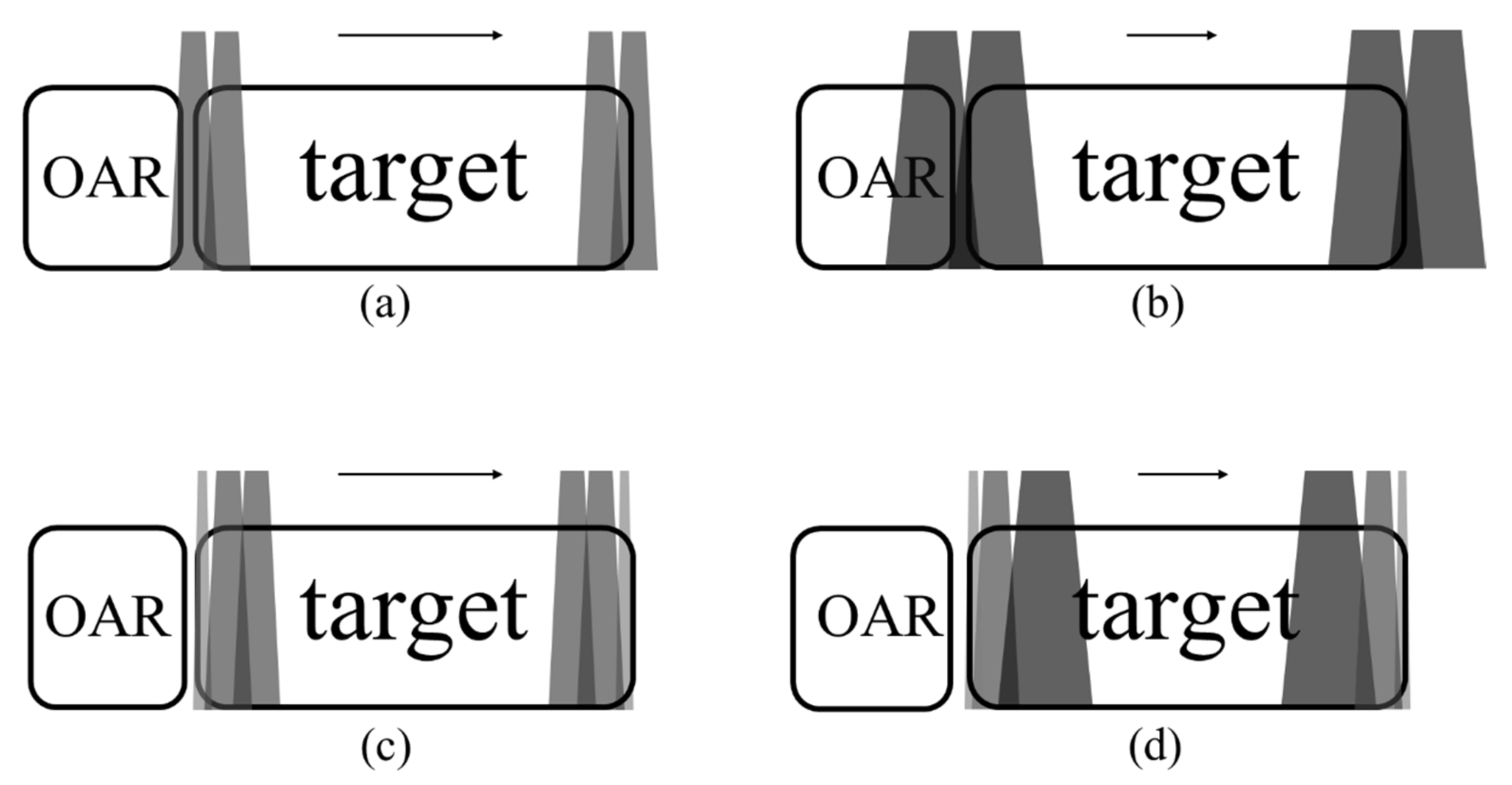
1. Introduction
2. Materials and Methods
2.1. Phantom and CT Scan Images
2.2. Treatment Planning
2.3. Evaluation
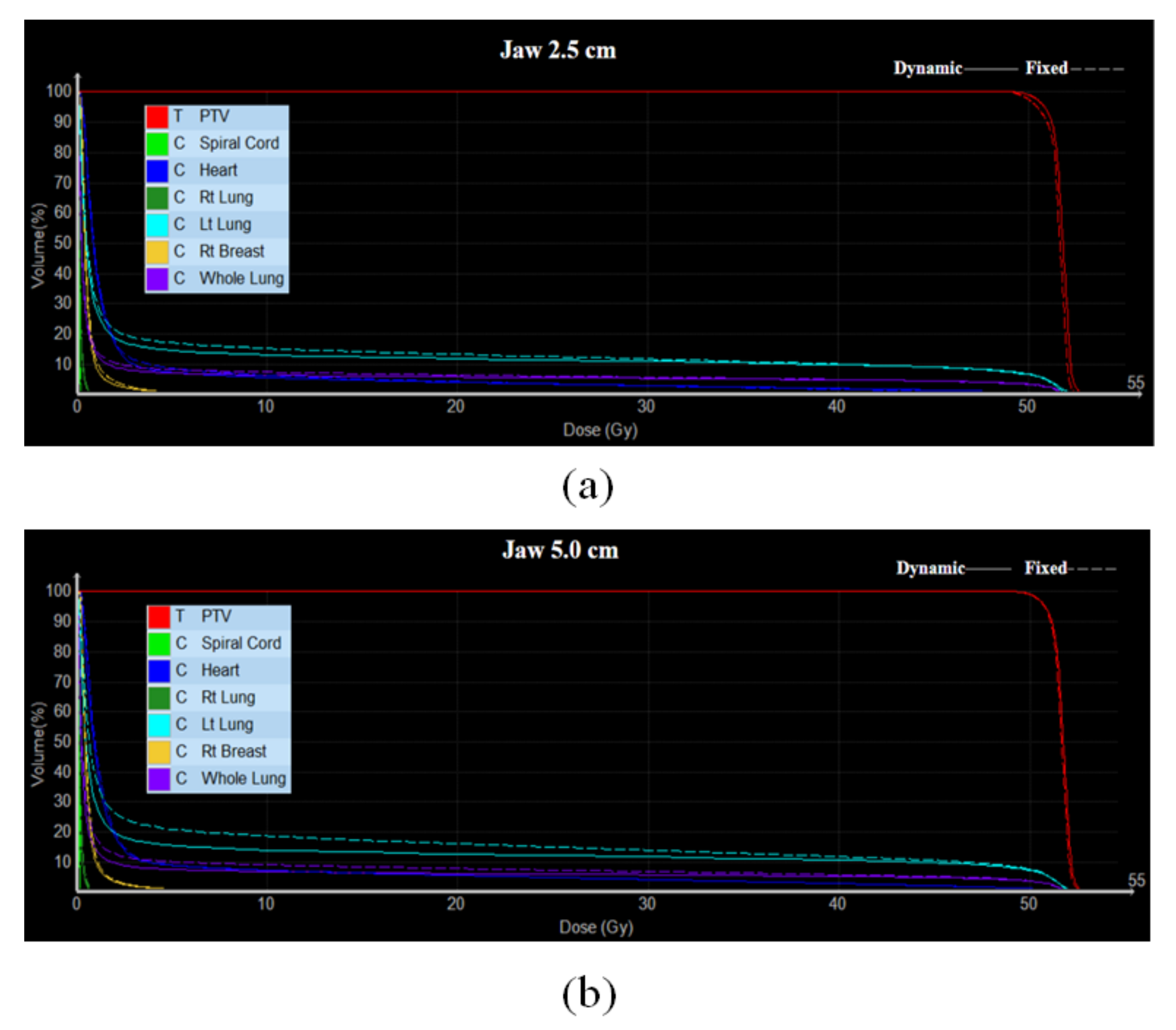
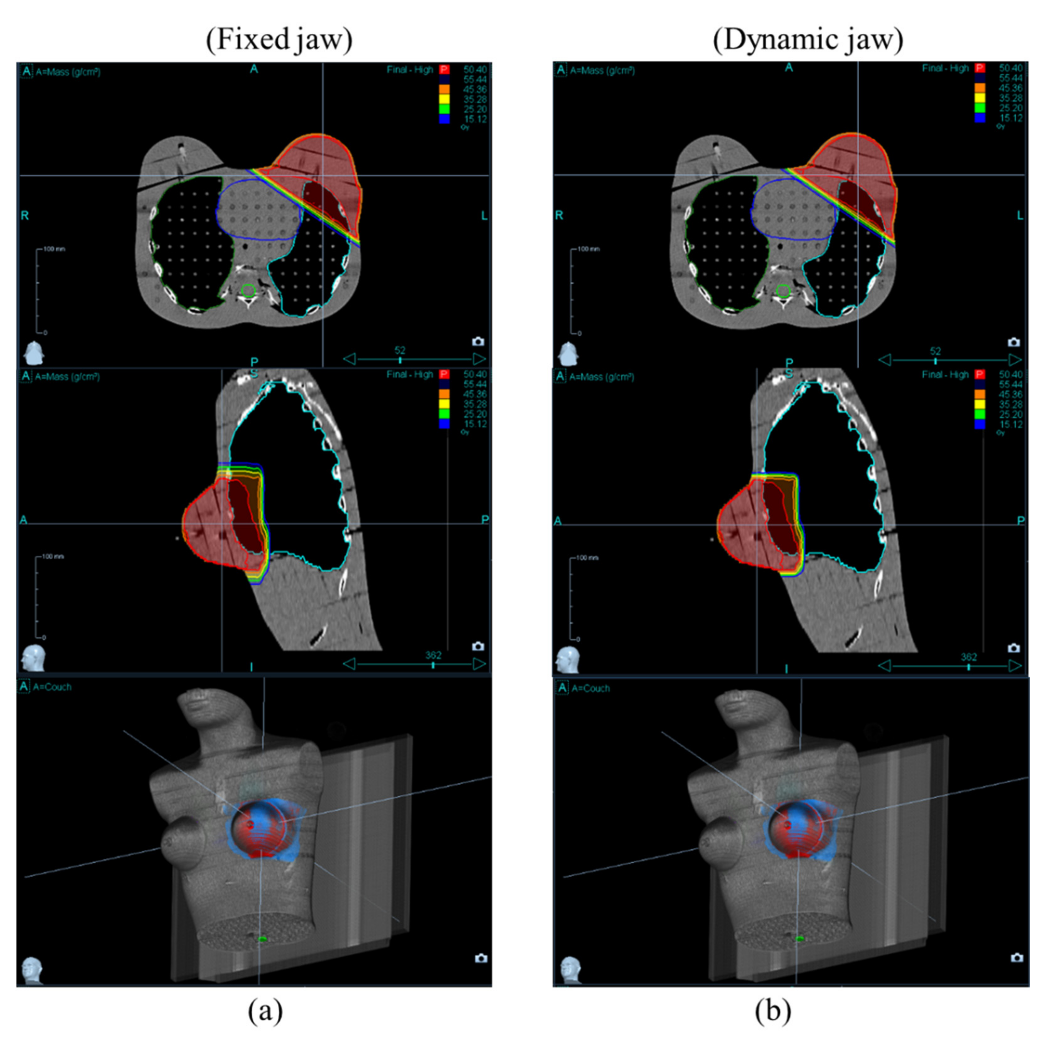
3. Results and Discussion
3.1. Target Coverage
3.2. OARs Dose
4. Conclusions
Author Contributions
Funding
Institutional Review Board Statement
Informed Consent Statement
Data Availability Statement
Conflicts of Interest
References
- Chae, S.M.; Lee, K.W.; Son, S.H. Dosimetric impact of multileaf collimator leaf width according to sophisticated grade of technique in the IMRT and VMAT planning for pituitary adenoma lesion. Oncotarget 2016, 7, 78119–78126. [Google Scholar] [CrossRef] [PubMed][Green Version]
- Bedford, J.L.; Warrington, A.P. Commissioning of volumetric modulated arc therapy (VMAT). Int. J. Radiat. Oncol. Biol. Phys. 2009, 73, 537–545. [Google Scholar] [CrossRef] [PubMed]
- Palma, D.A.; Verbakel, W.F.A.R.; Otto, K.; Senan, S. New developments in arc radiation therapy: A review. Cancer Treat. Rev. 2010, 36, 393–399. [Google Scholar] [CrossRef]
- Srivastava, R.P.; Vandeputte, K.; Wagter, C.D. Benefits and limitations of volumetric modulated arc therapy in treating bilateral breast cancer with regional lymph nodes. Adv. Breast Cancer Res. 2020, 9, 119–126. [Google Scholar] [CrossRef]
- Jabbari, K.; Akbari, M.; Tavakoli, M.B.; Amougeidari, A. Dosimetry and evaluating the effect of treatment parameters on the leakage of multi leaf collimators in ONCOR linear accelerators. Adv. Biomed. Res. 2016, 5, 193. [Google Scholar] [CrossRef] [PubMed]
- Takano, S.; Omura, M.; Suzuki, R.; Tayama, Y.; Matsui, K.; Hashimoto, H.; Hongo, H.; Nagata, H.; Tanaka, K.; Hata, M.; et al. Intensity-modulated radiation therapy using tomodirect for postoperative radiation of left-sided breast cancer including lymph node area: Comparison with tomohelical and three-dimensional conformal radiation therapy. J. Radiat. Res. 2019, 60, 694–704. [Google Scholar] [CrossRef]
- Squires, M.; Hu, Y.; Byrne, M.; Archibald-Heeren, B.; Cheers, S.; Bosco, B.; Teh, A.; Fong, A. Static beam tomotherapy as an optimisation method in whole-breast radiation therapy (WBRT). J. Med. Radiat. Sci. 2017, 64, 281–289. [Google Scholar] [CrossRef] [PubMed]
- Aras, S.; İkizceli, T.; Aktan, M. Dosimetric comparison of three-dimensional conformal radiotherapy (3D-CRT) and intensity modulated radiotherapy techniques (IMRT) with radiotherapy dose simulations for left-sided mastectomy patients. Eur. J. Breast Health 2019, 15, 85–89. [Google Scholar] [CrossRef] [PubMed]
- Ferini, G.; Molino, L.; Tipoli, A.; Valenti, V.; Illari, S.I.; Marchese, V.A.; Carvagno, I.R.; Borzi, G.R. Anatomical predictors of dosimetric advantages for deep-inspiration-breast-hold 3D-conformal radiotherapy among women with left breast cancer. Anticancer Res. 2021, 41, 1529–1538. [Google Scholar] [CrossRef] [PubMed]
- Ferini, G.; Valenti, V.; Viola, A.; Umana, G.E.; Martorana, E. A critical overview of predictors of heart sparing by deep-inspiration-breast-hold irradiation in left-sided breast cancer patients. Cancers 2022, 14, 3477. [Google Scholar] [CrossRef] [PubMed]
- Jiao, S.X.; Wang, M.I.; Chen, L.X.; Liu, X.W. Evaluation of dose-volume histogram prediction for organ-at risk and planning target volume based on machine learning. Sci. Rep. 2021, 11, 3117. [Google Scholar] [CrossRef] [PubMed]
- Lucia, F.; Bourbonne, V.; Visvikis, D.; Miranda, O.; Gujrral, D.M.; Gouders, D.; Dissaux, G.; Pradier, O.; Tixier, F.; Jaouen, V.; et al. Radiomics Analysis of 3D Dose Distributions to Predict Toxicity of Radiotherapy for Cervical Cancer. J. Personal. Med. 2021, 11, 398. [Google Scholar] [CrossRef] [PubMed]
- Thomas, T.O.; Refaat, T.; Choi, M.; Bacchus, I.; Sachdev, S.; Rademaker, A.W.; Sathiaseelan, V.; Karagianis, A.; Mittal, B.B. Brachial plexus dose tolerance in head and neck cancer patients treated with sequential intensity modulated radiation therapy. Radiat. Oncol. 2015, 10, 94. [Google Scholar] [CrossRef] [PubMed]
- Rudofsky, L.; Aynsley, E.; Beck, S.; Schuber, K.; Habl, G.; Krause, S.; Debus, J.; Sterzing, F. Lung and liver SBRT using helical tomotherapy—A dosimetric comparison of fixed jaw and dynamic jaw delivery. J. Appl. Clin. Med. Phys. 2014, 15, 114–121. [Google Scholar] [CrossRef] [PubMed]
- Drzymala, R.E.; Mohan, R.; Brewster, L.; Chu, J.; Goiteion, M.; Harms, W.; Urie, M. Dose-volume histograms. Int. J. Radiat. Oncol. Biol. Phys. 1991, 21, 71–78. [Google Scholar] [CrossRef] [PubMed]
- Montovano, M.; Zhang, M.; Oh, P.; Thor, M.; Crane, C.; Yorke, E.; Wu, A.J.; Jackson, A. Incidence and dosimetric predictors of radiation-induced gastric bleeding after chemoradiation for esophageal and gastroesophageal junction cancer. Adv. Radiat. Oncol. 2021, 6, 100648. [Google Scholar] [CrossRef] [PubMed]
- Boerma, M.; Sridharan, V.; Mao, X.; Nelson, G.A.; Cheema, A.K.; Koturbash, I.; Singh, S.P.; Tackett, A.J.; Hauer-Jensen, M. Effects of ionizing radiation on the heart. Mutat. Res. Rev. Mutat. Res. 2016, 770 (Pt B), 319–327. [Google Scholar] [CrossRef]
- Darby, S.C.; Ewertz, M.; McGale, P.; Bennet, A.M.; Blom-Goldman, U.; Brønnum, D.; Correa, C.; Cutter, D.; Gagliardi, G.; Gigante, B.; et al. Risk of ischemic heart disease in women after radiotherapy for breast cancer. N. Engl. J. Med. 2013, 368, 987–998. [Google Scholar] [CrossRef] [PubMed]
- Sugie, C.; Manabe, Y.; Hayashi, A.; Murai, T.; Takaoka, T.; Hattori, Y.; Iwata, H.; Takenaka, R.; Shibamoto, Y. Efficacy of the dynamic jaw mode in helical tomotherapy with static ports for breast cancer. Technol. Cancer Res. Treat. 2015, 14, 459–465. [Google Scholar] [CrossRef] [PubMed]


| Field Width (2.5 cm) | Field Width (5.0 cm) | |||
|---|---|---|---|---|
| Index | Fixed Jaw | Dynamic Jaw | Fixed Jaw | Dynamic Jaw |
| CI | 1.58 ± 0.06 | 1.56 ± 0.06 | 1.65 ± 0.06 | 1.61 ± 0.07 |
| HI | 1.07 ± 0.02 | 1.07 ± 0.01 | 1.07 ± 0.02 | 1.06 ± 0.01 |
| Beam on time (s) | 111.27 ± 2.28 | 125.64 ± 1.03 | 67.01 ± 1.08 | 73.19 ± 0.97 |
| OAR | Field Width (2.5 cm) | Field Width (5.0 cm) | ||
|---|---|---|---|---|
| Fixed Jaw | Dynamic Jaw | Fixed Jaw | Dynamic Jaw | |
| Heart | 2.72 ± 0.29 | 2.69 ± 0.29 | 3.43 ± 0.22 | 3.36 ± 0.22 |
| Right lung | 0.18 ± 0.01 | 0.16 ± 0.00 | 0.21 ± 0.01 | 0.17 ± 0.01 |
| Left lung | 6.22 ± 1.72 | 6.13 ± 0.28 | 8.06 ± 0.30 | 6.59 ± 0.34 |
| Right breast | 0.61 ± 0.07 | 0.59 ± 0.06 | 0.69 ± 0.09 | 0.63 ± 0.08 |
| Whole lungs | 3.26 ± 0.12 | 2.99 ± 0.13 | 3.93 ± 0.14 | 3.21 ± 0.16 |
Publisher’s Note: MDPI stays neutral with regard to jurisdictional claims in published maps and institutional affiliations. |
© 2022 by the authors. Licensee MDPI, Basel, Switzerland. This article is an open access article distributed under the terms and conditions of the Creative Commons Attribution (CC BY) license (https://creativecommons.org/licenses/by/4.0/).
Share and Cite
Kim, H.; Jung, J.; Jung, H.; Jeong, J.; Lee, D.; Jeong, H.-W.; Lee, Y. Comparison of Jaw Mode and Field Width for Left-Breast Cancer Using TomoDirect Three-Dimensional Conformal Radiation Therapy: A Phantom Study. Healthcare 2022, 10, 2431. https://doi.org/10.3390/healthcare10122431
Kim H, Jung J, Jung H, Jeong J, Lee D, Jeong H-W, Lee Y. Comparison of Jaw Mode and Field Width for Left-Breast Cancer Using TomoDirect Three-Dimensional Conformal Radiation Therapy: A Phantom Study. Healthcare. 2022; 10(12):2431. https://doi.org/10.3390/healthcare10122431
Chicago/Turabian StyleKim, Haneul, Jaehong Jung, Hyunseo Jung, Jibeom Jeong, Dohwa Lee, Hyun-Woo Jeong, and Youngjin Lee. 2022. "Comparison of Jaw Mode and Field Width for Left-Breast Cancer Using TomoDirect Three-Dimensional Conformal Radiation Therapy: A Phantom Study" Healthcare 10, no. 12: 2431. https://doi.org/10.3390/healthcare10122431
APA StyleKim, H., Jung, J., Jung, H., Jeong, J., Lee, D., Jeong, H.-W., & Lee, Y. (2022). Comparison of Jaw Mode and Field Width for Left-Breast Cancer Using TomoDirect Three-Dimensional Conformal Radiation Therapy: A Phantom Study. Healthcare, 10(12), 2431. https://doi.org/10.3390/healthcare10122431