Article

Differential Mechanical and Biological Contributions to Bone Mass Distribution—Insights from a Computational Model of the Human Femur

1
Grupo Biomecánica Computacional, Facultad de Ingeniería, Universidad Nacional de Entre Ríos, Ruta 10, Km 11, Oro Verde 3100, Argentina
2
Departamento Ingeniería Industrial, Facultad Regional Santa Fe, Universidad Tecnológica Nacional, Lavaisse 610, Santa Fe 3000, Argentina
3
Multiscale in Mechanical & Biological Engineering Research Group, Aragon Institute of Engineering Research (I3A), School of Engineering and Architecture, University of Zaragoza, 50009 Zaragoza, Spain
4
Centro Universitario de la Defensa de Zaragoza, 50090 Zaragoza, Spain
*
Author to whom correspondence should be addressed.
Mathematics 2025, 13(13), 2156; https://doi.org/10.3390/math13132156
Submission received: 2 May 2025 / Revised: 25 June 2025 / Accepted: 26 June 2025 / Published: 30 June 2025
(This article belongs to the Special Issue Advances in Biological Systems with Mathematics)

Abstract

Bone density distribution in the human femur is significantly influenced by mechanical forces that drive bone remodeling in response to physical demands. This study aims to assess how effectively mechanical factors alone explain femoral bone mass distribution and to identify areas where additional, non-mechanical influences may be required. We used a computational bone remodeling model to compare outcomes under two initial conditions: a uniform density distribution and one derived from tomographic imaging. Both conditions experienced identical mechanical loading, with the remodeling process simulated via finite element methods. Results demonstrated that mechanical loading substantially contributes to shaping bone density, but certain structural aspects, notably incomplete cortical bone formation in simulations starting from uniform density, suggest the involvement of other factors. The model also highlighted specific regions susceptible to bone loss under disuse scenarios, such as prolonged inactivity or microgravity. Our findings emphasize the need to incorporate non-mechanical factors and realistic initial conditions into computational models to enhance their applicability for personalized medical analyses.
MSC:
74-10; 92-10

1. Introduction

Bone remodeling is a dynamic process in which old bone tissue is replaced by new tissue, enabling the maintenance of structural integrity and adaptation to mechanical demands [1,2]. This process is primarily regulated by the coupled activity of osteoblasts and osteoclasts, which are influenced by mechanical loading, hormonal signaling, and cellular-level factors. Among the various bones in the human skeleton, the femur stands out for its key role in weight-bearing and locomotion, as well as its complex internal structure that reflects a fine balance between mechanical efficiency and biological regulation. Understanding the factors that govern femoral bone mass distribution is important for addressing clinical challenges such as osteoporosis, fractures, integration of orthopedic implants, prolonged periods of prostration, or the effects of microgravity during space travel.
Computational modeling has become central to the investigation of bone adaptation under varying mechanical and biological conditions. Most studies rely on two main initialization strategies to generate the adult human bone density distribution from which simulations typically begin: (1) a homogeneous density distribution and (2) CT-based initialization.
The homogeneous approach has been widely adopted in both fundamental research and clinical applications due to its simplicity and flexibility. Early mechanobiological models, such as that of Beaupré et al. [3], demonstrated that realistic femoral density patterns can emerge from homogeneous, isotropic initial states, reinforcing the explanatory power of load-driven remodeling frameworks. Building on this, Coelho et al. [4] introduced a multiscale model in which isotropic microscale material behavior gives rise to anisotropic macroscopic responses through the optimization of trabecular microstructure under local mechanical stress, effectively reproducing key features of femoral architecture. Subsequent developments incorporated additional physiological complexity, such as material anisotropy and microdamage accumulation and repair mechanisms (e.g., Doblaré and García-Aznar [5,6]; García-Aznar et al. [7]; Martínez-Reina et al. [8]). Martínez-Reina et al. [9] also addressed theoretical aspects such as the uniqueness and stability of remodeling solutions. Their results indicated that, starting from a uniform density distribution, convergence to a consistent final state required eliminating the so-called dead zone in the bone balance function (i.e., a range of mechanical stimuli that elicit no remodeling response), as well as imposing constraints on bone formation and resorption rates. However, their simulations used initial densities of 0.7 g/cm3 and 0.3 g/cm3, which are near typical cortical and trabecular thresholds, respectively. In contrast, Berli [10] showed that initializing with homogeneous densities above 0.9 g/cm3 leads to similar final bone mass and density distributions, suggesting that remodeling outcomes are relatively insensitive to the precise initial value within this range. Moreover, for these initial density values, the bone mass remains within a range consistent with measurements obtained from actual human specimens. More recent models have integrated cellular-level mechanisms into the remodeling framework, particularly focusing on basic multicellular units (BMUs) and the concept of focal bone balance. For instance, Berli et al. [11] developed a three-dimensional model coupling BMU activity to mechanical stress and local inhibitory signals, offering a more biologically grounded representation of bone adaptation. Similarly, Franco et al. [12] explored the effects of aging and hormonal changes on remodeling dynamics, emphasizing the interaction between mechanical and systemic regulatory factors.
The second major strategy (CT-based finite element (FE) initialization) has enabled subject-specific simulations that incorporate heterogeneous bone geometry and density distributions. For example, Bahia et al. [13] used CT-based modeling to develop accurate subject-specific finite element (FE) models that capture both anatomical geometry and spatial heterogeneity of bone material properties. This modeling approach enables simulations of structural behavior and adaptive bone remodeling, making it a valuable tool for applications such as fracture risk assessment and treatment planning. Bansod et al. [14] performed a finite element analysis of strain-adaptive bone remodeling in the human femur using CT-based geometry within an open-source framework. The simulations, initialized with homogeneous density, assessed both mechanical and piezoelectric effects. The results reproduced realistic density patterns and highlighted the potential of electrical stimulation to mitigate bone loss. In this work, they compared final results from different initial homogeneous density values with densities from CT, showing that there is convergence of densities in regions of high strain and moderate divergence in other areas of a coronal plane. These studies, among others [15,16,17,18], demonstrate how image-based reconstructions derived from CT scans are commonly used to initialize finite element simulations of bone remodeling, enabling anatomically and mechanically realistic predictions.
Despite their widespread use, systematic comparisons between these initialization strategies remain limited. Uniform-density initialization usually relies solely on mechanical cues to generate bone structure, while CT-based approaches incorporate intrinsic tissue heterogeneity and the complexity of pre-existing density distributions. A direct comparison is essential to assess the implications of each strategy for predictive accuracy and clinical applicability in subject-specific models of femoral remodeling.
Our study investigates whether mechanical loading alone can reproduce the complex bone density patterns observed in both the coronal and sagittal planes of the femur, as most previous studies have focused primarily on the coronal plane. This underscores the need to clarify the role of mechanical stimuli in shaping the internal structure of adult bone and to identify anatomical regions where additional biological or environmental factors, as proposed by García-Aznar et al. [7], may be necessary.

2. Materials and Methods

Although the bone remodeling process can be influenced by biological factors, mechanical factors, or their combined interaction, this study focuses exclusively on the effects of mechanical loads. The numerical model employed comprises two interconnected components, linked through time-evolving mechanical variables. The first component provides a mathematical representation of the biological process underlying bone remodeling, while the second addresses mechanical aspects, including the geometry, constraints, and applied loads on the femur. Both components are outlined briefly below, with further details available in our previous works [8,9,11,19].

2.1. Femur Geometry

The geometry of the femur was reconstructed from computed tomography (CT) images using 3D Slicer, a free and open-source software platform for visualizing and segmenting medical images. A CT scan of one patient’s femur was obtained from the Nuclear and Molecular Medicine Center Foundation of Entre Ríos (CEMENER, E.R., Argentina), with full patient consent and strict adherence to data anonymity protocols. To generate an initial, non-optimized geometry, the TotalSegmentator module of the 3D Slicer software, version 5.8.1 [20], an artificial intelligence-based method, was employed. This method is highly efficient and generates a preliminary bone geometry, which serves as the foundation for subsequent smoothing and refinement. Subsequently, a manual refinement process is required to optimize the bone reconstruction and exclude certain portions of adjacent tissues. Finally, the external and internal geometric reconstruction of the bone is completed, without incorporating information about its internal material or mechanical properties. This process is performed using the techniques described in Section 2.4.

2.2. Bone Remodeling Model

Bone is composed of a solid matrix and pores (see Figure 1). The solid matrix consists of an organic matrix, water, and mineral content, while the pores contain bone marrow, blood vessels, and nerves. In this study, it is assumed that the pores have no mechanical influence compared to the stiffness of the solid matrix. Thus, a bone sample of volume V t can be divided into a matrix tissue volume V b and an empty pore volume V p . Furthermore, the volume occupied by the solid matrix can be subdivided into a mineral phase ( V m ), an organic phase ( V o ), and water ( V w ). Predicting the evolution of each subvolume in response to mechanical loads is the main objective of the computational algorithm, maintaining the following equality:
V t = V b ( t ) + V p ( t ) = V m ( t ) + V o ( t ) + V w ( t ) + V p ( t )
Figure 1. Schematic illustration of the multiscale modeling framework for bone density adaptation in the human femur. At the macro scale, a mechanical problem is solved using FEM, considering patient-specific femoral geometry and initial bone density distribution. Mechanical loading conditions (top left, red arrows indicate walking and stair-climbing forces) correspond to those detailed numerically in Table 1. At the micro scale, the mechanical stimuli (strain) initiate biological signals, activating the BMUs to regulate local resorption and formation processes. The inset (top right) highlights the trabecular bone microstructure, showing tissue volume fractions (Vo: organic, Vm: mineralized bone, Vw: water). The iterative process continues until convergence is achieved.
Mathematics 13 02156 g001
The computational tracking of each subvolume requires the definition of variables in a relative form, that is,
v b ( t ) = V b ( t ) V t ; v m ( t ) = V m ( t ) V b ( t ) v o ( t ) = V o ( t ) V b ( t ) ; v w ( t ) = V w ( t ) V b ( t )
In Equation (2), it is assumed that v o has a constant value of 3/7 [2,11], while water is replaced in the mineralization. Thus, the following sum holds:
v m ( t ) + v o + v w ( t ) = 1
Equations (1)–(3) define spatially varying differential volumes, assuming that bone behaves as a continuous material with gradually changing properties. This approach is consistent with the characterization of bone as a functionally graded material (FGM), whose composition and mechanical properties vary smoothly throughout its structure [22]. Within this framework, volume fractions are defined in the infinitesimal limit (as the differential volume approaches zero), enabling the model to capture the continuous spatial heterogeneity of bone tissue.
Once the subvolumes are determined, the density of the solid tissue (material density), excluding pores, can be computed as follows:
ρ m a t ( t ) = ρ m v m ( t ) + ρ o v o + ρ w v w ( t )
where densities of mineral phase, organic phase and water are ρ m = 3.2 g/cm3, ρ o = 1.1 g/cm3, and ρ w = 1.0 g/cm3, respectively [23]. On the other hand, apparent density (density including pores), the main variable in most of the remodeling models, is related to material density as follows:
ρ a p p ( t ) = ρ m a t ( t ) v b ( t )
Another variable used to track the mineral content is the ratio of mineral mass and dry tissue mass:
α = ρ m v m ρ m v m + ρ o v o
Our remodeling algorithm is primarily based on evaluating the evolution of the different tissue components under the action of mechanical loads. Specifically, the BMU complex initiates the process by resorption carried out by osteoclasts, which remove a volume of tissue v r , and after a period of reversal, osteoblasts begin to form/deposit new non-mineralized osteoid tissue in the removed space until a volume v f is reached. At equilibrium, the volume remains constant ( v r = v f ), whereas during imbalances, it depends on the magnitude of the applied load. To describe bone tissue volume changes, the rates of bone deposition and resorption determine the net variation, as follows:
v ˙ b = v ˙ f v ˙ r
Although locally defined, these quantities reflect tissue-level averages, allowing the equation to be applied throughout the femur in a spatially distributed manner. This formulation assumes that bone geometry remains largely unchanged during remodeling. This assumption is reasonable under normal physiological conditions, where adaptation occurs gradually, and allows the model to focus on internal mass redistribution without requiring dynamic updates to the external geometry. While more detailed geometric tracking could be incorporated in future work, the current framework provides a robust and computationally efficient approach to simulate internal remodeling dynamics.
The volumetric rates of resorption or deposition will depend on the activation rates of the BMUs ( N ˙ B M U ). Specifically, BMUs participate in remodeling during their lifespan ( σ L ), which includes an initial resorption period ( T R ), an inactive reversal period ( T I ), and a subsequent formation phase ( T F ). The volume will depend on an average cross-sectional area, which has been estimated ( A B M U ) [8], and the resorption length related to the advancement speed of the BMU ( υ B M U ). Thus, the resorption and formation rates can be expressed as follows [11,24]:
v ˙ r ( t ) = t T R t t * σ L t * N ˙ B M U ( t * * ) d t * * A B M U T R f c ( t * ) v B M U d t *
v ˙ f ( t ) = t T R T I T F t T R T I t * σ L t * N ˙ B M U ( t * * ) d t * * A B M U T F f b ( t * ) v B M U d t *
In Equations (8) and (9), f c and f b are factors influencing resorption and formation, respectively, depending on the level of mechanical stimulus ξ . Their ratio, f b / f c , is referred to as the focal balance. In this work, a piecewise linear approximation is employed, as used in previous studies [11,12], based on the imbalance between the current stimulus ξ and the equilibrium stimulus ξ * . Therefore, when ξ = ξ * (equilibrium), there is no net bone gain/loss since v ˙ r ( t ) = v ˙ f ( t ) and v ˙ b ( t ) = 0 . If ξ > ξ * , bone formation ( v ˙ b ( t ) > 0 ) dominates, whereas if ξ < ξ * , bone resorption prevails. When the mechanical stimulus changes, bone tissue adapts to meet new mechanical demands. However, bone mass cannot continuously increase or decrease indefinitely if the stimulus persists over extended periods. Therefore, an exponential adaptation model is employed to recalibrate the equilibrium stimulus, as described in previous studies [24].
The activation frequency of the BMUs is related to biological and mechanical factors. In line with the theory proposed by Martin [25], this work proposes a model based on previous studies [4,18], in which the biological factor ( f b i o ) remains constant, while the mechanical influence is represented by an inhibitory signal, s, as follows:
N ˙ B M U = N B M U t N B M U m m 3 d a y = f b i o ( 1 s ) S v
In Equation (10), S v represents the specific surface of the tissue [12]. Following the approach of previous studies [11] and based on experimental measurements by Lukas et al. [26], it is assumed that the action of osteoclasts depends on the depth from the bone surface [27], thus suggesting that the activation of BMUs is closely related to this available surface, following a previously reported relationship [11,28]. On the other hand, the inhibitory signal will depend on the mechanical stimulus ξ :
s ( ξ ) = ξ ξ + c
where c is a parameter representing the homeostatic set point of the mechanical stimulus. This formulation ensures that the signal increases monotonically with the mechanical load: when the stimulus is low ( ξ →0), s approaches zero, indicating minimal signaling activity; conversely, at high levels of mechanical loading ( ξ ≫c), the signal approaches unity. The parameter c controls the sensitivity of the response. This inhibitory signal reduces the likelihood of bone resorption under high mechanical loading, thereby modulating BMU activation. This formulation offers a compact yet physiologically meaningful representation of osteocyte signaling and its role in mechanical adaptation of bone tissue. The mechanical stimulus ξ is a scalar quantity from the model proposed by Mikic and Carter [29], which represents the daily deformation history and, therefore, depends on the strain level and the number of cycles (N) for each loading case, as follows:
ξ = i N i ε ¯ i m 1 m
where “m” is an empirical exponent that weights the relative importance of the strain level and the number of cycles in the mechanical stimulus. Typically, a value of m = 4 is adopted [29]. The equivalent strain level ε ¯ is computed using the functional defined as:
ε ¯ = 2 U E
where U is the strain energy density. While bone is anisotropic at the microscale, it is commonly modeled as a locally isotropic, linear elastic continuum, particularly when using clinical CT data, which lacks directional information [30]. Therefore, in this study, we adopted a locally isotropic material model to remain consistent with the spatial resolution and data available from standard clinical CT images. It should be noted, however, that while the material is modeled as isotropic at the local level, the structure as a whole is not globally isotropic due to its complex geometry and heterogeneous density distribution. As noted by Geraldes and Phillips [31], while orthotropic models improve local accuracy, especially in regions such as the femoral neck, using isotropic assumptions still yields reliable predictions of global density patterns, particularly in the diaphyseal region. While this simplification may limit the accuracy of local predictions, it still allows the remodeling algorithm to reproduce physiologically relevant adaptation patterns, as shown in previous studies [11,23]. Accordingly, in Equation (13), E denotes the isotropic elastic modulus as a function of local tissue density and structure [23,24].
E = 84370 ( M P a ) v b 2.58 α 2.74
The mineralization process involves three phases: (i) a resting time, during which no mineral deposition takes place, (ii) a primary phase, characterized by a linear increase in mineral content; and (iii) a secondary phase, marked by exponential growth in mineral content. The modeling of these phases is thoroughly described in [11]. The main parameters used for the simulations are listed in Table 2.

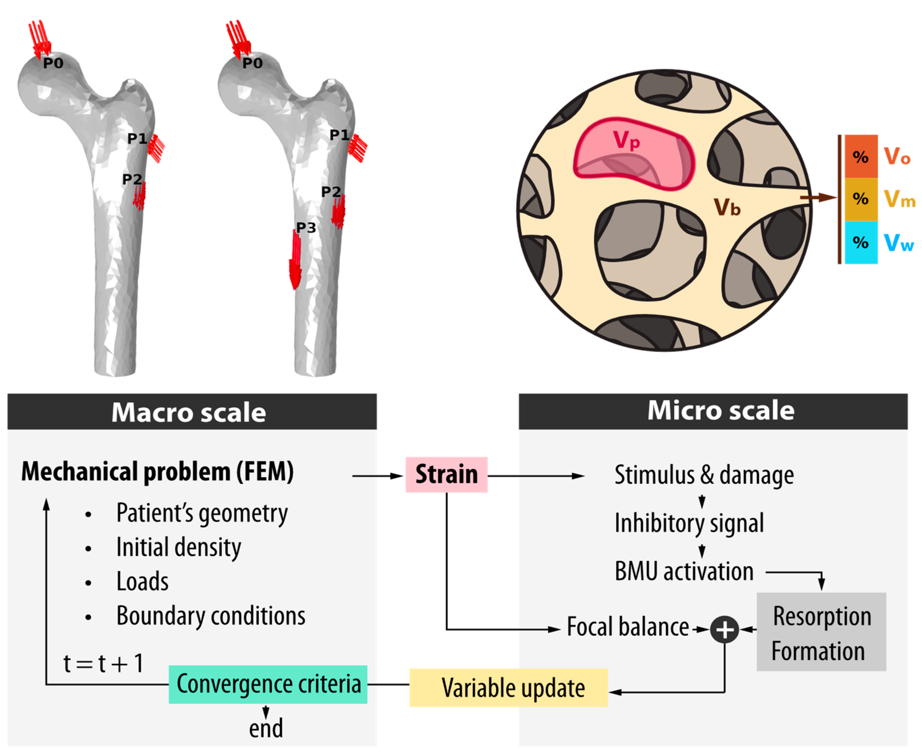
2.3. Mechanical Model

In order to compute the strain energy density, the bone is assumed to be a continuum, locally isotropic, linear elastic material with elastic modulus given by Equation (14) and constant Poisson’s ratio ν = 0.3. The displacement field is solved under the assumption of a stationary state with representative loads from normal walking and stair-climbing. While other daily activities, such as running, jumping, standing from a seated position, or carrying loads, may also influence bone adaptation, their inclusion was beyond the scope of the present study. Future work will consider these additional loading scenarios to provide a more comprehensive understanding of load-driven remodeling. Representative normal walking loads correspond to hip contact forces and forces from the most demanding muscles at the point of maximum hip contact force during the gait cycle. This instant was selected to capture the maximum bending moment at the midshaft of the femur. On the other hand, representative stair-climbing loads also correspond to hip contact forces and forces from the most demanding muscles at the point of maximum torsional moment during the stair-climbing cycle. The values of these forces are shown in Table 1, and their application points are depicted in Figure 1. These values are based on work by Heller et al. [21]. Each load pattern is computed separately, and the results are then combined to compute the strain level using Equation (12). The mechanical description is completed by considering the inferior cut section at the midshaft of the femur as fixed.
The mechanical model is solved using the finite element method. The finite element mesh consisted of 23,527 second-order Lagrange tetrahedral elements, with an average element size (mesh density) of 5.18 mm, which implies an approximate distance of 2.6 mm between integration points. A detailed mesh convergence study was performed and reported in a previous work [11], where the mesh resolution was systematically refined to ensure that the remodeling outcomes were stable and insensitive to further refinement. To avoid redundancy, a brief summary of the mesh convergence study can be found in the Supplementary Materials. The mechanical model was implemented in COMSOL Multiphysics 5.3, which iteratively interacts with the remodeling model implemented in Java 21.
The simulations were executed using the available resources at the Advanced Computing Laboratory, Faculty of Engineering, National University of Entre Ríos, ensuring efficient model resolution. The HPC cluster system consists of 10 nodes, each equipped with an Intel Xeon E5-2670 v3 2.5 GHz, manufactured by Intel Corporation (Santa Clara, CA, USA) processor and 128 GB of RAM running at 2133 MT/s. With this computing power, a simulation of 20 years of bone evolution using the described model takes approximately 38.5 h of computational time when executed on a single node (24 cores).

2.4. Bone Density Initialization

The objective of this work is to highlight the differences in the results obtained when initiating a femoral remodeling model with two distinct initial bone density distributions, as outlined in the introduction. This analysis also prompts a broader discussion on the factors governing bone’s internal organization. While it cannot be definitively concluded that the development of the femur is driven primarily by mechanical factors, numerous studies directly link its internal architecture to the stresses experienced by the bone. From Wolff’s early theories [32], which associated the orientation of trabeculae and the distribution of bone mass with mechanical loads, to more recent research, various authors have demonstrated a strong correlation between bone structure and mechanical forces [19]. However, it remains unclear whether the maturation of bone tissue to its adult state is entirely determined by mechanical loads or whether other factors also play a governing role. In this section, we explore how, according to the present model, certain regions of the bone are strongly influenced by mechanical loads, while others appear less dependent on their initial stages. Identifying which regions need to be carefully linked to their current mechanical load state is essential. However, this remains unclear in most remodeling models of the femur.
To conduct this analysis, we employed two methods for obtaining the initial state of a human femur, from which various clinically relevant studies can be performed. In the first approach, we generated a computational representation of the bone by segmenting computed tomographic (CT) images, identifying density distributions through a methodology detailed below. The reconstructed bone state was initialized under normal mechanical loads (e.g., walking and ascending/descending stairs) and then allowed to evolve to verify its stability under sustained loading conditions. In the second approach, we assumed an initial bone state with a uniform density distribution of 2 g/cm3, corresponding to compact bone tissue, and allowed the bone to evolve under the same loads. This value does not reflect the density of bone at any particular developmental stage, but rather serves to initialize the computational process from an arbitrary homogeneous state. Here, this particular value was chosen because previous works by the authors had shown faster convergence times [10]. Table 3 summarizes the key differences between both initialization methods.
The final states obtained from both approaches are treated as the “initial states” for any personalized study and are compared to highlight the key differences in how the BMU framework responds to mechanical inputs in each case. This comparison provides valuable insights into the mechanical and structural factors shaping bone development.

2.4.1. CT-Approach: Internal Bone Reconstruction Through CT-Image Segmentation

The first step in determining the density distribution involves interpreting the CT data. The grayscale values of the voxels, provided in DICOM (Digital Imaging and Communications in Medicine) format, must be converted into Hounsfield Units (HU) (see Figure 2a). This conversion was performed using a custom Python script (3.11 version), which also translates HU values into apparent (physical) densities. To calibrate the HU-to-apparent density relationship, a GAMMEX 467 phantom was initially used under the supervision of the CEMENER staff. This phantom includes the physical densities of various tissues, such as trabecular and cortical bone.
Since the remodeling algorithm requires volume fraction data to initialize the internal bone composition, an additional step is necessary. By using the apparent density values from the previous step, material densities are obtained relying on the apparent density–material density relationship reported in previous studies [11,23] and depicted in Figure 2b). Material density values are computed through a quadratic least-squares interpolation of the experimental data from Zioupos et al. [33]. Finally, the bone volume fraction is computed for each image voxel using Equation (5). However, voxel grid points do not necessarily align with the FEM mesh nodes. To resolve this, COMSOL Multiphysics® version 5.3 employs an internal interpolation algorithm that maps the CT-derived variable values from each voxel to the FEM mesh nodes. For the 10-node quadratic tetrahedral elements used in this model, four Gauss points are utilized to transfer variables of interest, such as volume fraction, between the CT reconstruction and the remodeling software. The remodeling algorithm, implemented in Java, interacts with COMSOL Multiphysicsthrough its Application Programming Interface (API).
After defining the bone’s geometric and material parameters, another important step involves initializing the bone’s homeostatic state. This initialization must be carefully conducted to ensure consistency between the bone’s density distribution and its mechanical stress state, as remodeling processes are driven by deviations from this equilibrium. To achieve this, the equilibrium state is first initialized by setting ξ * based on the equivalent strain (Equation (13)) under normal loading conditions. The remodeling model is then run until the convergence criterion is met, ensuring that internal bone mass changes are minimal compared to the initial state. The convergence criterion adopted is proposed by Martinez-Reina et al. [9], computed as:
V ρ i ρ i 1 d V V ρ i 1 d V < 0.0005
A key feature of this methodology is that the mass calculated at mechanical equilibrium remains unchanged from its initial state (see Figure 3a), as the tissue is guided toward mechanical equilibrium. Moreover, the computed mass values fall within the range reported by Singh S. and Singh S. P. [34], further validating the approach.

2.4.2. Homogeneous Approach: Internal Bone Reconstruction Through a Purely Mechano-Biological Method

The alternative approach for determining the internal bone mass distribution in the femur starts with an initial homogeneous density distribution, with a value close to that of the outer compact bone (see Figure 3b), while maintaining the external geometry unchanged. It is assumed that by applying the same mechanical loads as in the previous method, the BMU complex will remodel the internal bone structure, resulting in a final mass distribution that meets the mechanical demands. This method relies on the premise that the interaction between mechanical stimuli and the BMU complex will drive the remodeling process, guiding the bone’s internal composition toward that of an adult individual.
For this case, the initial mechanical stimulus was derived from previous studies, corresponding to the equilibrium state of compact bone. Notably, the final bone mass obtained through this method is approximately 30% lower than that achieved using the CT-approach (see Figure 3a). This discrepancy raises an important question: does the CT model include portions of tissue that are not fully represented in the homogeneous model, or is the difference purely a result of variations in density? This distinction is critical for understanding the limitations and implications of each method in accurately modeling bone mass distribution and will be discussed in detail.

3. Results

3.1. Normal Loads

This section presents a comparative analysis of the two bone density initialization approaches, highlighting their effect on the mechanical equilibrium states and structural adaptations. Figure 3c shows the frontal and sagittal sections after achieving equilibrium (density convergence) in both methodologies. Both the coronal and sagittal views reveal that the homogeneous initialization method results in regions of lower density and smoother transitions between areas of different densities. In contrast, the CT-approach produces sharper density gradients, particularly noticeable at the diaphysis, where the transition between high-density cortical bone and lower-density regions is more abrupt. Zioupos et al. [33] identified a value of 1.3 g/cm3 for apparent density as the transition threshold between cortical bone and the transition to porous tissue. In this context, Figure 3c clearly illustrates a significant difference in the spongy bone content between the two methods. Because the homogeneous model depends solely on mechanical loading, it fails to capture certain spongy bone regions that are present in the CT-based model. Certain regions of bone, particularly in the epiphyses, exhibit greater thickness than mechanically required, suggesting that biological factors, such as the presence of red marrow (source of blood cells), play a more significant role in their development than purely mechanical influences.
Figure 4 illustrates the von Mises stress distributions in both planes and models under two applied loading conditions at the onset of adaptation for each method (year zero in Figure 3a). Under typical loading conditions, bending and compressive stresses are dominant in the coronal plane, exhibiting minimal variation between the two models. The highest stresses appear in areas where the bone is expected to have a mechanically reinforced structure. Analysis of the medial-to-lateral cross-section in Figure 5 shows that cortical bone thicknesses are very similar between the two models. Figure 6 further highlights this alignment, showing that both thicknesses and apparent density values along the lateral–medial axis closely match, except for the bone marrow region in section A-A, where the CT-approach model shows the presence of a certain amount of trabecular bone that the homogeneous model is unable to replicate. The homogeneous model tends to smooth out density variations, better reflecting the gradual changes in mechanical stress. This link to mechanical loads justifies the thickness and densities of the CT model in this plane.
In contrast, the sagittal section reveals much more pronounced differences in density distribution. While the CT-approach model retains cortical bone in the sagittal section, the homogeneous model lacks compact bone entirely. Figure 7 and Figure 8 confirm this, as the homogeneous model’s anterior–posterior lines lack compact bone, with density values not exceeding 1.3 g/cm3 [33].
Figure 4 shows that sagittal stress values are nearly ten times lower than those in the coronal plane (106 Pa vs. 107 Pa). Despite this reduction, the CT-approach model retains a significant amount of cortical bone, comparable to that in the coronal plane, as clearly demonstrated in Figure 5. In contrast, the homogeneous model exhibits a drastic reduction in load-bearing stresses, resulting in an almost negligible amount of cortical tissue in this region. This stark difference underscores the limitations of the homogeneous initialization method in accurately representing cortical bone development under sagittal loading conditions.
Under the normal and daily loads considered in this section, it is evident that mechanical development alone (homogeneous model) fails to shape the compact bone content observed in the CT-approach model, which more closely resembles a real bone. Purely mechanical development lacks sufficient mechanical stimulus in the anterior and posterior regions to generate bone with densities corresponding to cortical tissue. Despite some variations in thickness, the densities in the CT-approach model exhibit maximum values in the sagittal section like those in the coronal section. It is worth noting that the highest von Mises stress values observed in the simulations are concentrated near the constrained midshaft boundary. These stress peaks likely result from the simplified boundary conditions applied in the model and may overestimate physiological stress in those regions. While they coincide with areas of high bone density, suggesting appropriate adaptive reinforcement, they should be interpreted with caution, as they may not accurately reflect in vivo loading distributions. Nevertheless, to confirm the consistency of these results, Figure 7 presents the porosity values in the middle zone of compact bone thickness as predicted by each model, compared to experimental data from Thomas et al. [35] for older adults. It can be observed that in the lateral and medial zones, both models accurately represent the cortical porosities. In contrast, the CT-based model closely matches experimental porosity in the anterior and posterior zones, whereas the homogeneous model fails to capture it.

3.2. Disuse and Microgravity

Studying bone evolution under prolonged disuse is key to understanding its effects in sedentary individuals, bedridden patients, and astronauts during long-duration space missions. In fact, although several studies, primarily experimental, have been conducted, the long-term effects of microgravity remain unclear, especially for postmenopausal women who experience significant bone mass loss. This loss can accelerate the onset of early osteoporosis and its associated, difficult-to-reverse, complications. This highlights the importance of accurate initialization in predictive models.
To evaluate the model under microgravity conditions, we simulated six months of 80% load reduction (a typical duration for astronauts on the International Space Station [36]) with both initializations (homogeneous and CT-based). Figure 8 illustrates the change in bone volume fraction ( v b ) of the cross-section at cut B-B (see Figure 5). Differences in bone loss patterns are evident between the two models. The homogeneous model shows increased porosity (bone loss) around the bone marrow and in the anterior and posterior regions, similar to patterns observed in osteoporosis [36], but not in disuse-related bone loss during the analyzed period. In contrast, the CT-based model shows increased porosity around the endocortical region, indicating progressive bone marrow expansion and a gradual reduction of the cortical area. These regions align with areas of higher initial porosity, revealing that the remodeling process is primarily driven by the available free surface, particularly in regions transitioning between high porosity and cortical bone. This behavior is closely linked to the tissue’s load-bearing capacity. For instance, the low porosity of cortical bone, which exhibits the highest mechanical resistance, restricts BMU activity and resorption, delaying structural reduction compared to more trabecular regions. As discussed in a previous study [11], it is reasonable for trabecular bone to exhibit higher remodeling activity, considering its role in hematopoiesis (red bone marrow) and intense ion exchange with the bloodstream. In this regard, the CT-based model aligns more closely with these theories than the homogeneous model during disuse. This is particularly relevant for preserving load-bearing regions over time—an aspect in which the homogeneous model clearly fails in the anterior and posterior regions.

4. Discussion

In this work, we have shown the differences between the homogeneous and CT initialization approaches, focusing on their ability to replicate realistic bone density distributions and structural adaptations using a previous computational model of bone remodeling. Emphasis is placed on the diaphysis, where mechanical demands play a critical role in shaping cortical thickness and density. By comparing these approaches, we highlight the limitations of homogeneous initialization and the advantages of CT-approach reconstruction for modeling femoral bone development and mechanical performance. While the homogeneous model may withstand typical loading, it fails to maintain structural integrity under occasional high-impact loads that generate significant torsion or flexion, especially in the anterior and posterior regions. Activities like jumping or crouching impose mechanical demands that the anterior–posterior regions of the homogeneous model are unlikely to sustain, as these areas develop tissue associated with lower mechanical stimulation. In contrast, the CT-approach model is better equipped to handle such scenarios, as its computationally generated bone more closely reflects the density and structural organization necessary for these stresses.
These results suggest that the development of the anterior–posterior regions of the femoral diaphysis from childhood to adulthood is strongly influenced by the overall growth of the bone, being able to withstand a potential mechanical demand similar to those routinely experienced by the lateral–medial regions or occasional loads of similar magnitude. Nonetheless, the differences in mechanical loading across the studied planes are evidenced by specific internal features of the cortical bone tissue. For example, Endo et al. [37] report variations in the type and density of osteons between lateral–medial and anterior–posterior regions, underscoring the intricate relationship between mechanical demands and bone composition. These findings call for further studies to analyze these variations in detail, which will be explored in future work. The results also indicate that cortical bone thickness along the femoral diaphysis is not solely dictated by mechanical demands, although some aspects of internal composition are. We hypothesize that the anterior–posterior cortical zones mirror the thickness and mechanically required densities of the lateral–medial regions to ensure sufficient load-bearing capacity. This hypothesis aligns with the observed ability of the CT-approach model to replicate the natural density distribution needed to endure mechanical stresses.
Our findings highlight the limitations of using a homogeneous initial state for reconstructing the internal structure of a computational femur for further simulations. Although simpler, this approach fails to capture the realistic bone mass distribution in the femoral diaphysis. In contrast, the CT-approach reconstruction method proposed in this work, combined with proper initialization of the internal homeostatic state, provides a more accurate representation of bone density distribution. Importantly, this approach ensures that the reconstructed structure remains stable under normal loading conditions (no bone mass loss during adaptation), maintaining consistency with the mechanical state for which the bone was initialized.
While our model is able to offer valuable insights into the mechanical and biological factors influencing femoral development, it is not without limitations. These arise primarily from the assumptions and simplifications necessary for computational modeling. For instance, the BMU model is based on continuum mechanics and, as such, cannot account for the detailed microstructural organization of bone tissue or the specific consequences of changes in trabecular thickness, orientation, or connectivity. This is a key limitation, as trabecular arrangement and morphology are essential for mechanical efficiency and adaptability, particularly in trabecular-rich regions. By neglecting these microstructural details, the model may oversimplify the relationship between mechanical loads and structural adaptation, potentially leading to an incomplete understanding of how local stresses influence bone remodeling. Another limitation, partially stemming from the exclusion of individual trabeculae in the model, is the assumption that local mechanical properties at the microscale are isotropic. This overlooks the heterogeneous and anisotropic nature of bone tissue, which plays an essential role in optimizing the bone’s resistance to complex loading scenarios, particularly in regions like the femoral neck where multidirectional forces act.
These limitations could affect the insights derived from the model in several ways. First, the inability to simulate trabecular adaptations might lead to underestimating the bone’s capacity to redistribute loads through microstructural changes. Second, the isotropic assumption might fail to capture the nuanced load-sharing mechanisms between cortical and trabecular bone, potentially influencing predictions about stress distribution and regions at risk of failure. Despite local limitations, the model performs well at the macroscopic level, reproducing observed trends in bone mass evolution and mechanical behavior [11,12]. This supports its suitability for analyzing bone’s macroscopic and integral properties, as its simplifications allow for the simulation of long-term remodeling (spanning decades) with minimal computational cost. Remarkably, each year of real-time remodeling can be simulated in under 2 h.
This study does not account for high-impact activities like jumping, squatting, or sitting and standing, which may significantly influence bone development during early childhood. However, the hypothesis presented here suggests that if these effects are mechanically generated, their influence would likely diminish over time as the bone adapts to the more routine and consistent mechanical loads experienced during adolescence and adulthood.
It is important to clarify that the development of the femur’s internal structure during childhood does not begin with a uniform density distribution [38], as assumed in the homogeneous model. Instead, the bone already exhibits a more complex and differentiated pattern of density. However, the use of a simplified homogeneous distribution in this study serves to demonstrate how the remodeling model—validated for its ability to adapt to both normal and altered loading conditions—gradually shapes the bone to meet mechanical demands.
Interestingly, the initial distribution of bone mass during childhood closely resembles the patterns obtained using CT-approach methods. This is significant because regions with low mechanical stimulation do not regress or transform into trabecular bone, as observed in amputee patients [39] or as predicted by the homogeneous model. Within this context, the homogeneous model is valuable for hypothesizing how distinct cortical regions might evolve, solely driven by the habitual loading patterns considered in this study. However, this condition would lead to unrealistic situations in the case of disuse states, where aggressive bone mass loss is observed in the anterior and posterior regions for the homogeneous model. This pattern is not seen in real scenarios during the disuse period, where bone loss mainly occurs in the endocortical region, as predicted by the CT model and consistent with Lerebours et al. [36]. This loss during the disuse period manifests as a ring in the endocortical zone. Although in the anterior and posterior regions the equilibrium stimulus is closer to a disuse state, porosity increases similarly to the lateral and medial regions, despite the equilibrium stimulus in the latter being further from disuse. However, in short periods (several months), the primary drivers of bone remodeling are the activity level and the free surface area or porosity. Therefore, due to disuse, bone activity remains elevated (see Equations (10) and (11)), with the highest porosity occurring in an endocortical ring surrounding the bone marrow. The imbalance persists as long as the mechanical stimulus adapts to the current condition. An interesting question arises: can bone loss occur after an adaptation period? We believe this depends on both the duration of disuse and the age of the subjects analyzed. If age exceeds 35 years old [40], it is likely that the focal balance will favor osteoclasts at equilibrium, and if disuse-induced activity is elevated, osteoclasts will resorb bone more aggressively, leading to continuous bone mass loss. While this phenomenon also occurs under normal physiological conditions, its effects can be mitigated by regular mechanical loading or exercise regimens, which suppress osteoclast activity and stimulate osteoblast function. This topic was partially addressed in a previous work [12]; however, age-related changes in BMU activity (such as altered turnover rates and imbalances between resorption and formation) remain key factors influencing bone mass and may affect the clinical relevance of simulation outcomes, particularly in older populations. Although a detailed investigation of these biological mechanisms lies beyond the scope of the present study, their relevance is acknowledged, and they will be addressed more thoroughly in future work through a more comprehensive, mechanobiologically informed modeling framework.
It is important to mention that this study does not include a fatigue-based remodeling mechanism or an explicit representation of microdamage accumulation. The remodeling stimulus was based solely on strain energy density, following classical formulations commonly used to capture the mechanical regulation of bone mass [6,7,24]. This choice allowed us to focus on large-scale effects of mechanical loading and initial density, while keeping the model computationally tractable. However, it is well established that fatigue-induced microdamage also plays a key role in initiating remodeling, particularly under repetitive loading conditions. Incorporating damage accumulation as a stimulus for bone turnover could enhance the model’s ability to predict long-term adaptation and the risk of stress-related bone failure. Similarly, the model does not explicitly represent trabecular microstructure or anisotropic material properties. These simplifications limit the resolution of local mechanical predictions, particularly in trabecular-rich regions where anisotropy plays a critical role. However, our modeling choices were guided by the nature of clinical CT data, which does not provide reliable directional information at the voxel level, and by the aim of capturing organ-scale patterns of bone adaptation. While this limits the model’s ability to resolve fine-scale architectural features, it does not preclude its use in clinically relevant applications. In particular, if extended to include fatigue-based damage accumulation, the model could still be used to estimate fracture risk under repetitive loading conditions, even in the absence of explicit trabecular geometry. Such an approach would provide a useful compromise between clinical feasibility and physiological realism, and represents a promising direction for future development.

5. Conclusions

This study examines two methods for determining the internal mass composition of a human femur, primarily for computational applications. The first method reconstructs the bone by segmenting CT images and converting grayscale values into external geometry and a continuous internal density distribution. The second method starts with the same external morphology obtained through segmentation but assumes a homogeneous internal distribution representative of compact bone. For both methods, a bone remodeling algorithm previously validated [11,23] is applied to predict the final state of the femur from its initial condition.
In the first case, the bone begins with an internal density distribution derived from the CT images, which is then allowed to evolve from this initial structure until it reaches internal equilibrium under normal walking or climbing stairs conditions. In the second case, the bone is exposed to the same mechanical loads and evolves under the hypothesis that the biological system adapts its internal architecture to meet these demands, reaching mechanical equilibrium similar to that of an adult human.
The results indicate that the pure mechanical model (second method) lacks the necessary stimuli to shape the femur’s internal structure according to the actual bone density distribution, especially in the diaphysis of a femur. Frequent loading scenarios, such as normal walking or climbing stairs, fail to sufficiently stimulate the anterior and posterior regions of the diaphysis to form cortical tissue. This discrepancy is not observed in the reconstruction based on tomographic data. Notably, in the CT-based model, proper mechanical initialization prevents excessive thinning of cortical tissue in the anterior and posterior diaphysis, despite low mechanical stimulation in these regions. In this regard, proper initialization of the computational model is crucial to ensure that bone tissue evolution aligns with its internal structure and initial homeostatic state. This is therefore key for conducting personalized studies with minimal deviations from real tissue properties. Proper initialization is also essential for monitoring bone adaptation after implant placement, since deviations from equilibrium directly affect local remodeling intensity. Additionally, based on the results under normal loading conditions, we hypothesize that the development of the human femur into adulthood depends on factors beyond mechanical loads, rather than being solely determined by them. Non-mechanical factors may help maintain bone robustness in the diaphysis, especially when normal-intensity loads act on less typical regions. This conclusion is supported by the work of Swan [38], who determined that in femoral development, in addition to mechanical loading, non-mechanical factors such as hormonal, nutritional, and genetic influences should also be considered. Although this remains a modeling-based hypothesis, it provides a valuable direction for future research, such as combining biomechanical simulation with longitudinal imaging in pediatric or adolescent populations (e.g., Magnetic Resonance Imaging or High-Resolution peripheral Quantitative Computed Tomography), or using animal models under controlled loading protocols to disentangle the relative contributions of mechanical and non-mechanical factors.
Based on the results of this study, the reconstruction method using segmentation with a properly initialized mechanical equilibrium state proves to be the most suitable for approximating the bone tissue’s condition at the onset of any remodeling process. These findings may help identify bone regions most sensitive to mechanical stimuli during disuse, as well as the most effective rehabilitation activities after density loss. This will be explored in more detail in future research. While homogeneous initialization may suit theoretical or coronal-plane studies, our results show that CT-based initialization offers a much more accurate representation for subject-specific analyses involving the full 3D architecture of the femur, especially the sagittal plane. This limitation of the first method is particularly relevant in contexts such as fracture risk assessment, implant design, or monitoring of treatment response, where accurate modeling of regional bone mass is essential.
Importantly, the current remodeling model captures the mechanical regulation of bone adaptation through local mechanosensing and BMU activity, yet it does not account for non-mechanical influences such as hormonal regulation, metabolic state, or cellular signaling, all of which are known to significantly modulate bone turnover. Future extensions of the model could incorporate these factors by introducing modulatory terms into the activation and bone balance equations to reflect the influence of systemic hormones (e.g., parathyroid hormone, estrogen), nutritional status (e.g., calcium or vitamin D availability), or pro-inflammatory mediators (e.g., TNF-α, IL-6). In parallel, spatially localized biochemical signaling could be modeled using diffusion-reaction frameworks, allowing the simulation of gradients of osteogenic or osteoclastogenic signals within the bone microenvironment. Integrating these non-mechanical drivers alongside mechanical stimuli would allow the model to capture the multifactorial nature of bone remodeling, improving its predictive capacity in pathological conditions (such as osteoporosis or inflammatory bone loss), aging, or in response to pharmacological treatments.
In this work, however, we intentionally isolate the mechanical component of the bone remodeling process to assess its capacity to reproduce realistic bone mass distributions under standard loading conditions. By comparing two initialization approaches in the same individual-specific geometry, we aimed to identify the extent to which mechanical stimuli alone explain internal femoral structure. This controlled setup provides a necessary baseline for adding biological and demographic variables in future model expansions.

Supplementary Materials

The following supporting information can be downloaded at: https://www.mdpi.com/article/10.3390/math13132156/s1.

Author Contributions

Methodology, F.F. and M.B.; Software, F.F.; Formal analysis, F.F., M.B. and C.B.; Writing—original draft, F.F., M.B. and C.B.; Writing—review & editing, F.F., C.B., J.D.P. and M.B.; Supervision, C.B., J.D.P. and M.B. All authors have read and agreed to the published version of the manuscript.

Funding

This research received no external funding.

Data Availability Statement

The original contributions presented in this study are included in the article/Supplementary Materials. Further inquiries can be directed to the corresponding author.

Acknowledgments

Marcelo Berli, Feliciano Franco, and José Di Paolo acknowledge the National University of Entre Ríos for supporting this work through PID 6265. Furthermore, these authors express their gratitude to the National Technological University for providing the necessary institutional framework and research support required for the completion of this study. We also express our gratitude to the Department of Teaching and Research of the Nuclear and Molecular Medicine Center Foundation of Entre Ríos (CEMENER) for their assistance with the acquisition and processing of tomographic images.

Conflicts of Interest

The authors declare no conflicts of interest. The funders had no role in the design of the study; in the collection, analyses, or interpretation of data; in the writing of the manuscript; or in the decision to publish the results.

References

  1. Allen, M.R.; Burr, D.B. Bone Modeling and Remodeling. Basic Appl. Bone Biol. 2013, 75–90. [Google Scholar] [CrossRef]
  2. Cowin, S.C.; Hegedus, D.H. Bone remodeling: A theory of adaptive elasticity. J. Elast. 1976, 6, 313–326. [Google Scholar] [CrossRef]
  3. Beaupré, G.S.; Orr, T.E.; Carter, D.R. An approach for time-dependent bone modeling and remodeling-application: A preliminary remodeling simulation. J. Orthop. Res. 1990, 8, 662–670. [Google Scholar] [CrossRef] [PubMed]
  4. Coelho, P.G.; Fernandes, P.R.; Rodrigues, H.C.; Cardoso, J.B.; Guedes, J.M. Numerical modeling of bone tissue adaptation-A hierarchical approach for bone apparent density and trabecular structure. J. Biomech. 2009, 42, 830–837. [Google Scholar] [CrossRef]
  5. Doblaré, M.; García, J.M. Application of an anisotropic bone-remodelling model based on a damage-repair theory to the analysis of the proximal femur before and after total hip replacement. J. Biomech. 2001, 34, 1157–1170. [Google Scholar] [CrossRef] [PubMed]
  6. Doblaré, M.; García, J.M. Anisotropic bone remodelling model based on a continuum damage-repair theory. J. Biomech. 2002, 35, 1–17. [Google Scholar] [CrossRef] [PubMed]
  7. García-Aznar, J.M.; Rueberg, T.; Doblare, M. A bone remodelling model coupling microdamage growth and repair by 3D BMU-activity. Biomech. Model. Mechanobiol. 2005, 4, 147–167. [Google Scholar] [CrossRef]
  8. Martínez-Reina, J.; Reina, I.; Domínguez, J.; García-Aznar, J.M. A bone remodelling model including the effect of damage on the steering of BMUs. J. Mech. Behav. Biomed. Mater. 2014, 32, 99–112. [Google Scholar] [CrossRef]
  9. Martínez-Reina, J.; Ojeda, J.; Mayo, J. On the use of bone remodelling models to estimate the density distribution of bones uniqueness of the solution. PLoS ONE 2016, 11, e0148603. [Google Scholar] [CrossRef]
  10. Berli, M. Computational Implementation of a New Bone Resorption Strategy Oriented to the Study of the Action of Multicellular Basic Units. Application to the Process of Human Bone Remodeling; National University of Entre Ríos: Concepción del Uruguay, Argentina, 2020. [Google Scholar]
  11. Berli, M.; Franco, F.; Di Paolo, J.; Zioupos, P.; Borau, C. The interplay between BMU activity linked to mechanical stress, specific surface and inhibitory theory dictate bone mass distribution: Predictions from a 3D computational model. Comput. Biol. Med. 2022, 148, 1–10. [Google Scholar] [CrossRef]
  12. Franco, F.; Borau Zamora, C.; Campana, D.M.; Berli, M.E. Computational Analysis of the Influence of Menopause and Ageing on Bone Mineral Density, Exploring the Impact of Bone Turnover and Focal Bone Balance—A Study on Overload and Underload Scenarios. Life 2023, 13, 2155. [Google Scholar] [CrossRef] [PubMed]
  13. Bahia, M.T.; Hecke, M.B.; Mercuri, E.G.F. Image-based anatomical reconstruction and pharmaco-mediated bone remodeling model applied to a femur with subtrochanteric fracture: A subject-specific finite element study. Med. Eng. Phys. 2019, 69, 58–71. [Google Scholar] [CrossRef]
  14. Bansod, Y.D.; Kebbach, M.; Kluess, D.; Bader, R.; van Rienen, U. Finite element analysis of bone remodelling with piezoelectric effects using an open-source framework. Biomech. Model. Mechanobiol. 2021, 20, 1147–1166. [Google Scholar] [CrossRef]
  15. Hölzer, A.; Schröder, C.; Woiczinski, M.; Sadoghi, P.; Scharpf, A.; Heimkes, B.; Jansson, V. Subject-specific finite element simulation of the human femur considering inhomogeneous material properties: A straightforward method and convergence study. Comput. Methods Programs Biomed. 2013, 110, 82–88. [Google Scholar] [CrossRef]
  16. Taddei, F.; Martelli, S.; Reggiani, B.; Cristofolini, L.; Viceconti, M. Finite-element modeling of bones from CT data: Sensitivity to geometry and material uncertainties. IEEE Trans. Biomed. Eng. 2006, 53, 2194–2200. [Google Scholar] [CrossRef] [PubMed]
  17. Idhammad, A.; Abdali, A.; Alaa, N. Computational simulation of the bone remodeling using the finite element method: An elastic-damage theory for small displacements. Theor. Biol. Med. Model. 2013, 10, 32. [Google Scholar] [CrossRef] [PubMed]
  18. Knowles, N.K.; Reeves, J.M.; Ferreira, L.M. Quantitative Computed Tomography (QCT) derived Bone Mineral Density (BMD) in finite element studies: A review of the literature. J. Exp. Orthop. 2016, 3, 36. [Google Scholar] [CrossRef]
  19. Martínez-Reina, J.; Ojeda, J.; Calvo-Gallego, J.L.; Pivonka, P.; Martelli, S. Assessment of mechanical variables best describing bone remodelling responses based on their correlation with bone density. J. Mech. Behav. Biomed. Mater. 2024, 160, 106773. [Google Scholar] [CrossRef]
  20. Available online: https://www.slicer.org (accessed on 17 January 2025).
  21. Heller, M.O.; Bergmann, G.; Kassi, J.P.; Claes, L.; Haas, N.P.; Duda, G.N. Determination of muscle loading at the hip joint for use in pre-clinical testing. J. Biomech. 2005, 5, 1155–1163. [Google Scholar] [CrossRef]
  22. Rho, J.Y.; Kuhn-Spearing, L.; Zioupos, P. Mechanical properties and the hierarchical structure of bone. Med. Eng. Phys. 1998, 20, 92–102. [Google Scholar] [CrossRef]
  23. Berli, M.; Borau, C.; Decco, O.; Adams, G.; Cook, R.B.; Aznar, J.M.G.; Zioupos, P. Localized tissue mineralization regulated by bone remodelling: A computational approach. PLoS ONE 2017, 12, e0173228. [Google Scholar] [CrossRef] [PubMed]
  24. Martínez-Reina, J.; García-Aznar, J.M.; Domínguez, J.; Doblaré, M. On the role of bone damage in calcium homeostasis. J. Theor. Biol. 2008, 254, 704–712. [Google Scholar] [CrossRef]
  25. Martin, R. Toward a unifying theory of bone remodeling. Bone 2000, 26, 1–6. [Google Scholar] [CrossRef] [PubMed]
  26. Lukas, C.; Ruffoni, D.; Lambers, F.M.; Schulte, F.A.; Kuhn, G.; Kollmannsberger, P.; Weinkamer, R.; Müller, R. Mineralization kinetics in murine trabecular bone quanti fi ed by time-lapsed in vivo micro-computed tomography. Bone 2013, 56, 55–60. [Google Scholar] [CrossRef] [PubMed]
  27. Eriksen, E.F. Cellular mechanisms of bone remodeling. Rev. Endocr. Metab. Disord. 2010, 11, 219–227. [Google Scholar] [CrossRef] [PubMed]
  28. Pivonka, P.; Buenzli, P.R.; Scheiner, S.; Hellmich, C.; Dunstan, C.R. The influence of bone surface availability in bone remodelling-A mathematical model including coupled geometrical and biomechanical regulations of bone cells. Eng. Struct. 2013, 47, 134–147. [Google Scholar] [CrossRef]
  29. Mikić, B.; Carter, D.R. Bone strain gage data and theoretical models of functional adaptation. J. Biomech. 1995, 28, 465–469. [Google Scholar] [CrossRef]
  30. Latypova, A.; Maquer, G.; Elankumaran, K.; Pahr, D.; Zysset, P.; Pioletti, D.P.; Terrier, A. Identification of elastic properties of human patellae using micro-finite element analysis. J. Biomech. 2016, 49, 3111–3115. [Google Scholar] [CrossRef]
  31. Geraldes, D.M.; Phillips, A.T.M. A comparative study of orthotropic and isotropic bone adaptation in the femur. Training 2014, 30, 873–889. [Google Scholar] [CrossRef]
  32. Wolff, J. The Law of Bone Remodelling; Springer: Berlin/Heidelberg, Germany, 1896. [Google Scholar]
  33. Zioupos, P.; Cook, R.B.; Hutchinson, J.R. Some basic relationships between density values in cancellous and cortical bone. J. Biomech. 2008, 41, 1961–1968. [Google Scholar] [CrossRef]
  34. Singh, S.; Singh, S.P. Weigth of the femur—A useful measurement for identification of sex. Acta Anat. 1974, 87, 141–145. [Google Scholar] [CrossRef]
  35. Thomas, C.D.; Feik, S.A.; Clement, J.G. Regional variation of intracortical porosity in the midshaft of the human femur: Age and sex differences. J. Anat. 2005, 206, 115–125. [Google Scholar] [CrossRef] [PubMed]
  36. Lerebours, C.; Buenzli, P.R.; Scheiner, S.; Pivonka, P. A multiscale mechanobiological model of bone remodelling predicts site-specific bone loss in the femur during osteoporosis and mechanical disuse. Biomech. Model. Mechanobiol. 2016, 15, 43–67. [Google Scholar] [CrossRef] [PubMed]
  37. Endo, D.; Ogami-Takamura, K.; Imamura, T.; Saiki, K.; Murai, K.; Okamoto, K.; Tsurumoto, T. Reduced cortical bone thickness increases stress and strain in the female femoral diaphysis analyzed by a CT-based finite element method: Implications for the anatomical background of fatigue fracture of the femur. Bone Rep. 2020, 13, 100733. [Google Scholar] [CrossRef] [PubMed]
  38. Swan, K.R.; Ives, R.; Wilson, L.A.B.; Humphrey, L.T. Ontogenetic changes in femoral cross-sectional geometry during childhood locomotor development. Am. J. Phys. Anthropol. 2020, 173, 80–95. [Google Scholar] [CrossRef]
  39. Michael, A.R. Histological estimation of age at death in amputated lower limbs: Issues of disuse, advanced age, and disease in the analysis of pathological bone. J. Forensic Leg. Med. 2018, 53, 58–61. [Google Scholar] [CrossRef]
  40. Zioupos, P. Ageing human bone: Factors affecting its biomechanical properties and the role of collagen. J. Biomater. Appl. 2001, 15, 187–229. [Google Scholar] [CrossRef]
Figure 2. (a) Hounsfield Units (HU) in a coronal plane. (b) Relationship between material density and apparent density, which is used to determine the material density values and, consequently, the volume fraction (Equation (5). Experimental data from Zioupos et al. [33].
Mathematics 13 02156 g002
Figure 3. (a) Evolution of total bone mass over time for both initialization methods. The homogeneous model converges to a final mass approximately 30% lower than that of the CT-based model (see results and discussion). (b) Coronal sections of the initial bone state (year 0 in panel a). The homogeneous model starts from a uniform density distribution equivalent to cortical bone (2.0 g/cm3) with an associated equilibrium stimulus. In contrast, the CT-based model is initialized from segmented grayscale data, converted into spatially varying densities, with local mechanical equilibrium achieved point by point. (c) Apparent density distributions at equilibrium in the sagittal (left) and coronal (right) planes. Notably, the homogeneous model fails to preserve cortical bone in the sagittal section, whereas the CT-based model retains compact bone structure in both planes.
Mathematics 13 02156 g003
Figure 4. Von Mises stresses for both activities (normal walking and stair climbing).
Mathematics 13 02156 g004
Figure 5. Apparent density in the cross-section at three areas of the proximal femur. Note that in the lateral–medial (L–M) axis, the cortical content is similar for both methods, while in the anterior–posterior (A–P) axis, differences are highly pronounced.
Mathematics 13 02156 g005
Figure 6. Apparent densities along the medial–lateral (left column) and anterior–posterior axes (right column) For the cross-sections of Figure 5. Note that in the lateral–medial section, the densities predicted by both methods are highly correlated, while for the anterior–posterior section, there are notable differences. Also, note that for this last section, the densities predicted by the homogeneous method are below the cortical threshold: 1.3 g/cm3 [33].
Mathematics 13 02156 g006
Figure 7. Mean porosity and confidence intervals (95%) in the mid-zone of cortical bone thickness, as predicted by each model across 11 horizontal slices between A–A and C–C planes along the femoral diaphysis, compared to experimental data (mean values) from Thomas et al. [35] for older adults. P: Posterior; L: Lateral; A: Anterior; M: Medial.
Mathematics 13 02156 g007
Figure 8. Variations in bone volume in a cross section of the diaphysis at the A-A cut (see Figure 5). A reduction in bone volume implies an increase in porosity after a disuse period of 6 months.
Mathematics 13 02156 g008
Table 1. Applied loads. Walking loads at maximum contact force. Stair-climbing at maximum torsional moment. Locations are depicted in Figure 1. Based on work by Heller et al. [21].
PatternLoad (N)FxFyFzLocation
WalkingHip contact−459.0−278.8−1948.2P0
Abductor493.036.6735.3P1
Tensor fascia latae, proximal61.298.6112.2P1
Tensor fascia latae, distal−4.3−6.0−161.5P1
Vastus lateralis−7.7157.3−789.7P2
Stair-climbingHip contact−504.1−515.1−2008.6P0
Abductor595.9244.8721.7P1
Iliotibial band, proximal89.3−25.5108.8P1
Iliotibial band, distal−4.3−6.8−142.8P1
Tensor fascia latae, proximal26.441.724.7P1
Tensor fascia latae, distal−1.7−2.6−55.3P2
Vastus lateralis−18.7190.4−1148.4P2
Vastus medialis−74.8336.6−2270.4P3
Table 2. Parameters value of the model [11].
Parameter Value
vBMUBMU advancement speed 0.04 mm/day
TRResorption period24 days
TIInversion period8 days
TFFormation period64 days
σLBMU Lifespan100 days
mWeighting exponent4
fbioBiological frequency factor0.005 NBMU/(mm3 day)
TnmMineralization lag time12 days
TprimLength of primary phase10 days
Tm,maxTime to reach the maximum mineral level4000 days
Table 3. Key differences between the two initialization methods.
FeatureCT-Based InitializationHomogeneous
Input sourceClinical CT scanSynthetic model with constant density
Initial densitySpatially heterogeneous (realistic)Uniform (2 g/cm3)
Biological relevanceClose to real adult bone structureArtificial starting point
PurposeAssess remodeling from a realistic baselineTest model ability to evolve structure purely from loading
Expected convergenceSmall adaptation from mature stateLarger changes required to reach physiological state
Disclaimer/Publisher’s Note: The statements, opinions and data contained in all publications are solely those of the individual author(s) and contributor(s) and not of MDPI and/or the editor(s). MDPI and/or the editor(s) disclaim responsibility for any injury to people or property resulting from any ideas, methods, instructions or products referred to in the content.

