Article

Morphological Variability of Pseudo-nitzschia pungens Clade I (Bacillariophyceae) in the Northwestern Adriatic Sea

1
Dipartimento di Scienze della Vita e dell’Ambiente, Università Politecnica delle Marche, via Brecce Bianche, 60131 Ancona, Italy
2
Istituto Zooprofilattico Sperimentale Umbria e Marche, Via Cupa di Posatora, 3, 60131 Ancona, Italy
3
Consorzio Interuniversitario per le Scienze del Mare, CoNISMa, ULR Ancona, Ancona, Italy
*
Author to whom correspondence should be addressed.
Plants 2020, 9(11), 1420; https://doi.org/10.3390/plants9111420
Submission received: 17 September 2020 / Revised: 17 October 2020 / Accepted: 19 October 2020 / Published: 23 October 2020
(This article belongs to the Special Issue Systematics and Ecology of Algae and Marine Plants)

Abstract

:
Pseudo-nitzschia pungens is a common component of the phytoplankton community of the northern Adriatic Sea. In this study, an in-depth morphological analysis of P. pungens was carried out in both cultured strains isolated in different periods and field samples, revealing a surprisingly wide variability in a number of details, with both the gross morphology and ultrastructural levels deviating from the nominal P. pungens. Colonies showed an overlap (from one-third to one-sixth) and a transapical axis (rarely reaching 3 µm), strongly differing from the original description of the species. Moreover, valves may be either symmetrical or slightly asymmetrical, with striae almost always biseriate but sometimes uniseriate or triseriate. Poroids’ morphology in cingular bands was characterized by a wide variability (square, circular, or rectangular poroids without or with up to two hymen sectors), with several combination of them, even within the same cingular band. Phylogenetic analyses based on ITS rDNA showed that the P. pungens of the northern Adriatic Sea belonged to clade I. Domoic acid was not detected.

1. Introduction

Planktonic diatom species of the genus Pseudo-nitzschia are recorded in coastal regions worldwide. Among the 53 known species [1], 26 have been shown to produce domoic acid (DA) [2,3], causing neurologic disorders and memory loss in vertebrates linked to the consumption of contaminated shellfish (Amnesic Shellfish Poisoning) [4,5]. In this genus, the number of genetic lineages is markedly higher than the number of taxa discernible by light microscope (LM). With the description of several new Pseudo-nitzschia species, detailing and comparing additional ultrastructural characters has become necessary [6,7,8]. Indeed, several species not easily distinguishable using LM have been described using electron microscopy (EM) coupled with molecular techniques [9,10,11,12,13,14,15,16,17].
Traditionally, species of the genus Pseudo-nitzschia have been subdivided in two LM-discernible groups based on cell width in valve view: All the species wider than 3 µm have been combined into the seriata group, while those less than 3 µm are in the delicatissima group [18]. Pseudo-nitzschia pungens, with its varieties, is a cosmopolitan species detected from temperate to tropical waters [8,19,20,21], ascribed to the seriata group. The combination of a few morphological characters (including cell size, overlap of cells in colonies [22]) has usually been enough to distinguish P. pungens from other Pseudo-nitzschia species within the seriata group (e.g., P. fraudulenta (Cleve) Hasle under LM [23]). Nevertheless, the global distribution of this species and its varieties have received increasing attention after Casteleyn et al. [24,25] recognized the existence of three closely related lineages (i.e., clade I, II, and III) based on ITS rDNA region analyses, which is also supported by morphological and mating studies [8]. These three clades approximately correspond to the morphological varieties P. pungens var. pungens (clade I), P. pungens var. cingulata (clade II), and P. pungens var. aveirensis (clade III) [24,26,27]. To date, among the P. pungens varieties, P. pungens var. cingulata and P. pungens var. aveirensis are not toxic, while P. pungens var. pungens is the only one containing both toxic and nontoxic strains [21,27,28,29,30]. There are several morphological features useful to distinguish those clades, such as (i) the shape of the poroids of the valvocopula, (ii) the number of fibulae and striae in 10 µm, (iii) the number of the poroids in 1 µm, (iv) the number of additional poroids in 10 striae, and (v) the transapical axis dimension [8,24]. Moreover, two subgroups of clade III (i.e., IIIa and IIIb) with apparently different geographic distribution (Western Pacific and Western/Northeastern Atlantic strains, respectively) were recently distinguished by ITS sequences [21], although no significant morphological differences have been detected [8]. Regarding the mating studies, an apparent reproductive isolation has been highlighted between clade I and clade III, while strains of clade I and II are sexually compatible and able to produce hybrid offspring with intermediate valve width and structure of valvocopula [31].
Pseudo-nitzschia pungens has been commonly recorded in the phytoplankton community of the Mediterranean Sea, although its geographical distribution is seemingly restricted to the northernmost areas of the basin in the NW [28,32,33,34,35], as well as in NE Mediterranean Sea [29], and it is a quite recurrent species in the diatom community of the northern Adriatic Sea [30,36,37,38]. Previous molecular studies carried out on the P. pungens from the northern Adriatic have highlighted that the Adriatic strain belonged to clade I [30]. However, an in-depth ultrastructural analysis on the Adriatic P. pungens population has not been carried out so far.
Since its first record in 2000 [39], in the Adriatic Sea, the presence of DA in shellfish has been detected only occasionally [7,40], with concentration always well below the EU regulatory limit of 20 mg kg−1 [41], despite blooms of several potentially toxic Pseudo-nitzschia species such as P. pungens, P. calliantha, P. delicatissima, P. fraudulenta, P. multistriata, and P. pseudodelicatissima [40,42], which commonly occur throughout the year in the study area [43]. High-performance liquid chromatography (HPLC) using UV detection is the most popular method used for the determination of DA in shellfish tissues, which was developed by Quilliam and Wright [44] and modified by AESAN [45]. However, as DA concentrations in Pseudo-nitzschia strains and phytoplankton field samples are often very low, more sensitive methods of detection are required. Among other developed methods, LC-MS exhibits good results in terms of sensitivity, accuracy, and selectivity [46], representing one of the best tools to investigate DA presence in cultured strains and phytoplankton field samples.
The aim of this study was to characterize the P. pungens population of northwestern Adriatic, based on molecular and ultrastructural features of both natural and culture samples, as well as the analysis of its toxin content.

2. Results

2.1. Morphology

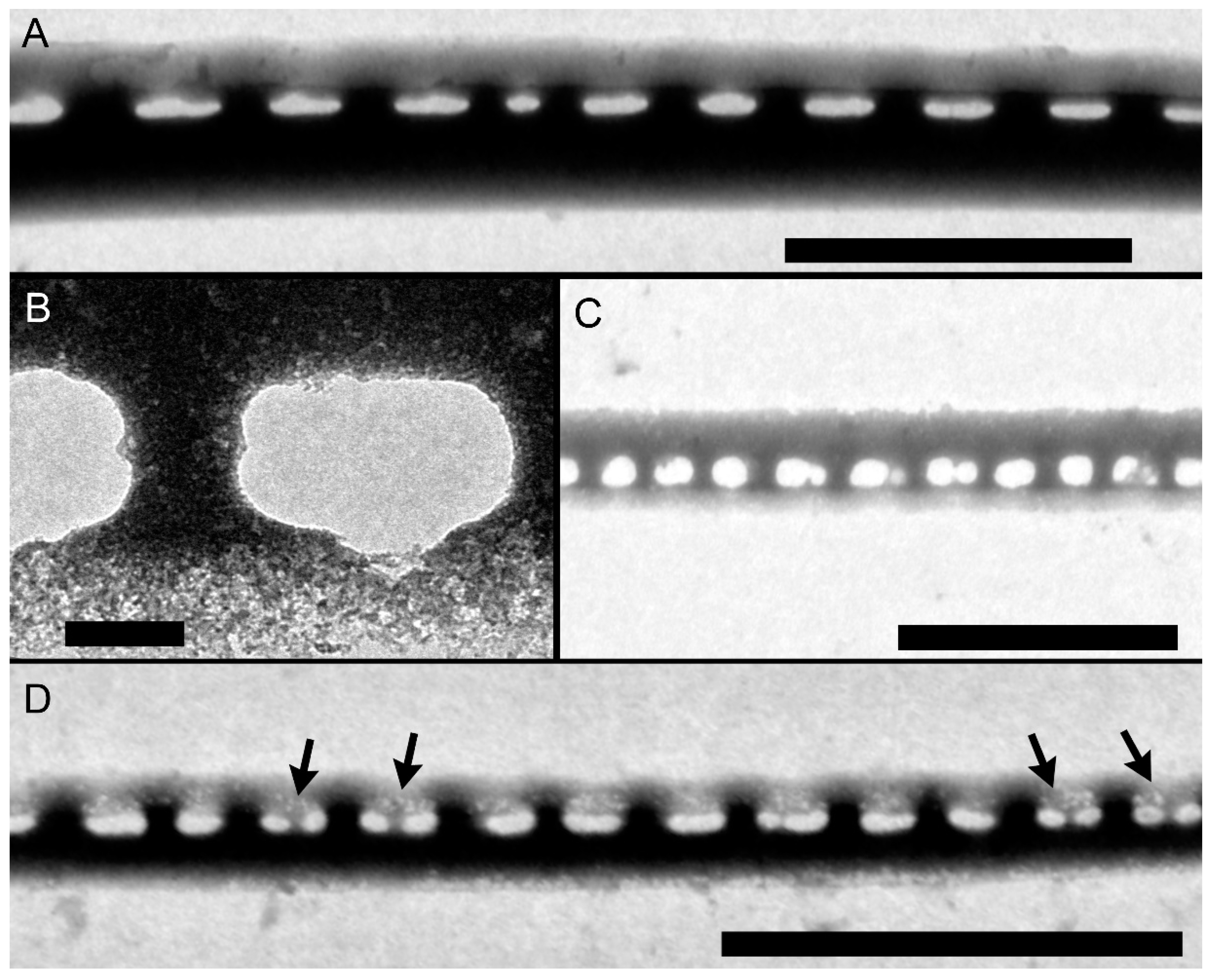
Cells of Pseudo-nitzschia pungens from culture material (Figures S1–S9) and from field samples showed very similar morphological and morphometric features. Cells had fusiform to lanceolate shape in both girdle (Figure 1A,B,E–G) and valve view (Figure 1C,D and Figure 2A). All the morphometric measurements have been performed with EM, except for the Apical Axis (AA) measured both in LM and SEM. The AA ranged from 51.1 µm to 127.6 µm, while the Transapical Axis (TA) ranged from 2.0 µm to 3.7 µm (Table 1 and Table S1). The central nodule was absent, and the raphe continued for the full length of the cells (Figure 3A).
Cells formed stepped colonies of several cells with an overlap that ranged from one-third to one-sixth of the AA length (Figure 1A,B, Table 1). Cells were strongly silicified. The interstriae and fibulae were discernible in LM (Figure 1A,C). Although colonies were generally symmetrical in valve view (Figure 1C), asymmetrical colonies were not rare (Figure 1D).
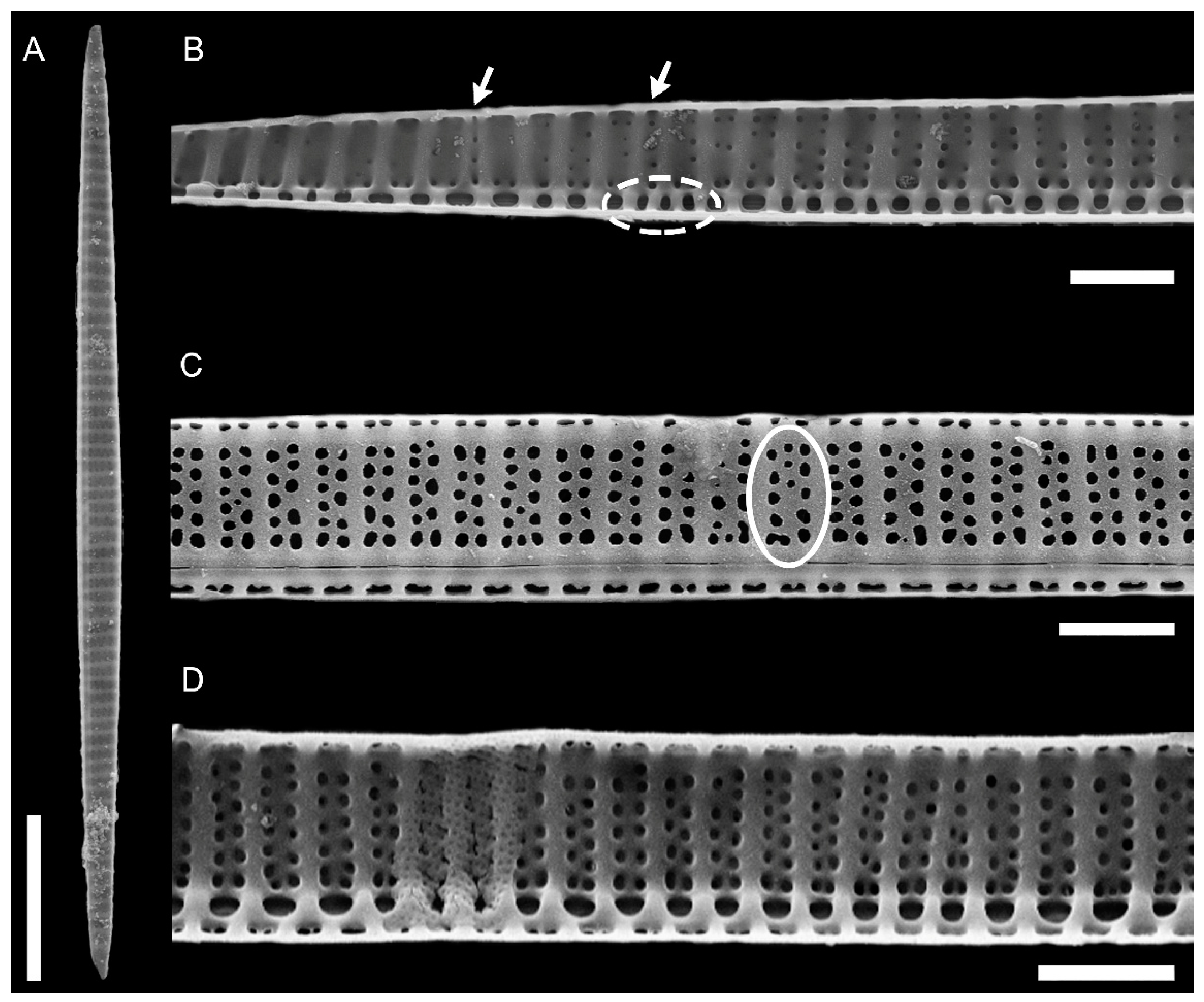
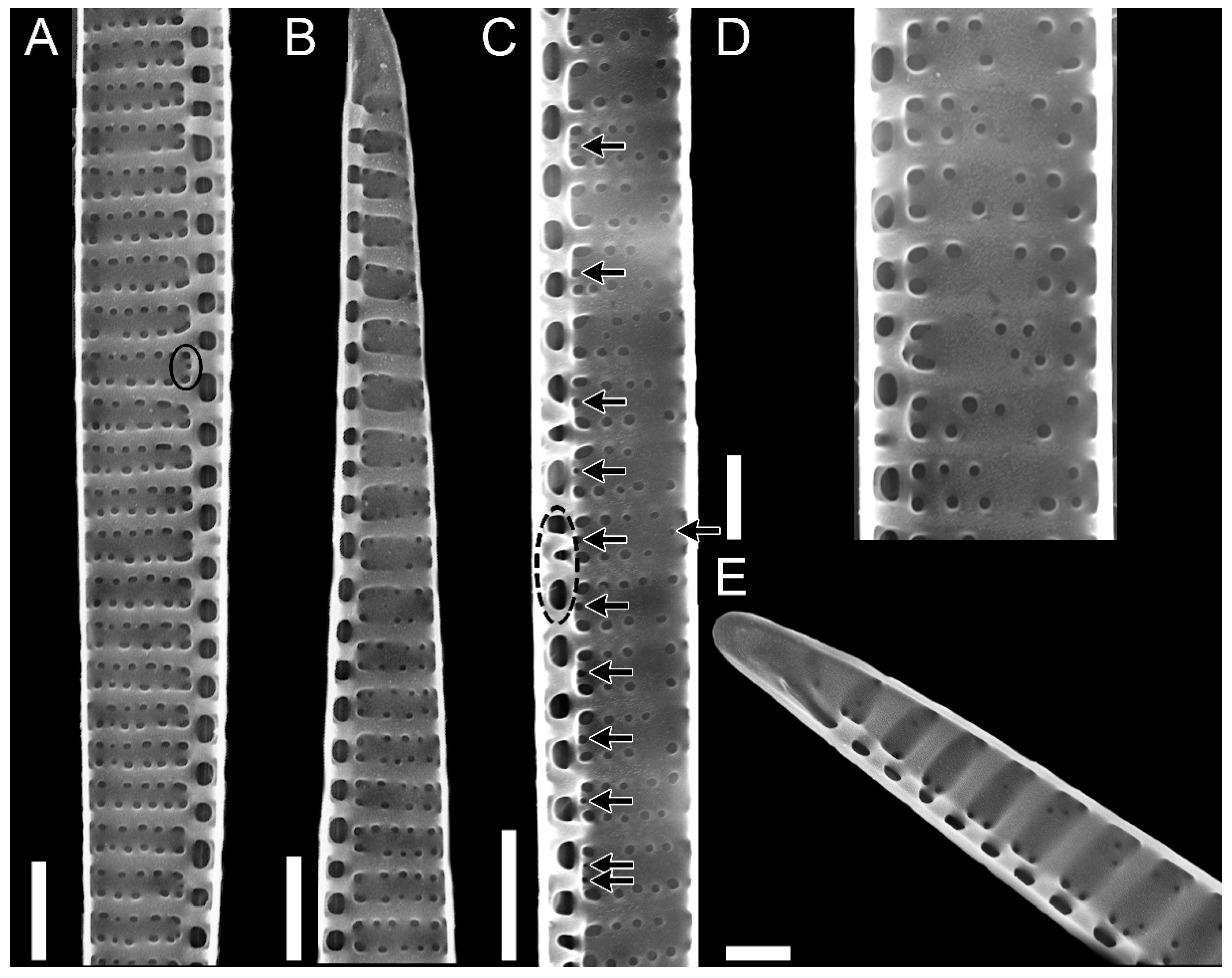
In valve view, valves were slightly asymmetrical with minor hemivalve to major hemivalve area ratios ranging from 0.56 to 0.99 (n = 49, 0.82 ± 0.12) and from 0.57 to 0.94 (n = 13, 0.80 ± 0.12) in cultured and field samples, respectively. The number of fibulae in 10 µm was 5–18, and the number of striae in 10 µm was 9–16 (Table 1). The number of fibulae was generally the same as the number of striae (Figure 2D and Figure 3A). At times, the number of fibulae was higher (Figure 2B and Figure 3C), but it was rarely lower.
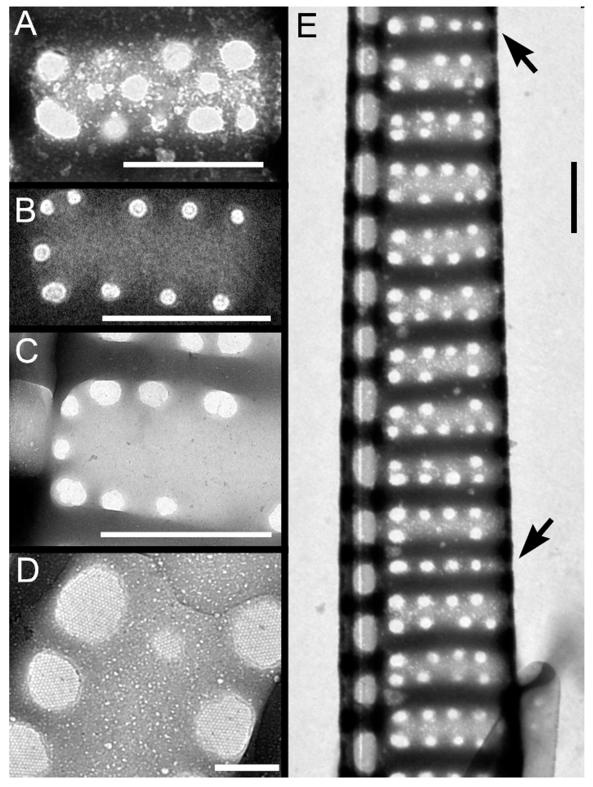
Striae were almost always (72% of the observations) biseriate (Figure 2C,D and Figure 3), with rounded poroids without hymen sectors (Figure 4A–D). Moreover, 3–4 additional poroids in each stria were observed in the 60% of the observations (additional poroids in 10 striae = 2.9 ± 2.79, n = 41) (Figure 3C and Figure 4B,C), forming an incomplete third row (Figure 2C and Figure 4A). The presence of additional poroids was more frequent in the part of the stria close to the raphe (Figure 3C and Figure 4B–D). Sometimes (28%), striae were uniseriate (Figure 2B and Figure 4E). The number of poroids in 1 µm ranged between 1 and 4 (Table 1). Cells with a very low density of poroids were not rare (22%, Figure 3D), and a decreasing density of poroids from the valve center toward the apices was often observed (Figure 2B and Figure 3B,E).
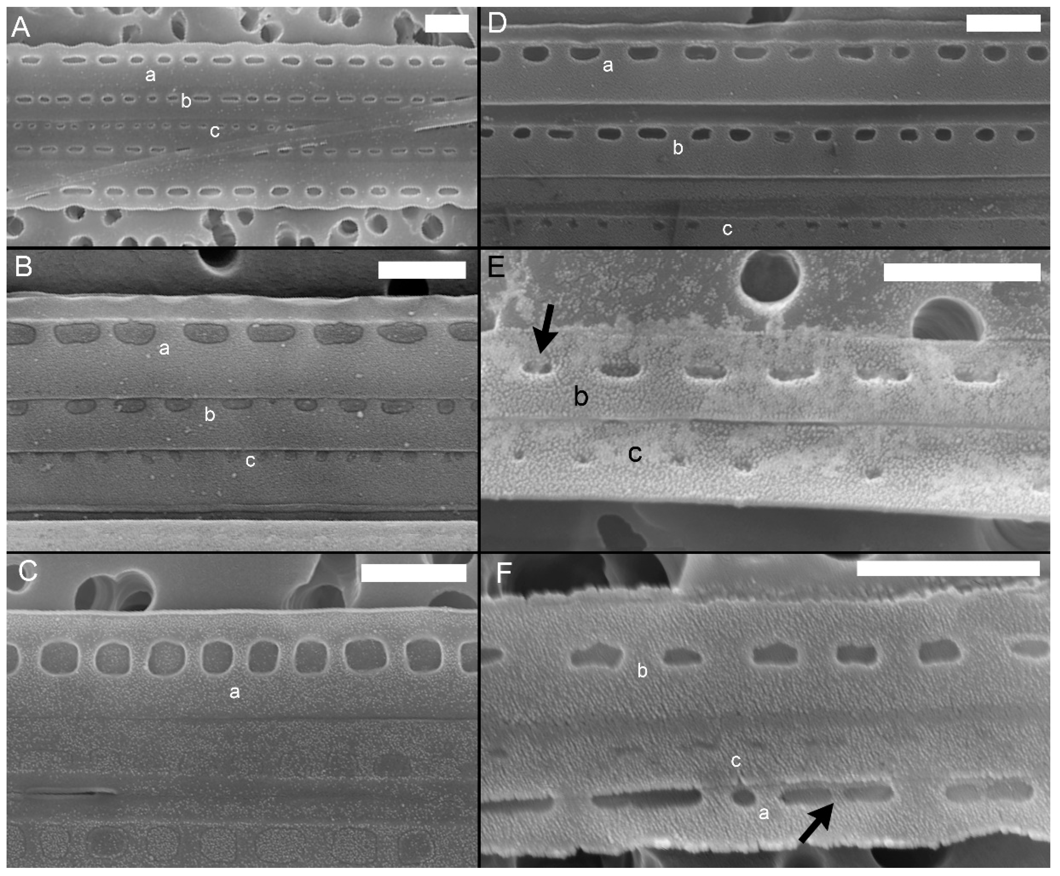
In girdle view, cingular bands showed a wide morphological variability both in the shape of poroids and in the number and size of sectors within them (Figure S10A,B, respectively), even within the same cingular band (Figure 5 and Figure 6). In general, poroids’ dimensions showed a decreasing trend in the abvalvar direction (i.e., poroids in the third cingular band were narrower than those in the first two, and sometimes (25%), no poroids were detected), even if, often (40%), the dimensions and the shape of poroids in the first two cingular bands did not differ so much (but the second band had more striae with smaller poroids than the valvocopula, e.g., Figure 5A). Band striae (12–23 in 10 µm, mean 16.2 ± 2.7, n = 88) were perforated, with (a) oval to rectangular, (b) square, or (c) circular poroids (Figure S10), showing one, two (partially to completely divided), or no hymen sectors (Figure 6). Within each cingular band, either poroids characterized by only one shape (Figure 5C or Figure 5B(a)) or poroids with different shapes (Figure 5D–F) could occur. Nevertheless, cingular bands with only circular poroids were never observed.
All the patterns of hymenation were observed in poroids (Figure 6), except for circular poroids that were observed only without hymen sectors (Figure 6A) or with one sector.

2.2. Molecular Analyses

BLAST results of the 13 LSU sequences confirmed the identification of Pseudo-nitzschia pungens (showing from 99 to 100% of identity with Pseudo-nitzschia pungens LSU sequences from GenBank).
The final alignment was obtained from a total 94 ITS1-5.8s-ITS2 rDNA sequences of P. pungens from different geographical locations, including four sequences from this study.
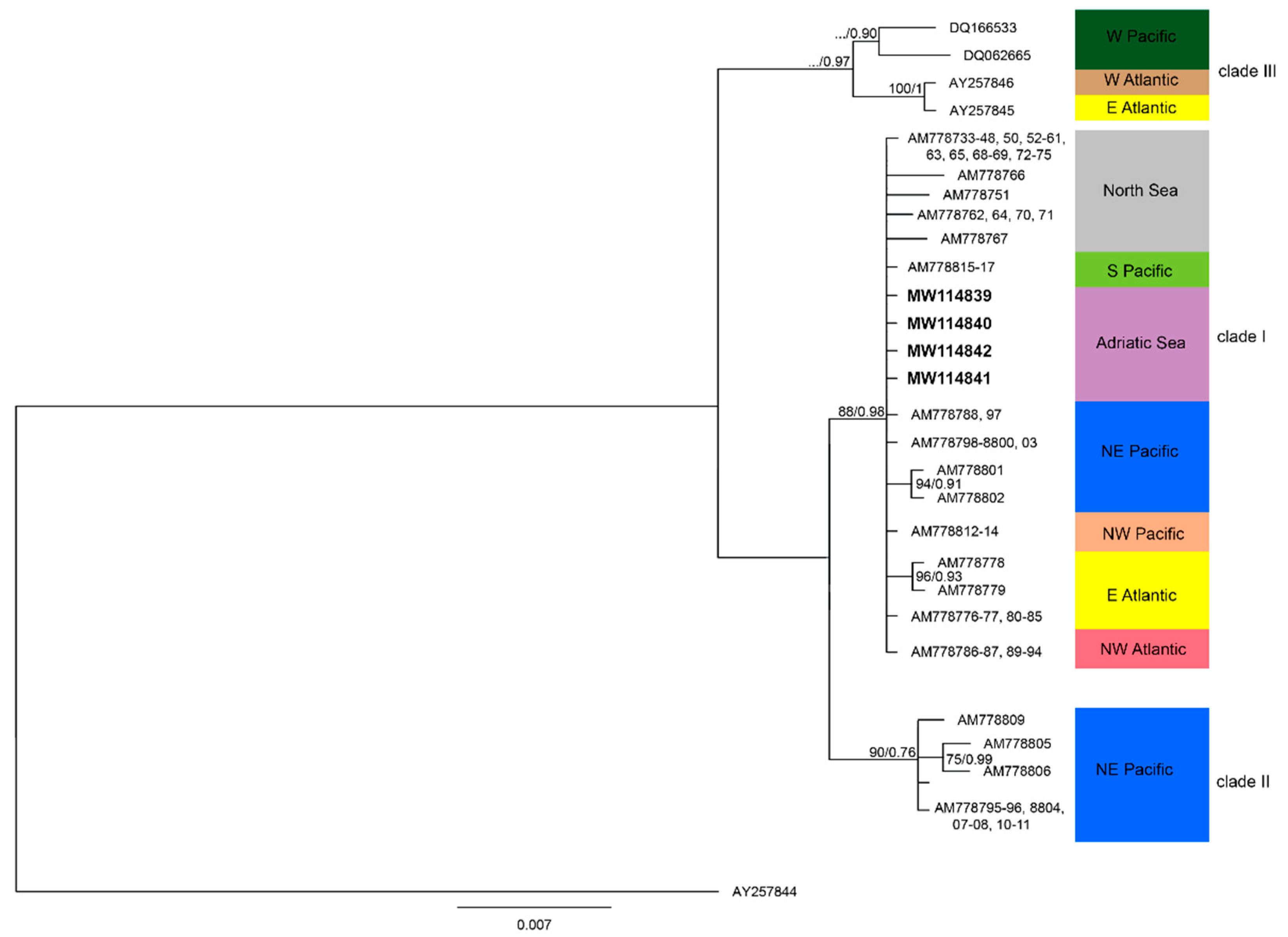
The complete alignment was rooted with P. multiseries (AY257844). The alignment comprised 632 characters, of which 116 were variable sites and 31 were parsimony-informative. Three clades were recovered (Figure 7).
ML and BI analysis revealed the P. pungens strains of this study fell into clade I with a strongly supported bootstrap value (88), and there was a p-distance value of 0.0004 between them and the other sequences of clade I. Clade I was closely related to clade II (mean p-distance value: 0.0143), while the highest p-distance was observed with clade III (mean p-distance value: 0.0319). The highest p-distance among P. pungens clades were observed between clade II and clade III (mean p-distance value: 0.0356).

2.3. Toxin Content

None of the tested strains by LC-MS/MS produced DA in detectable amounts. The LOD varied between 0.09 and 0.02 fg cell−1.

3. Discussion

In this study, a significant morphological variability in Pseudo-nitzschia pungens populations from the northern Adriatic Sea was highlighted by analyzing a consistent number of samples from both field and cultured material.
The molecular characterization revealed that the Adriatic population belonged to the clade I, as the P. pungens nominal variety [24]. However, a number of morphological and ultrastructural details differed from the original description. In the earliest description of P. pungens, an overlap of one-third of the cells in colony (or more) and a transapical axis (TA) wider 3 µm has been reported [47], as well as for all the other species belonging to the seriata group. Nevertheless, the P. pungens from this study revealed an overlap (from one-third to one-sixth) and a TA (rarely reaching 3 µm), strongly differing from the original description of the species, but in agreement with what previously reported for several P. pungens strains, irrespective of clade/variety (Table 1). Indeed, the increasing studies focusing on this species have shown that a number of morphological features, previously indicated as key characters, would be not strictly respected. Some authors have already reported an overlap much lower (ranging from one-fourth to one-sixth) in Canada, the Bay of Fundy [53], the northeastern Adriatic Sea [36], the North Sea [54], the Gulf of Mexico [56], the Danish coastal waters [48], and the Atlantic coast of Portugal (P. pungens var. aveirensis) [27], and a TA often less than 3 µm (down to 1.9), especially in the Mediterranean coast of Greece [29], the northeastern Adriatic Sea [36], and the Atlantic coast of Portugal (P. pungens var. aveirensis) [27] (Table 1).
Although striae are generally reported as biseriate, an incomplete third row of poroids was very common. Such third row of poroids has been detected in other strains included in all other clades/varieties [24,27,47,48]. On the contrary, the presence of uniseriate striae was reported for the first time in this study.
All the main morphological data of the Adriatic P. pungens matched with those of the other clades/varieties, except for (i) the density of band striae in 10 µm (12–23) that slightly diverges from that reported in Pacific coast of USA (California) and in Atlantic coast of Portugal (for P. pungens var. cingulata and P. pungens var. aveirensis, 20–24 and 21–25, respectively) [26,27], (ii) the density of fibulae in 10 µm (5–18) that slightly diverges from that reported in Danish coastal waters (10–20) [48], and (iii) the density of poroids in 1 µm (1–4) that slightly diverges from that reported in the Pacific coast of USA (Washington State) (4–5) [51] (Table 1).
In this study, the cell asymmetry of P. pungens in valve view has been highlighted for the first time, although it could be noticed looking at the TEM micrographs of P. pungens var. aveirensis ([27], Figure 47).
Several ultrastructural features have been indicated as useful to discriminate among P. pungens varieties, such as the shape and pattern of poroids in the cingular bands (Figure S11) [24,27]. While the nominal variety (P. pungens var. pungens) has three cingular bands, all with one row of oval to rectangular poroids [22], P. pungens var. cingulata is characterized by different cingular bands, i.e., the valvocopula has square poroids having two rows of 2–3 hymen sectors, while the second band has rectangular poroids characterized by 1–2 hymen sectors; no descriptions have been reported for the third band [26]. A further poroids’ pattern was described by Churro et al. [27] who established the variety P. pungens var. aveirensis, having two types of cingular bands, valvocopula with square poroids split into two to three parts and the second band with one row of oval (sometimes split) poroids ([27], Figure 51 and Figure 51 insert).
The morphology of the poroids in cingular bands of the Adriatic P. pungens was characterized by a wide variability among strains and within strains and among field samples, showing a number of different combinations of ultrastructural details previously used to discriminate the P. pungens varieties described so far. In fact, cingular bands could have square, circular, or rectangular poroids without or with 1–2 hymen sectors, with several combination of them, even within the same cingular band. As a consequence, this high variability in ultrastructural detail patterns makes such details uninformative for discriminating the N Adriatic P. pungens (belonging to clade I, as the P. pungens nominal variety [24]) from P. pungens var. aveirensis. On the contrary, N Adriatic P. pungens clearly differs from P. pungens var. cingulata.
Poroids’ dimensions in the cingular bands of P. pungens var. cingulata and P. pungens var. aveirensis showed a decreasing trend in the abvalvar direction (i.e., poroids in the third cingular band were narrower than those in the first two cingular bands) [26,27], as often observed in this study for P. pungens from the Adriatic Sea. However, differently from the other varieties, in our samples, it was not rare to observe that, in the dimensions of poroids, the first two cingular bands did not differ, so much that valvocopula and the second cingular band were not always easily discernable. In these cases, the second cingular band could be distinguished because of the slightly higher number of band striae in 10 µm.
Until now, the P. pungens varieties have been approximatively ascribed to the three clades: P. pungens var. pungens~clade I, P. pungens var. cingulata~clade II, and P. pungens var. aveirensis~clade III [24,26,27]. Nevertheless, results of this study suggest that a clade does not necessary correspond to a morphological variety and vice versa. Indeed, Adriatic P. pungens clade I showed a wide morphological variability, covering at least two varieties (i.e., P. pungens var. pungens and P. pungens var aveirensis).
Field and experimental studies showed that diatom frustules can be significantly modified by environmental conditions such that genetically identical individuals could be identified as different species [58,59]. For example, salinity and temperature, among other conditions, have strong effects on frustule morphology, clearly demonstrating the flexibility in diatom morphogenesis [58,60,61]. Some of the morphological features, defined as key characteristics for diatom taxonomic identification, have been shown to be more variable than previously thought [62,63]. In this regard, morphological variability should be investigated under different environmental conditions and in the highest possible number of individuals in order to cover the entire morphological variability within the same population.
The wide morphological and morphometrical variability observed in the P. pungens clade I population from Adriatic Sea, often overlapping characteristics proper of different varieties, could be explained by taking in account the great number of observations that were performed in this study compared with the previous ones (Table 1). Moreover, the analyses were conducted under a wide spectrum of conditions (i.e., from field and cultured samples sampled in different periods and with different strains of different ages). Nevertheless, this wide variability was detected also between different specimens belonging to the same sample, suggesting that this variability was intrinsic and only partially ascribable to the different environmental conditions.
Finally, although some strains of P. pungens clade I were recorded to be toxigenic [29], none of the cultured strains of P. pungens clade I from this study produced DA in detectable amounts, in accordance with what previously observed in NW Adriatic strains [30] and with the results from the official monitoring of shellfish production sites [64] in the NW Adriatic Sea, that only sporadically revealed the presence of DA in shellfish and at very low levels [7,40].

4. Materials and Methods

4.1. Study Area and Sampling

The study area is the coastal station SG01 (43°45.86′ N, 13°13.00′ E) of the Senigallia-Susak transect located in the southern part of the northern Adriatic subbasin, 1.2 nM from the Italian coastline (bottom depth: 12 m), and is included in the LTER (Long-Term Ecological Research) Italian sites, where phytoplankton and environmental parameters have been sampled since 1988.
Sampling was carried out with at about a monthly frequency, from January 2018 to December 2019. Water samples were collected at surface by Niskin bottles, in 250 mL dark glass bottles and preserved by adding 0.8% formaldehyde prefiltered and neutralized with hexamethylenetetramine [65], and stored at 4 °C until analysis was performed. Moreover, net (20 µm mesh) samples were collected for cell isolation (see below).

4.2. Pseudo-Nitzschia Strain Isolation

The isolation of single cells of Pseudo-nitzschia pungens was carried out in 24-well plates following the capillary pipette method [66]. Cultures were maintained at 21 °C with a 12:12 h of light:dark photoperiod and an irradiance of 100 µmol m2 s−1, in sterile filtered seawater enriched with f/2 nutrients [67]. Every month, the algal cultures were checked for their purity and quality and refreshed with fresh culture medium. A total of 14 Pseudo-nitzschia pungens strains were set up.

4.3. DNA Extraction from Algal Culture PCR Amplification and Sequencing

Of the total 14 strains set up, 13 were used for the molecular analyses. Algal cultures were harvested during their late exponential phase and centrifuged at 4000× g for 15 min in order to obtain the pellets. Pellets were extracted using CTAB (N-cetyl-N,N,N-trimethylammoniumbromide) buffer (2% CTAB, 1 M Tris pH 8.0, 0.5 M EDTA pH 8.0, 5M NaCl, 1%) modified from Doyle and Doyle [68].
Extracted DNA was amplified by Polymerase Chain Reaction (PCR) technique, carried out with a SimpliAmpTM Thermal Cycler.
The D1-D3 region was amplified using universal primers: forward primer D1R (5′-ACC CGC TGA ATT TAA GCA TA-3′) and reverse primer D3Ca (5′-ACG AAC GAT TTG CAG GTC AG-3′) [69]. The ITS region was amplified using ITS1 (5′-TCC GTA GGT GAA CCT GCG G-3′) and ITS4 (5′-TCC TCC GCT TAT TGA TAT GC-3′) [70].
PCR products were visualized with UV from an agarose gel (1%).
The PCR conditions for LSU and ITS region were 94 °C for 4 min, followed by 35 cycles of 94 °C for 30 sec, annealing at 60 and 58 °C (for LSU and ITS regions, respectively) for 45 sec, and elongation at 72 °C for 1 min, followed by further elongation at 72 °C for 5 min.

4.4. Sequence Analyses

Taxonomic assignation was performed by blasting each LSU sequences against the GenBank database (NCBI on-line BLAST web interface version 2.9.0+ [71]) to determine the closest known sequences.
Sequences were adjusted for the presence of double peaks by eye with BioEdit [72]. Among the ITS sequences from this study, 4 were aligned with 90 sequences retrieved from GenBank. Pseudo-nitzschia multiseries was the outgroup sequences (Table S2). The selection of outgroup sequences was based on the findings by Lim et al. [21].
Alignments were made with ClustalW [73] using the default setting and were then edited manually. Regions that did not fit with the others were excluded from the phylogenetic analyses. Two independent analyses were used to conduct the ITS1-5.8s-IT2 phylogeny: Maximum Likelihood (ML) and Bayesian Inference (BI). The best nucleotide substitution model was tested with Partitionfinder 2 [74]. The generalized time-reversible evolution model (GTR+G) was used for the construction of the RAxML phylogenetic analysis, and Kimura’s two-parameter model (K80 + I) was used for the Bayesian inference tree. ML analyses were carried out with RAxML [75] 1000 pseudo replicates through Cipress portal [76].
Bayesian analyses were carried out using MrBayes 3.2 [77] with 3,000,000 Markov chain and Monte Carlo generations, a sample frequency of 1500, and a diagnosing frequency of 1000. The 50% majority rule consensus tree was constructed discarding the first 25% of samples. Posterior probabilities were calculated to measure tree strength.
The distance estimation matrix between groups was calculated with the p-distance method using the default setting of MEGA 7 [78].

4.5. Morphological Characterization

4.5.1. Light Microscopy Analyses

Pseudo-nitzschia cells were measured at 1000× magnification using an inverted microscope (ZEISS Axiovert 135) equipped with phase contrast. The Apical Axis (AA) and the overlapping region of the cells in a chain were measured in at least 100 cells from cultured strains and field samples.

4.5.2. Ultrastructural Characterization (TEM and SEM)

Samples for TEM and SEM analyses were harvested from cultured strains in exponential growth phase, collected during 2018–2019 (Table S3) and from field net samples collected in February and May 2019.
Samples were acid-cleaned following von Stosch’s protocol [18]. A drop (2 µL) of the cleaned material was placed on a grid and on a stub and observed with a Philips TEM 400 microscope and a SEM (FE-SEM; Zeiss Supra 40, Carl Zeiss AG, Oberkochen, Germany), respectively.
Several cells were measured (see Table 1 for the number of cells used for each measurement) both from cultured and field samples for Transapical Axis (TA), fibulae, striae, and poroids’ density in both valves and cingular bands with particular focus to valvocopula.
A measurement of valval symmetry was performed on SEM micrographs, calculating the cell surface with an image analysis software and using the formula as follows: Cells in valval view were divided into two hemivalves by the apical axis, crossing the half of the transapical axis. Then, the symmetry was expressed as the ratio of the two hemivalve areas (minor hemivalve:major hemivalve, Figure S12). Valves were asymmetric when the ratio ≠ 1.

4.6. Toxin Content

4.6.1. Chemicals and Standards

The acetonitrile (MeACN) and formic acid (FA) were of LC-MS grade, and the methanol (MeOH) was of HPLC grade. Water was distilled and passed through a MilliQ water purification system (DIW) (Millipore Ltd., Bedford, MA, USA).
Certified reference material for DA, CRM-DA-g (103.3 µg mL−1), was purchased from the Institute of Biotoxin Metrology at the National Research Council of Canada (NRCC, Halifax, Nova Scotia, Canada). Calibration solutions of DA were prepared from serial dilutions of the reference material in DIW.

4.6.2. DA Extraction

Chemical analysis of Pseudo-nitzschia pungens needs a large quantity of cells, so each strain was grown in an increasing volume up to 2 L to achieve abundances, ranging from 17 × 104 to 61 × 104 cells mL−1 among the cultured strains.
The strains were grown in the same culture conditions reported above. Cells were harvested from the early stationary growth phase. Algal pellets of 4 P. pungens strains (Table S3) were extracted using a mixture of MeOH/H2O (50:50 v/v), following the official EU-RL RP-LC-UV method (EURLMB 2008), for the determination of DA in shellfish and finfish.
All culture volume (2 L) was centrifuged for 20 min at 2500× g (4 °C) in 40 centrifuge tubes (50 mL volume). Pellets were combined and extracted with 5 mL of MeOH/H2O (50:50 v/v), vortex-mixed for 1 min, and bath-sonicated for 10 min. After sonication, the aliquot was centrifuged for 10 min at 2500× g (4 °C), and the supernatant was transferred to a 100 mL evaporation flask. Pellet extraction was repeated three times, and the supernatants were combined and evaporated to dryness. The residue was reconstituted in 1 mL of MeOH/H2O (50:50 v/v) and filtered through a 0.2 µm syringe filter (Minisart, Sartorius, Germany) for LC-MS/MS analysis.

4.6.3. LC-MS/MS Analysis

LC-MS/MS analyses were performed using a hybrid triple-quadrupole/linear ion trap 3200 QTRAP mass spectrometer (AB Sciex, Darmstadt, Germany) equipped with a Turbo V source and an electrospray ionization (ESI) probe. The mass spectrometer was coupled to an Agilent model 1200 LC instrument (Palo Alto, CA, USA), which included a solvent reservoir, inline degasser, quaternary pump, refrigerated autosampler, and column oven.
The method was implemented following the conditions described by Mafra et al. [46], which were properly modified. LC separation was performed using a Gemini®® NX-C18 column (2 mm × 100 mm, 3 µm particle size; Phenomenex, Torrance, CA, USA), set at 40 °C, with a flowrate of 0.4 mL min−1. Mobile phase A was DIW and B MeACN, both containing 0.2% of FA. Gradient elution was adopted, as described below: From 10% to 20% B in 5 min, from 20% to 35% B in 1 min, then hold for 6 min, return to the original conditions at 13 min, and hold for 7 min before the next injection.
Infusion experiments were performed using CRM-DA-g to set the turbo IonSpray source parameters as follows: Nebulizer Gas (GS1) 50 psi, Auxiliary Gas (GS2) 60 psi, Temperature (TEM) 600 °C, Ion Spray Voltage (IS) 5000 V, Curtain Gas (CUR) 20 psi.
DA was detected using Multiple Reaction Monitoring (MRM) in positive ion mode by selecting the following transitions: m/z 312.2→266.1, m/z 312.2→220.1, and m/z 312.2→161.1. In addition, the pseudotransition m/z 334.2→334.2 of sodium adduct [DA + Na]+ was monitored to investigate ion suppression due to salts. A declustering potential (DP) of 60 V and a collision energy (CE) of 30 V were used for all transitions.
LOQ, calculated assuming a signal/noise (S/N) ratio of 10 was 10 ng mL−1, while LOD (S/N ratio of 3) was 3 ng mL−1.

Supplementary Materials

The following are available online at https://www.mdpi.com/2223-7747/9/11/1420/s1, Figure S1: Pseudo-nitzschia pungens SEM micrographs (Strain ID = 01185). (A) Valve view. (B) Detail of the biseriate striae with low density of poroids. (C) Girdle view. Scale bar = 2 µm, Figure S2: Pseudo-nitzschia pungens SEM micrographs (Strain ID = 01186). (A) Valve view. (B) Detail of the biseriate striae. (C) Girdle view: dimensions and shape of poroids in the first two cingular bands are similar. Scale bar = 2 µm, Figure S3: Pseudo-nitzschia pungens SEM micrographs (Strain ID = 01189). (A) Valve view. (B) Cingular bands showing square and rectangular poroids. Scale bar = 2 µm, Figure S4: Pseudo-nitzschia pungens SEM micrographs (Strain ID = 031832). (A–C) Valve view showing different patterns of symmetry (highlighted by the white lines). Scale bar = 10 µm, Figure S5: Pseudo-nitzschia pungens SEM micrographs (Strain ID = 04191). (A) Valve view. (B, C) Detail of the biseriate striae with different density of poroids. (D) Girdle view: dimensions and shape of poroids in the first two cingular bands are similar, while the third cingular band has smaller poroids. Scale bar = 10 µm (A); 2 µm (B, D); 1 µm (C), Figure S6: Pseudo-nitzschia pungens SEM micrographs (Strain ID = 04194). (A) Valve view. (B) Detail of the biseriate striae. (C, D) Girdle view: (C) dimensions and shape of poroids in the first two cingular bands are similar; (D) poroids with different shapes and hymenation occurring in the same cingular band. Scale bar = 10 µm (A); 2 µm (B–D), Figure S7: Pseudo-nitzschia pungens SEM micrographs (Strain ID = 04196). (A) Valve view of the internal valve face. (B) Detail of the biseriate striae with low density of poroids. (C) Girdle view showing a decreasing trend in abvalvar direction of the poroids’ dimensions. Scale bar = 10 µm (A); 2 µm (B–C), Figure S8: Pseudo-nitzschia pungens SEM micrographs (Strain ID = 05197). (A) Valve view. (B) Detail of the biseriate striae. (C, D) Girdle view: (C) dimensions and shape of poroids in the first two cingular bands are similar, while the third cingular band has smaller poroids; (D) poroids with different hymenation occurring in the same cingular band. Scale bar = 10 µm (A); 1 µm (B, D); 2 µm (C), Figure S9: Pseudo-nitzschia pungens SEM micrographs (Strain ID = 05199). (A) Valve view. (B) Detail of the biseriate striae. (C, D) Girdle view: (C) dimensions and shape of poroids in the first two cingular bands are similar; (D) poroids with different shape and hymenation occurring in the same cingular band. Scale bar = 10 µm (A); 2 µm (B, C); 0.3 µm (C), Figure S10: Schematic drawing describing the poroids’ morphologies of N Adriatic Sea Pseudo-nitzschia pungens. (A) Shapes of poroids: Type a: oval to rectangular. Type b: square poroids. Type c: circular. (B) Pattern of hymenation: Type I: no hymen sectors. Type II: one hymen sectors. Type III: two hymen sectors. Type IV: one partially divided hymen sector, Figure S11: Drawing representing the ultrastructure of poroids in the cingular bands of the three P. pungens varieties: (A) var. pungens [22]; (B) var. cingulata [26]; (C) var. aveirensis [27]. VC: valvocopula; II: second cingular band; III: third cingular band. No information about the third cingular band is available in var. cingulata and var. aveirensis, Figure S12: Pseudo-nitzschia pungens cell divided into two hemivalves. Green area: minor hemivalve; red area: major hemivalve, Table S1: Morphometric characteristics of Pseudo-nitzschia pungens strains from the coastal site SG1 of LTER Senigallia transect. n.r., not reported, Table S2: List of sequences retrieved from Genbank for the construction of the Bayesian consensus tree. Strains from this study are in bold. (c) strains of which PCR products were cloned, Table S3: List of Pseudo-nitzschia pungens strains from the coastal site SG1 of LTER Senigallia transect, cleaned for morphological characterization with TEM and SEM. Strain IDs indicated in bold are those analyzed for toxin content.

Author Contributions

Conceptualization, S.A. and C.T.; methodology, S.G., T.R., M.S. and S.B.; formal analysis, S.A. and S.G.; investigation, T.R., S.G., M.S. and S.B.; resources, S.A. and C.T.; data curation, S.A.; writing—original draft preparation, S.A.; writing—review and editing, S.A. and C.T.; visualization, S.A.; supervision, C.T.; project administration, S.A.; funding acquisition, S.A., M.S. and C.T. All authors have read and agreed to the published version of the manuscript.

Funding

This research was partially funded by the Italian Ministry of Health (Ricerca Finalizzata 2016), grant number GR-2016-02363211. The cruises carried out with the M/N Actea were entirely funded by the Department of Life and Environmental Sciences (Università Politecnica delle Marche).

Acknowledgments

We are grateful to LTER-ITALY (Italian Long-Term Ecological Research Network) and to ECOSS (Ecological Observing System in the Adriatic Sea) project. Thanks to the crew of M/N Actea for their support during sampling activities. Special thanks to Luigi Gobbi for assistance with the EM.

Conflicts of Interest

The authors declare no conflict of interest. The funders had no role in the design of the study; in the collection, analyses, or interpretation of data; in the writing of the manuscript, or in the decision to publish the results.

References

  1. Guiry, M.D.; Guiry, G.M. AlgaeBase Worldwide Electronic Publication, Nat. Univ. Ireland, Galway. 2020. Available online: http://www.algaebase.org (accessed on 10 September 2020).
  2. Bates, S.S.; Hubbard, K.A.; Lundholm, N.; Montresor, M.; Leaw, C.P. Pseudo-nitzschia, Nitzschia, and domoic acid: New research since 2011. Harmful Algae 2018, 79, 3–43. [Google Scholar]
  3. Lundholm, N. Bacillariophyceae. In IOC-UNESCO Taxonomic Reference List of Harmful Micro Algae. 2020. Available online: http://www.marinespecies.org/hab (accessed on 10 September 2020).
  4. Lefebvre, K.A.; Robertson, A. Domoic acid and human exposure risks: A review. Toxicon 2010, 56, 218–230. [Google Scholar] [PubMed]
  5. Trainer, V.L.; Bates, S.S.; Lundholm, N.; Thessen, A.E.; Adams, N.G.; Cochlan, W.P.; Trick, C.G. Pseudo-nitzschia physiological ecology, phylogeny, toxicity, monitoring and impacts on ecosystem health. Harmful Algae 2012, 14, 271–300. [Google Scholar]
  6. Lundholm, N.; Moestrup, Ø.; Hasle, G.R.; Hoef-Emden, K. A study of the Pseudo-nitzschia pseudodelicatissima/cuspidata complex (Bacillariophyceae): What is P. pseudodelicatissima? J. Phycol. 2003, 39, 797–813. [Google Scholar]
  7. Marić, D.; Ljubešić, Z.; Godrijan, J.; Viličić, D.; Ujević, I.; Precali, R. Blooms of the potentially toxic diatom Pseudo-nitzschia calliantha Lundholm, Moestrup & Hasle in coastal waters of the northern Adriatic Sea (Croatia). Estuar. Coast. Shelf Sci. 2011, 92, 323–331. [Google Scholar]
  8. Kim, J.H.; Park, B.S.; Kim, J.H.; Wang, P.; Han, M.S. Intraspecific diversity and distribution of the cosmopolitan species Pseudo-nitzschia pungens (Bacillariophyceae): Morphology, genetics, and ecophysiology of the three clades. J. Phycol. 2015, 51, 159–172. [Google Scholar] [PubMed]
  9. Amato, A.; Montresor, M. Morphology, phylogeny, and sexual cycle of Pseudo-nitzschia mannii sp. nov. (Bacillariophyceae): A pseudo-cryptic species within the P. pseudodelicatissima complex. Phycologia 2008, 47, 487–497. [Google Scholar]
  10. Lundholm, N.; Bates, S.S.; Baugh, K.A.; Bill, B.D.; Connell, L.B.; Léger, C.; Trainer, V.L. Cryptic and pseudo--cryptic diversity in diatoms with descriptions of Pseudo-nitzschia hasleana sp. nov. and P. fryxelliana sp. nov.1. J. Phycol. 2012, 48, 436–454. [Google Scholar]
  11. Orive, E.; Pérez-Aicua, L.; David, H.; García-Etxebarria, K.; Laza-Martínez, A.; Seoane, S.; Miguel, I. The genus Pseudo-nitzschia (Bacillariophyceae) in a temperate estuary with description of two new species: Pseudo-nitzschia plurisecta sp. nov. and Pseudo-nitzschia abrensis sp. nov.1. J. Phycol. 2013, 49, 1192–1206. [Google Scholar]
  12. Teng, S.T.; Lim, H.C.; Lim, P.T.; Dao, V.H.; Bates, S.S.; Leaw, C.P. Pseudo-nitzschia kodamae sp. nov. (Bacillariophyceae), a toxigenic species from the Strait of Malacca, Malaysia. Harmful Algae 2014, 34, 17–28. [Google Scholar]
  13. Teng, S.T.; Lim, P.T.; Lim, H.C.; Rivera-Vilarelle, M.; Quijano-Scheggia, S.; Takata, Y.; Quilliam, M.A.; Wolf, M.; Bates, S.S.; Leaw, C.P. A non-toxigenic but morphologically and phylogenetically distinct new species of Pseudo-nitzschia, P. sabit sp. nov. (Bacillariophyceae). J. Phycol. 2015, 51, 706–725. [Google Scholar] [PubMed]
  14. Percopo, I.; Ruggiero, M.V.; Balzano, S.; Gourvil, P.; Lundholm, N.; Siano, R.; Tammilehto, A.; Vaulot, D.; Sarno, D. Pseudo-nitzschia arctica sp. nov.; a new cold-water cryptic Pseudo-nitzschia species within the P. pseudodelicatissima complex. J. Phycol. 2016, 52, 184–199. [Google Scholar] [PubMed]
  15. Ajani, P.A.; Verma, A.; Lassudrie, M.; Doblin, M.A.; Murray, S.A. A new diatom species P. hallegraeffii sp. nov. belonging to the toxic genus Pseudo-nitzschia (Bacillariophyceae) from the East Australian Current. PLoS ONE 2018, 13, e0195622. [Google Scholar]
  16. Li, Y.; Dong, H.C.; Teng, S.T.; Bates, S.S.; Lim, P.T. Pseudo-nitzschia nanaoensis sp. nov. (Bacillariophyceae) from the Chinese coast of the South China Sea. J. Phycol. 2018, 54, 918–922. [Google Scholar]
  17. Lim, H.C.; Tan, S.N.; Teng, S.T.; Lundholm, N.; Orive, E.; David, H.; Quijano-Scheggia, S.; Leong, S.C.Y.; Wolf, M.; Bates, S.S.; et al. Phylogeny and species delineation in the marine diatom Pseudo-nitzschia (Bacillariophyta) using cox1, LSU, and ITS 2 rRNA genes: A perspective in character evolution. J. Phycol. 2018, 54, 234–248. [Google Scholar] [PubMed]
  18. Hasle, G.R.; Syvertsen, E.E. Marine diatoms. In Identifying Marine Phytoplankton; Thomas, C.R., Ed.; Academic Press: San Diego, CA, USA, 1997; pp. 5–385. [Google Scholar]
  19. Hasle, G.R. Are most of the domoic acid-producing species of the diatom genus Pseudo-nitzschia cosmopolites? Harmful Algae 2002, 1, 137–146. [Google Scholar]
  20. Anderson, C.R.; Sapiano, M.R.; Prasad, M.B.K.; Long, W.; Tango, P.J.; Brown, C.W.; Murtugudde, R. Predicting potentially toxigenic Pseudo-nitzschia blooms in the Chesapeake Bay. J. Mar. Syst. 2010, 83, 127–140. [Google Scholar]
  21. Lim, H.C.; Lim, P.T.; Teng, S.T.; Bates, S.S.; Leaw, C.P. Genetic structure of Pseudo-nitzschia pungens (Bacillariophyceae) populations: Implications of a global diversification of the diatom. Harmful Algae 2014, 37, 142–152. [Google Scholar]
  22. Hasle, G.R. Pseudo-nitzschia pungens and P. multiseries (Bacillariophyceae): Nomenclatural history, morphology, and distribution. J. Phycol. 1995, 31, 428–435. [Google Scholar]
  23. Hasle, G.R. Nomenclatural notes on marine planktonic diatoms. The family Bacillariaceae. Nova Hedwigia Beih. 1993, 106, 315–321. [Google Scholar]
  24. Casteleyn, G.; Chepurnov, V.A.; Leliaert, F.; Mann, D.G.; Bates, S.S.; Lundholm, N.; Lesley, R.; Koen, S.; Vyverman, W. Pseudo-nitzschia pungens (Bacillariophyceae): A cosmopolitan diatom species? Harmful Algae 2008, 7, 241–257. [Google Scholar]
  25. Casteleyn, G.; Leliaert, F.; Backeljau, T.; Debeer, A.E.; Kotaki, Y.; Rhodes, L.; Lundholm, N.; Sabbe, K.; Vyverman, W. Limits to gene flow in a cosmopolitan marine planktonic diatom. Proc. Natl. Acad. Sci. USA 2010, 107, 12952–12957. [Google Scholar] [PubMed] [Green Version]
  26. Villac, M.C.; Fryxell, G.A. Pseudo-nitzschia pungens var. cingulata var. nov. (Bacillariophyceae) based on field and culture observations. Phycologia 1998, 37, 269–274. [Google Scholar]
  27. Churro, C.I.; Carreira, C.C.; Rodrigues, F.J.; Craveiro, S.C.; Calado, A.J.; Casteleyn, G.; Lundholm, N. Diversity and abundance of potentially toxic Pseudo-nitzschia Peragallo in Aveiro coastal lagoon, Portugal and description of a new variety, P. pungens var. aveirensis var. nov. Diatom Res. 2009, 24, 35–62. [Google Scholar]
  28. Quijano-Scheggia, S.; Garcés, E.; Andree, K.B.; De la Iglesia, P.; Diogène, J.; Fortuño, J.M.; Camp, J. Pseudo-nitzschia species on the Catalan coast: Characterization and contribution to the current knowledge of the distribution of this genus in the Mediterranean Sea. Sci. Mar. 2010, 74, 395–410. [Google Scholar]
  29. Moschandreou, K.K.; Baxevanis, A.D.; Katikou, P.; Papaefthimiou, D.; Nikolaidis, G.; Abatzopoulos, T.J. Inter-and intra-specific diversity of Pseudo-nitzschia (Bacillariophyceae) in the northeastern Mediterranean. Eur. J. Phycol. 2012, 47, 321–339. [Google Scholar]
  30. Penna, A.; Casabianca, S.; Perini, F.; Bastianini, M.; Riccardi, E.; Pigozzi, S.; Scardi, M. Toxic Pseudo-nitzschia spp. in the northwestern Adriatic Sea: Characterization of species composition by genetic and molecular quantitative analyses. J. Plankton Res. 2013, 35, 352–366. [Google Scholar]
  31. Casteleyn, G.; Adams, N.G.; Vanormelingen, P.; Debeer, A.E.; Sabbe, K.; Vyverman, W. Natural hybrids in the marine diatom Pseudo-nitzschia pungens (Bacillariophyceae): Genetic and morphological evidence. Protist 2009, 160, 343–354. [Google Scholar]
  32. Quiroga, I. Pseudo-nitzschia blooms in the Bay of Banjuls-sur-mer, northwestern Mediterranean Sea. Diatom Res. 2006, 21, 91–104. [Google Scholar]
  33. Andree, K.B.; Fernández-Tejedor, M.; Elandaloussi, L.M.; Quijano-Scheggia, S.; Sampedro, N.; Garcés, E.; Camp, J.; Diogene, J. Quantitative PCR coupled with melt curve analysis for detection of selected Pseudo-nitzschia spp. (Bacillariophyceae) from the northwestern Mediterranean Sea. Appl. Environ. Microbiol. 2011, 77, 1651–1659. [Google Scholar]
  34. Quijano-Scheggia, S.; Garcés, E.; Sampedro, N.; Flo, E.; Fernandez-Tejedor, M.; Diogène, J.; Camp, J. Bloom dynamics of the genus Pseudo-nitzschia (Bacillariophyceae) in two coastal bays (NW Mediterranean Sea). Sci. Mar. 2008, 72, 577–590. [Google Scholar]
  35. Quijano-Scheggia, S.; Garcés, E.; Sampedro, N.; van Lenning, K.; Flo, E.; Andree, K.; Fortuño, J.M.; Camp, J. Identification and characterisation of the dominant Pseudo-nitzschia species (Bacillariophyceae) along the NE Spanish coast (Catalonia, NW Mediterranean). Sci. Mar. 2008, 72, 343–359. [Google Scholar]
  36. Ljubešić, Z.; Bosak, S.; Viličić, D.; Borojević, K.K.; Marić, D.; Godrijan, J.; Ujević, I.; Peharec, P.; Ɖakovac, T. Ecology and taxonomy of potentially toxic Pseudo-nitzschia species in Lim Bay (north-eastern Adriatic Sea). Harmful Algae 2011, 10, 713–722. [Google Scholar]
  37. Pugliese, L.; Casabianca, S.; Perini, F.; Andreoni, F.; Penna, A. A high-resolution melting method for the molecular identification of the potentially toxic diatom Pseudo-nitzschia spp. in the Mediterranean Sea. Sci. Rep. 2017, 7, 4259. [Google Scholar]
  38. Dermastia, T.T.; Cerino, F.; Stanković, D.; Francé, J.; Ramšak, A.; Tušek, M.Ž.; Beran, A.; Natali, V.; Cabrini, M.; Mozetič, P. Ecological time series and integrative taxonomy unveil seasonality and diversity of the toxic diatom Pseudo-nitzschia H. Peragallo in the northern Adriatic Sea. Harmful Algae 2020, 93, 101773. [Google Scholar]
  39. Ciminiello, P.; Dell’Aversano, C.; Fattorusso, E.; Forino, M.; Magno, G.S.; Tartaglione, L.; Quilliam, M.A.; Tubaro, A.; Poletti, R. Hydrophilic interaction liquid chromatography/mass spectrometry for determination of domoic acid in Adriatic shellfish. Rapid Commun. Mass Spectrom. 2005, 19, 2030–2038. [Google Scholar]
  40. Arapov, J.; Ujević, I.; Pfannkuchen, D.M.; Godrijan, J.; Bakrač, A.; Gladan, Ž.N.; Marasović, I. Domoic acid in phytoplankton net samples and shellfish from the Krka River estuary in the Central Adriatic Sea. Mediterr. Mar. Sci. 2016, 17, 340–350. [Google Scholar]
  41. European Council Regulation No 853/2004 of the European Parliament and of the Council of 29 April 2004 laying down specific hygiene rules for on the hygiene of foodstuffs. Off. J. Eur. Union 2004, 47, 55–205. Available online: http://data.europa.eu/eli/reg/2004/853/oj (accessed on 21 October 2020).
  42. Arapov, J.; Skejić, S.; Bužančić, M.; Bakrač, A.; Vidjak, O.; Bojanić, N.; Ujević, I.; Gladan, Ž.N. Taxonomical diversity of Pseudo-nitzschia from the Central Adriatic Sea. Phycol. Res. 2017, 65, 280–290. [Google Scholar]
  43. Totti, C.; Romagnoli, T.; Accoroni, S.; Coluccelli, A.; Pellegrini, M.; Campanelli, A.; Grilli, F.; Marini, M. Phytoplankton communities in the northwestern Adriatic Sea: Interdecadal variability over a 30-years period (1988–2016) and relationships with meteoclimatic drivers. J. Mar. Syst. 2019, 193, 137–153. [Google Scholar]
  44. Quilliam, M.A.; Wright, J.L.C. The amnesic shellfish poisoning mystery. Anal. Chem. 1989, 61, 1053–1059. [Google Scholar] [CrossRef]
  45. AESAN EU-Harmonised Standard Operating Procedure for Determination of Domoic Acid in Shellfish and Finfish by RP-HPLC Using UV Detection; Version 1 June 2008; Agencia Española de Seguridad Alimentaria y Nutrición: Vigo, Spain, 2008.
  46. Mafra, L.L., Jr.; Léger, C.; Bates, S.S.; Quilliam, M.A. Analysis of trace levels of domoic acid in seawater and plankton by liquid chromatography without derivatization, using UV or mass spectrometry detection. J. Chromatogr. A 2009, 1216, 6003–6011. [Google Scholar] [CrossRef] [PubMed]
  47. Hasle, G.R.; Lange, C.B.; Syvertsen, E.E. A review of Pseudo-nitzschia, with special reference to the Skagerrak, North Atlantic, and adjacent waters. Helgol. Meeresunt. 1996, 50, 131–175. [Google Scholar] [CrossRef] [Green Version]
  48. Skov, J.; Lundholm, N.; Moestrup, Ø.; Larsen, J. Potentially toxic phytoplankton 4. The diatom genus Pseudo-nitzschia (Diatomophyceae/BaciIlariophyceae). In ICES Identification Leaflets for Plankton; Lindley, A., Ed.; International Council for the Exploration of the Sea: Copenhagen, Denmark, 1999; Leaflet no. 185; pp. 1–23. [Google Scholar]
  49. Stonik, I.V.; Orlova, T.Y.; Shevchenko, O.G. Morphology and ecology of the species of the genus Pseudo-nitzschia (Bacillariophyta) from Peter the Great Bay, Sea of Japan. Russ. J. Mar. Biol. 2001, 27, 362–366. [Google Scholar] [CrossRef]
  50. Stonik, I.V.; Orlova, T.Y.; Lundholm, N. Diversity of Pseudo-nitzschia H. Peragallo from the western North Pacific. Diatom Res. 2011, 26, 121–134. [Google Scholar] [CrossRef]
  51. Stehr, C.M.; Connell, L.; Baugh, K.A.; Bill, B.D.; Adams, N.G.; Trainer, V.L. Morphological, toxicological and genetic differences among Pseudo-nitzschia (Bacillariophyceae) species in inland embayments and outer coastal waters of Washington State, USA. J. Phycol. 2002, 38, 55–65. [Google Scholar] [CrossRef]
  52. Fryxell, G.A.; Hasle, G.R. Taxonomy of harmful diatoms. In Manual on Harmful Marine Microalgae, 2nd ed.; Monographs on Oceanographic Methodology, Hallegraeff, G.M., Andersen, D.M., Cembella, A.D., Eds.; UNESCO: Paris, France, 2004; pp. 465–509. [Google Scholar]
  53. Kaczmarska, I.; LeGresley, M.M.; Martin, J.L.; Ehrman, J. Diversity of the diatom genus Pseudo-nitzschia Peragallo in the Quoddy Region of the Bay of Fundy, Canada. Harmful Algae 2005, 4, 1–19. [Google Scholar] [CrossRef]
  54. Casteleyn, G. Species Structure and Biogeography of Pseudo-nitzschia pungens. Ph.D. Dissertation, Ghent University, Ghent, Belgium, January 2009. [Google Scholar]
  55. Fernandes, L.F.; Brandini, F.P. The potentially toxic diatom Pseudo-nitzschia H. Peragallo in the Paraná and Santa Catarina states, southern Brazil. Iheringia Ser. Bot. 2010, 65, 47–65. [Google Scholar]
  56. Parsons, M.L.; Okolodkov, Y.B.; Castillo, J.A.A. Diversity and morphology of the species of Pseudo-nitzschia (Bacillariophyta) of the National Park Sistema Arrecifal Veracruzano, SW Gulf of Mexico. Acta Bot. Mex. 2012, 98, 51–72. [Google Scholar] [CrossRef]
  57. Fernandes, L.F.; Hubbard, K.A.; Richlen, M.L.; Smith, J.; Bates, S.S.; Ehrman, J.; Léger, C.; Mafra, L.L.; Kulis, D.; Quillam, M.; et al. Diversity and toxicity of the diatom Pseudo-nitzschia Peragallo in the Gulf of Maine, Northwestern Atlantic Ocean. Deep-Sea Res. II 2014, 103, 139–162. [Google Scholar] [CrossRef] [Green Version]
  58. Trobajo, R.; Cox, E.J.; Quintana, X.D. The effects of some environmental variables on the morphology of Nitzschia frustulum (Bacillariophyta), in relation its use as a bioindicator. Nova Hedwigia 2004, 79, 433–445. [Google Scholar] [CrossRef]
  59. Cox, E.J. Diatom identification in the face of changing species concepts and evidence of phenotypic plasticity. J. Micropalaeontol. 2014, 33, 111–120. [Google Scholar] [CrossRef]
  60. Trobajo, R.; Rovira, L.; Mann, D.G.; Cox, E.J. Effects of salinity on growth and on valve morphology of five estuarine diatoms. Phycol. Res. 2011, 59, 83–90. [Google Scholar] [CrossRef]
  61. Javaheri, N.; Dries, R.; Burson, A.; Stal, L.J.; Sloot, P.M.A.; Kaandorp, J.A. Temperature affects the silicate morphology in a diatom. Sci. Rep.-UK 2015, 5, 11652. [Google Scholar] [CrossRef] [PubMed]
  62. Kooistra, W.H.C.F.; Sarno, D.; Balzano, S.; Gu, H.; Andersen, R.A.; Zingone, A. Global diversity and biogeography of Skeletonema species (Bacillariophyta). Protist 2008, 159, 177–193. [Google Scholar] [CrossRef]
  63. Amato, A.; Kooistra, W.H.C.F.; Montresor, M. Cryptic diversity: A long-lasting issue for diatomologists. Protist 2019, 170, 1–7. [Google Scholar] [CrossRef]
  64. European Council Regulation No. 854/2004 of the European Parliament and of the Council of 29 April 2004 laying down specific rules for the organisation of official controls on products of animal origin intended for human consumption. Off. J. Eur. Union. 2004, pp. 206–320. Available online: http://data.europa.eu/eli/reg/2004/854/oj (accessed on 21 October 2020).
  65. Throndsen, J. Preservation and storage. In Phytoplankton Manual; Monographs on Oceanographic Methodology 6; Sournia, A., Ed.; UNESCO: Paris, France, 1978; pp. 69–74. [Google Scholar]
  66. Hoshaw, R.W.; Rosowski, J.R. Methods for microscopic algae. In Handbook of Phycological Methods; Stein, J.R., Ed.; Cambridge University Press: New York, NY, USA, 1973; pp. 53–67. [Google Scholar]
  67. Guillard, R.R.L.; Ryther, J.H. Studies of marine planktonic diatoms: I. Cyclotella nana Hustedt, and Detonula confervacea (Cleve) Gran. Can. J. Microbiol. 1962, 8, 229–239. [Google Scholar] [CrossRef]
  68. Doyle, J.J.; Doyle, J.L. A rapid DNA isolation procedure for small quantities of fresh leaf tissue. Phytochem. Bull. 1987, 19, 11–15. [Google Scholar]
  69. Lenaers, G.; Maroteaux, L.; Michot, B.; Herzog, M. Dinoflagellates in evolution. A molecular phylogenetic analysis of large subunit ribosomal RNA. J. Mol. Evol. 1989, 29, 40–51. [Google Scholar] [CrossRef]
  70. White, T.J. Amplification and direct sequencing of fungal ribosomal RNA genes for phylogenetics. In PCR Protocols: A Guide to Methods and Applications; Innis, M.A., Gelfand, D.H., Sninsky, J.J., White, T.J., Eds.; Academic Press: San Diego, CA, USA, 1990; pp. 315–322. [Google Scholar]
  71. Altschul, S.F.; Madden, T.L.; Schäffer, A.A.; Zhang, J.; Zhang, Z.; Miller, W.; Lipman, D.J. Gapped BLAST and PSI-BLAST: A new generation of protein database search programs. Nucleic Acids Res. 1997, 25, 3389–3402. [Google Scholar] [CrossRef] [Green Version]
  72. Hall, T.A. BioEdit: A user-friendly biological sequence alignment editor and analysis program for Windows 95/98/NT. Nucleic Acids Symp. Ser. 1999, 41, 95–98. [Google Scholar]
  73. Thompson, J.D.; Higgins, D.G.; Gibson, T.J. Improving the sensitivity of progressive multiple sequence alignment through sequence weighting, position-specific gap penalties and weight matrix choice. Nucleic Acids Res. 1994, 22, 4673–4680. [Google Scholar] [CrossRef] [Green Version]
  74. Lanfear, R.; Frandsen, P.B.; Wright, A.M.; Senfeld, T.; Calcott, B. PartitionFinder 2: New methods for selecting partitioned models of evolution for molecular and morphological phylogenetic analyses. Mol. Biol. Evol. 2016, 34, 772–773. [Google Scholar] [CrossRef] [PubMed] [Green Version]
  75. Stamatakis, A. RAxML-VI-HPC: Maximum likelihood-based phylogenetic analyses with thousands of taxa and mixed models. Bioinformatics 2006, 22, 2688–2690. [Google Scholar] [CrossRef]
  76. Miller, M.A.; Pfeiffer, W.; Schwartz, T. The CIPRES science gateway: A community resource for phylogenetic analyses. In Proceedings of the 2011 TeraGrid Conference: Extreme Digital Discovery, Salt Lake City, UT, USA, 18–21 July 2011. Article No. 41. [Google Scholar] [CrossRef]
  77. Ronquist, F.; Teslenko, M.; Van Der Mark, P.; Ayres, D.L.; Darling, A.; Höhna, S.; Larget, B.; Liu, L.; Suchard, M.A.; Huelsenbeck, J.P. MrBayes 3.2: Efficient Bayesian phylogenetic inference and model choice across a large model space. Syst. Biol. 2012, 61, 539–542. [Google Scholar] [CrossRef] [PubMed] [Green Version]
  78. Kumar, S.; Stecher, G.; Tamura, K. MEGA7: Molecular Evolutionary Genetics Analysis version 7.0. Mol. Biol. Evol. 2016, 33, 1870–1874. [Google Scholar] [CrossRef] [PubMed] [Green Version]
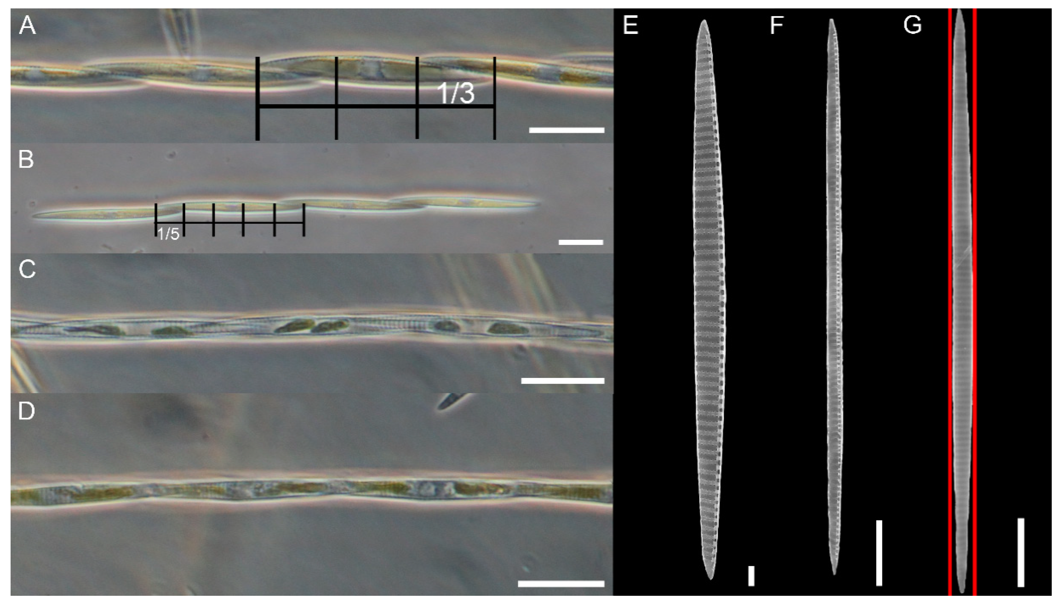
Figure 1. Pseudo-nitzschia pungens LM (AD) and SEM images (EG). (A,B) Colonies in girdle view, showing a different overlap indicated by the grid (one-third and one-fifth of the total cell length, respectively). (C) Colony in valve view. (D) Colony in valve view with cells slightly expanding one a side making asymmetrical colony. (EG) Valve view of three cells showing the wide Transapical Axis (TA) variability. Red lines mark the asymmetry of the valve. Scale bar = 20 µm (AD); 2 µm (E); 10 µm (F,G). Images obtained from the following strains: (A,C,D) 01181; (B) 01186; (E) 01189; (F,G) 04196.
Plants 09 01420 g001
Figure 2. Pseudo-nitzschia pungens SEM micrographs of valves. (A) Cell in valve view. (B) Detail of valve showing the irregular density of fibulae (interrupted circle) and striae (arrows). The decreasing density of poroids toward the apical end is shown. (C) Detail of the central part of the valve with an incomplete third row of poroids (white circle). (D) Detail of the central part of the valve showing the most common striae pattern with two rows of poroids. Scale bar = 10 µm (A); 2 µm (BD). Images obtained from field samples.
Plants 09 01420 g002
Figure 3. Pseudo-nitzschia pungens SEM micrographs of internal valve faces. (A) Detail of valve with regular fibulae and striae and an additional poroid (black circle). (B) Detail of valve showing lower number of poroids toward the apical part. (C) Detail of valve showing irregular density of fibulae (dotted black circle) with several additional poroids (black arrows). (D) Detail of the central part of the valve with very low density of poroids. (E) Detail of apical part with very low density of poroids. Scale bar = 2 µm (AC); 1 µm (D,E). Images obtained from the following strains: (A,B) 01186; (C,D) 04191; (E) 01185.
Plants 09 01420 g003
Figure 4. Pseudo-nitzschia pungens TEM micrographs of valves. (AD) Detail of striae showing (A) an incomplete third row of poroids, (BC) two additional poroids in the area of the stria close to the raphe, (D) additional poroid in the internal part of the stria. (E) Detail of valve face with regular density of fibulae and irregular density of striae (arrows indicate striae with only one row of poroids). Scale bar = 1 µm (AC,E); 0.2 µm (D). Images obtained from the following strains: (A,E) 031832; (B,D) 01185; (C) 01186.
Plants 09 01420 g004
Figure 5. Pseudo-nitzschia pungens SEM micrographs of cingular bands: (a) Valvocopula; (b) second cingular band; (c) third cingular band. Band striae was perforated by (A,B) oval to rectangular, (C) square, or (E,F) circular poroids. (A,D,F) Different types or (B,C) only one type of poroids could occur in the valvocopula. Black arrows indicate sectors dividing poroids. Scale bar = 1 µm (AF). Images obtained from the following strains: (A) 04196; (B) 01186; (C) 01189; (D) 04191; (E) 04194; (F) 05199.
Plants 09 01420 g005
Figure 6. Pseudo-nitzschia pungens TEM micrographs of valvocopulae with different poroid pattern: (A,B) with no sectors, (C) one entire or partially divided sector, (D) two sectors (black arrows). Scale bar = 2 µm (A); 0.2 µm (B), 3 µm (C,D). Images obtained from the following strains: (A) 01185; (B) 01186; (C,D) 031832.
Plants 09 01420 g006
Figure 7. Bayesian consensus tree based on ITS1-5.8s-ITS2 of Pseudo-nitzschia pungens rooted with P. multiseries (AY257844). Sequences from this study are in bold. Only ML bootstraps values ≥ 70%, and BI posterior probabilities (PP) ≥ 0.90 are shown. (ML/PP). Scale bar = substitutions/site.
Plants 09 01420 g007
Table 1. Morphometric characteristics of Pseudo-nitzschia pungens reported in literature and in this study. n.r., not reported.
Apical Axis (µm)Transapical Axis (µm)Fibulae in 10 µmStriae in 10 µmPoroids in 1 µmBand Atriae (in 10 µm)Apical Axis/OverlapLocationType of SamplesClade/VarietyReferences
74–1422.9–4.59–159–153–4n.r.3n.r. n.r.[47]
71–1402.8–4.510–1410–143–4.520–24n.r.Pacific coast of USA (California)Field and culture samplesII/cingulata[26]
100–155
(116.1 ± 13.2)
n = 74
1.8–4.0
(2.9 ± 0.5)
n = 60
10–20
(12.3 ± 1.6)
n = 78
10–14
(12.0 ± 1.1)
n = 82
2–4
(3.3 ± 0.4)
n = 81
n.r.3–5
(4)
n = 70
Danish coastal watersn.r.n.r.[48]
72–135
(99.1 ± 21.5)
2.4–4.5
(3.9 ± 0.8)
9–13
(10.8 ± 1.9)
9–13
(10.8 ± 1.6)
3–4
(3.4 ± 0.3)
17–18
(17.5 ± 0.4)
n.r.Sea of JapanField samplesn.r.[49,50]
94–1602–410–1610–164–5n.r.n.r.Pacific coast of USA (Washington State)Field samplesn.r.[51]
74–1742.4–5.39–169–16n.r.n.r.3Gulf of Mexico
Field samplesn.r.[52]
92–156
(113 ± 31)
n = 3
3.5–4.2
(4.0 ± 0.3)
n = 3
10–11
(11 ± 0.3)
n = 4
10–11
(10.5 ± 0.5)
n = 5
1–3
(2.5 ± 0.6)
n = 6
4Canada, Bay of FundyField samplesn.r.[53]
24.4–121.0
(79.1 ± 8.7)
n = 70
2.4–3.8
(3.2 ± 0.4)
n = 70
9–13
(11.9 ± 0.3)
n = 70
10–14
(11.1 ± 0.5)
n = 70
2–4
(3.0 ± 0.5)
n = 50
n.r.n.r.North SeaCulture samplesI/pungens[24]
87.9–108.7
(101.5 ± 27.2)
n = 42
3.4–4.7
(3.9 ± 0.1)
n = 42
11–15
(12.7 ± 0.2)
n = 50
10–13
(11.6 ± 0.3)
n = 42
3–5
(4.2 ± 0.5)
n = 50
n.r.n.r.North SeaCulture samplesII/cingulata[24]
74–147
(112 ± 17.6)
n = 35
2.6–4.5
(3.4 ± 0.5)
n = 35
10–13
(11 ± 1.2)
n = 5
9–13
(11.4 ± 1.5)
n = 5
2.5–3
(2.9 ± 0.2)
n = 5
n.r.n.r.Catalonia, NW MediterraneanField samplesn.r.[34]
70–156
116.9 ± 24.6
n = 81
2.2–4.8
(3.75 ± 0.57)
n = 81
9–13
(11.2 ± 1.3)
n = 10
9–13
(11.4 ± 1.4)
n = 10
2.5–3
(3.0 ± 0.2)
n = 10
n.r.n.r.Catalonia, NW MediterraneanField samplesn.r.[35]
86.3–160.8
(104.6 ± 10.42)
n = 50
3.7–5.3
(4.5 ± 0.35)
n = 50
10–16
(12.8 ± 1.4)
n = 50
10–13
(11.1 ± 0.68)
n = 50
n.r.n.r.3–5North SeaField samplesn.r.[54]
47–100
(67.7 ± 14.8)
2.7–3.7
(3.3 ± 0.6)
13–16
(14.8 ± 0.7)
13–16
(14.9 ± 0.7)
3–5
(4.0 ± 0.0)
21–25
(23.0 ± 1.1)
5–6Atlantic Ocean, PortugalCulture samplesIII/ aveirensis[27]
84–1653.0–5.013–18Striae n.r.
Interstriae: 13–16
2–3n.r.n.r.Atlantic Ocean, southern Brazil Field samplespungens[55]
89–1223.0–4.013–14Striae n.r.
Interstriae: 12–14
3–4n.r.n.r.Atlantic Ocean, southern Brazil Field samplescingulata[55]
n.r.1.9–3.2
(2.5 ± 0.41)
n = 28
10–17
(13 ± 2.3)
n = 8
8–15
(13 ± 2.3)
n = 8
2–4
(3 ± 0.6)
n = 8
n.r.4.5–4.8NE Adriatic SeaField samplesn.r.[36]
n.r.2.5–3.6
(2.9 ± 0.3)
n = 75
11–14
(12.2 ± 0.9)
n = 71
11–14
(12.0 ± 0.8)
n = 71
2–4
(3.5 ± 0.6)
n = 105
14–20
(16.5 ± 1.6)
n = 45
n.r.Northern Aegean SeaCulture samplesI[29]
93–1262.8–3.211–1511–143–3.4n.r.3–4Gulf of MexicoField samplesn.r.[56]
72–1493.0–4.111–1310–123–415–23n.r.Atlantic Ocean, Gulf of MaineCulture samplesn.r.[57]
80–92
(86.21 ± 3.66)
n = 24
2.4–4.2
(3.75 ± 0.96)
n = 24
9–13
(11.4 ± 1.2)
n = 10
10–13
(11.2 ± 0.9)
n = 10
2–4
(2.97 ± 0.45)
n = 35
n.r.n.r.NE Adriatic Sea (Gulf of Trieste)Culture samplesI[38]
51.1–99.4
(78.9 ± 11.7)
n = 213
2.0–3.6 (2.82 ± 0.32)
n = 80
5–18
(11.6 ± 1.9)
n = 79
9–16
(11.3 ± 1.2)
n = 81
1–4
(3 ± 0.6)
n = 90
12–23
(16.2 ± 2.7)
n = 88
2.9–6.0
(3.9 ± 0.6)
n = 130
NW Adriatic Sea (Senigallia LTER station)Culture samplesIthis study
57.2–127.6
(93.9 ± 25.8)
n = 26
2.4–3.7
(2.8 ± 0.33)
n = 15
9–16
(11.6 ± 1.98)
n = 13
9–14
(11.3 ± 1.3)
n = 14
3–4
(3.4 ± 0.5)
n = 15
n.r.3.7–5.2
(4.6 ± 0.5)
n = 14
NW Adriatic Sea (Senigallia LTER station)Field samplesIthis study
Publisher’s Note: MDPI stays neutral with regard to jurisdictional claims in published maps and institutional affiliations.