Article

An Integrated Approach for Efficient and Accurate Medicinal Cuscutae Semen Identification

Herbal Medicine Resources Research Center, Korea Institute of Oriental Medicine, Naju 58245, Korea
*
Authors to whom correspondence should be addressed.
Plants 2020, 9(11), 1410; https://doi.org/10.3390/plants9111410
Submission received: 11 September 2020 / Revised: 11 October 2020 / Accepted: 20 October 2020 / Published: 22 October 2020
(This article belongs to the Section Plant Systematics, Taxonomy, Nomenclature and Classification)

Abstract

:
To guarantee the safety and efficacy of herbal medicines, accurate identification and quality evaluation are crucial. The ripe dried seeds of Cuscuta australis R.Br. and C. chinensis Lam. are known as Cuscutae Semen (CS) and are widely consumed in Northeast Asia; however, the seeds of other species can be misidentified as CS owing to morphological similarities, leading to misuse. In this report, we propose a multilateral strategy combining microscopic techniques with statistical analysis and DNA barcoding using a genus-specific primer to facilitate the identification and authentication of CS. Morphology-based identification using microscopy revealed that the useful diagnostic characteristics included general shape, embryo exudation, hairiness, and testa ornamentation, which were used to develop an effective identification key. In addition, we conducted DNA barcoding-based identification to ensure accurate authentication. A novel DNA barcode primer was produced from the chloroplast rbcL gene by comparative analysis using Cuscuta chloroplast genome sequences, which allowed four Cuscuta species and adulterants to be discriminated completely. Therefore, this investigation overcame the limitations of universal DNA barcodes for Cuscuta species with high variability. We believe that this integrated approach will enable CS to be differentiated from other species, thereby improving its quality control and product safety in medicinal markets.

1. Introduction

Medicinal plants and plant-derived medicines are commonly used worldwide in traditional Chinese medicine and are recognized as natural alternatives or supplements to synthetic chemicals from a modern pharmacological perspective [1]; however, there have been repeated reports of contamination of herbal products due to inaccurate identification and poor quality control, i.e., substitution, adulteration, and/or admixture of other species, at national [2,3,4], continental [5,6], or global markets [7,8]. As the quality of herbal medicines directly affects treatment efficacy and drug safety, ensuring the quality, safety, and effectiveness of these medicinal materials has become a serious issue [9,10,11,12]. Thus, pharmacovigilance is increasingly important for developing reliable information on the safety of herbal medicines [13,14].
According to the Korean Herbal Pharmacopoeia, the dried ripe seeds of Cuscuta chinensis Lam. (Convolvulaceae) are a herbal medicine known as Cuscutae Semen (CS), or “To-Sa-Ja” in Korean [15,16]. However, the Pharmacopoeia of the Democratic People’s Republic of Korea lists seeds from both C. australis R.Br. and C. japonica Choisy as sources of the same herbal medicine [15,17], while the Pharmacopoeia of the People’s Republic of China and the Taiwan Herbal Pharmacopoeia list seeds from C. chinensis and C. australis as authentic CS, known as “Tu-Si-Zi” in Chinese [15,18,19]. The differences between these definitions have resulted in the circulation of adulterants or counterfeits in herbal markets [20]; therefore, it is important to obtain information regarding species related to CS.
Modern pharmacological studies have revealed that CS can act in various parts of the body, including the reproductive [21,22] and immune [23,24,25] systems, and exerts biological functions such as anti-oxidant [26,27,28] and anti-cancer [29,30] effects. In addition, some reports have suggested that CS can prevent and treat liver [28,31,32,33,34] and neurodegenerative-related [35] diseases.
Unfortunately, seeds with similar morphological traits from southern regions of Korea are misused as CS. For example, seeds from Perilla frutescens (L.) Britton var. frutescens and P. frutescens var. crispa (Thunb.) H.Deane (family Lamiaceae, Labiatae) are often found as adulterants in CS due to their morphological similarities [20,36,37]. Moreover, similar congeneric species such as C. australis, C. japonica, and C. pentagona are also found throughout Korea [38,39].
Cuscuta (dodder) species are parasitic vines with reduced or almost absent vegetation and very small floral organs. Due to their peculiar morphology and parasitism, the species have received interest from various fields, including agriculture [40], conservation [41], taxonomy (including morphology) [42,43,44,45], and genomics [46,47,48]. However, no studies have yet investigated the accurate identification and authentication of CS as medicinal seeds among morphologically similar species.
Seeds are commonly used as traditional herbal medicines; however, commercial markets often sell mixed seeds from different species, including adulterants, within a single package, while some authentic medicinal seeds can be difficult to distinguish [49,50]. Therefore, multilateral approaches including microscopic analysis have been developed to authenticate and distinguish genuine species from congeneric species and adulterants such as Lepidii Seu Descurainiae Semen [51] and Pharbitidis Semen [52]. Microscopic authentication of medicinal materials has proven to be a highly reliable and accurate identification tool [8,51,52,53,54]. DNA barcoding has also been recognized as a powerful tool for identifying herbal medicines [55,56] because it is convenient, efficient, and accurate. Therefore, the technique has a wide range of potential applications for identifying adulterants in commercial herbal products [56] and for inputting barcoding sequences into the medicinal database [55,57].
In this study, we aimed to elucidate detailed morphological and micromorphological characteristics of CS and adulterants using microscopic analysis, thereby developing a convenient and effective identification key. In addition, we designed a specific primer to distinguish between C. chinensis and congeneric species using DNA barcodes. Together, these data will facilitate the quality control of the valuable medicinal seed, CS.

2. Results

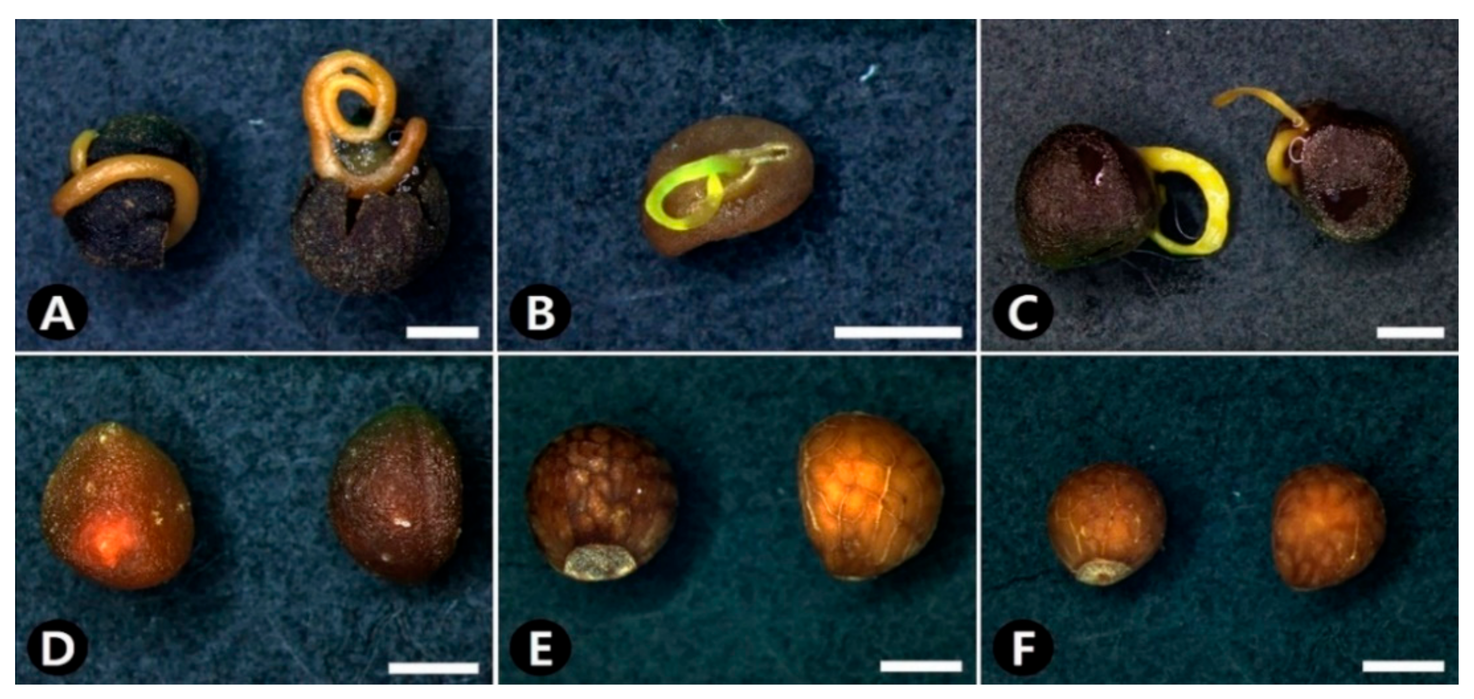
2.1. Morphological Characteristics

All seeds in the genus Cuscuta, including CS, were ovoid in shape and triangular in cross-sectional shape (Figure 1A–D, Table 1); however, the seeds of the two adulterants had a globose to sub-globose shape and a circular to broadly triangular cross-sectional shape (Figure 1E,F, Table 1).
The seeds of the studied species varied from 1.17–3.26 mm in length and 0.98–2.78 mm in width, with C. australis having the smallest seeds (average length × width: 1.48 × 1.12 mm) and C. japonica having the largest (average length × width: 2.71 × 2.16 mm; Table 1). The seeds were dull brown (200A-D), gray-brown (199A-B, N199A-B, N199A-C, N199C-D), and black (203) in color. In particular, C. australis, C. chinensis, and C. japonica had darker seeds, whereas C. pentagona, P. frutescens var. frutescens, and P. frutescens var. crispa had lighter seeds (Figure 1, Table 1). In addition, the embryos of C. australis, C. chinensis, and C. japonica exuded from their seeds when placed into boiling water for 10 min (Figure 1A–C, Table 1); however, no changes were observed for C. pentagona, P. frutescens var. frutescens, or P. frutescens var. crispa seeds following the same treatment (Figure 1D–F, Table 1).

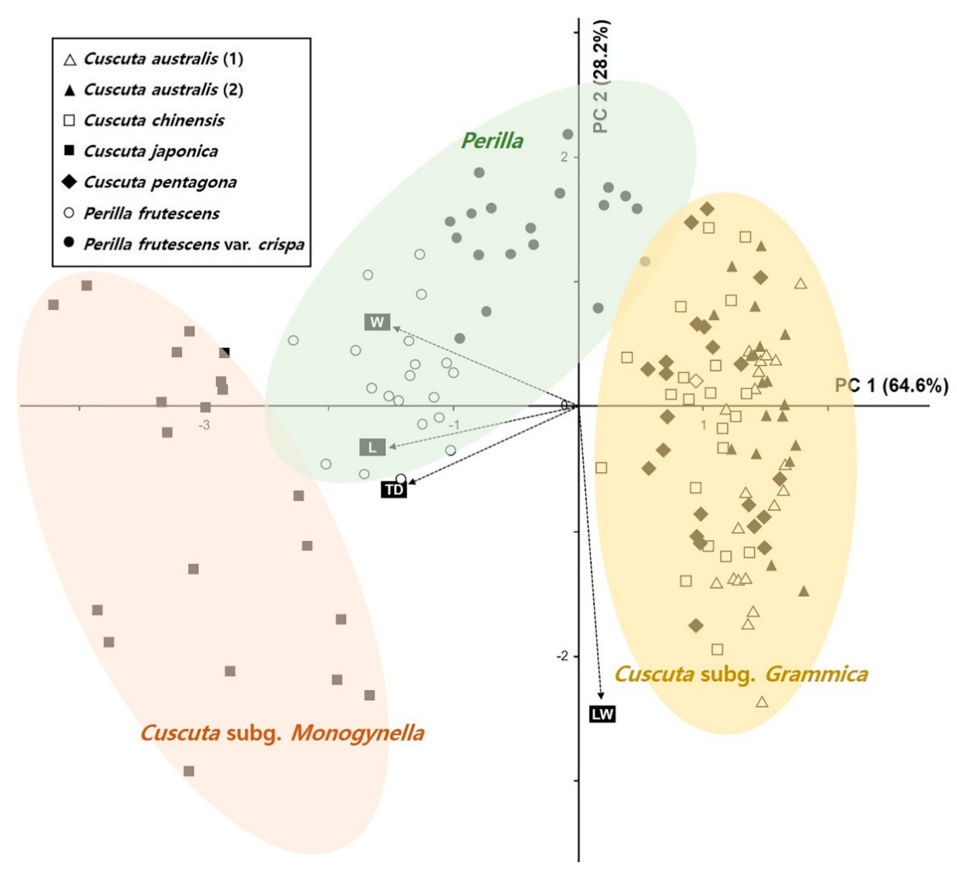
2.2. Statistical Analysis of Morphological Characteristics

Next, we explored the relationships between the quantitative data for each species of seed using principal component analysis (PCA; Figure 2). The first two principal components (PC1 and PC2) explained 92.80% of the total variance, whereas PC1 explained 64.60% of the variance in seed length (L) and width (W) and testa cell (TD) size and PC2 accounted for 28.20% of the variance in the seed size ratio (L/W). The PCA biplot split the operational taxonomic units (OTUs) into three main groups (Figure 2). The OTUs for C. australis including commercial CS, C. chinensis, and C. pentagona, which belong to the subgenus Grammica, were grouped on the positive side of the PC1 axis, whereas those for C. japonica, which belongs to the subgenus Monogynella, were grouped on the negative side of the PC1 axis (Figure 2). The adulterant OTUs (P. frutescens vars. frutescens and crispa) were on the central to positive side of the PC2 axis (Figure 2).

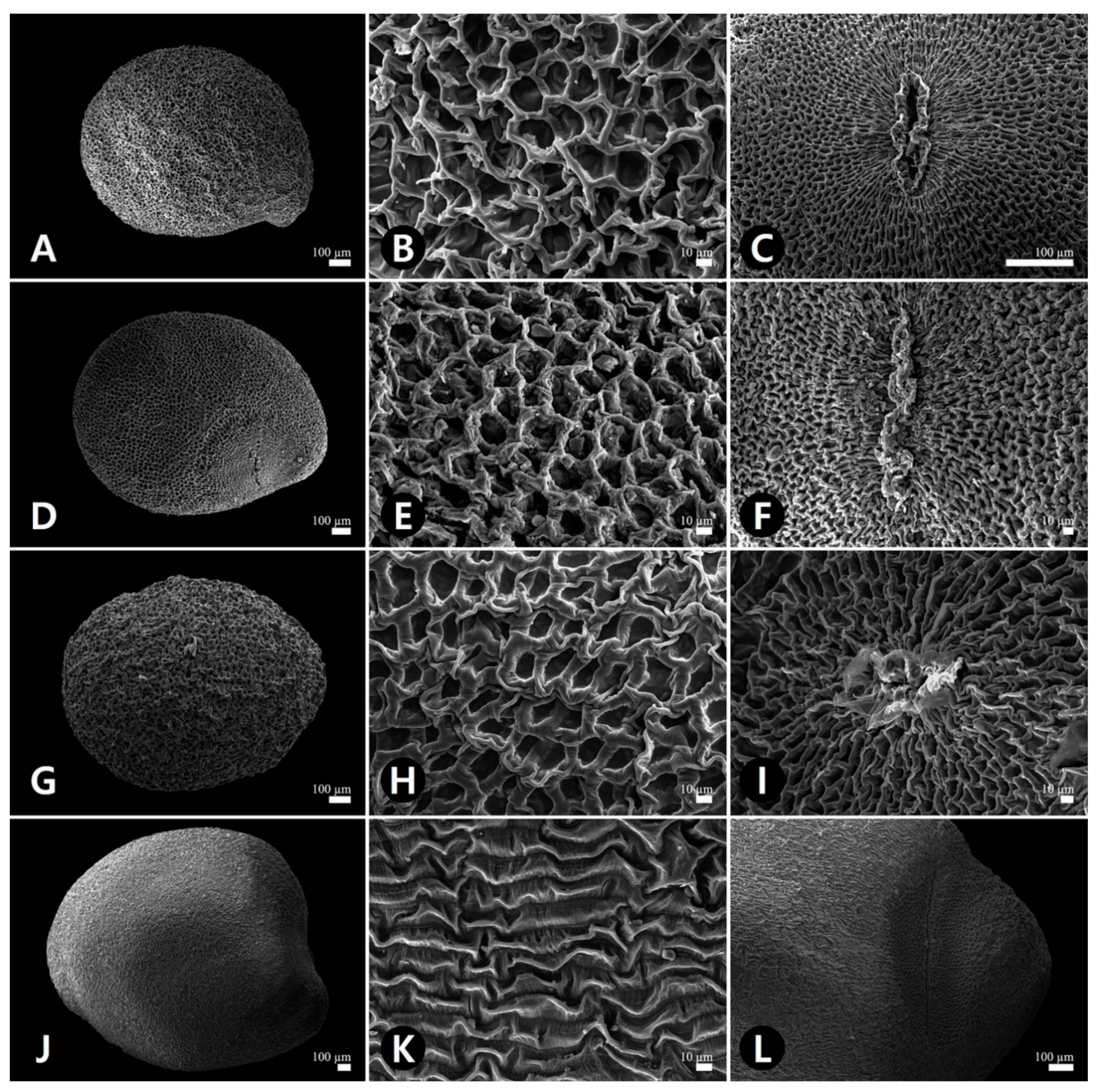
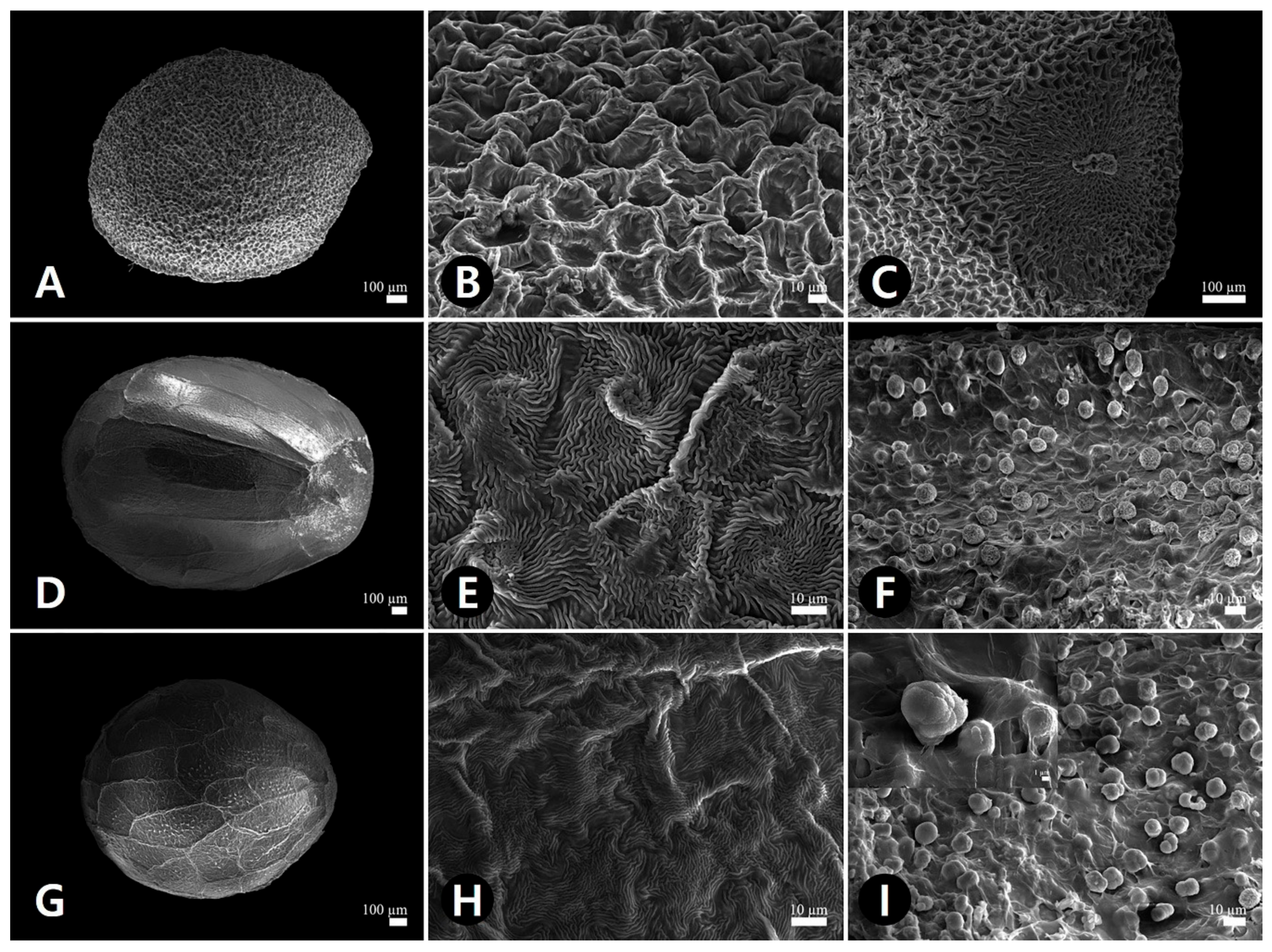
2.3. Micromorphological Characteristics

Observing the seeds using scanning electron microscopy revealed remarkable variation in testa surface patterns (Figure 3 and Figure 4, Table 2). Therefore, we categorized seeds into three major types based on qualitative characteristics such as ornamentation, epidermal cell pattern, cell wall shape, hilum, and hairiness. The epidermal cell outline was either isodiametric or elongated, while the boundary of the anticlinal cell wall was straight or sinuate and relief of the boundary was channeled or raised. The curvature of the outer periclinal cell walls was either concave or convex. The epidermal cell diameter ranged from 11.0–99.2 μm among all studied species, with C. australis having the smallest cell (average 20.8 μm) and C. japonica having the largest (average 70.8 μm; Table 2).
Besides C. japonica, all Cuscuta seeds displayed type I reticulate (net-like) ornamentation with isodiametric cell arrangement, a straight and raised anticlinal cell wall, and a concave outer periclinal cell wall (Figure 3A,B,D,E,G,H, and Figure 4A,B). C. japonica seeds displayed type II rugulate (puzzle-like) ornamentation with elongated cell arrangement, a straight and channeled anticlinal cell wall, and a convex outer periclinal cell wall (Figure 3J,K). P. frutescens vars. frutescens and crispa seeds showed type III rugose-colliculate ornamentation, a anticlinal cell wall, and a convex periclinal cell wall (Figure 4D,E,G,H). These seeds were channeled divided into subtypes based on anticlinal cell wall curvature: type III-1, P. frutescens var. frutescens, straight cell walls (Figure 4E); type III-2, P. frutescens var. crispa, sinuate cell walls (Figure 4H). Only the hilum surfaces of type III seeds were multicellular with stalked glandular trichomes (Figure 4F,I, Table 2).

2.4. Identification Key

Based on our general morphological and micromorphological analyses of CS and two adulterant seed types, we developed a key for the accurate identification of seeds according to shape, cross-sectional shape, the presence or absence of hairs on the hilum, embryo exudation, and combined micromorphological characteristics, such as testa ornamentation, epidermal cell pattern, and anticlinal and periclinal cell wall shape, as described below:
1.
Seeds ovoid in shape; triangular cross-section; glabrous on hilum; rugulate and reticulate testa ornamentation ------------------------------------------------------------------------------------------------------------------------------------------------------------------ 2
1′.
Seeds globose to subglobose in shape; circular to broadly triangular in cross-section; glandular hairs on hilum; rugose-colliculate testa ornamentation ----------------------------------------------------------------------------------------------------------------------------------------------------------------- 3
2.
Embryo exudation in boiling water ------------------------------------------------------------------------------------------------------------------- 4
2′.
No embryo exudation in boiling water ---------------------------------------------------- Cuscuta pentagona
3.
Straight anticlinal cell wall boundary ------------------------------------- Perilla frutescens var. frutescens
3′.
Sinuate anticlinal cell wall boundary ------------------------------------------- Perilla frutescens var. crispa
4.
Seeds 2.17–3.26 × 1.57–2.78 mm; elliptic hilum; elongated epidermal cell outline; rugulate testa ornamentation; channeled anticlinal cell wall boundary; convex outer periclinal cell wall ----------------------------------------------------------------------------------------------------- Cuscuta japonica
4′.
Seeds 1.17–1.82 × 1.04–1.46 mm; circular-ovate hilum; isodiametric epidermal cell outline; reticulate testa ornamentation; raised anticlinal cell wall boundary; concave outer periclinal cell wall ------------------------------------------------------------------------------ Cuscuta australis and C. chinensis

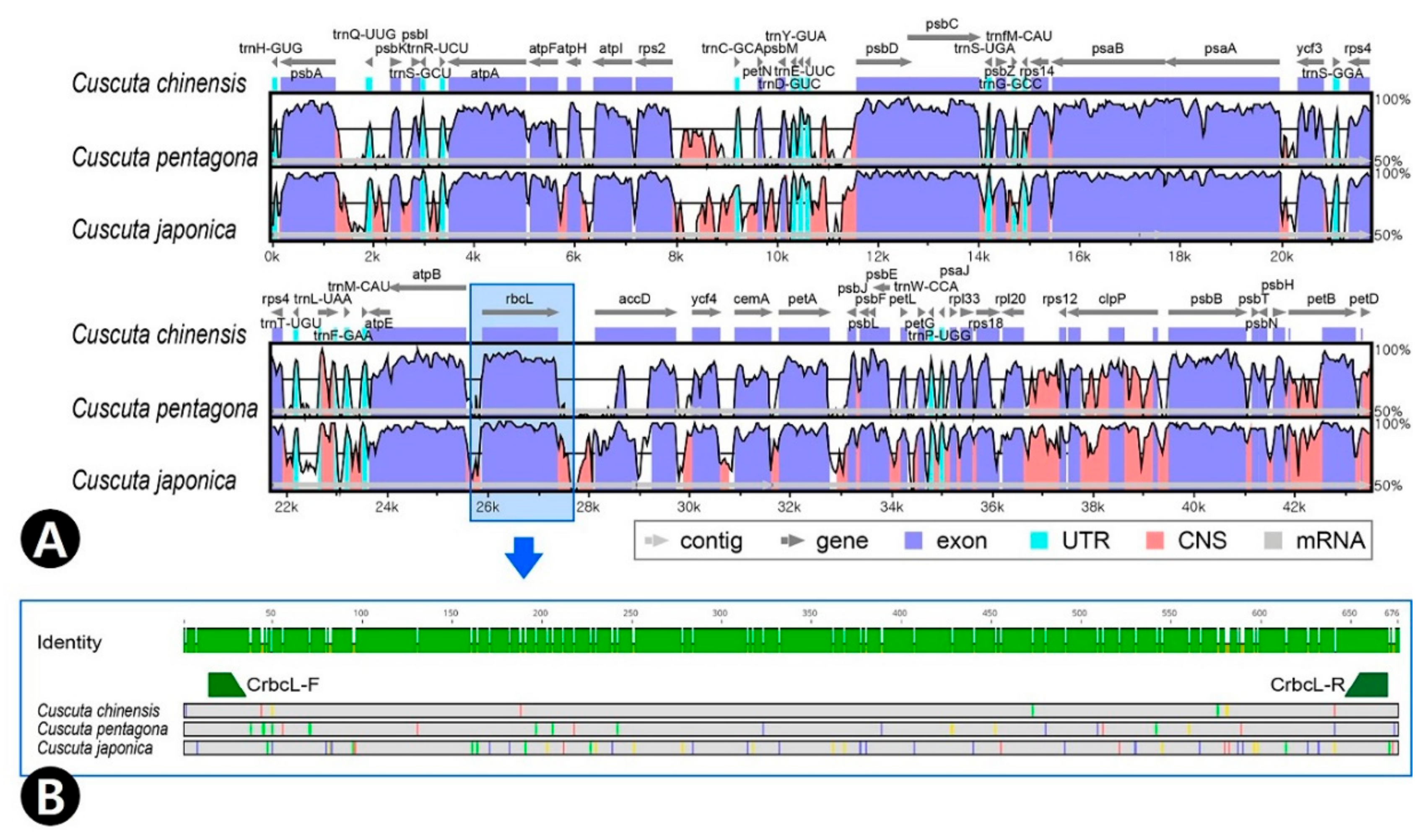
2.5. Comparision of rbcL Using Cuscuta

To evaluate the rbcL gene as a potential DNA barcode to discriminate Cuscuta species and to test primer universality, we compared the whole chloroplast genome sequences of three Cuscuta species using the mVISTA program (Figure 5A). The Cuscuta chloroplast genomes were downloaded from Genbank (C. chinensis: MH780079; C. pentagona: NC_039759; C. japonica; MH780080). As the rbcL gene length varied in C. chinensis (1443 bp), C. pentagona (1446 bp), and C. japonica (1497 bp), we aligned their rbcL sequences and found that the region from 190 to 846 bp was relatively variable for species identification, with approximately 89–95% similarity. The flanking region is conserved and hence primers were designed from those regions. Therefore, we selected a 676 bp sequence from this region to design the forward and reverse primers (Figure 5B).
We confirmed the utility of the rbcL gene as a DNA barcode and developed novel DNA barcode primers using rbcL-generated marker sequences (Table 3). The Cuscuta subgenus Monogynella including C. chinensis and C. pentagona chloroplast genomes showed high sequence variability and lacked the matK gene, which is a universal DNA barcode region. Consequently, it is difficult to distinguish between Cuscuta species using universal DNA barcodes; however, the developed primer (CrbcL) had good discriminatory efficiency for Cuscuta species.

2.6. Comparision of ITS and rbcL Using Cuscuta and Perilla

To distinguish between the four Cuscuta species and two species of P. frutescens, we performed DNA barcode analysis using the nuclear rDNA ITS and rbcL regions. ITS sequences for P. frutescens var. frutescens and var. crispa were downloaded from Genbank (1: FJ513160; 2: KP644065; 3: KT220688; 4: KX397889; 5: MG223657; 6: MG224544; 7: KY624981). Similarly, the sequence data were obtained for 15 samples among the four Cuscuta species and aligned (Table 4). The ITS alignment region was 798 bp long in four Cuscuta species and 809 bp long in all studied taxa including P. frutescens. The rbcL alignment region was 656 bp long in all studied taxa. Among all taxa, there were 287 (35.5%) parsimony-informative sites in ITS and 102 (15.5%) in rbcL, whereas the ITS region was more variable than the rbcL region (ITS nucleotide diversity Pi = 0.17662; rbcL Pi = 0.06212). Intra-species variation in the four Cuscuta species was shown 0–3.4% in ITS, but not found in rbcL. Nine ITS haplotypes were detected in C. pentagona and two in C. japonica, indicating ambiguous species identification; however, only one of the four Cuscuta species shared a rbcL haplotype with P. frutescens. Thus, rbcL is more suitable for Cuscuta species identification than ITS, allowing complete discrimination of all four Cuscuta species.

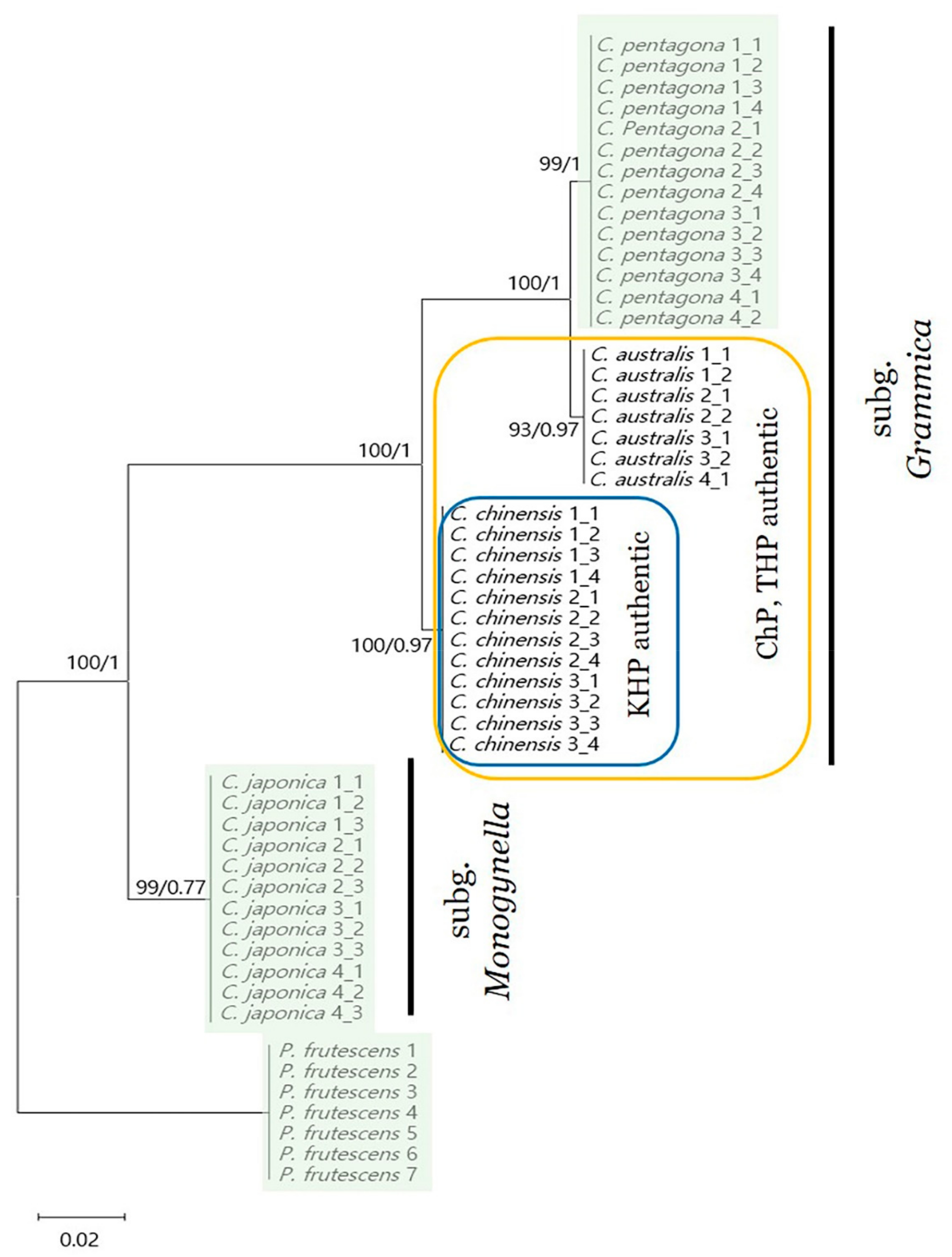
2.7. Phylogenetic Analysis

Finally, we analyzed the phylogenetic relationships between the rbcL sequences of four Cuscuta and one P. frutescens species using the maximum likelihood (ML) and Bayesian inference (BI) methods. All species clustered into monophyletic groups; however, C. australis and C. pentagona were more closely related to the other species (Figure 6 and Supplementary Materials Figure S1). This phylogeny could be further separated into two distinct clusters, one of which contained C. australis, C. pentagona, and C. chinensis with 100% bootstrap (BS) values, whereas the other only contained C. japonica. Phylogenetic analysis using ITS indicated an ambiguous node in both ML and BI trees (Figures S2 and S3): despite analyzing the same C. pentagona sequences, its phylogenetic position was varied and complex. However, rbcL clearly classified monophyletic relationships for each Cuscuta species.

3. Discussion

The increasing pharmacological and clinical importance of herbal medicine has resulted in issues related to misidentification and failed quality control. In this study, we used three different approaches, namely morphological and micromorphological analyses with statistical analysis and DNA barcoding, to accurately identify the widely consumed medicinal seed, CS. In addition, we created an effective identification key from the results of microscopic analysis to distinguish authentic medicinal seeds from adulterants and designed a specific and sensitive novel DNA barcode primer for the rbcL gene

3.1. Morphology-Based Identification

CS are small seeds that are difficult to distinguish from adulterants using the naked eye; in particular, three congeneric species (C. australis, C. japonica, and C. pentagona) in Korea have a very similar external seed morphology [58]. According to a dispensatory on the visual and organoleptic examination of herbal medicines, testa color and hilum position are important characteristics for authenticating CS [36]. In this study, we found that all Cuscuta seeds shared the same hilum position and that, although C. australis seeds were somewhat dark and blackish brown, their color varied widely (Gr-Br to Bl; Table 1), thus limiting the utility of these key characteristics. Ji et al. [49] described C. australis and C. chinensis seeds as having a “vomiting thread shape” and C. japonica seeds as “not vomiting thread shape” when put in water. However, we consistently observed embryo exudation (“vomiting thread”) in all Cuscuta seeds except for C. pentagona when placed into boiling water. The seed coat consists of three layers: the outer epidermis, two different palisade layers, and an inner multi-parenchymal layer. In most Cuscuta seeds, the endosperm surrounds the coiled embryo [59]; therefore, we hypothesize that embryo exudation is associated with seed coat thickness and the degree of endosperm embedding. Interestingly, the C. campestris embryo is only embedded in the endosperm in small regions [59], suggesting that C. pentagona may display a different internal structure to other Cuscuta species. Further comparative ultrastructural studies are required to evaluate the seed coat stratification and internal structure of seeds from different Cuscuta species.

3.2. Micromorphology-Based Identification

Our previous study suggested that the morphological characteristics of reproductive organs, particularly testa ornamentation, can be used to distinguish between C. chinensis and C. japonica [60]. Recently, the availability of detailed seed micromorphology has provided valuable diagnostic characteristics for authenticating small medicinal seeds [51,52] and taxonomic identification [61,62]. In this study, micromorphological analysis using a scanning electron microscope revealed that Cuscuta seeds including CS (types I, II, and glabrous hilum) are easily distinguishable from the seeds of adulterant Perilla species (type III and glandular hairs on hilum). In addition, the two types of testa ornamentation observed were consistent with subgenus classification [42]; C. australis, C. chinensis, and C. pentagona (subgenus Grammica, type I seeds) and C. japonica (subgenus Monogynella, type II seeds), suggesting that testa ornamentation may be stable at the subgenus level. Although seed micromorphology was a useful diagnostic characteristic, C. australis and C. chinensis could not be clearly distinguished and were difficult to differentiate using morphology or statistical analysis. Therefore, multilateral approaches including morphology with statistical analysis and DNA barcoding are required to accurately identify medicinal materials [53].

3.3. DNA Barcoding-Based Identification

Since 2010, the definition of CS in the Chinese Pharmacopoeia has changed to include both C. chinensis and C. australis [19]; however, quality control remains a major problem for CS due to the morphological similarity of these species, as C. australis and C. chinensis have significantly different chemical constituents [63]. To improve the pharmacological and clinical activities of CS, it is therefore important that these two similar medicinal seeds can be accurately identified. Molecular identification based on DNA barcoding is an efficient and accurate tool that has been used to authenticate medicinal species [55,56,57]; however, the method has limitations when using universal chloroplast DNA primers in closely related species [64,65]. As such, it is preferable to design species-specific primers or use next generation sequencing (NGS) and NGS-based DNA-metabarcoding to overcome this limitation of DNA-based analysis [66,67]. In this study, we accurately identified four Cuscuta species and adulterant Perilla species using novel genus-specific DNA barcode primers (CrbcL-F, CrbcL-R). Moreover, phylogenetic analysis yielded a phylogeny that was consistent with previous studies [42,48].

4. Materials and Methods

4.1. Plant Materials

Authentic species of CS (C. chinensis) and three congeneric species (C. australis, C. japonica, and C. pentagona) were collected from natural populations in Korea during the fruiting season (July 2016 to May 2019). All samples collected in this study were identified based on their macroscopic morphology and microscopic characteristics by two authors (Sungyu Yang and Jun-Ho Song) using relevant literature [38,39,42,58,68,69]. Commercial CS was purchased in medicinal markets from commercial suppliers (Kwang Myung Dang Co., Ulsan, Korea). The identity of the commercial CS (manufacturer’s No. K0412050015KE15) was carefully confirmed under a stereomicroscope (Olympus SZX16, Olympus, Tokyo, Japan). Adulterant seeds from P. frutescens var. frutescens and P. frutescens var. crispa were sampled from voucher specimens. The morphological and micromorphological characteristics of mature seeds from all species were analyzed. To ensure consistency, at least two samples were examined for each species. All seed samples were deposited in the Korean Herbarium of Standard Herbal Resources at the Korea Institute of Oriental Medicine (KIOM), Naju, Korea. Detailed information about the plant materials used in this study is summarized in Table 5, including the collection site, collection date, and voucher number of specimens deposited in the KB and KIOM.

4.2. General Morphology and Embryo Exudation

Twenty seeds per species (a total of 140 seeds) were measured and subjected to optical observation. Seed length, width, and the length/width ratio were measured using a digital vernier caliper (CD-15CP, Mitutoyo, Kawasaki, Japan). Shape, cross-sectional shape, color, hilum hair, and embryo exudation were observed using a stereomicroscope and images captured using a digital camera (Olympus DP21, Olympus, Tokyo, Japan). To observe embryo exudation, seeds were placed into boiling water for 10 min. Seed color was determined according to the Royal Horticultural Society Mini-color Chart [70].

4.3. Statistic Analysis

To verify whether four quantitative variables (seed length, seed width, L/W, and epidermal cell diameter) grouped the species, principal component analysis (PCA) was performed using PC-ORD version 5.31 [71].

4.4. Micromorphology

To fully dry the samples, the seeds were placed in silica gel desiccators for 10 days to remove any moisture. For micromorphological observation, seeds were directly mounted on aluminum stubs using a double sided adhesive conductive carbon disk (05073-BA, SPI Supplies, West Chester, PA, USA). The stubs were coated with gold using a sputter coater (208HR, Cressington Scientific Instruments Ltd., Watford, UK) and testa were observed using a low voltage field emission scanning electron microscope (JSM-7000F, JEOL, Tokyo, Japan) at an accelerating voltage of 5–10 kV and a working distance of 10–13 mm. The terminology for seed micromorphology followed that of Barthlott [72,73].

4.5. Sequence Analysis

DNA was extracted from all seed samples using a DNeasy Plant Mini kit (Qiagen, Valencia, CA, USA) according to the manufacturer’s instructions. Genomic DNA (20 ng) was amplified in a 20-µL PCR mixture (SolgTM 2X Taq PCR smart mix 1, Solgent, Daegeon, Korea) with 10 pmol of each primer (Bioneer, Daejeon, Korea). The ITS region was amplified using ITS1 (TCC GTA GGT GAA CCT GCG G) and ITS4 (TCC TCC GCT TAT TGA TAT GC) primers, as described previously [74]. The conserved rbcL region in chloroplast genomes (C. chinensis: MH780079; C. pentagona: NC_039759; C. japonica; MH780080) was detected using mVISTA [75]. CrbcL primers were designed using Geneious Prime (Biomatters, Auckland, New Zealand) and tested by PCR amplification with 20 ng of genomic DNA from 15 Cuscuta samples in a 20 μL PCR mixture with 10 pmol of CrbcL primers using a Pro Flex PCR system (Applied Biosystems, Waltham, MA, USA) with the following parameters: initial denaturation at 95 °C for 2 min; 35 cycles at 95 °C for 50 s, 60 °C for 50 s, and 72 °C for 50 s; final extension at 72 °C for 5 min. The PCR products were separated on a 2% agarose gel for 40 min at 150 V. Each PCR product was isolated using a gel extraction kit (Qiagen), subcloned into a pGEM-T Easy vector (Promega, Madison, WI, USA), and sequenced using a DNA sequence analyzer (ABI 3730, Applied Biosystems Inc., Foster City, CA, USA). The newly sequenced chloroplast genome sequences in this study were deposited in the NCBI GenBank database under the accession numbers MT982734-MT982740, MT982754-MT982757, MT982776-MT982784, MT982792-982799, MT984385-MT984395, and MT998850-MT998863.

4.6. Comparative Analysis and Phylogenetic Analysis

The newly sequenced ITS and rbcL sequences of 15 samples from four Cuscuta species, eight Perilla ITS sequences, and seven rbcL sequences from Genbank were obtained and aligned using Multiple Alignment using Fast Fourier Transform. Gaps in the alignment were stripped using BioEdit [76]. DNA SP 6 [77] was used to calculate nucleotide variability (Pi), variable sites, and haplotypes. Phylogenetic analysis was carried out using a best-fit model based on Akaike Information Content using JModeltest V2.1.10 [78]. The GTR + I model was applied to ITS sequences (Table S1) and CrbcL (Table S2). ML analysis was performed using MEGA 6 [79] and branch support was calculated with 1000 bootstrap replicates. BI analysis was carried out using MrBayes 3.2.2 [80] with the following settings: two independent Markov Chain Monte Carlo runs performed for one million generations with samples every 1000 generations, the first 25% of trees were discarded as a burn-in.

5. Conclusions

To more accurately identify and authenticate herbal medicines, a multilateral strategy combining macroscopic and microscopic techniques with statistical analysis and DNA barcoding is essential. This is the first comprehensive and integrated study to accurately identify medicinal CS and adulterant seeds. Morphology- and micromorphology-based analyses revealed that general shape, embryo exudation, hairiness, and testa ornamentation are useful characteristics for identifying and authenticating the studied species. Furthermore, we were able to completely discriminate between four Cuscuta species and two Perilla adulterants species using novel genus-specific DNA barcode primers. Therefore, our morphological and molecular data should allow the accurate identification and quality control of CS.

Supplementary Materials

The following are available online at https://www.mdpi.com/2223-7747/9/11/1410/s1. Figure S1: Phylogenetic analysis of five species. The phylogenetic tree was constructed from Cuscuta species with the Bayesian inference (BI) method. The rbcL sequences of Perilla frutescens were downloaded from GenBank and used as outgroups. BI posterior probability (PP) values at each node. Figure S2: Phylogenetic analysis of five species. The phylogenetic tree was constructed from Cuscuta species with the maximum likelihood (ML) method. The ITS sequences of Perilla frutescens were downloaded from GenBank and used as outgroups. ML posterior probability (PP) values at each node. Figure S3: Phylogenetic analysis of five species. The phylogenetic tree was constructed from Cuscuta species with the Bayesian inference (BI). The ITS sequences of Perilla frutescens were downloaded from GenBank and used as outgroups. BI posterior probability (PP) values at each node. Table S1: Selection of the best-fitting substitution model for ITS using jModelTest. Table S2: Selection of the best-fitting substitution model for rbcL using jModelTest.

Author Contributions

Conceptualization, J.-H.S. and I.P.; methodology, J.-H.S. and I.P.; software, J.-H.S. and I.P.; validation, S.Y.; formal analysis, J.-H.S. and I.P.; investigation, J.-H.S., I.P., and G.C.; resources, J.-H.S., B.C.M., G.C., and S.Y.; microscopic analysis, J.-H.S.; genomic analysis, I.P.; writing—Original draft preparation, J.-H.S. and I.P.; writing—Review and editing, B.C.M., G.C., and S.Y.; visualization, J.-H.S. and I.P.; supervision, J.-H.S. and B.C.M.; project administration, B.C.M.; funding acquisition, B.C.M. All authors have read and agreed to the published version of the manuscript.

Funding

This work was supported by a grant for the Development of Sustainable Applications for Standard Herbal Resources (KSN2012320) from the Korea Institute of Oriental Medicine (KIOM), Korea.

Acknowledgments

We thank the directors of the herbarium KB for permission to examine the specimens.

Conflicts of Interest

The authors declare that there are no conflicts of interest.

References

  1. Van Wyk, B.-E.; Wink, M. Medicinal Plants of the World: An Illustrated Scientific Guide to Important Medicinal Plants and Their Uses, 1st ed.; Briza Pub.: Pretoria, South Africa, 2004; 480p. [Google Scholar]
  2. Han, J.; Pang, X.; Liao, B.; Yao, H.; Song, J.; Chen, S. An authenticity survey of herbal medicines from markets in China using DNA barcoding. Sci. Rep. 2016, 6, 18723. [Google Scholar] [CrossRef] [PubMed]
  3. Joharchi, M.R.; Amiri, M.S. Taxonomic evaluation of misidentification of crude herbal drugs marketed in Iran. Avicenna J. Phytomed. 2012, 2, 105–112. [Google Scholar] [PubMed]
  4. Zhao, Z.; Yuen, J.P.; Wu, J.; Yu, T.; Huang, W. A systematic study on confused species of Chinese materia medica in the Hong Kong market. Ann. Acad. Med. Singap. 2006, 35, 764–769. [Google Scholar] [PubMed]
  5. Newmaster, S.G.; Grguric, M.; Shanmughanandhan, D.; Ramalingam, S.; Ragupathy, S. DNA barcoding detects contamination and substitution in North American herbal products. BMC Med. 2013, 11, 222. [Google Scholar] [CrossRef] [Green Version]
  6. Seethapathy, G.S.; Raclariu-Manolica, A.C.; Anmarkrud, J.A.; Wangensteen, H.; De Boer, H.J. DNA metabarcoding authentication of Ayurvedic herbal products on the European market raises concerns of quality and fidelity. Front. Plant Sci. 2019, 10, 68. [Google Scholar] [CrossRef] [Green Version]
  7. Ichim, M.C. The DNA-based authentication of commercial herbal products reveals their globally widespread adulteration. Front. Pharmacol. 2019, 10, 1227. [Google Scholar] [CrossRef]
  8. Ichim, M.C.; Häser, A.; Nick, P. Microscopic authentication of commercial herbal products in the globalized market: Potential and limitations. Front. Pharmacol. 2020, 11, 876. [Google Scholar] [CrossRef]
  9. Jamshidi-Kia, F.; Lorigooini, Z.; Amini-Khoei, H. Medicinal plants: Past history and future perspective. J. Herbmed. Pharmacol. 2018, 7, 7. [Google Scholar] [CrossRef]
  10. Wu, L.; Jiang, W.; Huang, T.; Li, M.; Li, W.; Zhong, H.; Yu, S. Study on the Quality of Chinese Herbal Pieces and the Development of Enterprises. Adv. Econ. Bus. Manag. Res. 2019, 91, 509–513. [Google Scholar]
  11. Perharic, L.; Shaw, D.; Léon, C.; De Smet, P.A.; Murray, V.S. Possible association of liver damage with the use of Chinese herbal medicine for skin disease. Vet. Hum. Toxicol. 1995, 37, 562–566. [Google Scholar]
  12. Nortier, J.L.; Vanherweghem, J.L. For patients taking herbal therapy—Lessons from aristolochic acid nephropathy. Nephrol. Dial. Transplant. 2007, 22, 1512–1517. [Google Scholar] [CrossRef] [PubMed] [Green Version]
  13. Shaw, D.; Graeme, L.; Pierre, D.; Elizabeth, W.; Kelvin, C. Pharmacovigilance of herbal medicine. J. Ethnopharmacol. 2012, 140, 513–518. [Google Scholar] [CrossRef]
  14. World Health Organisation (WHO). WHO Guidelines on Safety Monitoring of Herbal Medicines in Pharmacovigilance Systems; WHO: Geneva, Switzerland, 2004. [Google Scholar]
  15. Korea Institute of Oriental Medicine. Defining Dictionary for Medicinal Herbs (Korean, ‘Hanyak Giwon Sajeon’). 2020. Available online: http://https://oasis.kiom.re.kr/herblib/hminfo/hbmcod/hbmcodList.do (accessed on 21 February 2020).
  16. Korea Food and Drug Administration. The Korean Herbal Pharmacopoeia, 4th ed.; Korea Food and Drug Administration: Seoul, Korea, 2012. [Google Scholar]
  17. Pharmacopoeia Committee of the DPRK. Pharmacopoeia of Democratic People’s Republic of Korea, 7th ed.; Medicine and Science Press: Pyeongyang, Korea, 2011. [Google Scholar]
  18. Committee on Chinese Medicine and Pharmacy. Taiwan Herbal Pharmacopeia, 2nd ed.; English Version; Ministry of Health and Welfare: Taipei, Taiwan, 2013. [Google Scholar]
  19. Chinese Pharmacopoeia Committee. Pharmacopoeia of the People’s Republic of China; Medical Science and Technology Press: Beijing, China, 2015. [Google Scholar]
  20. Moon, B.C.; Choi, G.; Yuan, Y. Origins of Herbal Medicines and Adulterants in Korea and China; Korea Institute of Oriental Medicine: Daejeon, Korea, 2017. [Google Scholar]
  21. Qin, D.N.; She, B.R.; She, Y.C.; Wang, J.H. Effects of flavonoids from Semen Cuscutae on the reproductive system in male rats. Asian J. Androl. 2000, 2, 99–102. [Google Scholar] [PubMed]
  22. Qin, D.; She, B.; She, Y. Effects of flavonoids of Semen Cuscutae on reproductive function of animals. Trad. Chin. Drug Res. Clin. Pharmacol. 2000, 11, 349–351. [Google Scholar]
  23. Pan, H.J.; Sun, H.X.; Pan, Y.J. Adjuvant effect of ethanol extract of Semen Cuscutae on the immune responses to ovalbumin in mice. J. Ethnopharmacol. 2005, 99, 99–103. [Google Scholar] [CrossRef] [PubMed]
  24. Lin, M.K.; Yu, Y.L.; Chen, K.C.; Chang, W.T.; Lee, M.S.; Yang, M.J.; Cheng, H.C.; Liu, C.H.; Chen Dz, C.; Chu, C.L. Kaempferol from Semen Cuscutae attenuates the immune function of dendritic cells. Immunobiology 2011, 216, 1103–1109. [Google Scholar] [CrossRef]
  25. Liu, X.; Zeng, Y.Q.; Liang, Y.Z.; Zou, C.; Liu, H.; Qiu, F.; Liang, C.L.; Jin, X.W.; Su, Z.R.; Dai, Z. Medicinal herbs Fructus corni and Semen cuscutae suppress allograft rejection via distinct immune mechanisms. Oncotarget 2016, 7, 35680–35691. [Google Scholar] [CrossRef] [Green Version]
  26. Bao, X.; Wang, Z.; Fang, J.; Li, X. Structural features of an immunostimulating and antioxidant acidic polysaccharide from the seeds of Cuscuta chinensis. Planta Med. 2002, 68, 237–243. [Google Scholar] [CrossRef]
  27. Liu, J.H.; Ho, S.C.; Lai, T.H.; Liu, T.H.; Chi, P.Y.; Wu, R.Y. Protective effects of Chinese herbs on D-galactose-induced oxidative damage. Methods Find. Exp. Clin. Pharmacol. 2003, 25, 447–452. [Google Scholar] [CrossRef]
  28. Yen, F.L.; Wu, T.H.; Lin, L.T.; Lin, C.C. Hepatoprotective and antioxidant effects of Cuscuta chinensis against acetaminophen-induced hepatotoxicity in rats. J. Ethnopharmacol. 2007, 111, 123–128. [Google Scholar] [CrossRef]
  29. Nisa, M.; Akbar, S.; Tariq, M.; Hussain, Z. Effect of Cuscuta chinensis water extract on 7,12-dimethylbenz[a]anthracene-induced skin papillomas and carcinomas in mice. J. Ethnopharmacol. 1986, 18, 21–31. [Google Scholar] [CrossRef]
  30. Lee, J.B.; Lee, B.R. Study on the anti-cancer, anti-metastasis and effects of immune-response of aqua-acupuncture with Cuscutae Semen infusion solution. J. Acupunct. Res. 2001, 18, 94–104. [Google Scholar]
  31. Kim, E.Y.; Kim, E.K.; Lee, H.S.; Sohn, Y.; Soh, Y.; Jung, H.S.; Sohn, N.W. Protective effects of Cuscutae semen against dimethylnitrosamine-induced acute liver injury in Sprague-Dawley rats. Biol. Pharm. Bull. 2007, 30, 1427–1431. [Google Scholar] [CrossRef] [PubMed] [Green Version]
  32. Yen, F.L.; Wu, T.H.; Lin, L.T.; Cham, T.M.; Lin, C.C. Nanoparticles formulation of Cuscuta chinensis prevents acetaminophen-induced hepatotoxicity in rats. Food Chem. Toxicol. 2008, 46, 1771–1777. [Google Scholar] [CrossRef]
  33. Peng, W.H.; Chen, Y.W.; Lee, M.S.; Chang, W.T.; Tsai, J.C.; Lin, Y.C.; Lin, M.K. Hepatoprotective Effect of Cuscuta campestris Yunck. Whole Plant on Carbon Tetrachloride Induced Chronic Liver Injury in Mice. Int. J. Mol. Sci. 2016, 17, 2056. [Google Scholar] [CrossRef] [Green Version]
  34. Moon, J.; Ha, M.J.; Shin, M.J.; Kim, O.Y.; Yoo, E.H.; Song, J.; Chung, J.H. Semen Cuscutae Administration Improves Hepatic Lipid Metabolism and Adiposity in High Fat Diet-Induced Obese Mice. Nutrients 2019, 11, 3035. [Google Scholar] [CrossRef] [Green Version]
  35. Ye, M.; Lee, S.G.; Chung, E.S.; Lim, S.J.; Kim, W.S.; Yoon, H.; Kim, S.K.; Ahn, K.S.; Jang, Y.P.; Bae, H. Neuroprotective effects of cuscutae semen in a mouse model of Parkinson’s disease. Evid. Based Complement. Altern. Med. 2014, 2014, 150153. [Google Scholar] [CrossRef] [Green Version]
  36. National Institute of Food and Drug Safety Evaluation. The Dispensatory on the Visual and Organoleptic Examination of Herbal Medicine; National Institute of Food and Drug Safety Evaluation: Cheongwon, Korea, 2012. [Google Scholar]
  37. Choi, G. List of Fake Korean Medicinal Herbs; Korea Institute of Oriental Medicine: Daejeon, Korea, 2016. [Google Scholar]
  38. Choi, B.H. Cuscuta L. In The Genera of Vascular Plants of Korea; Flora of Korea Editorial Committee; Academic Publishing Co.: Seoul, Korea, 2007; pp. 793–794. [Google Scholar]
  39. Park, S.H. New Illustrations and Photographs of Naturalized Plants of Korea; Ilchokak: Seoul, Korea, 2009. [Google Scholar]
  40. Dawson, J.H.; Musselman, L.J.; Wolswinkel, P.; Dörr, I. Biology and control of Cuscuta. Weed Sci. 1994, 6, 265–317. [Google Scholar]
  41. Costea, M.; Stefanovic, S. Cuscuta jepsonii (Convolvulaceae): An invasive weed or an extinct endemic? Am. J. Bot. 2009, 96, 1744–1750. [Google Scholar] [CrossRef] [Green Version]
  42. Costea, M.; Garcia, M.A.; Stefanovic, S. A phylogenetically based infrageneric classification of the parasitic plant genus Cuscuta (Dodders, Convolvulaceae). Syst. Bot. 2015, 40, 269–285. [Google Scholar] [CrossRef]
  43. Welsh, M.; Stefanovic, S.; Costea, M. Pollen evolution and its taxonomic significance in Cuscuta (dodders, Convolvulaceae). Plant Syst. Evol. 2010, 285, 83–101. [Google Scholar] [CrossRef]
  44. Wright, M.A.R.; Welsh, M.; Costea, M. Diversity and evolution of the gynoecium in Cuscuta (dodders, Convolvulaceae) in relation to their reproductive biology: Two styles are better than one. Plant Syst. Evol. 2011, 296, 51–76. [Google Scholar] [CrossRef]
  45. Riviere, S.; Clayson, C.; Dockstader, K.; Wright, M.A.R.; Costea, M. To attract or to repel? Diversity, evolution and role of the “most peculiar organ” in the Cuscuta flower (dodder, Convolvulaceae)-the infrastaminal scales. Plant Syst. Evol. 2013, 299, 529–552. [Google Scholar] [CrossRef]
  46. Funk, H.T.; Berg, S.; Krupinska, K.; Maier, U.G.; Krause, K. Complete DNA sequences of the plastid genomes of two parasitic flowering plant species, Cuscuta reflexa and Cuscuta gronovii. BMC Plant Biol. 2007, 7, 45. [Google Scholar] [CrossRef] [Green Version]
  47. McNeal, J.R.; Kuehl, J.V.; Boore, J.L.; de Pamphilis, C.W. Complete plastid genome sequences suggest strong selection for retention of photosynthetic genes in the parasitic plant genus Cuscuta. BMC Plant Biol. 2007, 7, 57. [Google Scholar] [CrossRef] [Green Version]
  48. Park, I.; Yang, S.; Kim, W.J.; Noh, P.; Lee, H.O.; Moon, B.C. The complete chloroplast genome of Cuscuta pentagona Engelm. Mitochondrial DNA B 2018, 3, 523–524. [Google Scholar] [CrossRef] [Green Version]
  49. Ji, C.Y.; Kim, T.J.; Lee, S.H.; Kim, Y.S.; Lee, G.S.; Kim, J.H.; Choi, G.; Ju, Y.S. Identification of morphological appearance of fine seed herbs using stereoscope (Report I)—Celosiae Semen, Celoisae Cristatae Semen, Cuscutae Semen, Perillae Semen. J. Korean Med. 2013, 34, 1–12. [Google Scholar] [CrossRef]
  50. Kim, Y.S.; Ju, Y.S. Identification of morphological appearance of minute seed herbs using stereoscope (Report II)—Alli Tuberosi Semen, Alli Fistulosi Semen, Malvae Semen, Abutili Semen, Plantaginis Semen, Schizonepetae Semen. Korean J. Herbol. 2016, 31, 61–66. [Google Scholar] [CrossRef]
  51. Song, J.H.; Moon, B.C.; Choi, G.; Yang, S. Morphological identification of Lepidii Seu Descurainiae Semen and adulterant seeds using microscopic analysis. Appl. Sci. 2018, 8, 2134. [Google Scholar] [CrossRef] [Green Version]
  52. Song, J.H.; Yang, S.; Choi, G.; Moon, B.C. An external and micromorphological identification for Pharbitidis Semen and its congeneric species. Korea J. Herbol. 2018, 33, 43–51. [Google Scholar] [CrossRef]
  53. Song, J.H.; Cha, J.M.; Moon, B.C.; Kim, W.J.; Yang, S.; Choi, G. Mantidis Oötheca (mantis egg case) original species identification via morphological analysis and DNA barcoding. J. Ethnopharmacol. 2020, 252, 112574. [Google Scholar] [CrossRef] [PubMed]
  54. Song, J.H.; Yang, S.; Choi, G. Taxonomic implications of leaf micromorphology using microscopic analysis: A tool for identification and authentication of Korean Piperales. Plants 2020, 9, 566. [Google Scholar] [CrossRef] [PubMed]
  55. Chen, S.; Pang, X.; Song, J.; Shi, L.; Yao, H.; Han, J.; Leon, C. A renaissance in herbal medicine identification: From morphology to DNA. Biotechnol. Adv. 2014, 32, 1237–1244. [Google Scholar] [CrossRef] [PubMed]
  56. Tnah, L.H.; Lee, S.L.; Tan, A.L.; Lee, C.T.; Ng, K.K.S.; Ng, C.H.; Farhanah, Z.N. DNA barcode database of common herbal plants in the tropics: A resource for herbal product authentication. Food Control 2019, 95, 318–326. [Google Scholar] [CrossRef]
  57. Mishra, P.; Kumar, A.; Nagireddy, A.; Mani, D.N.; Shukla, A.K.; Tiwari, R.; Sundaresan, V. DNA barcoding: An efficient tool to overcome authentication challenges in the herbal market. Plant Biotechnol. J. 2016, 14, 8–21. [Google Scholar] [CrossRef]
  58. Kim, C.C.; Chung, Y.J.; Oh, S.M. Taxonomic evaluation of selected Cuscuta species (Cuscutaceae) based on seed morphology. Korean J. Weed Sci. 2000, 20, 255–263. [Google Scholar]
  59. Lyshede, O.B. Studies on mature seeds of Cuscuta pedicellata and C. campestris by electron microscopy. Ann. Bot. 1992, 69, 365–371. [Google Scholar] [CrossRef]
  60. Park, I.; Song, J.H.; Yang, S.; Kim, W.J.; Choi, G.; Moon, B.C. Cuscuta species identification based on the morphology of reproductive organs and complete chloroplast genome sequences. Int. J. Mol. Sci. 2019, 20, 2726. [Google Scholar] [CrossRef] [Green Version]
  61. Abdel Khalik, K.N. Seed morphology of Cuscuta L. (Convolvulaceae) in Egypt and its systematic significance. Feddes Repert. 2006, 117, 217–224. [Google Scholar] [CrossRef]
  62. Abdel Khalik, K. Systematic implications of seed coat diversity in some representatives of the genus Ipomoea (Convolvulaceae). Turk. J. Bot. 2013, 37, 811–824. [Google Scholar] [CrossRef]
  63. He, X.H.; Yang, W.Z.; Ye, M.; Wang, Q.; Guo, D. Differentiation of Cuscuta chinensis and Cuscuta australis by HPLC-DAD-MS Analysis and HPLC-UV Quantitation. Planta Med. 2011, 77, 1950–1957. [Google Scholar] [CrossRef] [PubMed] [Green Version]
  64. Kshirsagar, P.; Umdale, S.; Chavan, J.; Gaikwad, N. Molecular authentication of medicinal plant, Swertia chirayita and its adulterant species. Proc. Natl. Acad. Sci. India Sect. B 2017, 87, 101–107. [Google Scholar] [CrossRef]
  65. Li, M.; Cao, H.; But, P.P.H.; Shaw, P.C. Identification of herbal medicinal materials using DNA barcodes. J. Syst. Evol. 2011, 49, 271–283. [Google Scholar] [CrossRef]
  66. Parveen, I.; Gafner, S.; Techen, N.; Murch, S.J.; Khan, I.A. DNA barcoding for the identification of botanicals in herbal medicine and dietary supplements: Strengths and limitations. Planta Med. 2016, 82, 1225–1235. [Google Scholar] [CrossRef] [PubMed] [Green Version]
  67. Raclariu, A.C.; Heinrich, M.; Ichim, M.C.; de Boer, H. Benefits and limitations of DNA barcoding and metabarcoding in herbal product authentication. Phytochem. Anal. 2018, 29, 123–128. [Google Scholar] [CrossRef]
  68. Yuncker, T.G.; Cuscuta, L. Cuscuta L. In North American Flora II; Part 4; The New York Botanical Garden Press: New York, NY, USA, 1965; pp. 1–51. [Google Scholar]
  69. Fang, R.; Lytton, J.M.; Uzi, P. Cuscuta Linnaeus. In Flora of China; Wu, Z.Y., Raven, P.H., Eds.; Science Press: Beijing, China; Missouri Botanical Garden Press: St. Louis, MO, USA, 1995; Volume 16, pp. 322–325. [Google Scholar]
  70. Griesbach, R.J.; Austin, S. Comparison of the Munsell and Royal Horticultural Society’s color charts in describing flower color. Taxon 2005, 54, 771–773. [Google Scholar] [CrossRef]
  71. Grandin, U. PC-ORD version 5: A user-friendly toolbox for ecologists. J. Veg. Sci. 2006, 17, 843–844. [Google Scholar] [CrossRef]
  72. Barthlott, W. Epidermal and seed surface characters of plants: Systematic applicability and some evolutionary aspects. Nord. J. Bot. 1981, 1, 345–355. [Google Scholar] [CrossRef]
  73. Barthlott, W. Microstructural features of seed surface. In Current Concepts in Plant Taxonomy; Heywood, V.H., Moore, D.C., Eds.; Academic Press: London, UK, 1984; pp. 95–105. [Google Scholar]
  74. White, T.J.; Bruns, T.; Lee, S.; Taylor, J. Amplification and direct sequencing of fungal ribosomal RNA Genes for phylogenetics. In PCR Protocols; Academic Press, Inc.: Cambridge, MA, USA, 1990; pp. 315–322. [Google Scholar]
  75. Frazer, K.A.; Pachter, L.; Poliakov, A.; Rubin, E.M.; Dubchak, I. VISTA: Computational tools for comparative genomics. Nucleic Acids Res. 2004, 32, W273–W279. [Google Scholar] [CrossRef]
  76. Hall, T.A. BioEdit: A user-friendly biological sequence alignment editor and analysis program for Windows 95/98/NT. Nucleic Acids Symp. Ser. 1999, 41, 95–98. [Google Scholar]
  77. Rozas, J.; Ferrer-Mata, A.; Sanchez-DelBarrio, J.C.; Guirao-Rico, S.; Librado, P.; Ramos-Onsins, S.E.; Sanchez-Gracia, A. DnaSP 6: DNA sequence polymorphism analysis of large data sets. Mol. Biol. Evol. 2017, 34, 3299–3302. [Google Scholar] [CrossRef] [PubMed]
  78. Darriba, D.; Taboada, G.L.; Doallo, R.; Posada, D. jModelTest 2: More models, new heuristics and parallel computing. Nat. Methods 2012, 9, 772. [Google Scholar] [CrossRef] [PubMed] [Green Version]
  79. Tamura, K.; Stecher, G.; Peterson, D.; Filipski, A.; Kumar, S. MEGA6: Molecular evolutionary genetics analysis version 6.0. Mol. Biol. Evol. 2013, 30, 2725–2729. [Google Scholar] [CrossRef] [PubMed] [Green Version]
  80. Ronquist, F.; Teslenko, M.; van der Mark, P.; Ayres, D.L.; Darling, A.; Hohna, S.; Larget, B.; Liu, L.; Suchard, M.A.; Huelsenbeck, J.P. MrBayes 3.2: Efficient Bayesian phylogenetic inference and model choice across a large model space. Syst. Biol. 2012, 61, 539–542. [Google Scholar] [CrossRef] [Green Version]
Figure 1. Stereomicrographs showing seed morphology and embryo exudation. (A) Cuscuta australis. (B) C. chinensis. (C) C. japonica. (D) C. pentagona. (E) Perilla frutescens var. frutescens. (F) P. frutescens var. crispa. Scale bars = 1 mm.
Plants 09 01410 g001
Figure 2. Principal component analysis (PCA) with four quantitative variables for the studied taxa. L, length; LW, L/W; TD, testa diameter; W, width.
Plants 09 01410 g002
Figure 3. Scanning electron micrographs showing the seed (A,D,G,J), detailed testa ornamentation (B,E,H,K), and the hilum (C,F,I,L). (AC) Cuscuta australis (1). (DF) C. australis (2). (GI) C. chinensis. (JL) C. japonica.
Plants 09 01410 g003
Figure 4. Scanning electron micrographs showing the seed (A,D,G), detailed testa ornamentation (B,E,H), and the hilum (C,F,I). (AC) Cuscuta pentagona. (DF) Perilla frutescens var. frutescens. (GI) P. frutescens var. crispa. (I) Detailed glandular trichomes.
Plants 09 01410 g004
Figure 5. Schematic diagram of Cuscuta chloroplast genomes and novel DNA barcode primer design using rbcL. (A) Comparative analysis of the chloroplast genomes of three Cuscuta species using mVISTA, with the C. japonica chloroplast genome as the reference. Blue block, conserved genes; sky-blue blocks, tRNA and rRNA genes; red blocks, conserved non-coding sequences (CNSs); white blocks, polymorphic regions among the three Cuscuta species. A 50% identity cut-off was used for the plots. The Y-axis represents 50–100% identity. (B) Green arrows indicate novel DNA barcode primers. Colored regions indicate variable sequences in the three Cuscuta species.
Plants 09 01410 g005
Figure 6. Phylogenetic analysis of five species using the maximum likelihood (ML) and Bayesian inference (BI) methods with Cuscuta rbcL sequences and Perilla frutescens rbcL sequences downloaded from Genbank as outgroups (1: FJ513160; 2: KP644065; 3: KT220688; 4: KX397889; 5: MG223657; 6: MG224544; 7: KY624981). ML topology is shown with bootstrap (BS) values (%) and BI posterior probability (PP) values at each node. ChP, Pharmacopoeia of the China; KHP, Korean Herbal Pharmacopoeia; THP, Taiwan Herbal Pharmacopeia. Green shaded species indicate adulterants of CS.
Plants 09 01410 g006
Table 1. Seed morphological characteristics of original species of Cuscutae Semen, congeneric, and adulterant species.
TaxaShape aCross-Sectional Shape bLength (mm)Width (mm)L/WColor cEmbryo d
Cuscuta australis (1)ovotri1.17-(1.48)-1.721.06-(1.12)-1.241.08-(1.32)-1.56Gr-Br (199A-B)+
Cuscuta australis (2)ovotri1.25-(1.39)-1.530.98-(1.15)-1.341.06-(1.22)-1.43Gr-Br (N199A-B), Bl (202A)+
Cuscutachinensisovotri1.29-(1.56)-1.821.04-(1.25)-1.461.03-(1.24)-1.50Br (200A-D), Gr-Br (199A-B)+
Cuscutajaponicaovotri2.17-(2.71)-3.261.57-(2.16)-2.781.05-(1.27)-1.57Gr-Br (N199C-D),
Bl (203)
+
Cuscutapentagonaovotri1.36-(1.65)-1.871.09-(1.32)-1.471.03-(1.24)-1.48Br (200A-D)
Perilla frutescens var. frutescensglo to subglocir to broadly tri2.19-(2.40)-2.831.79-(2.00)-2.281.06-(1.20)-1.39Gr-Br (199A-B, N199A-C)
Perilla frutescens var. crispaglo to subglocir to broadly tri1.58-(1.97)-2.451.54-(1.87)-2.110.96-(1.05)-1.20Gr-Br (199A-B, N199A-C)
a glo, globose; subglo, subglobose; ovo, ovoid. b cir, circular; tri, tri-angular. c Bl, black; Br, brown; Gr, gray. d Embryo exudation when the seeds were boiled in water for 10 min, −, no exudation; +, exudation.
Table 2. Seed micromorphological characteristics of original species of Cuscutae Semen, congeneric, and adulterant species.
TaxaOrnamentation a (Type)Cell Outline bEpidermal Cell Diameter (μm)Anticlinal Cell Wall cPericlinal Cell Wall dHilum Shape eHairy f
Cuscuta australis (1)ret (I)iso11.0-(21.5)-27.4str, rsdccvcir-ova-
Cuscuta australis (2)ret (I)iso15.7-(20.2)-26.3str, rsdccvcir-ova-
Cuscutachinensisret (I)iso19.5-(25.4)-35.0str, rsdccvcir-ova-
Cuscutajaponicarug (II)elg49.3-(70.8)-99.2str, chncvxell-
Cuscutapentagonaret (I)iso15.2-(19.5)-24.3str, rsdccvcir-ova-
Perilla frutescens var. frutescensrgs-col (III-1)elg32.6-(40.5)-53.0str, chncvxcirgl
Perilla frutescens var. crispargs-col (III-2)elg20.8-(24.8)-39.8sin, chncvxcirgl
a col, colliculate; ret, reticulate; rgs, rugose; rug, rugulate. b elg, elongated; iso, isodiametric. c chn, channeled; sin, sinuate; str, straight; rsd, raised. d ccv, concave; cvx, convex. e cir, circular; ell, elliptic; ova, ovate. f, absent; gl, present gland on hilum.
Table 3. Novel DNA barcode primers for Cuscuta species.
Primer NamePrimer Sequence (5′ –> 3′)Position
CrbcL-FGGTACATGGACAACTGTGTGGrbcL
CrbcL-RTGAGCCAAAGAAGTATTTGCAGTG
Table 4. Comparative analysis of DNA barcodes for four Cuscuta species and two Perilla frutescens.
RegionSpeciesAlignment LengthParsimony Informative SiteVariable siteNucleotides Diversity (Pi)No. of Haplotypes
Number%Number%
ITSFour Cuscuta79820225.3%20525.7%0.0888313
C. pentagona784253.2%273.4%0.016619
C. australis78400.0%00.0%01
C. chinensis78400.0%00.0%01
C. japonica79300.0%10.1%0.000422
P. frutescens61550.8%00.0%0.004614
Total80928735.5%28935.7%0.1766217
rbcLFour Cuscuta6567010.7%7010.7%0.046454
C. pentagona65600.00%00.0%01
C. australis65600.00%00.0%01
C. chinensis65600.00%00.0%01
C. japonica65600.00%00.0%01
P. frutescens65600.00%00.0%01
Total65610215.5%10215.50%0.062125
Table 5. List of plant species used in this study along with the collection site, collection date, and voucher number.
Scientific NameCollection Site (Commercial Suppliers)Collection DateVoucher No.
Cuscuta australis (1) †,‡Hapcheon-gun, Gyeongsangnam-do, Korea5 August 2017NIBRVP0000652021 M
Hapcheon-gun, Gyeongsangnam-do, Korea20 August 2019KIOM201901022449 M
Hapcheon-gun, Gyeongsangnam-do, Korea20 August 2019KIOM201901022450 B,M
Cuscuta australis (2) †,‡China (Kwang Myung Dang Co., Ulsan, Korea)12 May 20172-19-0369 M
Cuscutachinensis *,Hallim-eup, Jeju-si, Jeju, Korea29 August 2016KIOM201601017927 B,M
Hallim-eup, Jeju-si, Jeju, Korea29 August 2016KIOM201601017928 B,M
Yeongcheon-si, Gyeongsangbuk-do, Korea2 August 2016NIBRGR0000431983 B,M
CuscutajaponicaDanyang-gun, Chungcheongbuk-do, Korea28 July 2016KIOM201701018784 B,M
Bonghwa-gun, Gyeongsangbuk-do, Korea25 August 2016MBC_KIOM-2016-279 B,M
Gujwa-eup, Jeju-si, Jeju, Korea29 August 2016NIBRVP0000646517 M
Jeongseon-gun, Gangwon-do, Korea13 October 2016NIBRVP0000591879 M
CuscutapentagonaAewol-eup, Jeju-si, Jeju, Korea29 August 2016KIOM201601017931 B,M
Yuseong-gu, Daejeon, Korea27 July 2016KIOM201701018523 B,M
Jinan-gun, Jeollabuk-do, Korea3 October 2016MBC_KIOM-2016-365 B
Danyang-gun, Chungcheongbuk-do, Korea28 July 2016KIOM201701018786 M
Perilla frutescens var. frutescens §Inje-gun, Gangwon-do, Korea8 October 2010NIBRVP0000272211 M
Perilla frutescens var. crispa §Buan-gun, Jellabuk-do, Korea13 October 2006VP-CNNU-356073-5524 M
Daejeong-eup, Seogwipo-si, Jeju, Korea29 August 2016KOSPVP0000238074 M
* Official species designated as materials of CS in the Korean Herbal Pharmacopoeia. Official species designated as materials of CS in both Pharmacopoeia of the People’s Republic of China and Taiwan Herbal Pharmacopeia. Official species designated as materials of CS in the Pharmacopoeia of Democratic People’s Republic of Korea. § Adultrants. Commercial CS were purchased from the medicinal material market. B DNA barcoding analysis; M morphological analysis.
Publisher’s Note: MDPI stays neutral with regard to jurisdictional claims in published maps and institutional affiliations.

