Article

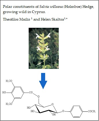
Polar Constituents of Salvia willeana (Holmboe) Hedge, Growing Wild in Cyprus

Department of Pharmacognosy & Chemistry of Natural Products, School of Pharmacy, National and Kapodistrian University of Athens, Panepistimiopolis, Zografou, 157 71 Athens, Greece
*
Author to whom correspondence should be addressed.
Plants 2018, 7(1), 18; https://doi.org/10.3390/plants7010018
Submission received: 29 January 2018 / Revised: 27 February 2018 / Accepted: 1 March 2018 / Published: 6 March 2018
(This article belongs to the Special Issue Lamiaceae Species: Biology, Ecology and Practical Uses)

Abstract

:
Twenty compounds were isolated from the aerial parts of Salvia willeana (Holmboe) Hedge, growing wild in Cyprus. These compounds comprise one new and one known acetophenone, one megastigmane glucoside, five phenolic derivatives, two caffeic acid oligomers, three flavonoids, two lignans, two triterpene acids, one monoterpene glucoside, and two fatty acids. The structures of the isolated compounds were established by means of NMR [(Rotating-frame OverhauserEffect SpectroscopY) (1H-1H-COSY (COrrelation SpectroscopΥ), 1H-13C-HSQC (Heteronuclear Single Quantum Correlation), HMBC (Heteronuclear Multiple Bond Correlation), NOESY (Nuclear Overhauser Effect SpectroscopY), ROESY (Rotating-frame Overhauser Effect SpectroscopY)] and MS spectral analyses. This is the first report of the natural occurrence of 4-hydroxy-acetophenone 4-O-(3,5-dimethoxy-4-hydroxybenzoyl)-β-d-glucopyranoside. A chemical review on the non-volatile secondary metabolites has been carried out. Based on the literature data, the analysis revealed that the chemical profile of S. willeana is close to that of S. officinalis L.

Graphical Abstract

1. Introduction

The Lamiaceae family consists of more than 250 genera; Salvia L. is the largest genus within this family due to the presence of approximately 900 species. Salvia L. spreads in the warm and temperate regions of both the northern and southern hemispheres, and some species of this genus have been cultivated worldwide for use in folk medicine, in perfumery and cosmetics industries, as well as for culinary purposes, like flavoring and aromatic agents [1,2]. Some of its many interesting biological and pharmacological properties are its antioxidant [3], antimicrobial [3,4], cytotoxic [3,4], anti-HIV [3], and antiplasmodial effects [4], as well as others [3]. It is noteworthy that the name of the genus, Salvia, is derived from the Latin word “salvare”, which means “to save”, in reference to the curative properties of the plant [5]. The genus has attracted such great interest, that it has become the subject of numerous chemical studies, giving evidence that these plants are a rich source of a wide variety of secondary metabolites, such as polyphenols and terpenoids [1].
Salvia willeana (syn. S. grandiflora subsp. willeana Holmboe and S. grandiflora subsp. albiflora Lindb.) is a low-growing, strongly aromatic suffruticose herb, sometimes carpeting the ground [6]. This species is endemic to Cyprus, where it grows on moist, rocky mountainsides of the Troodos range at 1150–1950 m altitude and it flowers from May to October [7]. Its extracts possess different pharmacological properties and the plant has been used to halt milk production in nursing mothers, as well as for its antiseptic activity [8]. As S. willeana is locally used in aqueous preparations, the purpose of our study was the investigation of secondary metabolites obtained from the polar extract of its aerial parts. Our previous study of its lipophilic extract, revealed the presence of camphor, lupeol, and oleanolic acid, and demonstrated their anti-inflammatory effect [8]. It is of interest to note that there is only one more report regarding the chemical constituents of a polar extract of S. willeana, which revealed the presence of the triterpenoids ursolic and oleanolic acids, the diterpenoids carnosic acid and isorosmanol, and the flavonoid salvigenin [9].

2. Results

The polar extract (MeOH:H2O 5:1) of S. willeana was fractionated by MPLC (medium pressure liquid chromatography), CC (column chromatography), followed by semi-preparative HPLC, preparative TLC, and yielded two triterpenoids, namely ursolic acid (1) and maslinic acid (2), one monoterpene glucoside, (1S,2R,4R)-1,8-epoxy-p-menthan-2-yl-O-β-d-glucopyranoside (3), one megastigmane glucoside, (6R,9S)-3-oxo-α-ionol β-d-glucopyranoside (4), five phenolic compounds, i.e., hydroxy-tyrosol (5), p-anisic acid (6), eleutheroside B (syringin) (7), 1-O-p-hydroxybenzoyl-β-d-apiofuranosyl-(1→6)-β-d-glucopyranoside (8) and eugenylglucoside (9), two acetophenones, 4-O-β-d-glucopyranosyl acetophenone (picein) (10) and 4-hydroxy-acetophenone 4-O-(3,5-dimethoxy-4-hydroxybenzoyl)-β-d-glucopyranoside (11), two caffeic acid oligomers, rosmarinic acid (12) and salvianolic acid K (13), three flavonoids, luteolin-7-O-β-d-glucoside (cynaroside) (14), 6-hydroxyluteolin 7-O-β-d-glucoside (15) and hesperidin (16), two lignans, syringaresinol-4-O-β-d-glucopyranoside (17), pinoresinol-4-O-β-d-glucopyranoside (18), and two fatty acids: linoleic acid (19) and methyl α-linolenate (20) (Figure 1).
Compound 11 was obtained as a white amorphous powder. [ a ] D 20 − 4.71° (c 0.25 MeOH); UV (CH3OH) λmax: 272 nm. The HREIMS of 11 established its molecular formula as C23H26O11 (found 477.1405 [M − H], calcd. 478.1449). The 1H-NMR spectrum (Table 1) showed signals at δH 7.69 (2H, d, J = 9.0 Hz) and 7.05 (2H, d, J = 9.0 Hz), which were indicative of a 1,4-bisubsituted phenyl group. Thus, these proton signals were recognized belonging to the aromatic ring of the acetophenone moiety [10]. In the upfield region of the 1H-NMR spectrum, a singlet at δH 2.47 (3H, s) was ascribed to the methyl group (CH3-8) attached on the carbonyl group of the acetophenone [10]. Moreover, the presence of a singlet at δH 7.33 with an integration of two aromatic protons, indicative of a pair of equivalent methine protons, revealed the occurrence of the syringic acid ester structure in the molecule [11]. In the 1H-NMR spectrum, the presence of a β-d-glucopyranose unit was evident based on a characteristic doublet signal with a coupling constant of 7.9 Hz at δH 5.04, assignable to the anomeric proton of the sugar moiety. Moreover, in the 1H-1H-COSY spectrum the correlation peaks between the vicinal protons of the sugar ring were observed. Furthermore, from the 13C-NMR data (Table 1) the carbon signals of the glucose moiety were assigned at δC 101.7 (C-1′), 77.5 (C-3′), 75.5 (C-5′), 74.5 (C-2′), 71.3 (C-4′), and 64.8 (C-6′), matching the reported data of 1,6-disubstituted-β-d-glucose [12]. The structural assignment was further confirmed by HSQC experiments, due to the carbon signals at δC 130.1 (C-2/C-6), 116.2 (C-3/C-5), and 26.5 (C-8), indicative of a 4-hydroxy-acetophenone moiety [9,13], while the syringic acid ester was confirmed by the presence of the carbon signals at δC 108.3 (C-2″/C-6″), 56.0 (3″, 5″-OCH3) [11]. The existence of the acetophenone moiety was corroborated by the HMBC experiment. This spectrum revealed a long-range cross peak between the carbonyl group at δC 199.1 (C-7) with the equivalent protons H-2/H-6 (δH 7.69), as well as an interaction between the carbonyl group (δC 199.1) and the methyl group CH3-8 (δH 2.47). In addition, the linkage of the syringyl moiety with the glucosyl moiety was substantiated by the observation of an HMBC correlation between the carbonyl group at δC 167.3 and the 6′-methylene protons at δH 4.46 (dd, J = 11.7, 8.0). Furthermore, the position of the attachment of the carboxyl group to the quaternary carbon C-1″ of the syringic ester was determined by a diagnostic HMBC cross peak between the equivalent methine protons H-2″/H-6″ at δH 7.33 and the carbonyl carbon C-7″ at δC 167.3, while a long-range coupling between the methyl protons of the methoxy groups at δH 3.83 and the benzylic carbons C-3″/C-5″ at δC 149.0 was also observed. Moreover, the position of the attachment of glucose to the 4-hydroxy-acetophenone moiety was revealed by a ROESY experiment, which displayed correlations between the anomeric proton H-1′ (δH 5.04) and the equivalent protons H-3/H-5 (δH 7.05) of the acetophenone (Figure 2). On the basis of the information above and by comparison with the data for compounds of similar structures [10,11,13] compound 11 was identified as 4-hydroxy-acetophenone 4-O-(3,5-dimethoxy-4-hydroxybenzoyl)-β-d-glucopyranoside, which is a new natural product, to the best of our knowledge.
The identification of the known flavonoids luteolin-7-O-β-d-glucoside (14) [14,15,16], 6-hydroxyluteolin 7-O-β-d-glucoside (15) [17], and hesperidin (16) [18,19,20,21,22,23,24] was based on UV–VIS and NMR spectroscopic analyses, as well as by comparing their spectroscopic data with those reported in the literature. The structure of the two fatty acids, linoleic acid (19) [25,26] and methyl α-linolenate (20) [26,27], has been deduced by the interpretation of NMR and GC-MS data. The 1H-NMR chemical shifts for compounds 1 [28,29,30], 2 [31,32,33], 3 [34,35], 4 [35,36,37], 7 [38,39,40,41,42], 9 [35,43], 10 [10,35,44], 12 [35,45,46,47,48], 17 [49,50], and 18 [51,52,53,54] presented in our study are in agreement with the data previously reported in the literature.
However, the NMR data of compounds 5, 6, 8, and 13 are not fully recorded in the literature, therefore, they are presented here below.

3. Discussion

The genus Salvia L. is characterized by the presence of several different secondary metabolites, mainly phenolic derivatives and terpenoids [3].
In the present study, overall, 20 compounds were isolated from S. willeana polar extracts, i.e., two triterpenoids, namely ursolic acid (1) and maslinic acid (2), one monoterpene glucoside, (1S,2R,4R)-1,8-epoxy-p-menthan-2-yl-O-β-d-glucopyranoside (3), one megastigmane glucoside, (6R,9S)-3-oxo-α-ionol β-d-glucopyranoside (4), five simple phenolic compounds, i.e., hydroxy-tyrosol (5), p-anisic acid (6), eleutheroside B (syringin) (7), 1-O-p-hydroxybenzoyl-β-d-apiofuranosyl-(1→6)-β-d-glucopyranoside (8) and eugenylglucoside (9), two acetophenones, 4-O-β-d-glucopyranosyl acetophenone (picein) (10) and 4-hydroxy-acetophenone 4-O-(3,5-dimethoxy-4-hydroxybenzoyl)-β-d-glucopyranoside (11), two caffeic acid oligomers, rosmarinic acid (12) and salvianolic acid K (13), three flavonoids, luteolin-7-O-β-d-glucoside (cynaroside) (14), 6-hydroxyluteolin 7-O-β-d-glucoside (15) and hesperidin (16), two lignans, syringaresinol-4-O-β-d-glucopyranoside (17), pinoresinol-4-O-β-d-glucopyranoside (18) and two fatty acids: linoleic acid (19) and methyl α-linolenate (20).
It is interesting to point out that compounds 7, 16, 17, and 18 had not been previously detected in Salvia L. Syringin (7) is reported, here, as a component of the Lamiaceae family for the first time. Moreover, compounds 3, 5, 6, and 810 had been previously mentioned only once in the genus, as follows: 3, 9, 10 [35], 6 [55], 8 [10] and 14, 15 [3] in S. officinalis L., 5 in S. digitaloides Diels [56], while compound 4 twice in S. nemorosa and S. officinalis L. [35,37]. As for the two triterpenoids 1 and 2, these have previously been isolated from the acetone extract of the aerial parts of S. willeana [9]. So far, only 134 Salvia species of the over 1000 species suggested have been investigated [3]. Based on our results, concerning the polar secondary metabolites, among these species S. willeana showed many similarities to S. officinalis L., since most of the isolated simple phenols (5, 6, 810), as well as the flavonoids 14 and 15, are found only in these two species (Supplementary Materials Table S1 and the references herein).

4. Materials and Methods

4.1. Plant Material

Aerial parts of Salvia willeana (Holmboe) Hedge were collected on Troodos Mountain in Cyprus in April 2004 [8]. A voucher specimen has been deposited in the Agricultural Research Institute Herbarium of Nicosia [no. ARI 3213].

4.2. Equipment and Reagents

1H, 13C, and 2D-NMR spectra were recorded in CDCl3 and CD3OD on Bruker DRX 400 (399.95 MHz for 1H-NMR) and Bruker AC 200 (200.13 MHz for 1H-NMR and 50.3 MHz for 13C-ΝMR) instruments at 295 K (Bruker BioSpin GmbH, Silberstetten, Germany). Chemical shifts are given in ppm (δ) and were referenced to the solvent signals at 7.24/3.31 and 77.0/49.5 ppm for 1H and 13C-NMR, respectively. COSY, HSQC, HMBC, NOESY, and ROESY (mixing time 950 ms) were performed using standard Bruker microprograms. High-resolution mass spectra were measured on a Q-TOF 6540 UHD (Aligent Technologies, Santa Clara, California, USA). The solvents used were of spectroscopic grade (Merck KGaA, Darmstadt, Germany). UV spectra were recorded on a Shimadzu UV-160A spectrophotometer (Shimadzu; Kyoto, Japan), according to Mabry et al. [57]. Optical rotations were determined using a Perkin-Elmer Polarimeter 341 (Perkin-Elmer, GmbH, Überlingen, Germany). GC-MS (Gas Chromatography-Mass Spectrometry) analyses were performed on a Hewlett-Packard 5973–6890 system (Palo Alto, California) operating in EI mode (70 eV) equipped with a split/splitless injector (220 °C), a split ratio 1/10, using a fused silica HP-5 MS capillary column (30 m × 0.25 mm (i.d.), film thickness: 0.25 µm) with a temperature program for HP-5 MS column from 60 °C (5 min) to 280 °C, at a rate of 4 °C/min and helium as a carrier gas at a flow rate of 1.0 mL/min. Preparative HPLC (High-Performance Liquid Chromatography) was performed using a C18 25 cm × 10 mm Kromasil column on a HPLC system (Jasco PU-2080; JASCO, Tokyo, Japan) equipped with an RI detector Shimadzu 10A (Shimadzu, Kyoto, Japan); flow rate: 1.0 mL/min; concentration of the samples: 3.5–7.0 mg/mL. All solvents used were of HPLC grade (Merck). MPLC (Medium Pressure Liquid Chromatography) was performed using Büchi C-615 and Büchi 688 chromatographic pump; columns: Büchi Borosilikat 3.3, (41.0 cm × 4.0 cm), flow rate: 10 mL/min; (15.0 cm × 1.5 cm) flow rate: 3 mL/min; vacuum liquid chromatography (VLC): silica gel 60H (Merck, Art. 7736) [58]. Column chromatography (CC): silica gel (Merck, Art. 9385), silica gel 60 (230–400 mesh ASTM, SDS 2050044) gradient elution with the solvent mixtures indicated in each case; Sephadex LH-20 (Pharmacia Fine Chemicals); cellulose (Avicel, Merck, Art. 2330). Preparative TLC (Thin Layer Chromatography) was performed using pre-coated silica gel 60 plates (Merck, Art. 5721). Fractionation was always monitored by TLC silica gel 60 F-254, (Merck, Art. 5554) with visualization under UV (254 and 365 nm) and spraying with vanillin-sulfuric acid reagent (vanillin Merck, Art. No. S26047 841) [59] and Neu’s reagent for phenolics [60]. Analytical solvents were obtained from Panreac Quimica SA (Barcelone, Spain, Italy), while deuterated solvents were purchased from Merck, KGaA (Darmstadt, Germany). Di-phosphorus pentoxide was purchased from Chemlab, Belgium.

4.3. Extraction and Chromatography

The air-dried powdered aerial parts of S. willeana (0.43 kg) were successively extracted at room temperature with cyclohexane, dichloromethane, MeOH, and MeOH:H2O (5:1) (2 L of each solvent, twice for 48 h) [8]. A portion of the latter extract (9.0 g) was fractionated on a RP18-MPLC (41.0 × 4.0 cm) using a H2O: MeOH gradient system (100% H2O → 100% MeOH; steps of 10% MeOH; 50 min each; 50% MeOH: 50% EtOAc 50 min; 100% EtOAc 50 min) to yield twenty three fractions (A–V) of 500 mL each. Fraction D (598.8 mg; H2O:MeOH 85:15) was similarly purified by RP18-MPLC (15 cm × 1.5 cm) to obtain three sub-fractions (1–3). The first two sub-fractions were combined together (sub-fraction DA, 364.5 mg), subjected to a Sephadex LH-20 column and eluted with 100% methanol to afford 61 fractions combined in 13 groups (DAA–DAM). Group DAJ (4.2 mg) was identified as salvianolic acid K (13). Group DAC (12.9 mg) was subjected to RP18-HPLC (RID; isocratic elution using MeOH:CH3COOH 5% 30:70; flow-rate: 1.0 mL/min) and afforded 1-O-p-hydroxybenzoyl-β-d-apiofuranosyl-(1→6)-β-d-glucopyranoside (8) (Rt = 14.0 min, 0.5 mg) and 4-O-β-d-glucopyranosyl-acetophenone (picein) (10) (Rt = 17.2 min, 0.7 mg). Group DAD (9.3 mg) was purified by prep. TLC on silica gel using CHCl3:MeOH:AcOH (7:1.5:1.5) as the eluent and yielded hydroxytyrosol (6.3 mg) (5). Fraction H (718.3 mg) was fractionated by CC on a Sephadex LH-20 (25.0 cm × 3.2 cm) using H2O:MeOH (20:80 to 0:100) for gradient elution to afford luteolin 7-O-β-d-glucoside (0.7 mg) (14). Groups HC to HG were combined (HC′; 193.7 mg) and purified by CC (12.2 cm × 2.2 cm) over silica gel with cyclohexane: DM:EtOAc:MeOH mixtures of increasing polarity to yield nine groups (HC′A–HC′I). Group HC′C (14.2 mg; eluted with EtOAc:MeOH 97:3 to 94:6) was purified by RP18-HPLC (RID; MeOH; H2O 40:60; flow rate: 1 mL/min) to obtain syringin (Rt = 56.4 min, 0.1 mg) (7), (1S,2R,4R)-1,8-epoxy-p-menthan-2-yl-O-β-d-glucopyranoside (Rt = 74.2 min, 0.9 mg) (3), (6R,9S)-3-oxo-α-ionol-β-d-glucopyranoside (Rt = 116.5 min, 0.5 mg) (4), eugenyl-glucoside (Rt = 130.1 min, 0.3 mg) (9). Combined groups HJ to HL (HJ′; 48.9 mg; eluted with H2O:MeOH 50:50) were subjected to CC over silica gel using cyclohexane: DM:EtOAc:MeOH mixtures of increasing polarity (60 fractions). Purification of fraction HJ′E (2.1 mg; eluted with EtΟAc:MeOH 90:10) was carried out by prep. TLC on silica gel, using CHCl3:MeOH:AcOH (9.0:1.0:0.1) and afforded syringaresinol-4-O-β-d-glucopyranoside (0.9 mg) (17) and pinoresinol-4-O-β-d-glucopyranoside (1.2 mg) (18). Combined groups HM to HP (HM′; 86.7 mg) were fractionated by RP18-HPLC (MeOH 58%:H2O 42%; flow rate: 1 mL/min) and yielded rosmarinic acid (Rt = 10.6 min, 2.2 mg) (12) and p-anisic acid (Rt = 18.9 min, 0.1 mg) (6), and the sub-fraction HM′14b (Rt = 16.7 min, 5.9 mg), which was further purified by prep. TLC on silica gel with EtOAc:AcOH:H2O (65:15:20) and led to the isolation of hesperidin (3.0 mg) (16) and of 4-hydroxyacetophenone 4-Ο-(3,5-dimethoxy-4-hydroxybenzoyl)-β-d-glucopyranoside (2.8 mg) (11). Combined groups HT to HV (HT′; 176.5 mg), subjected to CC on cellulose (11.0 cm × 3.2 cm) using as eluent AcOH:H2O (30:70) afforded 79 fractions. Fraction HT′H (4.8 mg) was purified by prep. TLC on silica gel with EtOAc:AcOH:H2O (65:15:20) to obtain 6-hydroxyluteolin 7-O-β-d-glucoside (0.9 mg) (15). The purification of fraction N (248.5 mg) was performed on silica gel CC (15.2 cm × 2.0 cm) using mixtures of cyclohexane: DM:EtOAc:MeOH of increasing polarity and afforded methyl α-linolenate (C18:3) (1.4 mg) (20) and ursolic acid (4.7 mg) (1). Groups NB′ (7.4 mg) and NF (19.6 mg) were subjected to prep. TLC on silica gel with CHCl3:MeOH:AcOH (9.5:0.5:0.05) and yielded linoleic acid (C18:2) (0.5 mg) (19) and maslinic acid (18.3 mg) (2), respectively. All obtained extracts, fractions, and isolated compounds were evaporated to dryness in vacuum under low temperature and then were put in activated desiccators with P2O5 until their weights had stabilized.

4.4. NMR Data of 5, 8, and 13

Compound 5: Yellow amorphous powder; 1H-NMR (CD3OD, 400 MHz): 2.66 (2H, t, J = 7.3, H-7), 3.66 (2H, t, J = 7.3, H-8), 6.52 (1H, dd, J = 8.0, 2.0, H-6), 6.65 (1H, d, J = 2.0, H-2), 6.67 (1H, d, J = 8.0, H-5).
Compound 6: White amorphous powder; 1H-NMR (CD3OD, 400 MHz): 3.91 (3H, s, OCH3), 6.85 (2H, d, J = 8.5, H-3/H-5), 7.89 (2H, d, J = 8.5, H-2/H-6).
Compound 8: White amorphous powder; 1H-NMR (CD3OD, 400 MHz): δ 3.38 (1H, m, H-5′), 3.47 (2H, m, H-2′, H-3′), 3.56 (2H, s, H-5a,b″), 3.61 (1H, m, H-6b′), 3.73 (1H, d, J = 9.6, H-4b″), 3.90 (1H, d, J = 2.2, H-2″), 3.96 (1H, d, J = 9.6, H-4a″), 3.99 (1H, d, J = 11.5, H-6a′), 4.96 (1H, d, J = 2.2, H-1″), 5.64 (1H, d, J = 7.9, H-1′), 6.84 (2H, d, J = 8.7, H-3, H-5), 7.96 (2H, d, J = 8.7, H-2, H-6).
Compound 13: Yellow amorphous powder; [ a ] D 20 + 0.36° (c 0.350 MeOH); 1H-NMR (CD3OD, 400 MHz): δ 2.90 (1H, dd, J = 14.0, 9.8, H-7′), 3.07 (1H, dd, J = 14.0, 3.8, H-7′), 4.24 (1H, d, J = 6.5, H-8″), 4.88 (1H, d, J = 6.5, H-7″), 5.02 (1H, dd, J = 9.8, 3.8, H-8′), 6.31 (1H, d, J = 16.0, H-8), 6.38 (1H, d, J = 8.0, H-5), 6.62 (1H, dd, J = 8.0, 1.6, H-6′), 6.66 (1H, d, J = 8.0, H-5′), 6.74 (1H, d, J = 8.0, H-5″), 6.76 (1H, s, H-2′), 6.82 (1H, dd, J = 8.0, 2.0, H-6), 6.84 (1H, dd, J = 8.0, 2.0, H-6″), 6.98 (1H, d, J = 2.0, H-2″), 7.01 (1H, d, J = 2.0, H-2), 7.47 (1H, d, J = 16.0, H-7).

Supplementary Materials

The following are available online at https://www.mdpi.com/2223-7747/7/1/18/s1. Table S1: Non-volatile secondary metabolites of Salvia L.

Acknowledgments

The authors thank J. Heilmann (University of Regensburg, Pharmaceutical Biology), as well as J. Kiermaier for recording the MS spectra (all central analytics of NWF IV, University of Regensburg, Germany).

Author Contributions

T.M. contributed to the writing and carried out all chemical analyses. H.S. conceived and designed the experiments, contributed to the writing, and also supervised the chemical analyses.

Conflicts of Interest

The authors declare no conflict of interest.

References

  1. Lu, Y.; Foo, L.Y. Polyphenolics of Salvia—A review. Phytochemistry 2002, 59, 117–140. [Google Scholar] [CrossRef]
  2. Walker, J.B.; Systma, K.J. Staminal Evolution in the genus Salvia (Lamiaceae). Ann. Bot. 2007, 100, 375–391. [Google Scholar] [CrossRef] [PubMed]
  3. Wu, Y.-B.; Ni, Z.-Y.; Shi, Q.-W.; Dong, M.; Kiyota, H.; Gu, Y.-C.; Cong, B. Constituents from Salvia Species and Their Biological Activities. Chem. Rev. 2012, 112, 5967–6026. [Google Scholar] [CrossRef] [PubMed]
  4. Kamatou, G.P.P.; Makunga, N.P.; Ramogola, W.P.N.; Viljoena, A.M. South African Salvia species: A review of biological activities and phytochemistry. J. Ethnopharmacol. 2008, 119, 664–672. [Google Scholar] [CrossRef] [PubMed]
  5. Grieve, M. A Modern Herbal; Savas Publishing: El Dorado Hills, CA, USA, 1984; ISBN 13 9780486227986. [Google Scholar]
  6. Dereboylu, A.; Şengonca, N.; Güvensen, A.; Gücel, S. Anatomical and palynological characteristics of Salvia willeana (Holmboe) Hedge and Salvia veneris Hedge endemic to Cyprus. Afr. J. Biotechnol. 2010, 9, 2076–2088. [Google Scholar]
  7. Bellomaria, B.; Arnold, N.; Valentini, G. Contribution to the study of the essential oils from three species of Salvia growing wild in the eastern Mediterranean region. J. Essent. Oil Res. 1992, 4, 607–614. [Google Scholar] [CrossRef]
  8. Vonaparti, A.; Karioti, A.; Recio, M.; Máñez, S.; Ríos, J.; Skaltsa, E.; Giner, R. Effects of terpenoids from Salvia willeana in delayed-type hypersensitivity, human lymphocyte proliferation and cytokine production. Nat. Prod. Commun. 2008, 3, 1953–1958. [Google Scholar]
  9. De la Torre, M.; Bruno, M.; Savona, F.; Rodriquez, B.; Apostolides Arnold, N. Terpenoids from Salvia willeana and S. virgata. Phytochemistry 1990, 29, 668–670. [Google Scholar] [CrossRef]
  10. Wang, M.; Kikuzaki, H.; Lin, C.; Kahyaoglu, A.; Huang, M.; Nakatani, N.; Ho, C. Acetophenone Glycosides from Thyme (Thymus vulgaris L.). J. Agric. Food Chem. 1999, 47, 1911–1914. [Google Scholar] [CrossRef] [PubMed]
  11. Machida, K.; Yogiashi, Y.; Matsuda, S.; Suzuki, A.; Kikuchi, M. A new phenolic glycoside syringate from the bark of Juglans mandshurica MAXIM. var. sieboldiana MAKINO. J. Nat. Med. 2009, 63, 220–222. [Google Scholar] [CrossRef] [PubMed]
  12. Wang, M.; Kikuzaki, H.; Zhu, N.; Sang, S.; Nakatani, N.; Ho, C.-T. Isolation and structural elucidation of two new glycosides from sage (Salvia officinalis L.). J. Agric. Food Chem. 2000, 48, 235–238. [Google Scholar] [CrossRef] [PubMed]
  13. Olennikov, D.; Chekhirova, G.V. 6″-Galloylpicein and other phenolic compounds from Arctostaphylos uva-ursi. Chem. Nat. Compd. 2013, 49, 1–7. [Google Scholar] [CrossRef]
  14. Özgen, U.; Sevindik, H.; Kazaz, C.; Yigit, D.; Kandemir, A.; Secen, H.; Calis, I. A new sulfated a-Ionone glycoside from Sonchus erzincanicus Matthews. Molecules 2010, 15, 2593–2599. [Google Scholar] [CrossRef] [PubMed]
  15. Benayache, F.; Boureghda, A.; Ameddah, S.; Marchioni, E.; Benayache, S. Flavonoids from Thymus numidicus Poiret. Pharm. Lett. 2014, 6, 50–54. [Google Scholar]
  16. Rashed, K.; Ćirić, A.; Glamočlija, J.; Calhelha, R. Antimicrobial and cytotoxic activities of Alnus rugosa L. aerial parts and identification of the bioactive components. Ind. Crops Prod. 2014, 59, 189–196. [Google Scholar] [CrossRef]
  17. Lu, Y.; Foo, L.Y. Flavonoid and phenolic glycosides from Salvia officinalis. Phytochemistry 2000, 55, 263–267. [Google Scholar] [CrossRef]
  18. Ikan, R. Natural Products. A Laboratory Guide, 2nd ed.; Academic Press Inc.: Cambridge, MA, USA, 1991; pp. 9–11. [Google Scholar]
  19. Carvalho, M.G.; Costa, P.M.; Santos Abreu, H. Flavones from Vernonia diffusa. J. Braz. Chem. Soc. 1999, 10, 163–166. [Google Scholar] [CrossRef]
  20. Marder, M.; Viola, H.; Wasowski, C.; Fernández, S.; Medina, J.; Paladini, A. 6-Methylapigenin and hesperidin, new valeriana flavonoids with activity on the CNS. Pharmacol. Biochem. Behav. 2003, 75, 537–545. [Google Scholar] [CrossRef]
  21. Aghel, N.; Ramezani, Z.; Beiranvard, S. Hesperidin from Citrus sinensis Cultivated in Dezful, Iran. Pak. J. Biol. Sci. 2008, 11, 2451–2453. [Google Scholar] [CrossRef] [PubMed]
  22. Nizamutdinova, I.T.; Jeong, J.J.; Xu, G.H.; Lee, S.; Kang, S.S.; Kim, Y.S.; Chang, K.C.; Kim, H.J. Hesperidin, hesperidin methyl chalone and phellopterin from Poncirus trifoliata (Rutaceae) differentially regulate the expression of adhesion molecules in tumor necrosis factor-α-stimulated human umbilical vein endothelial cells. Int. Immunopharmacol. 2008, 8, 670–678. [Google Scholar] [CrossRef] [PubMed]
  23. Maltese, F.; Erkelens, C.; Kooy, F.; Choi, Y.; Verpoorte, R. Identification of natural epimeric flavanone glycosides by NMR spectroscopy. Food Chem. 2009, 116, 575–579. [Google Scholar] [CrossRef]
  24. Han, S.; Mok, S.; Kim, H.; Lee, J.; Lee, D.; Lee, S.Y.; Kim, J.; Kim, S.; Lee, S. Determination of hesperidin in mixed tea by HPLC. J Agric. Sci. 2011, 38, 295–299. [Google Scholar]
  25. Lee, C.K.; Chang, M.H. The chemical constituents from the heartwood of Eucalyptus citriodora. J Chin. Chem. Soc. 2000, 47, 555–560. [Google Scholar] [CrossRef]
  26. Hatzakis, E.; Agiomyrgianaki, A.; Kostidis, S.; Dais, P. High-resolution NMR spectroscopy, an alternative fast tool for Qualitative and Quantitative Analysis of Diacylglycerol (DAG) oil. J. Am. Oil Chem. Soc. 2011, 88, 1695–1708. [Google Scholar] [CrossRef]
  27. Horst, M.; Urbin, S.; Burton, R.; MacMillan, C. Using proton nuclear magnetic resonance as a rapid response research tool for methyl ester characterization in biodiesel. Lipid Technol. 2009, 21, 1–3. [Google Scholar] [CrossRef]
  28. Güvenalp, Z.; Özbek, H.; Kuruüzüm-Uz, A.; Kazaz, C.; Demirezer, Ö.L. Secondary metabolites from Nepeta heliotropifolia. Turk. J. Chem. 2009, 33, 667–675. [Google Scholar]
  29. Ragasa, Y.; Ng, V.A.S.; Ebajo, V.D.; Fortin, D.R.; De Los Reyes, M.M.; Shen, C.-C. Triterpenes from Shorea negrosensis. Pharm. Lett. 2014, 6, 453–458. [Google Scholar]
  30. Raza, R.; Ilyas, Z.; Ali, S.; Muhammad, N.M.; Muhammad, Y.; Khokhar, M.Y.; Iqbal, J. Identification of Highly Potent and Selective α-Glucosidase Inhibitors with Antiglycation Potential, Isolated from Rhododendron arboreum. Rec. Nat. Prod. 2015, 9, 262–266. [Google Scholar]
  31. Tanaka, J.C.A.; Vidotti, G.J.; da Silva, C.C. A New Tormentic Acid Derivative from Luehea divaricata Mart. (Tiliaceae). J. Braz. Chem. Soc. 2003, 14, 475–478. [Google Scholar] [CrossRef]
  32. Rungsimakan, S. Phytochemical and Biological Activity Studies on Salvia viridis L. Ph.D. Thesis, Department of Pharmacy and Pharmacology, University of Bath, Bath, UK, 2011. [Google Scholar]
  33. Woo, W.K.; Han, Y.J.; Un Choi, S.; Ki, H.; Kim, K.H.; Lee, K.R. Triterpenes from Perilla frutescens var. acuta and Their Cytotoxic Activity. Nat. Prod. Sci. 2014, 20, 71–75. [Google Scholar]
  34. Manns, D. Linalool and cineole type glucosides from Cunila spicata. Phytochemistry 1995, 39, 1115–1118. [Google Scholar] [CrossRef]
  35. Wang, M.; Shao, Y.; Huang, T.; Wei, G.; Ho, C. Isolation and structural elucidation of aroma constituents bound as glucosides from Sage (Salvia officinalis). J. Agric. Food Chem. 1998, 46, 2509–2511. [Google Scholar] [CrossRef]
  36. Pabst, A.; Barron, D.; Sémon, E.; Schreier, P. Two diastereomeric 3-oxo-α-ionol β-d-glucosides from Raspberry fruit. Phytochemistry 1992, 31, 1649–1652. [Google Scholar] [CrossRef]
  37. Takeda, Y.; Zhang, H.; Matsumoto, T.; Otsuka, H.; Oosio, Y.; Honda, G.; Tabata, M.; Fujita, T.; Su, H.; Sezik, E.; et al. Megastigmane glycosides from Salvia nemorosa. Phytochemistry 1997, 44, 117–120. [Google Scholar] [CrossRef]
  38. Sugiyama, M.; Nagayama, E.; Kikuchi, M. Lignan and Phenylpropanoid glycosides from Osmanthus asiaticus. Phytochemistry 1993, 33, 1215–1219. [Google Scholar] [CrossRef]
  39. Kim, M.; Moon, H.; Lee, D.; Woo, E. A new lignan glycoside from the Stem bark of Styrax japonica S. et Z. Arch. Pharm. Res. 2007, 30, 425–430. [Google Scholar] [CrossRef] [PubMed]
  40. Qin, Y.; Yin, C.; Cheng, Z. A new tetrahydrofuran lignan diglycoside from Viola tianshanica Maxim. Molecules 2013, 18, 13636–13644. [Google Scholar] [CrossRef] [PubMed]
  41. Lee, J.; Lee, M.; Pae, S.; Oh, K.; Jung, C.; Baek, I.; Lee, S. Analysis of yield of eleutherosides B and E in Acanthopanax divaricatus and A. koreanum Grown with varying cultivation methods. Sci. World J. 2014. [Google Scholar] [CrossRef]
  42. Lall, N.; Kishore, N.; Binneman, B.; Twilley, D.; Venter, M.; Plessis-Stoman, D.; Boukes, G.; Hussein, A. Cytotoxicity of Syringin and 4-methoxycinnamyl alcohol isolated from Foeniculum vulgare on selected human cell lines. Nat. Prod. Res. 2015, 1752–1756. [Google Scholar] [CrossRef] [PubMed]
  43. Mulkens, A.; Kapetanidis, I. Eugenylglucoside, a new natural phenylpropanoid heteroside from Melissa officinalis. J. Nat. Prod. 1988, 51, 496–498. [Google Scholar] [CrossRef] [PubMed]
  44. Ushiyama, M.; Furuya, T. Glycosylation of phenolic compounds by root culture of Panax ginseng. Phytochemistry 1989, 28, 3009–3013. [Google Scholar] [CrossRef]
  45. Lu, Y.; Foo, L.Y. Rosmarinic acid derivatives from Salvia officinalis. Phytochemistry 1999, 51, 91–94. [Google Scholar] [CrossRef]
  46. Woo, E.; Piao, M. Antioxidative Constituents from Lycopus lucidus. Arch. Pharm. Res. 2004, 27, 173–176. [Google Scholar] [CrossRef] [PubMed]
  47. Özgen, U.; Mavi, A.; Terzi, Z.; Kazaz, C.; Aşçi, A.; Kaya, Y.; Seçen, H. Relationship between chemical structure and antioxidant activity of luteolin and its glyxosides isolated from Thymus sipyleus subsp. sipyleus var. sipyleus. Rec. Nat. Prod. 2011, 5, 12–21. [Google Scholar]
  48. Dapkevicius, A.; Beek, T.; Lelyveld, G.; Veldhuizen, A.; Groot, A.; Linssen, J.; Venskutonis, R. Isolation and structure Elucidation of radican scavengers from Thymus vulgaris Leaves. J. Nat. Prod. 2002, 65, 892–896. [Google Scholar] [CrossRef] [PubMed]
  49. Shahat, A.A.; Abdel-Azim, N.S.; Pieters, L.; Vlietinck, A.J. Isolation and NMR spectra of syringaresinol-β-d-glucoside from Cressa cretica. Fitoterapia 2004, 75, 771–773. [Google Scholar] [CrossRef] [PubMed]
  50. Park, H.B.; Lee, K.H.; Kim, K.H.; Lee, I.K.; Noh, H.J.; Choi, S.U.; Lee, K.R. Lignans from the roots of Berberis amurensis. Nat. Prod. Sci. 2009, 15, 17–21. [Google Scholar]
  51. Klimek, B.; Tokar, M. Biologically active compounds from the flowers of Forsythia suspensa Vahl. Acta Pol. Pharm. 1998, 55, 499–504. [Google Scholar]
  52. Ouyang, M.; Shung, Y.; Zhang, Z.K.; Kuo, Y.H. Inhibitory Activity against Tobacco Mosaic Virus (TMV) Replication of Pinoresinol and Syringaresinol Lignans and Their Glycosides from the Root of Rhus javanica var. roxburghiana. J. Agric. Food Chem. 2007, 55, 6460–6465. [Google Scholar] [CrossRef] [PubMed]
  53. Chiou, W.; Shen, C.; Lin, L. Anti-inflammatory principles from Balanopora laxiflora. J. Food Drug Anal. 2011, 19, 502–508. [Google Scholar]
  54. Kim, A.R.; Ko, H.J.; Chowdhury, M.A.; Chang, Y.; Woo, E. Chemical constituents of the aerial parts of Artemisia selengensis and their IL-6 inhibitory activity. Arch. Pharm. Res. 2015, 38, 1059–1065. [Google Scholar] [CrossRef] [PubMed]
  55. Jerković, I.; Mastelić, J.; Marijanović, Z. A variety of volatile compounds as Markers in Unifloral Honey from Dalmatian Sage (Salvia officinalis L.). Chem. Biodivers. 2006, 3, 1307–1316. [Google Scholar] [CrossRef] [PubMed]
  56. Wu, S.; Chan, Y. Five new iridoids from roots of Salvia digitaloides. Molecules 2014, 19, 15521–15534. [Google Scholar] [CrossRef] [PubMed]
  57. Mabry, T.G.; Markham, K.R.; Thomas, M.B. The Systematic Identification of Flavonoids; Springer: New York, NY, USA, 1970. [Google Scholar]
  58. Coll, J.C.; Bowden, B.F. The application of Vacuum Liquid Chromatography to the separation of terpene mixtures. J. Nat. Prod. 1986, 49, 934–936. [Google Scholar] [CrossRef]
  59. Stahl, E. Thin-Layer Chromatography, a Laboratory Handbook, 2nd ed.; Springer: Berlin/Heideiberg, Germany, 1969. [Google Scholar]
  60. Neu, R. Chelate von Diarylborsäuren mit aliphatischen Oxyalkylaminen als Reagenzien für den Nachweis von Oxyphenyl-benzo-γ-pyronen. Naturwissenschaften 1957, 44, 181–183. [Google Scholar] [CrossRef]
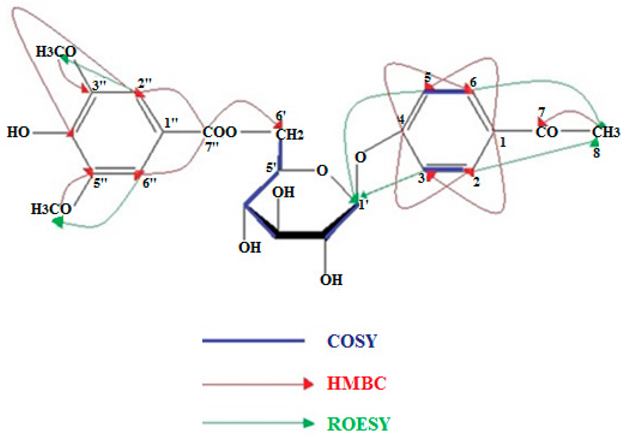
Figure 1. Structures of isolated compounds from Salvia willeana.
Plants 07 00018 g001aPlants 07 00018 g001bPlants 07 00018 g001c
Figure 2. COSY, HMBC, and ROE signals of compund 11.
Plants 07 00018 g002
Table 1. 1H-NMR and 13C-NMR spectrum of 11.
δCCδHHJ (Hz)
1131.5C---
2130.1CH7.691d (J = 9.0)
3116.2CH7.051d (J = 9.0)
4162.5C---
5116.2CH7.051d (J = 9.0)
6130.1CH7.691d (J = 9.0)
7199.1C---
826.5CH32.473s
1′101.7CH5.041d (J = 7.8)
2′74.5CH3.532m
3′77.5CH
4′71.3CH3.431m
5′75.5CH3.891dd (J = 8.0, 2.3)
6a′64.8CH24.712dd (J = 11.7, 2.3)
6b′4.46dd (J = 11.7, 8.0)
1″-C---
2″108.3CH7.331s
3″149.0C---
4″142.3C---
5″149.0C---
6″108.3CH7.331s
7″167.3C---
3″, 5″-OCH356.0CH33.836s

