Article

Leaf Morpho-Anatomy of Twelve Cymbidium (Orchidaceae) Species from China and Their Taxonomic Significance

1
College of Forestry, Southwest Forestry University, Kunming 650224, China
2
School of Chemistry, Biology and Environment, Yuxi Normal University, Yuxi 653100, China
3
Yunnan Academy of Biodiversity, Southwest Forestry University, Kunming 650224, China
*
Author to whom correspondence should be addressed.
These authors contributed equally to this work.
Plants 2025, 14(9), 1396; https://doi.org/10.3390/plants14091396
Submission received: 17 February 2025 / Revised: 14 March 2025 / Accepted: 25 March 2025 / Published: 6 May 2025
(This article belongs to the Special Issue Plant Taxonomy, Phylogeny, and Evolution)

Abstract

:
Cymbidium are endangered and ornamental orchids, and the taxonomy and species identification of this genus have been debated due to some overlapping morphological features between taxa and limited data being available. The leaf morpho-anatomy of 12 Cymbidium species from China was investigated using light microscopy and paraffin sectioning. Based on a comparative analysis, some leaf morphological features that varied between species were selected and used for taxonomic differentiation as follows: (1) The shape and structure of leaves were varied and could be used for species delimitation. (2) Microscopic characteristics show that the leaves lacked trichomes and displayed polygonal to rectangular epidermal cells on both surfaces, with larger adaxial cells and more abaxial stigmata. Stomata were mostly distributed only on the abaxial side, but on both sides in Cymbidium mastersii, which exhibited a rare amphistomatic type. The stomatal complex was uniformly tetracytic in 11 species, while it was observed to be anomocytic in C. floribundum. (3) Anatomically, two distinct midrib configurations were identified, a shallow V-shape and V-shape. The mesophyll cells were homogeneous in 10 species, with the exception of a layer of parenchyma cells resembling palisade cells occurring in C. lancifolium and C. qiubeiense. The thickness of the cuticle varied between species, with the adaxial surface covered by a thicker cuticle than the abaxial surface and displaying either a smooth or corrugated surface. A fiber bundle was observed in six species, but absent in the other six. In the former group, the fiber bundle occurred adjacent to both epidermal cells in C. mastersii and C. hookerianum, while it was adjacent to the abaxial epidermis in four other species. The stegmata, with conical, spherical silica bodies, were associated with fiber bundles and mesophyll in seven species, but absent in the other five (C. kanran, C. defoliatum, C. floribundum, C. lancifolium, and C. serratum). Three kinds of crystals were identified, namely the terete bundle, the long tube bundle, and the raphide. (4) It was suggested that some of these variable features could be selected and used for the delimitation of the species and taxonomy of Cymbidium. In addition, a key to the 12 Cymbidium species based on their leaf morpho-anatomic features was proposed, which could lead to a better understanding of the taxonomy and conservation of Orchidaceae.

1. Introduction

Cymbidium Sw. (Orchidaceae), comprising about 52 species, is predominantly found in the tropical and subtropical regions of Asia, with some species extending to Papua New Guinea and Australia [1]. There are around 49 species (19 endemic) that grow in China, the distribution center of Cymbidium [2]. Cymbidium species demonstrate diverse ecological adaptations such as epiphytic, lithophytic, or terrestrial orchids, which are characterized by ovoid to spindle-shaped pseudobulbs, distichous narrowly ligulate to elliptic leaves, showy and often fragrant flowers with trilobed labella, and two or four pollinia [1,3]. The species of Cymbidium found in China, known as ‘Guolan’ in Chinese, have been widely cultivated for over two hundred years due to their high ornamental value [2,3]. However, wild populations of Cymbidium have experienced a significant decline due to overexploitation and habitat fragmentation and are listed as rare and endangered species [4,5,6,7].
The infrageneric taxonomy of Cymbidium (the category of a section or subgenus of the genus and identification of different species) has been problematic and controversial due to the addition of newly described species [1,2,8,9,10]. Three subgenera of Cymbidium have been identified—subgen. Cymbidium (L.) Sw., subgen. Cyperorchis (Bl.), and subgen. Jensoa (Rafin.)—based on their pollinium number and labellum–column fusion [11]. This classification system has been adopted by several subsequent taxonomists [1,2,9]. Previously, 29 were species recorded in China, which were classified into three subgenera containing 14 sections [3]. Later, the number of Cymbidium species identified increased to 68 species, and the genus was classified into three subgenera containing with 16 sections, with 49 of these species found in China [9]. Later, Cymbidium was thought to contain 52 species and was divided into 11 sections based on molecular data from ITS and matK [1]. In addition, another study recognized 55 Cymbidium species (including the 49 from China), without further classifying them into subgenera or sections [2].
The taxonomy of Cymbidium has been problematic at the level of infrageneric categories, including species, sections, and subgenera, due to some overlapping floral morphological features, natural and artificial hybridization, and the discovery of new species [3,12,13,14]. There are some species that can be easily confused based on their morphological features (Figure 1), such as Cymbidium eburneum Lindl. (Figure 1A) and Cymbidium mastersii Griff. ex Lindl. (Figure 1B), due to both possessing white and showy flowers with long and large leaves, and Cymbidium qiubeiense K. M. Feng & H. Li (Figure 1E) and Cymbidium kanran Makino (Figure 1I), which share small and greenish flowers with narrow strap leaves [9]. However, these easily confused species should be recognized as separate taxa based on genomic and anatomical data from their chloroplasts [9,15,16,17]. Furthermore, the taxonomy of Cymbidium members from China has been controversial; they were divided into three ‘stable’ subgenera, namely subgen. Cyperorchis, subgen. Jensoa, and subgen. Cymbidium, but with each containing different types of sections [9]. Compared with the previous system [3], this one proposed some new sections species whose identification was previously uncertain [9]. For example, in subgen. Cyperorchis, Cymbidium wenshanense Y. S. Wu & F. Y. Liu was raised to sect. Annamaea [9] from sect. Iridorchis [3]. However, the categories of subgenus and section were neglected or missed in other classification systems when it came to these confusing differentiations [1,2]. Recently, molecular phylogenetic evidence has provided new insights into the taxonomy of Cymbidium, but significant controversy has remained [14]. It was suggested that Cymbidium should not be monophyletic and that it could be divided into two or three subgenera using the DNA markers matK and nrITS gathered from 34 species [15]. Subsequently, molecular systematic research revealed that there were paraphyletic/polyphyletic relationships among the three subgenera and that sect. Cymbidium, sect. Cyperorchis, and sect. Iridorchis were non-monophyletic by using ITS, matK, trnK, and 18S-26S sequence data from 37 taxa [16]. Later, it was shown that the species identification of Cymbidium was not fully resolved based on DNA barcodes from 237 chloroplast genomes, which came from the sampling of 50 species, indicating that establishing the taxonomy of Cymbidium required more evidence and further effort [17].
Leaf morpho-anatomy varies greatly between taxa and has played a key role in the taxonomy and phylogeny of Orchidaceae [2,8]. It was revealed that foliar characteristics, including leaf shape, cross-sectional outline, epidermal cell structure, cuticular thickness, crystal morphology, and vascular bundle arrangement, could be valuable for the identification of orchid species such as in Bulbophyllum Thouars [18] and Dendrobium Sw. [19]. However, there are limited and scattered data on the leaf morpho-anatomy of some Cymbidium species [8,20,21,22], which should be expanded through the collection of more samples. Although other studies have been published on the vegetative anatomy of Cymbidium, including analyses of 21 species [22] and 30 species [8], they primarily described leaf anatomic features while lacking detailed illustrations for each species. It was noted that some leaf morphological features varied between species of Cymbidium and should receive close attention, including stomatal complex types (tetracytic or cyclocytic–tetracytic, superficial, or depressed), cuticular patterns (smooth or uneven), hypodermal distribution (present or absent adaxially/abaxially), mesophyll organization (homogeneous or heterogeneous), vascular bundle arrangement (collateral or uniseriate), fiber bundle distribution, stegmata presence, and crystal morphology [8,22].
Therefore, the leaf morpho-anatomy of 12 Cymbidium species from China (Table 1, Figure 1) was investigated using light microscopy and paraffin sectioning. These species represent three subgenera—subgen. Cyperorchis (five species), subgen. Jensoa (six species), and subgen. Cymbidium (one species)—and include four easily confused species (Cymbidium mastersii vs. C. eburneum, C. kanran vs. C. qiubeiense). This study aimed to (1) investigate the leaf morpho-anatomy of these 12 species in detail, including the shape, margin, apex, and the base structure of the leaf epidermal cell, stomatal apparatus, and stigmata; the outline of midrib cross-section and cuticle; the organization of the mesophyll; fiber bundle arrangements; stegmata; and crystal types. Further aims were (2) to select some taxonomic features used for species delimitation based on a comparison of leaf morpho-anatomy and (3) provide a better understanding of the species and taxonomy of Cymbidium based on leaf morpho-anatomic evidence, which should be useful for the conservation of Orchidaceae.

2. Results

2.1. Leaf Morphology

The leaves of the 12 investigated Cymbidium species (Figure 1, Table 2) exhibited characteristic monocotyledonous features, displaying predominantly lorate or oblanceolate–oblong shapes with parallel venation patterns. Their vascular architecture consisted of primary and secondary veins extending acropetally from the leaf base to the apex (Figure 1R), consistent with typical monocot leaf morphology. Some leaf morphological features varied between species, including leaf shape, size, margin morphology, apex characteristics, and base structure (the presence or absence of an articulate and petiole). The oblanceolate–oblong leaf shape was observed only in Cymbidium lancifolium (Figure 1K), while a lorate leaf was observed in the remaining 11 species (Figure 1A–J,L). Leaf size varied greatly among species. The largest leaf (60–90 cm × 1.3–1.7 cm, L × W) was observed in C. wenshanense (Figure 1E), while the smallest leaf (6–17 cm × 4–7 cm, L × W) was that of C. lancifolium (Figure 1K). Entire (Figure 1Q) and toothed (Figure 1P) leaf blade margins were seen. Entire leaf margins occurred in seven species, whereas toothed leaf margins were seen in C. qiubeiense (Figure 1F), C. faberi (Figure 1G), C. kanran (Figure 1I), C. serratum (Figure 1J), and C. lancifolium (Figure 1K). An acute leaf apex was observed in 10 species, while a slightly two-lobed apex (Figure 1O) was seen in C. eburneum and C. mastersii and a slightly oblique apex (Figure 1M) in C. floribundum. Furthermore, an acute leaf apex with a finely toothed apex was found only in C. lancifolium (Figure 1P). The leaf base was articulate (Figure 1S) in 10 species, with the exception of C. faberi and C. serratum. A petiolate (Figure 1N) leaf was exclusively observed in C. qiubeiense and C. lancifolium, while the remaining 10 species had leaves without petiole (Table 1). In addition, trichomes were absent from both leaf surfaces in 12 species (Figure 1, Figure 2 and Figure 3).
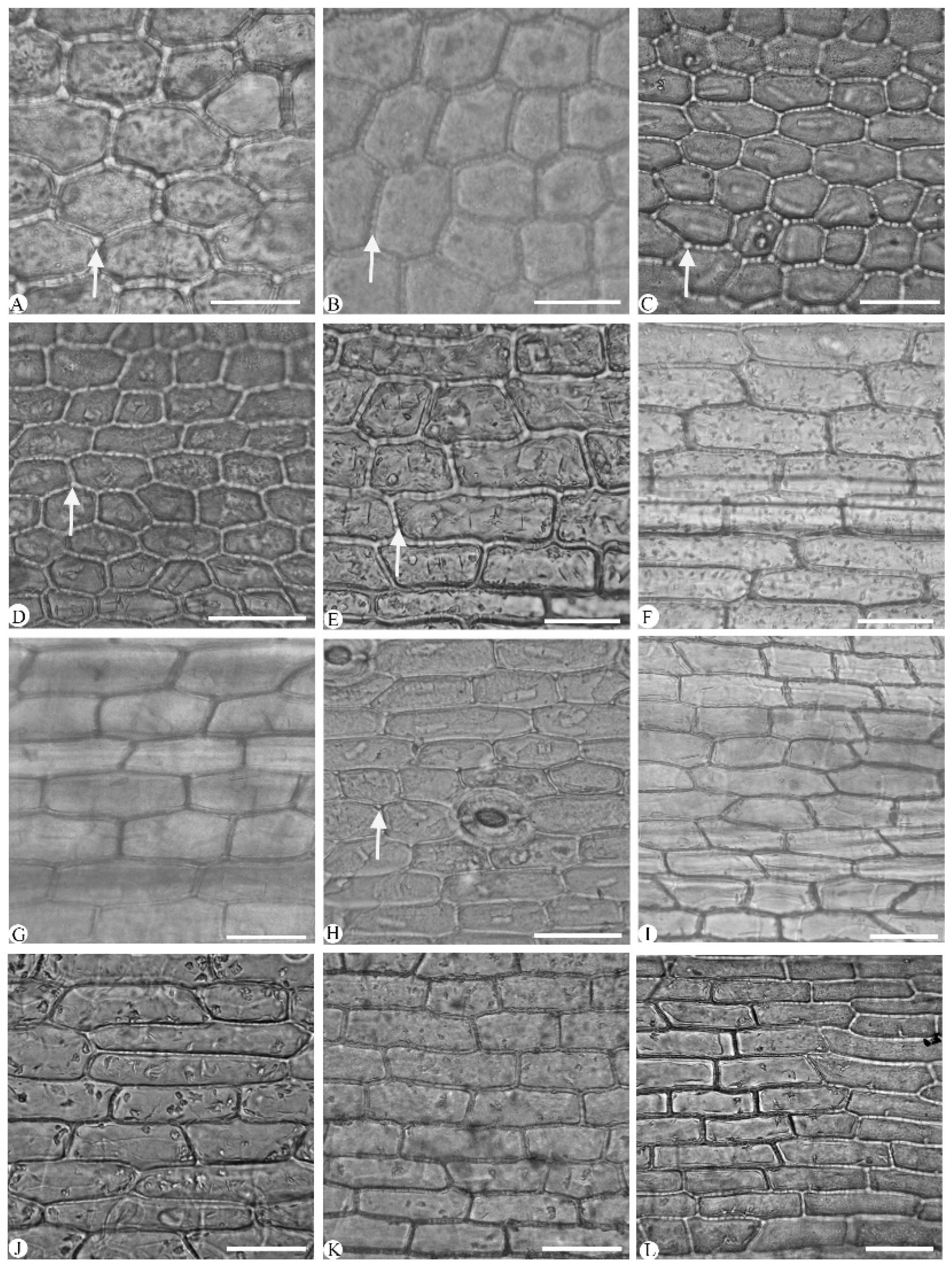
The epidermal cells were polygonal or rectangular on both leaf surfaces, with straight-arched and significantly thickened anticlinal walls (Figure 2 and Figure 3; Table 3). The adaxial epidermal cells displayed were polygonal in nine species (Figure 2A–I) and rectangular in Cymbidium hookerianum, C. defoliatum, and C. serratum (Figure 2J–L). The abaxial epidermal cells uniformly were polygonal, though variation in their cellular dimensions and proportions was evident. Based on length-to-width (L/W) ratios, these cells were categorized into two distinct types: broad polygonal (1.20–1.65 L/W) and narrow polygonal (1.70–3.40 L/W) (Figure 3, Table 3). This variation is attributed to cellular displacement during stomatal ontogeny (Figure 3 and Figure 4). The broad polygonal cell was found in eight species, mixed with a few rectangular cells (Figure 3E–L), while the narrow polygonal cell was observed in the other four species, which were C. floribundum (Figure 3A), C. lancifolium (Figure 3B), C. eburneum (Figure 3C), and C. wenshanense (Figure 3D). The size of the epidermal cells was different in the 12 species (Table 3). The largest epidermal cells were recorded in C. lancifolium (62.10 ± 0.87 μm × 59.34 ± 0.90 μm, L × W), while the smallest cells were found in C. faberi (23.69 ± 1.11 μm × 13.15 ± 0.48 μm, L × W).
Stigmata were easily observed in the 12 species, consistently appearing along the anticlinal walls of both adaxial and abaxial epidermal cells in a characteristic moniliform arrangement (Figure 2 and Figure 3). Stigmata were universally present in the abaxial epidermal cells of the 12 species (Figure 3); they occurred also on the adaxial surfaces in six species: Cymbidium lancifolium (Figure 2A), C. floribundum (Figure 2B), C. wenshanense (Figure 2C), C. tracyanum (Figure 2D), C. faberi (Figure 2E), and C. mastersii (Figure 2H).
The distribution and morphology of stomatal cells showed significant interspecific variation. All species exhibited abaxial stomata, with the exception of the amphistomatic stomata seen in Cymbidium mastersii (Figure 2H). The predominant stomatal type was tetracytic (Figure 3), though occasional anomocytic stomata were identified in C. floribundum (Figure 3A). Stomatal complexes were typically surrounded by (4–)5–7 epidermal cells and exhibited two distinct shapes based on their length-to-width (L/W) ratios: elliptic stomata (1.20–1.30 μm, L/W) and round stomata (1.10–1.20 μm, L/W) (Table 3). Elliptic stomata were observed in C. floribundum (Figure 3A), C. lancifolium (Figure 3B), C. eburneum (Figure 3C), C. faberi (Figure 3F), C. mastersii (Figure 3H), and C. serratum (Figure 3I), while round stomata were found in the other six species. Notably, C. faberi exhibited sunken stomata (Figure 3F), which was in contrast to the stomatal position observed in the other 11 species.
Our data analysis revealed substantial variation in stomatal dimensions, indices, and densities across the 12 species (Table 3). The largest stomata were observed in Cymbidium lancifolium (39.37 ± 0.63 × 29.68 ± 0.56 μm, SL × SW), while the smallest were those in C. tracyanum (24.68 ± 0.67 × 20.18 ± 0.39 μm, SL × SW). The stomatal index varied significantly, from 3.98 ± 0.11 μm (C. serratum) to 8.01 ± 0.20 μm (C. eburneum). Density measurements reached their maximum value in C. kanran (1.80 ± 0.15 stomata/μm2) and minimum value in C. lancifolium (1.10 ± 0.56 stomata/μm2).

2.2. Leaf Section

Outline of cross-section of a leaf midrib: The leaf midrib anatomy of 12 Cymbidium species includes a uniseriate epidermis on both the adaxial and abaxial surfaces. The outline of the cross-section of the species’ leaf midrib varied and included V-shape (0–90°) and shallow V-shape (90–180°) types (Figure 4). The shallow V-shape was found in Cymbidium tracyanum (Figure 4A), C. mastersii (Figure 4B), C. wenshanense (Figure 4C), C. serratum (Figure 4D), and C. eburneum (Figure 4E). In contrast, the V-shaped midrib was characteristic of the remaining seven species, including C. kanran (Figure 4F), C. hookerianum (Figure 4G), C. floribundum (Figure 4H), C. faberi (Figure 4I), C. lancifolium (Figure 4J), C. qiubeiense (Figure 4K), and C. defoliatum (Figure 4L). Moreover, the adaxial side was relatively flat in terms of the shape of the cross-section of the leaf midrib in C. tracyanum, while the abaxial epidermis displayed a relatively acute angle in C. serratum (Figure 4D). The cross-section of the leaf midrib was flat in C. eburneum, while it was semicircular in the remaining 11 species.
Leaf blade thicknesses: The leaves’ blade thicknesses averaged about 300 μm, with significant variation based on species. The thinnest leaf blade was observed in Cymbidium kanran (203.90 ± 1.23 μm) and the thickest in C. qiubeiense (490.81 ± 3.60 μm), which is a nearly two-fold difference (Table 4).
Thickness of the leaf midrib: The thickness of the leaf midrib averaged 350 μm, with significant variation between species. The midrib thickness ranged from 211.83 ± 3.41 μm in Cymbidium serratum to 697.75 ± 18.88 μm in C. qiubeiense, which is nearly a three-fold difference. Based on thickness, the midribs could be categorized as either thick (>350 μm) or thin (≤350 μm).
Mesophyll: The mesophyll was predominantly homogeneous, consisting of round to elliptical parenchyma cells (Figure 4). Homogeneous mesophyll cells were seen in 10 species, whereas palisade mesophyll cells were present only in Cymbidium lancifolium and C. qiubeiense (Figure 4J–K). The thickness of the mesophyll averaged 260.43 μm; the largest thickness was found in C. qiubeiense (440.36 ± 3.47 μm) and the thinnest in C. kanran (177.27 ± 1.10 μm) (Table 4).
Vascular bundles: Vascular bundles composed of xylem and phloem were distributed throughout the mesophyll of the 12 species and arranged in a single row at the center of the mesophyll (Figure 4). The size of the vascular bundles varied significantly, with most occupying up to half of the total leaf thickness, except those in Cymbidium mastersii (Figure 4B), C. wenshanense (Figure 4C), and C. serratum (Figure 4D) (Table 4). The thickest vascular bundles averaged 166.09 μm. The smallest vascular bundle was found in C. serratum (92.88 ± 3.61 μm) and the largest in C. qiubeiense (293.73 ± 22.40 μm), with the thickness of the latter’s vascular bundles up to three times that of the former’s.
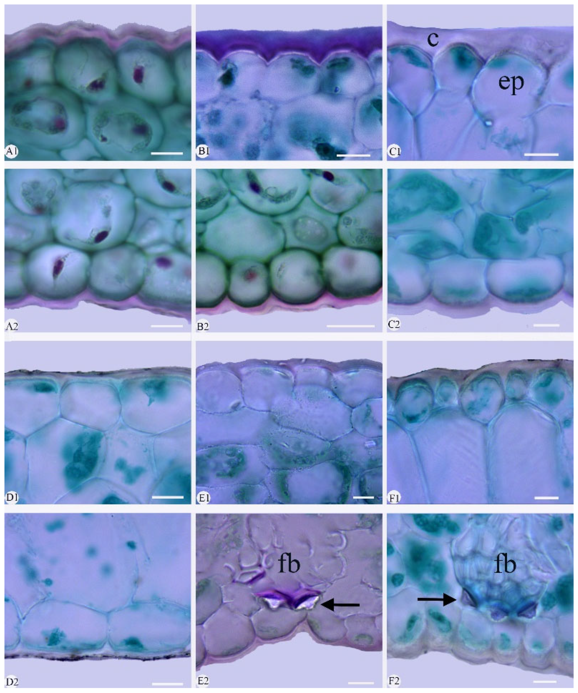
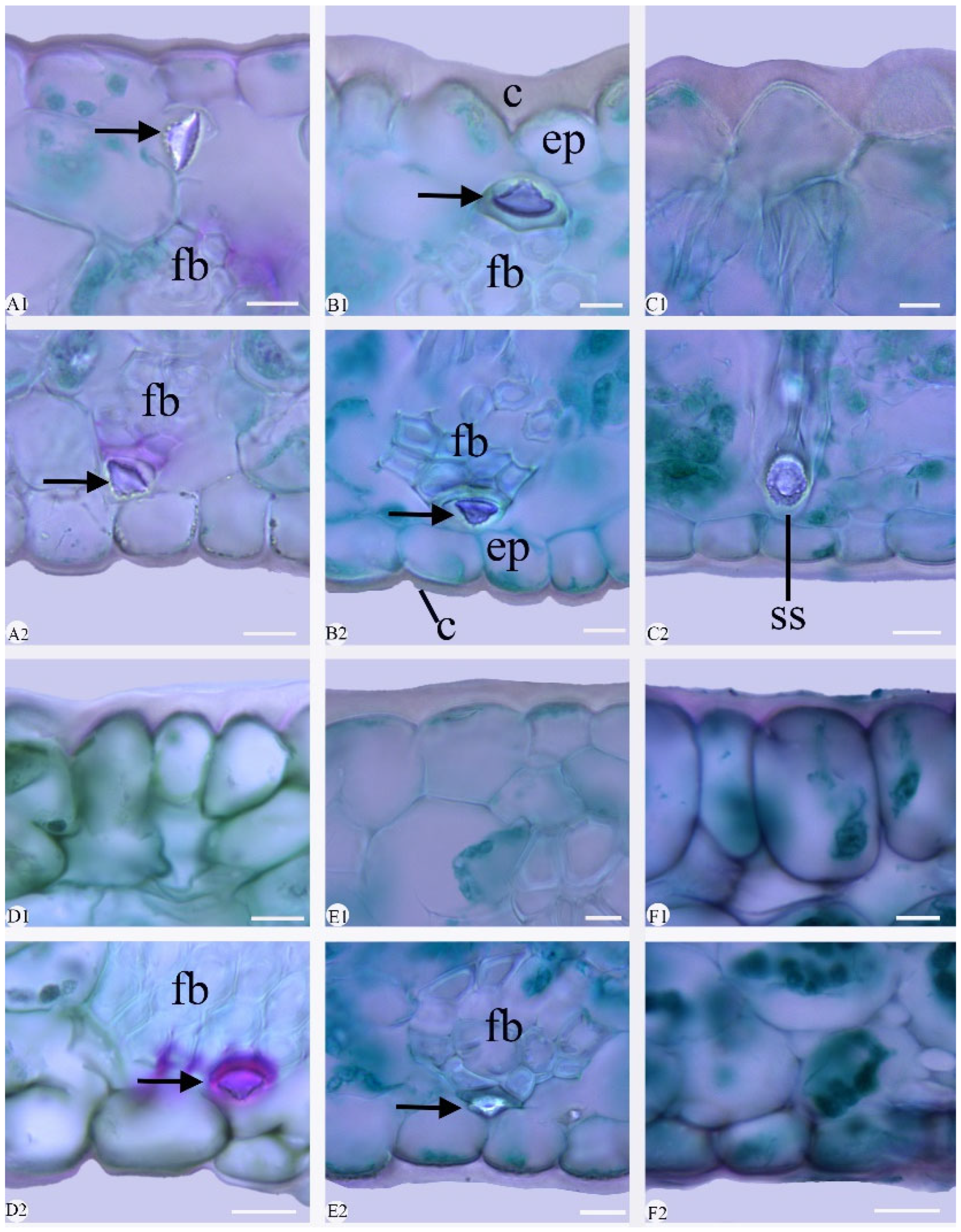
Cuticle: The adaxial cuticle exhibited a greater thickness compared to that of the abaxial surface in the 12 species (Figure 5 and Figure 6). The cuticle was smooth (adaxial and abaxial surface) or corrugated. The cuticle was corrugated in nine species: C. kanran (Figure 5A), C. defoliatum (Figure 5B), C. floribundum (Figure 5C), C. faberi (Figure 5E), C. qiubeiense (Figure 5F), C. mastersii (Figure 6A), C. hookerianum (Figure 6B), C. tracyanum (Figure 6C), and C. eburneum (Figure 6D). The cuticle was smooth in C. lancifolium (Figure 5D), C. wenshanense (Figure 6E), and C. serratum (Figure 6F).
The thicknesses of the cuticles were significantly different between taxa (Figure 5 and Figure 6; Table 4). The thickest cuticle (9.03 ± 0.60 μm) occurred in C. tracyanum (Figure 6C), while the thinnest (3.39 ± 0.14 μm) was in C. mastersii (Figure 6A). Cuticle thicknesses of up to one-third of the height of the epidermal cells was found in four species, namely C. defoliatum (Figure 5B), C. hookerianum (Figure 6B), C. tracyanum (Figure 6C), and C. wenshanense (Figure 6E). A cuticle thickness less than one-third of the height of the epidermal cell occurred in the other eight species: C. kanran (Figure 5A), C. floribundum (Figure 5C), C. lancifolium (Figure 5D), C. faberi (Figure 5E), C. qiubeiense (Figure 5F), C. mastersii (Figure 6A), C. eburneum (Figure 6D), and C. serratum (Figure 6F).
Epidermis: The 12 species possessed a single-layer epidermis on both sides of their leaves (Figure 5 and Figure 6, Table 4). Anatomically, the epidermal cells were of oval (Figure 5A–C,F), polygonal (Figure 5D), and conical (Figure 6B,C); crowded, with small triangular inter-cellular spaces; and sometimes anticlinal or periclinal. Adaxial and abaxial epidermal cells nearly identical in size (1.20–2.00, L/W) were found in six species: Cymbidium kanran (Figure 5A), C. defoliatum (Figure 5B), C. floribundum (Figure 5C), C. lancifolium (Figure 5D), C. faberi (Figure 5E), and C. qiubeiense (Figure 5F). The adaxial epidermal cells were clearly larger than the abaxial epidermal cells (2.01–4.10, L/W) in the other six species: C. mastersii (Figure 6A), C. hookerianum (Figure 6B), C. tracyanum (Figure 6C), C. eburneum (Figure 6D), C. wenshanense (Figure 6E), and C. serratum (Figure 6F). The largest epidermal cells were observed in C. serratum (4.02, ETad/ETab) and were approximately four times the size of the abaxial ones.
Fiber bundle: Fiber bundles were irregularly distributed within the mesophyll, typically aligned parallel to the vascular bundles and separated from the epidermis by one or two cells (Figure 5 and Figure 6). Two distribution patterns were observed: (1) the fiber bundle was adjacent to both epidermises in Cymbidium mastersii (Figure 6A) and C. hookerianum (Figure 6B), and (2) the fiber bundle wasrestricted to the abaxial epidermis in C. faberi (Figure 5E), C. qiubeiense (Figure 5F), C. eburneum (Figure 6D), and C. wenshanense (Figure 6E). Notably, six species lacked fiber bundles entirely: C. kanran (Figure 5A), C. defoliatum (Figure 5B), C. floribundum (Figure 5C), C. lancifolium (Figure 5D), C. tracyanum (Figure 6C), and C. serratum (Figure 6F).
Stegmata: Stegmata, with conical, spherical silica bodies associated with fiber bundles and mesophyll, were observed in seven species, but absent in the other five: Cymbidium kanran (Figure 5A), C. defoliatum (Figure 5B), C. floribundum (Figure 5C), C. lancifolium (Figure 5D), and C. serratum (Figure 6E). In the former group, conical silica bodies associated with fiber bundles were present in six species: C. faberi (Figure 5E), C. qiubeiense (Figure 5F), C. mastersii (Figure 6A), C. hookerianum (Figure 6B), C. eburneum (Figure 6D), and C. wenshanense (Figure 6E). Stegmata with spherical silica bodies immersed in the mesophyll were found only in C. tracyanum (Figure 6C).
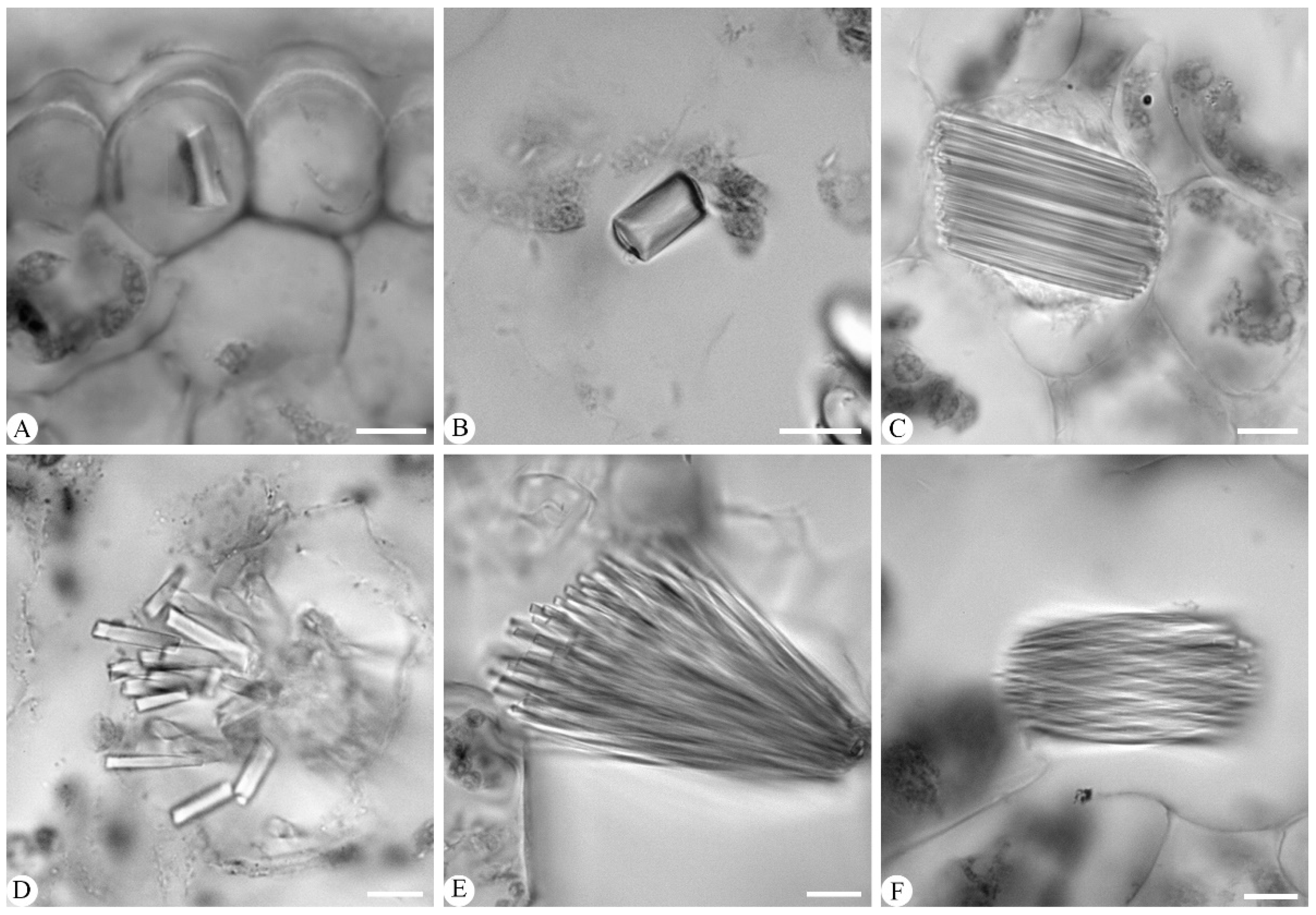
Crystals: Crystals were observed in six species, but absent in the remaining six. In the six species containing crystals, three crystals were identified, including the terete crystal in Cymbidium faberi (Figure 7A) and C. tracyanum (Figure 7B); long tube crystal bundles in C. qiubeiense (Figure 7C), C. kanran (Figure 7D), and C. mastersii (Figure 7E); and raphide, which was found exclusively in C. lancifolium (Figure 7F). The crystals were primarily distributed within heteromorphic mesophyll cells, except in the epidermal cells of C. faberi (Figure 7A).

3. Discussion

3.1. Taxonomic Significance of Leaf Morpho-Anatomy in Cymbidium

Some data on the leaf morpho-anatomy of some Cymbidium species have been reported, but without detailed illustrations [21,22]. Here, the leaf morpho-anatomy of 12 species was investigated thoroughly, although that of five species had previously been reported, including C. faberi, C. lancifolium, C. floribundum, and C. tracyanum [21,22]. It was shown that most features of the leaf morpho-anatomy of 12 Cymbidium species were consistent with those from other orchids that have been investigated [18,19,23,24,25,26]. However, some features were different between taxa, exhibiting taxonomic significance, including the shape of the epidermal cell, stomata, vascular bundle, cuticle, mesophyll, fiber bundle, and stegmata, as discussed below.

3.1.1. Leaf Epidermal Cell

The leaf epidermal cells were polygonal or rectangular, with straight anticlinal walls. While this is common in Orchidaceae, it varies between taxa [23,24,25,26]. Here, polygonal cells were predominant on both leaf surfaces in nine species, while rectangular cells were restricted to the adaxial surface in Cymbidium hookerianum, C. defoliatum, and C. serratum, and rectangular adaxial epidermal cells were seen in C. hookerianum and C. kanran. It was confirmed that polygonal epidermal cells were observed on both surfaces of the leaves of C. lancifolium, C. faberi, C. floribundum, and C. tracyanum [21,22]. However, both adaxial and abaxial epidermal cells were found to be polygonal in C. kanran, which is in contrast to the rectangular epidermal cells previously recorded for the species [22]. In addition, trichome on the leaf epidermis was recorded in C. eburneum and C. lancifolium [27], but it was not observed here, nor was it in other research [22].

3.1.2. Stomata

Stomata and their distribution vary among orchid taxa, which could be of taxonomic significance [27,28,29,30]. Five types of stomata have been identified in Orchidaceae, including anomocytic, cyclocytic, paracytic, pentacytic, and tetracytic stomata [21,22,27,28]. Among them, tetracytic stomata are predominantly observed in the 23 genera from the tribe Cymbidieae [24]. Here, tetracytic stomata were observed in 11 species, while anomocytic stomata were seen only in Cymbidium floribundum. Tetracytic stomata were also recorded in six other Cymbidium species: Cymbidium ensifolium (L.) Sw., C. aloifolium (L.) Sw. [29], C. cyperifolium Wall. ex Lindl., C. goeringii (Rchb. f.) Rchb. F., C. devonianum Paxton, and C. bicolor Lindl. [22]. Meanwhile, abaxial stomata were observed in 11 species, while amphistomatic stomata were found only in C. mastersii. Amphistomatic stomata were previously recorded in Cymbidium canaliculatum R.Br. [22]. Therefore, it was proven that tetracytic stomata are more common than anomocytic stomata in Cymbidium [22,28]. Similarly, it was suggested that abaxial stomata were more predominant than anomocytic stomata in Cymbidium. In addition, anomocytic stomata were identified in C. floribundum, which is in contrast to the tetracytic type previously described [22].

3.1.3. Vascular Bundles

The amphivasal vascular bundles, composed of xylem and phloem, were distributed throughout the mesophyll and arranged in a single row at the center of the mesophyll in the 12 species observed, which was consistent with the vascular bundles of other Cymbidium species [22]. However, both amphivasal and bicollateral vascular bundles were recorded in Dendrobium [19]. Vascular bundles were evident and accounted for approximately half of the total leaf blade thickness in nine species, but not in Cymbidium mastersii, C. wenshanense, and C. serratum. This feature was also noted in C. ensifolium, C. goeringii, and C. sinense (Jack. ex Andr.) Willd. [21].

3.1.4. Cuticle

Meanwhile, the shape and thickness of the cuticle varied between orchid species [31]. Commonly, plant cuticles play an important role in the interaction between plants and their environment, reducing the solar radiation absorbed and the temperature of the plant by reflecting sunlight and reducing transpiration [32]. Here, a corrugated cuticle was observed in nine species, whereas a smooth cuticle was seen in C. lancifolium, C. wenshanense, and C. serratum. Notably, corrugated cuticles were found in C. kanran, C. floribundum, and C. hookerianum, which is in contrast to the smooth cuticle documented in previous data [22]. In addition, the thickness of the cuticle varied between species, which was not mentioned in previous research [22]. A thick cuticle, observed to reach up to one-third of the epidermal cell height, was observed in four species (C. defoliatum, C. hookerianum, C. wenshanense, and C. tracyanum), while thin cuticles were observed in the other eight species studied.

3.1.5. Mesophyll

The mesophyll cells are often homogeneous but occasionally heterogeneous in Orchidaceae [21,22,23]. Here, the mesophyll was homogeneous in 10 Cymbidium species, but heterogeneous, with a pronounced palisade mesophyll cell, in C. qiubeiense and C. lancifolium. It was noted that the latter two species were distinguished from the other 10 by the presence of a petiole. It seemed that the differentiation of the heterogenous mesophyll might be related to the presence of a petiole in Cymbidium. A heterogeneous mesophyll with a likely palisade layer was also recorded in 3 out of 21 species Cymbidium elsewhere (C. aloifolium, C. bicolor, and C. finlaysonianum Lindl.), which was presumed to be the result of evolution for adaption to high-light-level habitats, since members of Cymbidium grow on isolated trees in tropical lowland forests or on rocks [22]. However, heterogeneous mesophyll is not present in other epiphytic orchids, such as Bulbophllyum [18] and Dendrobium [19].

3.1.6. Fiber Bundle

The presence and position of fiber bundles varied between the species of Cymbidium [21,22,24]. Here, fiber bundles occurred in six species but were absent in the other six. In the former group, fiber bundles adjacent to the ad- and abaxial epidermises was observed in C. mastersii and C. hookerianum, while they were restricted to the abaxial epidermis in C. faberi, C. qiubeiense, C. eburneum, and C. wenshanense. It was reconfirmed that fiber bundles occurring on both surfaces were previously recorded in C. floribundum, C. tracyanum, C. hookerianum, and C. kanran, but thought to be absent in C. lancifolium [22]. However, fiber bundles were clearly absent in C. floribundum and C. tracyanum in this research but described as present, without illustration, in older data [22]. Interestingly, fiber bundles were observed to be very numerous on the abaxial surface but completely absent on the adaxial surface in C. ensifolium [29]. It was revealed that fiber bundles were absent in Bulbophyllum [18]. However, the state of the fiber bundles varies between taxa at the levels of genus and subtribe in Orchidaceae. For example, fiber bundles are absent in Dendrobium section Stachyobium [19] but present in Dendrobium arachnoideum Schltr. and D. guttulatum Schltr [31]. And fiber bundles have been observed in some members of Oncidiinae (Maxillarieae), such as Ionopsis utricularioides (Sw.) Lindl., Aspasia lunata Lindl., and Oncidium boothianum Rchb.f., although they are notably absent in Erycina echinata (Kunth) Lindl. [33].

3.1.7. Stegmata

The state, type, and position of the stegmata are different in different Orchidaceae [22,34,35]. The shape of the stegmata varied, but included spherical, conical, and rough-surfaced silica bodies associated with vascular bundles and fiber bundles [22,24,34]. Here, stegmata with conical and spherical silica bodies that were associated with fiber bundles and mesophyll were observed in seven species, but absent in the other five species (C. kanran, C. defoliatum, C. floribundum, C. lancifolium, and C. serratum). The stegmata associated with fiber bundles were common in six species, but those with spherical silica bodies immersed in the mesophyll were found only in Cymbidium tracyanum. In addition, stegmata were evidently absent from C. kanran and C. lancifolium, although they were previously recorded as present, without illustrations, in previous data [22].

3.1.8. Crystals

Crystals such as raphide crystals, spherical silica body crystals, and prismatic crystals are common in leaf anatomy and vary in shape and size among orchid taxa [19,36]. Here, crystals were present in six species, but absent in the other six. The crystals were mainly observed in heteromorphic mesophyll cells, except in Cymbidium faberi, where they were found in epidermal cells. In the former cells, three types of crystals were identified: terete crystals, long tube crystal bundles, and raphide crystals. Terete crystals were present in C. faberi and C. tracyanum and long tube crystal bundles in C. qiubeiense, C. kanran, and C. mastersii. Raphide crystals were only found in C. lancifolium, while they have been recorded in C. ensifolium and C. aloifolium [29]. The crystals varied between orchid taxa. In Dendrobium section Stachyobium, spherical silica body crystals were found in D. sinominutiflorum but prismatic crystals were found in D. compactum, D. monticola, D. porphyrochilum, and D. strongylanthum. Crystals such as raphide bundles and druse were observed in some species but absent in four species from Dendrobium sect. Rhizobium [37]. Three crystals were varied and co-occurring among six species of Phalaenopsis [38], including prismatic crystals, spherical silica bodies, and raphide.

3.1.9. Stigmata

Stigmata were evidently observed along the anticlinal wall of polygonal and rectangular epidermal cells in the 12 Cymbidium species studied, but they were either not noticed or their taxonomic implications were neglected in previous research [21,22]. Stigmata were predominantly observed on the abaxial epidermis in the 12 Cymbidium species, and they were also detected on both the adaxial and abaxial epidermal surfaces in 6 species (Cymbidium lancifolium, C. floribundum, C. wenshanense, C. tracyanum, C. faberi, and C. mastersii). Stigmata were previously observed and noted in Dendrobium kingianum Bidwill ex Lindl. and Bulbophyllum mentosum Barb. Rodr [18], but without further investigation into this uncommon feature. It was clearly observed, with detailed illustrations, that stigmata were totally absent in five Dendrobium species [19] and six Phalaenopsis species [38] by the same authors, implying that they paid more attention to their taxonomic significance with larger numbers of samples.

3.2. Implications for Infrageneric Taxonomy for Cymbidium

Three subgenera were recognized, subgen. Cymbidium, Cyperorchis, and Jensoa, based on their pollinium number and labellum–column fusion characteristics [11]. This classification was widely adopted by subsequent taxonomists [1,2,9]. However, the delimitation of the sections and species within these subgenera has been unclear when using previously gathered floral morphological and molecular data, meaning more evidence is needed to clarify the delimitation of taxa in Cymbidium [1,9]. The leaf morpho-anatomic features of 12 species representing the three subgenera of Cymbidium varied greatly, which means they could be used to make taxonomic distinctions as follows below.

3.2.1. Delimitation of Species in subgen. Cyperorchis

Five species from subgen. Cyperorchis were investigated, including two species (Cymbidium eburneum and C. mastersii) in sect. Eburnea and three (C. hookerianum, C. tracyanum, and C. wenshanense) from sect. Iridorchis [3]. Here, it was shown that the shape of the leaf blade tip varied between the two sections; a lorate leaf with a slightly two-lobed tip was common in sect. Eburnea, compared to the complete tip seen in sect. Iridorchis.
Furthermore, Cymbidium wenshanense was moved out of sect. Iridorchis and transferred into sect. Annamaea based on some subtle floral features [9]. It was shown that C. wenshanense could be distinguished from the other two species from sect. Iridorchis by its large leaf, smooth cuticle, the fiber bundles on its abaxial epidermis, and its lack of crystals. In contrast, C. hookerianum and C. tracyanum (sect. Iridorchis) shared some common features, such as a moderate leaf size and corrugated cuticles, but each of them could be discriminated between by some leaf morpho-anatomic features. Cymbidium hookerianum was characterized by polygonal adaxial epidermal cells, fiber bundles on both epidermal surfaces, and conical silica bodies, whereas C. tracyanum featured polygonal adaxial and abaxial epidermal cells and lacked fiber bundles and spherical silica bodies.
Cymbidium mastersii and C. eburneum were placed in sect. Eburnea, which seemed to contain species that were easily confused due to their overlapping morphological features, such as the size and the shape of the plant, leaf, and flowers. However, each of them was a separate species, since they formed sister clades based on their molecular data [2,3]. Each of them could be recognized by their comparative leaf morpho-anatomy. Cymbidium mastersii is characterized by the presence of stigmata on both surfaces, hypostomatous stomata, fiber bundles near both epidermis layers, the thinnest cuticle, and long tube crystal bundles. In contrast, C. eburneum features amphistomatic stomata, the highest stomatal index, stigmata on its abaxial surface, and fiber bundles confined to the abaxial epidermis, while lacking these crystals.

3.2.2. Species Delimitation in subgen. Jensoa

Five species were taken from subgen. Jensoa, including Cymbidium defoliatum, C. qiubeiense, C. kanran, C. faberi, and C. serratum, from sect. Jensoa and C. lancifolium from sect. Geocymbidium [3]. It was proposed that C. faberi and C. serratum should be moved from sect. Jensoa to sect. Nanula, considering their lack of an articulate leaf base [9]. However, no clear evidence from leaf morpho-anatomy could be used for the delimitation of these two sections. There were some morpho-anatomic features that could easily be used for a clear discrimination of sect. Geocymbidium from the other two sections, such as the maximal stomatal size, minimal stomatal density, and an articulate leaf shape, among the 12 species studied. Of course, it seemed reasonable to place C. lancifolium in the monotypic sect. Geocymbidium, because it was distinguished by an oblanceolate–oblong leaf with a petiole. In addition, leaves with petioles and heterogenous mesophyll were not seen in most Cymbidium members, but present in C. lancifolium and C. qiubeiense, which could be used as a diagnostic feature for them.
And then there exist two species which are easily confused, namely Cymbidium faberi and C. serratum, due to the similar morphology of their plants, leaves, and flowers [3,9]. However, it was possible to separate them using molecular evidence [2] and leaf morpho-anatomic features in this research. Cymbidium faberi was characterized by a smaller epidermal cell, stigmata on both surfaces, sunken stomata, a V-shaped midrib, fiber bundles restricted to the abaxial epidermis, and the presence of crystals. In contrast, C. serratum possessed a larger epidermal cell, stigmata on the adaxial epidermis, flat stomata, a shallow V-shape midrib cross-section, and an absence of both fiber bundles and crystals.
In addition, there are three species which share some common features and are easily confused: Cymbidium defoliatum, C. qiubeiense and C. kanran [3,9]. Each of them could be identified using molecular data [2], and also by their leaf morphology, including the leaf blade margin (entire or toothed) and the state of their petiole. C. defoliatum was characterized by an entire leaf margin, while the latter two had toothed leaf margins. C. qiubeiense was characterized by a petiolate leaf with heterogenous mesophyll, a thicker leaf blade, larger vascular bundles, and the presence of stegmata and crystals. Conversely, C. defoliatum and C. kanran were distinguished by a leaf without a petiole and with homogenous mesophyll, smaller vascular bundles, and the absence of both stegmata and crystals.

3.2.3. Species Delimitation in subgen. Cymbidium

There was only one species (Cymbidium floribundum) sampled from Subgen. Cymbidium, which should be investigated further using more samples. Cymbidium floribundum was greatly distinguished from the other 11 species by having the only anomocytic stomata, which have rarely been observed or noticed in the other orchid data available.

3.2.4. Species Delimitation of Cymbidium Based on Leaf Morpho-Anatomic Features

It was shown that some leaf morpho-anatomic features varied between taxa and could be used for species delimitation in Cymbidium. Accordingly, a key to the 12 Cymbidium species observed was proposed based on their leaf morpho-anatomic features (Appendix A). This would provide a better understanding of the species delimitation and taxonomy of Cymbidium.

4. Materials and Methods

Taxa selected: Twelve species of Cymbidium from China (Table 1 and Table 2, Figure 1) were selected, representing three subgenera and five or seven sections, based on previous taxonomic references [3,9]. Six species were from the subgenera Jensoa and covered its two sections (Cymbidium defoliatum Y. S. Wu & S. C. Chen; C. faberi Rolfe; C. kanran Makino; C. qiubeiense K. M. Feng & H. L; and C. serratum Schltr. from sect. Jensoa and C. lancifolium from sect. Geocymbidium Hook). Five species were from subgen. Cyperorchis (C. hookerianum Hook., C. tracyanum L. Castle and C. wenshanense Y. S. Wu & F. Y. Liu from sect. Iridorchis; C. eburneum Lindl. and C. mastersii Griff. ex Lindl. from sect. Eburnea), and one species (C. floribundum Lindl.) was from subgen. Cymbidium, according to previous taxonomic data [3].
Sample preparation: Samples from plants of these 12 Cymbidium species were prepared as follows. Plants of these 12 Cymbidium species have been cultivated and have bloomed every year for nearly twenty years in the Kunming Institute of Botany, Chinese Academy of Sciences, and in the Orchid Germplasm Resource Nursery, Yunnan Fengchunfang Biotechnology Company Limited, Fumin, Yunnan, China (25° 20′ 01″ N, 102° 27′ 26″ E). Three to five mature leaves from several plants per species were cut into pieces, fixed in FAA (50% alcohol/acetic acid/formaldehyde = 90:5:5) [39], and preserved in 50% alcohol (Tianjin Zhiyuan Chemical Reagent Co., Ltd., Tianjin, China). The length and width of the fully expanded leaves were measured in the field with the help of a glass scale ruler and graph paper.
Observation of leaf morpho-anatomic features: The epidermis was disassociated using the method described by Sun and Jiang [40]. Leaf epidermal characteristics were manually recorded from leaf edge fragments measuring approximately 1 cm × 1 cm, which were immersed in 30% H2O2–CH2COOH (Xilong Scientific Co., Ltd., Guangzhou, China) at 60 °C for 12–36 h and stained with 1% safranin for 2–3 min. Transversal sections (0.5 cm × 0.5 cm) from between the mid-vein and leaf edge were dehydrated, embedded in paraffin wax (melting point = 57 °C), sectioned into 8–30 μm thicknesses using a manual rotary microtome (Leica RM225RTS, Wetzlar, Germany), and stained with 1% safranin and 1% fast green (HeFei BoMei Biotechnology Co., Ltd., HeFei, China). The samples were mounted on permanent slides using Canada balsam (Beijing Labgic Technology Co., Ltd., Beijing, China) and observed and photographed using a Nikon E100 light microscope (Nikon, Tokyo, Japan). The terminology used for the stomatal complex types followed that of Patel [41]. Thirty measurements were taken for each documented value—the lengths and widths of adaxial and abaxial leaf epidermal cells, stomata (index and density), leaf thickness, midrib thickness, mesophyll thickness, vascular bundle thickness (measuring only the thickness of the phloem and xylem of midvein vascular bundles), and epidermis and cuticle thicknesses of both adaxial and abaxial surfaces—using ImageJ software (https://imagej.nih.gov/ij/download.html) (NIH, Bethesda, MD, USA). Stomatal index = number of stomata/(number of stomata + number of ordinary epidermal cells) × 100%.
Data Analysis: Statistical analyses were performed using Excel 2016 (Microsoft, Redmond, WA, USA), and means and standard deviations were calculated (mean ± SD) while retaining two decimal places. To evaluate significant differences in morphoanatomical features among the 12 species of Cymbidium, the data were tested separately via a one-way analysis of variance (ANOVA) using SPSS 25.0 software (IBM, Armonk, NY, USA). The means were separated using Duncan’s multiple range test, and differences between the mean were considered significant at a p < 0.05.

5. Conclusions

The leaf morpho-anatomy of 12 Cymbidium species was investigated using a microscope and paraffin sections. Their comparative leaf morpho-anatomy indicated that some leaf morpho-anatomical features of the 12 Cymbidium species varied between species, including the leaf epidermal cell, stomata, mesophyll, fiber bundle, stegmata, and type of crystal present. These features could be used for species identification. Accordingly, a taxonomic key to the 12 Cymbidium species from China was proposed based on these leaf morpho-anatomical features. However, the taxonomic implications of these aspects of leaf morpho-anatomy for different sections and subgenera of Cymbidium could not be definitely proposed since the data available were limited. Therefore, further investigation with extended sampling should be conducted, which would provide a better understanding of the taxonomy of Cymbidium.

Author Contributions

Conceptualization, L.L.; formal analysis, X.H.; funding acquisition, L.L.; investigation, X.H.; methodology, L.L.; project administration, L.L.; supervision, L.L.; writing—original draft, X.H. and L.T.; collection of materials and identification of species, L.L. and J.H.; writing—review and editing, X.H., L.T., D.M., K.T. and L.L. All authors have read and agreed to the published version of the manuscript.

Funding

This study was funded by the National Natural Science Foundation of China (NSFC 32060049).

Data Availability Statement

The data that support the findings of this study are available from the corresponding author upon reasonable request.

Acknowledgments

This study was supported by the National Nature Science Foundation of China (NSFC 32060049). We thank Zhi-feng Xu and Xiao-yun Wang from the Orchid Conservation Center of Yunnan Fengchunfang Biotechnology Co., Ltd., for providing sample materials. This research was carried out at the Yunan Academy of Biodiversity, Southwest Forestry University.

Conflicts of Interest

The authors declare that they have no conflicts of interest.

Appendix A. A Key to Twelve Cymbidium Species Based on Leaf Morpho-Anatomic Features

1. Leaf shape oblanceolate to oblongC. lancifolium
1. Leaf lorate(2)
2. Leaf margin toothed(3)
2. Leaf margin entire(6)
3. Leaf with petioleC. qiubeiense
3. Leaf without petiole(4)
4. Leaf base with articulateC. kanran
4. Leaf base without articulate(5)
5. Stomata sunkenC. faberi
5. Stomata flatC. serratum
6. Leaf apex with slightly two-lobed tip(7)
6. Leaf apex without two-lobed tip(8)
7. Stomata amphistomatic; stomatal index largestC. eburneum
7. Stomata hypostomatousC. mastersii
8. The leaf midrib with shallow V-shape(9)
8. The leaf midrib with V-shape(10)
9. Polygonal ad-abaxial epidermal cellsC. hookerianum
9. Polygonal ad-abaxial epidermal cellsC. defoliatum
10. Flat leaf mid-rib adaxial side cross-section outline; the cuticle thickestC. tracyanum
10. Semicircular leaf mid-rib adaxial side cross-section outline(11)
11. Lacked fiber bundles; without stegmataC. floribundum
11. With fiber bundles and stegmataC. wenshanense

References

  1. Cribb, P.J.; Du Puy, D.J. The Genus Cymbidium; Royal Botanic Garden: Kew, UK, 2007; 359p. [Google Scholar]
  2. Liu, Z.J.; Chen, X.Q.; Cribb, J.P. Cymbidium Swartz. In Flora of China: Orchidaceae; Science Press: Beijing, China, 2009; Volume 25, pp. 260–280. [Google Scholar]
  3. Chen, X.Q.; Liu, Z.J.; Zhu, G.H.; Lang, K.Y.; Ji, Z.H.; Luo, Y.B.; Jin, X.H.; Cribb, P.J.; Wood, J.J.; Gale, S.W.; et al. Flora of China (Orchidaceae); Science Press: Beijing, China, 1999; Volume 18, pp. 191–194. [Google Scholar]
  4. Hinsley, A.; De Boer, H.J.; Fay, M.F.; Gale, S.W.; Gardiner, L.M.; Gunasekara, R.S.; Kumar, P.; Masters, S.; Metusala, D.; Roberts, D.L.; et al. A review of the trade in orchids and its implications for conservation. Bot. J. Linn. Soc. 2018, 186, 435–455. [Google Scholar] [CrossRef]
  5. Liu, H.; Liu, Z.; Jin, X.; Gao, J.; Chen, Y.; Liu, Q.; Zhang, D.-Y. Assessing conservation efforts against threats to wild orchids in China. Biol. Conserv. 2020, 243, 108484. [Google Scholar] [CrossRef]
  6. National Forestry and Grassland Administration. List of National Key Protected Wild Plants in China. 2021. Available online: https://www.forestry.gov.cn/c/www/lczc/10746.jhtml (accessed on 20 January 2025).
  7. Balilashaki, K.; Vahedi, M.; Ho, T.-T.; Niu, S.-C.; Cardoso, J.C.; Zotz, G.; Khodamzadeh, A.A. Biochemical, cellular and molecular aspects of Cymbidium orchids: An ecological and economic overview. Acta Physiol. Plant. 2022, 44, 24. [Google Scholar] [CrossRef]
  8. Du Puy, D.; Cribb, P. The Classification of Cymbidium; Kew Publishing, Royal Botanic Garden: Kew, UK, 1988; pp. 50–194. [Google Scholar]
  9. Liu, Z.J.; Chen, S.C.; Ru, Z.Z. The Genus Cymbidium in China; Science Press: Beijing, China, 2006; 237p. [Google Scholar]
  10. Averyanov, L.V.; Nguyen Van, C.; Nguyen, K.; Maisak, T.; Truong, V. New Orchids (Orchidaceae) in the Flora of Vietnam I Epidendroideae. Taiwania 2019, 64, 176–188. [Google Scholar] [CrossRef]
  11. Seth, C.J.; Cribb, P.J. A reassessment of the sectional limits in the genus Cymbidium Swartz. In Orchid Biology: Reviews and Perspectives; Arditti, J., Ed.; Cornell University Press: London, UK, 1984; pp. 283–322. [Google Scholar]
  12. Trias-Blasi, A.; Vorontsova, M. Plant identification is key to conservation. Nature 2015, 521, 161. [Google Scholar] [CrossRef]
  13. Zhang, S.-B.; Dai, Y.; Hao, G.-Y.; Li, J.-W.; Fu, X.-W.; Zhang, J.-L. Differentiation of water-related traits in terrestrial and epiphytic Cymbidium species. Front. Plant Sci. 2015, 6, 260. [Google Scholar] [CrossRef]
  14. Zhou, Z.; Shi, R.; Zhang, Y.; Xing, X.; Jin, X. Orchid conservation in China from 2000 to 2020: Achievements and perspectives. Plant Divers. 2021, 43, 343–349. [Google Scholar] [CrossRef]
  15. van den Berg, C.; Ryan, A.; Cribb, P.J.; Chase, M.W. Molecular phylogenetics of Cymbidium (Orchidaceae: Maxillarieae): Sequence data from internal transcribed spacers (ITS) of nuclear ribosomal DNA and plastid matK. Lindleyana 2002, 17, 102–111. [Google Scholar]
  16. Yukawa, T.; Miyoshi, K.; Yokoyama, J. Molecular Phylogeny and Character Evolution of Cymbidium (Orchidaceae). Bull. Natl. Sci. Mus. Tokyo Ser. B 2002, 28, 129–139. [Google Scholar]
  17. Zhang, L.; Huang, Y.-W.; Huang, J.-L.; Ya, J.-D.; Zhe, M.-Q.; Zeng, C.-X.; Zhang, Z.-R.; Zhang, S.-B.; Li, D.-Z.; Li, H.-T. DNA barcoding of Cymbidium by genome skimming: Call for next-generation nuclear barcodes. Mol. Ecol. Resour. 2023, 23, 424–439. [Google Scholar] [CrossRef]
  18. Smidt, E.C.; Gallo, L.W.; Scatena, V.L. Leaf anatomical and molecular studies in Bulbophyllum section Micranthae (Orchidaceae) and their implications for systematics. Braz. J. Bot. 2013, 36, 123–143. [Google Scholar] [CrossRef]
  19. Xie, Y.X.; Wang, Y.; Tao, L.; Liu, W.; Luo, Y.; Li, L. Leaf morpho-anatomy and taxonomic significance in five Dendrobium sect. Stachyobium species from China. Flora 2024, 317, 152560. [Google Scholar] [CrossRef]
  20. Song, J.Y.; Luo, T.; Zhang, N. Leaf structure characteristics of five species of Cymbidium. Plant Sci. J. 2019, 37, 422–433. [Google Scholar] [CrossRef]
  21. Sun, A.C. Investigations on leaves of Cymbidium, Paphiopedilum, Dendrobium under scanning electron microscope. J. Wuhan Bot. Res. 1995, 13, 289–294. [Google Scholar]
  22. Yukawa, T.; Stern, W.L. Comparative vegetative anatomy and systematics of Cymbidium (Cymbidieae: Orchidaceae). Bot. J. Linn. Soc. 2002, 138, 383–419. [Google Scholar] [CrossRef]
  23. Stern, W.L.; Whitten, W.M. Comparative vegetative anatomy of Stanhopeinae (Orchidaceae). Bot. J. Linn. Soc. 1999, 129, 87–103. [Google Scholar] [CrossRef]
  24. Stern, W.L.; Judd, W.S. Systematic and comparative anatomy of Cymbidieae (Orchidaceae). Bot. J. Linn. Soc. 2002, 139, 1–27. [Google Scholar] [CrossRef]
  25. Sathapattayanon, A. Taxonomic Revision of Orchids in the Genus Dendrobium Sw. Section Formosae (Benth. & Hook. f.) Hook. f. Thailand and Adjacent Areas. Ph.D. Thesis, Chulalongkorn University, Bangkok, Thailand, 2008; pp. 29–43. [Google Scholar]
  26. Singh, M.; Vimala, Y.; Lavania, S.; Verma, D. Leaf epidermal features in relation to taxonomy of some species of Bulbophyllum (Orchidaceae) from Northeast India. Rheedea 2020, 30, 427–443. [Google Scholar] [CrossRef]
  27. Saensouk, S.; Saensouk, P. Comparative leaf surfaces of Orchidaceae species from Thailand. Suranaree J. Sci. Technol. 2019, 27, 030032. [Google Scholar]
  28. Yukawa, T.; Ando, T.; Karasawa, K.; Hashimoto, K. Existence of two stomatal shapes in the genus Dendrobium (Orchidaceae) and its systematic significance. Am. J. Bot. 1992, 79, 946–952. [Google Scholar] [CrossRef]
  29. Krüger, V.P. Die oherirflischen Vegetationsorgane der Orchideen in ihren Beziehungen zu Klima und Standort. Flora 1883, 66, 435–523. [Google Scholar]
  30. Tian, Q.; Duan, H.; Wang, Y.; Li, H.; Li, L. Leaf morphology and taxonomic significance of five species in Hypoxidaceae from China. Guihaia 2022, 42, 1661–1674. [Google Scholar] [CrossRef]
  31. Rindyastuti, R.; Nurfadilah, S.; Rahadiantoro, A.; Hapsari, L.; Abywijaya, I.K. Leaf anatomical characters of four epiphytic orchids of Sempu Island, East Java, Indonesia: The importance in identification and ecological adaptation. Biodivers. J. Biol. Divers. 2018, 19, 1906–1918. [Google Scholar] [CrossRef]
  32. Domínguez, E.; Cuartero, J.; Heredia, A. An overview on plant cuticle biomechanics. Plant Sci. 2011, 181, 77–84. [Google Scholar] [CrossRef]
  33. Stern, W.L.; Carlsward, B.S. Comparative vegetative anatomy and systematics of Oncidiinae (Maxillarieae, Orchidaceae). Bot. J. Linn. Soc. 2006, 152, 91–107. [Google Scholar] [CrossRef]
  34. Møller, J.D.; Rasmussen, H. Stegmata in Orchidales: Character state distribution and polarity. Bot. J. Linn. Soc. 1984, 89, 53–76. [Google Scholar] [CrossRef]
  35. Prychid, C.J.; Rudall, P.J.; Gregory, M. Systematics and biology of silica bodies in monocotyledons. Bot. Rev. 2003, 69, 377–440. [Google Scholar] [CrossRef]
  36. Riverón-Giró, F.B.; Damon, A.; García-González, A.; Solís-Montero, L.; Aguilar-Romero, O.; Ramírez-Marcial, N.; Nieto, G. Anatomy of the invasive orchid Oeceoclades maculata: Ecological implications. Bot. J. Linn. Soc. 2017, 184, 94–112. [Google Scholar] [CrossRef]
  37. Stern, W.L.; Morris, M.W.; Judd, W.S. Anatomy of the thick leaves in Dendrobium section Rhizobium (Orchidaceae). Int. J. Plant Sci. 1994, 155, 716–729. [Google Scholar] [CrossRef]
  38. Liu, W.; Xie, Y.; Zou, L.; Luo, Y.; Li, L. Leaf morpho-anatomy and taxonomic significance in six Phalaenopsis s.l. (Orchidaceae) species from China. Flora 2025, 326, 152710. [Google Scholar] [CrossRef]
  39. Johansen, D.A. Plant Microtechnique; McGraw Hill Book Company: New York, NY, USA, 1940. [Google Scholar]
  40. Sun, T.X.; Jiang, X.S. H2O2-CH2COOH maceration, a simple and perfect method for leaf epidermis. Guihaia 2009, 29, 44–47. [Google Scholar]
  41. Patel, J.D. A new morphological classification of stomatal complexes. Phytomorphology 1981, 29, 218–229. [Google Scholar]
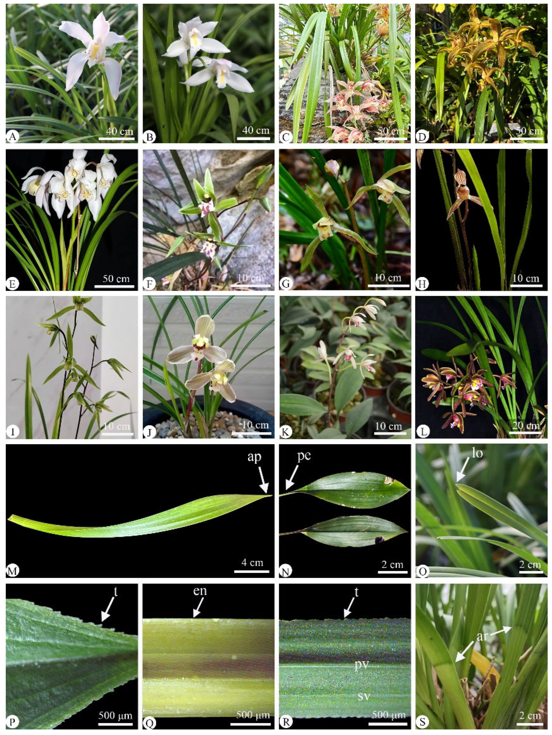
Figure 1. Plants of twelve Cymbidium species, showing their flower, leaf morphology and detailed leaf features. (AL) Flower and leaf of twelve Cymbidium species; (MS) detailed leaf features; (AE) subgen. Cyperorchis; (FK) subgen. Jensoa; (L) subgen. Cymbidium. (A) C. eburneum; (B) C. mastersii; (C) C. hookerianum; (D) C. tracyanum; (E) C. wenshanense; (F) C. qiubeiense; (G) C. faberi; (H) C. defoliatum; (I) C. kanran; (J) C. serratum; (K) C. lancifolium; (L) C. floribundum; (M) Lorate leaf of (L) C. floribundum with slightly oblique apex (enlarged image); (N) oblanceolate–oblong leaf; (O) leaf apex and two-lobed leaf; (P) leaf of (N) C. lancifolium (enlarged image) with apex margin and finely toothed margin; (Q) entire leaf margin entire of (J) C. defoliatum (enlarged image); (R) toothed leaf margin of (H) C. faberi (enlarged image); (S) articulate leaf of (C) C. hookerianum (enlarged image). ap, apex; pe, petioles; lo, lobed; t, toothed; en, entire; pv, primary veins; sv, secondary veins; ar, articulate.
Plants 14 01396 g001
Figure 2. Adaxial leaf epidermis of twelve Cymbidium species under light microscope, with epidermal cells and stigmata present (arrow). (AI) Polygonal epidermal cells; (JL) rectangular epidermal cells. (A) C. lancifolium; (B) C. floribundum; (C) C. wenshanense; (D) C. tracyanum; (E) C. faberi; (F) C. eburneum; (G) C. qiubeiense; (H) C. mastersii; (I) C. kanran; (J) C. hookerianum; (K) C. defoliatum; (L) C. serratum. Scale bars = 50 µm.
Plants 14 01396 g002
Figure 3. Abaxial leaf epidermis of twelve Cymbidium species under light microscope, with epidermal cells, stigmata (arrow), and stomatal shape clearly observable. (AD) Broad polygonal epidermal cells; (EL) narrow polygonal epidermal cells. (A) C. floribundum; (B) C. lancifolium; (C) C. eburneum; (D) C. wenshanense; (E) C. kanran; (F) C. faberi; (G) C. defoliatum; (H) C. mastersii; (I) C. serratum; (J) C. qiubeiense; (K) C. hookerianum; (L) C. tracyanum. as, anomocytic stomata; ts, tetracytic stomata. Scale bars = 50 μm.
Plants 14 01396 g003
Figure 4. Two types of midrib cross-sections seen in twelve Cymbidium species. (AE) Shallow V-shape leaf midrib outline; (FL) V-shape leaf midrib outline. (A) C. tracyanum; (B) C. mastersii; (C) C. wenshanense; (D) C. serratum; (E) C. eburneum; (F) C. kanran; (G) C. hookerianum; (H) C. floribundum; (I) C. faberi; (J) C. lancifolium; (K) C. qiubeiense; (L) C. defoliatum. ad, adaxial surface; ab, abaxial surface; c, cuticle; vb, vascular bundles; pmc, palisade mesophyll cells. Scale bars = 200 μm.
Plants 14 01396 g004
Figure 5. Micro-morphology of cuticle, epidermis, fiber bundle, and stegmata, seen in cross-sections of six Cymbidium species. 1, adaxial surface; 2, abaxial surface. (A1,A2) C. kanran; (B1,B2) C. defoliatum; (C1,C2) C. floribundum; (D1,D2) C. lancifolium; (E1,E2) C. faberi; (F1,F2) C. qiubeiense. Conical of stegmata = arrow; c, cuticle; ep, epidermis cell; fb, fiber bundles. Scale bars = 10 μm.
Plants 14 01396 g005
Figure 6. Micro-morphology of cuticle, epidermis, fiber bundle, and stegmata seen in cross-sections of a blade from the other six Cymbidium species. 1, adaxial surface; 2, abaxial surface. (A1,A2) C. mastersii; (B1,B2) C. hookerianum; (C1,C2) C. tracyanum; (D1,D2) C. eburneum; (E1,E2) C. wenshanense; (F1,F2) C. serratum. Conical of stegmata = arrow. fb, fiber bundles; ss, spherical of stegmata; c, cuticle; ep, epidermis cell. Scale bars = 10 μm.
Plants 14 01396 g006
Figure 7. Three types of crystal seen in six Cymbidium species. (A,B) Terete crystal in (A) C. faberi and (B) C. tracyanum; (CE) long tube crystal bundles in (C) C. qiubeiense, (D) C. kanran, and (E) C. mastersii; and (F) raphide crystals in C. lancifolium. Scale bars = 10 μm.
Plants 14 01396 g007
Table 1. Taxonomic data of twelve Cymbidium species investigated.
SpeciesChen et al., (1999) [3]Liu et al., (2006) [9]DistributionVoucher
——Subgen. CyperorchisSubgen. Cyperorchis————
C. eburneumSect. EburneaSect. EburneaChina, India, Myanmar, Nepal, VietnamLilu20190228
C. mastersiiSect. EburneaSect. EburneaAsiaLilu20220725
C. hookerianumSect. IridorchisSect. IridorchisChina, India, Nepal, VietnamLilu20200526
C. tracyanumSect. IridorchisSect. IridorchisChina, Myanmar,
Thailand
Lilu20181218
C. wenshanenseSect. IridorchisSect. AnnamaeaChina, VietnamLilu20200324
——Subgen. JensoaSubgen. Jensoa————
C. defoliatum *Sect. JensoaSect. JensoaChinaLilu20201112
C. kanranSect. JensoaSect. JensoaChina, Japan, KoreaLilu20201111
C. qiubeiense *Sect. JensoaSect. JensoaChinaLilu20220114
C. faberiSect. JensoaSect. NanulaChina, India, NepalLilu20170725
C. serratum *Sect. JensoaSect. NanulaChinaLilu20220715
C. lancifoliumSect. GeocymbidiumSect. GeocymbidiumAsia, OceaniaLilu20180728
——Subgen. CymbidiumSubgen. Cymbidium————
C. floribundumSect. FloribundaSect. FloribundaChina, VietnamLilu20180730
* Chinese endemic species.
Table 2. Leaf morphology of twelve Cymbidium species.
SpeciesShapesSize (L × W), cmMarginApexBasePetiole
C. defoliatumlorate10–40 × 0.5–1entireacutearticulateabsent
C. eburneumlorate57–65 × 1.4–2.1entireacute, slightly two
lobed
articulateabsent
C. faberilorate25–80 × 0.4–1.2toothedacuteabsentabsent
C. floribundumlorate22–50 × 0.8–1.8entireacute, slightly obliquearticulateabsent
C. hookerianumlorate35–60 × 1.4–2.3entireacutearticulateabsent
C. kanranlorate40–70 × 0.9–1.7toothedacutearticulateabsent
C. lancifoliumoblanceolate
oblong
6–17 × 4–7toothedacutearticulatepetiole
C. mastersiilorate24–75 × 1.1–2.5entireacute, slightly two
lobed
articulateabsent
C. qiubeienselorate30–80 × 5–10toothedacutearticulatepetiole
C. serratumlorate20–40 × 0.5–0.9toothedacuteabsentabsent
C. tracyanumlorate55–80 × 1.5–3.4entireacutearticulateabsent
C. wenshanenselorate60–90 × 1.3–1.7entireacutearticulateabsent
Table 3. Leaf epidermal features of twelve Cymbidium species.
SpeciesAdaxial EpidermisAbaxial Epidermis
ELadEWadL/WELabEWabL/WSLSWSL/SWSISD
C. defoliatum45.48 ± 2.11 e17.35 ± 0.40 e2.6242.85 ± 1.71 bc18.17 ± 0.54 bc2.3632.49 ± 0.63 a27.70 ± 0.37 ab1.175.21 ± 0.23 d1.37 ± 0.10 bcd
C. eburneum56.32 ± 3.42 abc19.49 ± 0.47 d2.8925.91 ± 1.25 f16.13 ± 0.24 cdef1.6031.71 ± 0.56 bcd25.99 ± 0.60 a1.228.01 ± 0.20 a1.67 ± 0.13 ab
C. faberi58.54 ± 2.06 ab13.26 ± 0.59 f4.4145.61 ± 2.10 ab16.80 ± 0.72 cde2.7130.65 ± 0.55 cd24.41 ± 0.37 a1.256.87 ± 0.29 bc1.27 ± 0.82 cd
C. floribundum23.69 ± 1.11 g13.15 ± 0.48 f1.8020.81 ± 0.73 g17.14 ± 0.50 cde1.2124.68 ± 0.67 f20.18 ± 0.39 d1.226.37 ± 0.21 c1.47 ± 0.10 bcd
C. hookerianum54.02 ± 1.80 bc15.00 ± 1.10 f3.6031.72 ± 1.10 e13.67 ± 0.60 de2.3232.33 ± 0.63 bc27.54 ± 0.62 a1.186.60 ± 0.24 bc1.36 ± 0.10 bcd
C. kanran52.30 ± 2.14 bcd17.92 ± 0.33 de2.9236.64 ± 1.40 d16.71 ± 0.67 cde2.1930.19 ± 0.61 cd26.22 ± 0.42 ab1.156.79 ± 0.30 bc1.80 ± 0.15 a
C. lancifolium62.10 ± 0.87 a59.34 ± 0.90 a1.0538.59 ± 2.06 cd26.65 ± 0.68 a1.4539.37 ± 0.63 f29.68 ± 0.56 d1.334.19 ± 0.21 e1.10 ± 0.56 d
C. mastersii50.73 ± 2.50 cde22.91 ± 0.45 c2.2140.81 ± 2.34 cd16.93 ± 0.56 cde2.4133.34 ± 0.48 ab27.24 ± 0.69 a1.254.00 ± 0.20 e1.36 ± 0.10 bcd
C. qiubeiense47.54 ± 1.80 de14.12 ± 0.46 f3.3630.10 ± 0.73 f15.87 ± 0.38 e1.8929.50 ± 0.53 d25.19 ± 0.65 c1.176.38 ± 0.20 c1.37 ± 0.10 bcd
C. serratum56.08 ± 2.92 abc13.43 ± 0.50 f4.1748.17 ± 2.34 a14.59 ± 0.33 fg3.3030.33 ± 0.47 d24.14 ± 0.45 c1.273.98 ± 0.11 e1.30 ± 0.98 cd
C. tracyanum27.98 ± 1.40 g14.10 ± 0.49 f1.9825.65 ± 0.43 f13.68 ± 0.54 b1.8926.60 ± 0.53 e24.53 ± 0.52 c1.086.45 ± 0.21 c1.50 ± 0.12 abc
C. wenshanense34.38 ± 1.04 f26.30 ± 0.75 b1.3122.40 ± 2.08 cd17.78 ± 0.63 cd1.2530.15 ± 0.36 d27.61 ± 0.48 a1.097.23 ± 0.21 b1.50 ± 0.10 abc
Mean ± SE (n = 30). Different letters in the same line indicate statistical differences p < 0.05 (ANOVA). ELad, length of adaxial ordinary epidermal cell; Wad, width of adaxial ordinary epidermal cell; ELab, length of abaxial ordinary epidermal cell; EWab, width of abaxial ordinary epidermal cell; SL, length of stomata; SW, stomata width; SI, stomatal index; SD, stomatal density.
Table 4. Leaf anatomic features of twelve Cymbidium species.
SpeciesLTTMMTVBTETadETabETad/ETabCadCab
C.lancifolium317.71 ± 6.73 c371.92 ± 3.32 d268.93 ± 4.09 de141.47 ± 3.66 ef27.56 ± 0.61 a17.68 ± 0.37c1.585.65 ± 0.31 cde3.99 ± 0.21 bc
C. serratum217.16 ± 4.69 f211.83 ± 3.41 fg185.63 ± 7.60 h88.98 ± 7.12 g44.70 ± 2.00 bc11.10 ± 0.79 b4.024.14 ± 0.29 fg1.85 ± 0.09 g
C. mastersii215.24 ± 2.67 f273.61 ± 1.43 f183.55 ± 2.23 h92.88 ± 3.61 g13.77 ± 1.06 bcd11.45 ± 0.33 b1.073.39 ± 0.14 g2.19 ± 0.08 fg
C. kanran203.90 ± 1.23 g239.62 ± 10.58 g177.27 ± 1.10 h119.83 ± 4.86 fg18.01 ± 0.62 f10.13 ± 0.40 b1.793.54 ± 0.13 g1.88 ± 0.07 g
C. Wenshanense344.64 ± 7.47 b423.01 ± 12.86 bc277.75 ± 3.74 cd158.16 ± 10.79 de23.75 ± 0.85 b10.89 ± 0.20 a2.187.61 ± 0.35 b4.31 ± 0.21 a
C. hookerianum293.52 ± 3.29 d325.29 ± 13.44 f254.94 ± 1.86 f180.50 ± 2.23 g16.57 ± 0.52 h9.13 ± 0.60 de1.817.48 ± 0.41 b3.75 ± 0.19 c
C. tracyanum245.87 ± 2.66 e454.14 ± 16.41 b207.54 ± 2.21 g126.77 ± 4.90 ef22.14 ± 1.21 cdef10.82 ± 0.91 c2.059.03 ± 0.60 a2.34 ± 0.12 ef
C. faberi347.06 ± 3.14 b449.16 ± 10.23 b287.52 ± 2.34 c250.86 ± 15.87 b21.00 ± 0.63 b cde14.04 ± 0.39 c1.544.93 ± 0.17 ef3.64 ± 0.10 c
C. eburneum293.45 ± 4.68 d312.34 ± 4.56 e253.19 ± 3.53 f150.82 ± 4.91 ef23.43 ± 0.89 b10.26 ± 0.29 a2.285.21 ± 0.29 cde2.65 ± 0.12 de
C. defoliatum344.26 ± 2.26 b410.61 ± 14.31 c313.80 ± 3.07 b214.75 ± 15.32 c15.00 ± 0.33 g9.76 ± 0.41 c1.585.94 ± 0.19 cd4.31 ± 0.16 ab
C. qiubeiense490.81 ± 3.60 a697.75 ± 18.88 a440.36 ± 3.47 a293.73 ± 22.40 a28.18 ± 0.68 ef12.65 ± 0.32 cd2.236.18 ± 0.35 c4.51 ± 0.26 ab
C. floribundum302.90 ± 5.14 d421.45 ± 6.10 bc262.67 ± 4.39 ef174.31 ± 6.50 d20.26 ± 0.36 def16.33 ± 0.50 e1.224.84 ± 0.18 ef2.87 ± 0.12 d
Mean ± SE (n = 30). Different letters in the same line indicate statistical differences p < 0.05 (ANOVA). LT, leaf thickness; TM, thickness of midrib; MT, mesophyll thickness; VBT, vascular bundle thickness; ETad, thickness of adaxial epidermis; ETab, thickness of abaxial epidermis; Cad, adaxial cuticle thickness; Cab, abaxial cuticle thickness.
Disclaimer/Publisher’s Note: The statements, opinions and data contained in all publications are solely those of the individual author(s) and contributor(s) and not of MDPI and/or the editor(s). MDPI and/or the editor(s) disclaim responsibility for any injury to people or property resulting from any ideas, methods, instructions or products referred to in the content.