Article

Phytophthora inundata: A New Root Pathogen of Citrus in Europe and the Mediterranean Region

Department of Agriculture, Food and Environment, University of Catania, Via S. Sofia 100, 95123 Catania, Italy
*
Authors to whom correspondence should be addressed.
These authors contributed equally to this work.
Plants 2025, 14(9), 1333; https://doi.org/10.3390/plants14091333
Submission received: 7 April 2025 / Accepted: 24 April 2025 / Published: 28 April 2025
(This article belongs to the Collection Plant Disease Diagnostics and Surveillance in Plant Protection)

Abstract

:
Citruses are one of the major fruit crops globally. Among Mediterranean citrus producers, Sicily (southern Italy) is renowned for its high-quality fresh fruit production. Phytophthora diseases are a serious issue for citrus production worldwide and Phytophthora nicotianae is a prevalent causal agent of root rot in most citrus growing areas globally and particularly in the Mediterranean region. This study reports the occurrence of Phytophthora inundata as a root pathogen of declining mature citrus trees in eastern Sicily in association with P. nicotianae. This is the first record of P. inundata on citrus in Europe and the Mediterranean region. The species was identified on the basis of a morphology and multi-gene phylogenetic analysis, which included the internal transcribed spacer, β-tubulin and cytochrome c oxidase subunit 1. Pathogenicity tests on citrus saplings showed P. inundata was a less aggressive pathogen than P. nicotianae. However, the co-inoculation of both species produced more severe symptoms than inoculation with a single species, indicating an additive effect of these two pathogens and suggesting that opportunistic secondary pathogens like P. inundata may have a crucial role in complex diseases.

1. Introduction

Citrus, encompassing oranges, lemons, limes, mandarins, pomelos, grapefruits, citrons and kumquats, is one of the most important fruit crop groups worldwide, with cultivation spanning over 140 countries and an annual production exceeding 150 million tons [1]. Among citrus producers in the Mediterranean region, Sicily (southern Italy) is renowned for its primacy in the production of blood oranges [2], which are valuable for the fresh fruit market as well as for juice extraction. Citrus orchards are exposed to numerous biotic and abiotic stresses, many of which can lead to substantial yield losses [3,4,5]. Among the most serious threats are diseases caused by soil-borne oomycetes of the genus Phytophthora. These pathogens induce a broad spectrum of symptoms, including root and foot rot, trunk and branch gummosis, canopy blight and fruit brown rot [6,7,8,9]. Gummosis and root rot are particularly severe, often leading to rapid death in young citrus trees, whereas in mature trees the disease typically progresses more slowly and chronically [10]. In almost all major citrus producing countries, the use of tolerant rootstocks is the most popular and effective management option to prevent Phytophthora root and foot rot as well as trunk gummosis [10,11,12,13]. The genus Phytophthora encompasses over 250 formally described species, known for their ecological plasticity and broad host range, spanning from agricultural crops to forest ecosystems [14,15,16,17,18]. Around 15 species of Phytophthora have been reported to infect or to be associated with citrus [17,19,20,21,22,23,24,25,26,27]. In the Mediterranean region, the most common species causing diseases in citrus orchards are Phytophthora nicotianae and P. citrophthora [4,25].
Our study focused on a commercial citrus orchard in eastern Sicily, where several mature trees exhibited symptoms of decline, including leaf chlorosis starting from the main veins and expanding to the entire leaf blade, defoliation and root rot, indicative of a possible soil-borne Phytophthora infection. To determine the causal agents, soil and fine root samples were collected from symptomatic trees. During this investigation, a Phytophthora species with a petaloid colony pattern on potato dextrose agar (PDA), a maximum in vitro growth temperature of 30 °C, and non-papillate sporangia as its most distinctive phenotypic traits was consistently isolated. This species was found in association with P. nicotianae, suggesting a potential role in citrus decline.
The objectives of this study were as follows: (I) to identify this Phytophthora species previously unreported on citrus in Sicily; (II) to evaluate its pathogenicity on citrus; and (III) to determine its involvement in etiology of root rot and tree decline.

2. Results

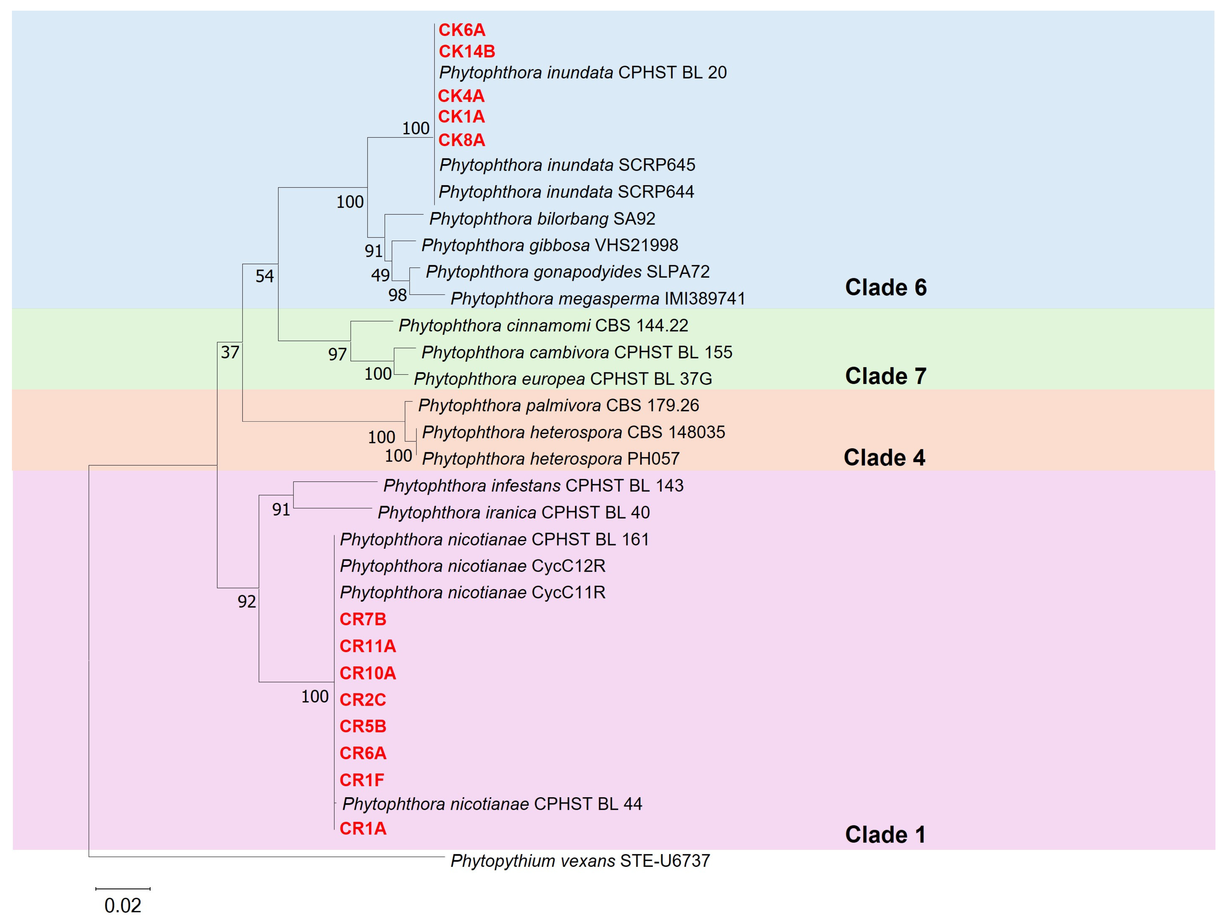
2.1. Isolation and Identification of the Phytophthora Species

The Phytophthora species, new for citrus in Sicily, was recovered in a commercial citrus orchard from the rhizosphere of five declining mature trees with symptoms of root rot and decline. It was isolated from soil collected under the tree canopy using leaf baits and directly from fine roots by plating root segments on PARPNH agar medium. Of a total of 42 Phytophthora isolates recovered from these five symptomatic trees, 13 were of this species while 39 were tentatively identified as P. nicotianae on the basis of the morphological characteristics. The two species showed clearly discernible morphotypes. The isolates of the unidentified species formed petaloid colonies on PDA and colonies without an obvious pattern on V8 agar. Sporangia were non-papillate and persistent, ovoid or obpyriform (30–68 µm in length and 20–52 µm in width, n = 50), sometimes with tapered bases and internal proliferation. They originated from unbranched or simple sympodial sporangiophores, which occasionally displayed swellings. Hyphal swellings were globose, subglobose, irregular, and either individual or catenulated. Chlamydospores were absent. None of the isolates exhibited sexual structures in single culture. Two subsets, one of five isolates of this unidentified species and the other of eight putative isolates of P. nicotianae, respectively, were randomly selected for phylogenetic analysis of ITS-rDNA, β-tubulin, and COI sequences. The sequences of isolates of Phytophthora species from phylogenetic Clades 1, 4, 6 and 7 were used as references, whereas a Phytopythium vexans isolate served as the outgroup (Table 1).
Isolates identified tentatively as P. nicotianae were grouped within Clade 1 along with the reference isolates of P. nicotianae (CPHST BL 44, CycC11R, CycC12R and CPHST BL 161), confirming their identity. Conversely, the other five isolates were grouped in Clade 6, together with the reference isolates of P. inundata (CPHST BL 20, SCRP645 and SCRP644), and were assigned to this species (Figure 1). The sequences of all isolates characterized in this study were deposited in GenBank (Table 2).

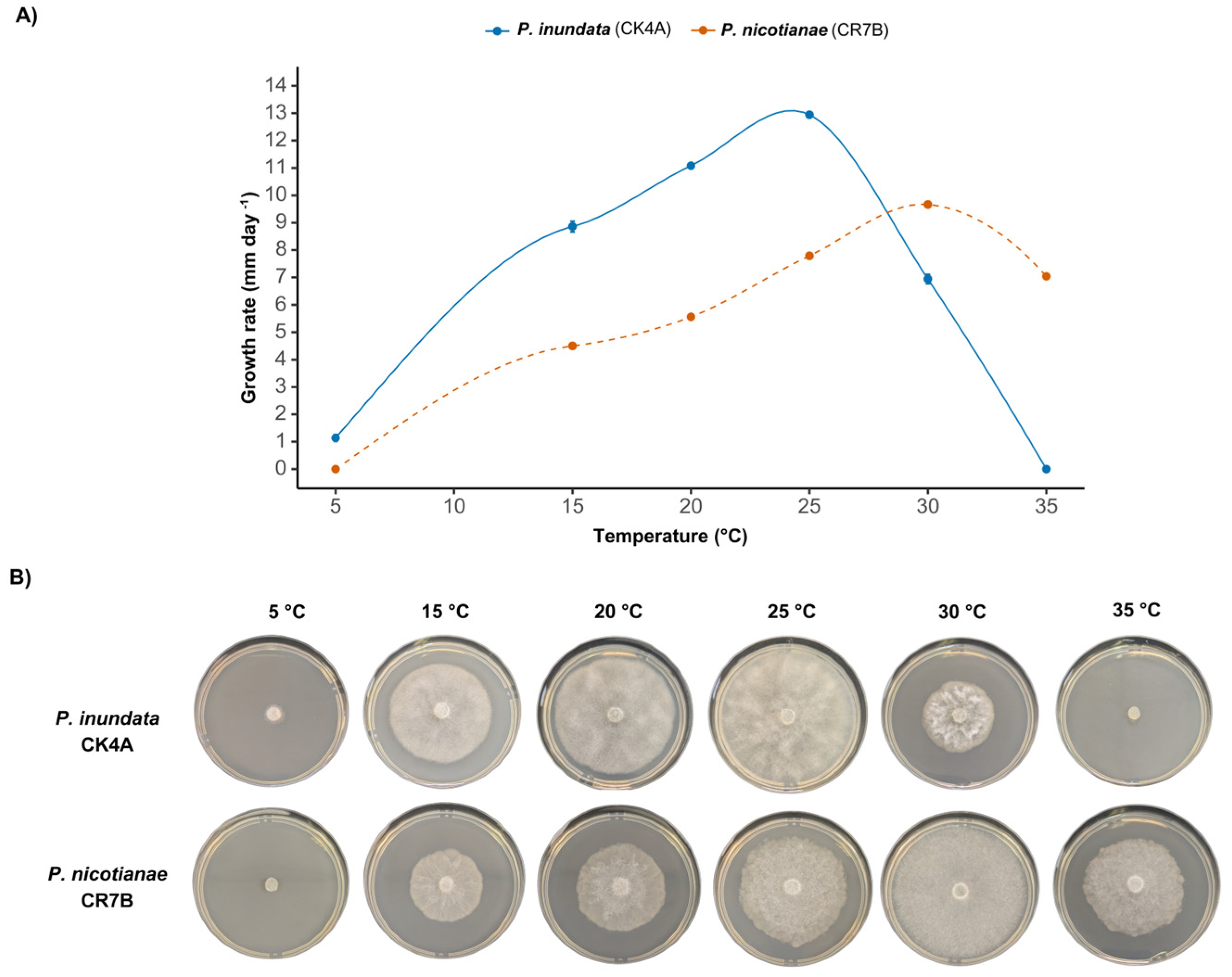
2.2. Radial Growth of Phytophthora spp. at Different Temperatures

The radial growth rates of P. inundata (CK4A) and P. nicotianae (CR7B) were evaluated at six different temperatures: 5, 15, 20, 25, 30 and 35 °C (Figure 2). Growth rate measurements, expressed in mm/day, were calculated as the mean radial growth along two perpendicular axes.
Both species exhibited distinct temperature-dependent growth patterns. P. inundata reached its maximum growth rate after six days at 25 °C, with an average radial extension of 12.04 ± 0.07 mm/day. In contrast, P. nicotianae exhibited its highest growth rate at 30 °C after eight days, with a mean radial extension of 9.66 ± 0.06 mm/day.
At extreme temperatures, differential species responses were observed. At 5 °C, P. inundata was able to grow, reaching a radial extension of 1.13 ± 0.13 mm/day after six days, whereas P. nicotianae showed no detectable growth. Conversely, at 35 °C, P. inundata showed no detectable growth, while P. nicotianae was able to grow, reaching a radial extension of 7.04 ± 0.06 mm/day.

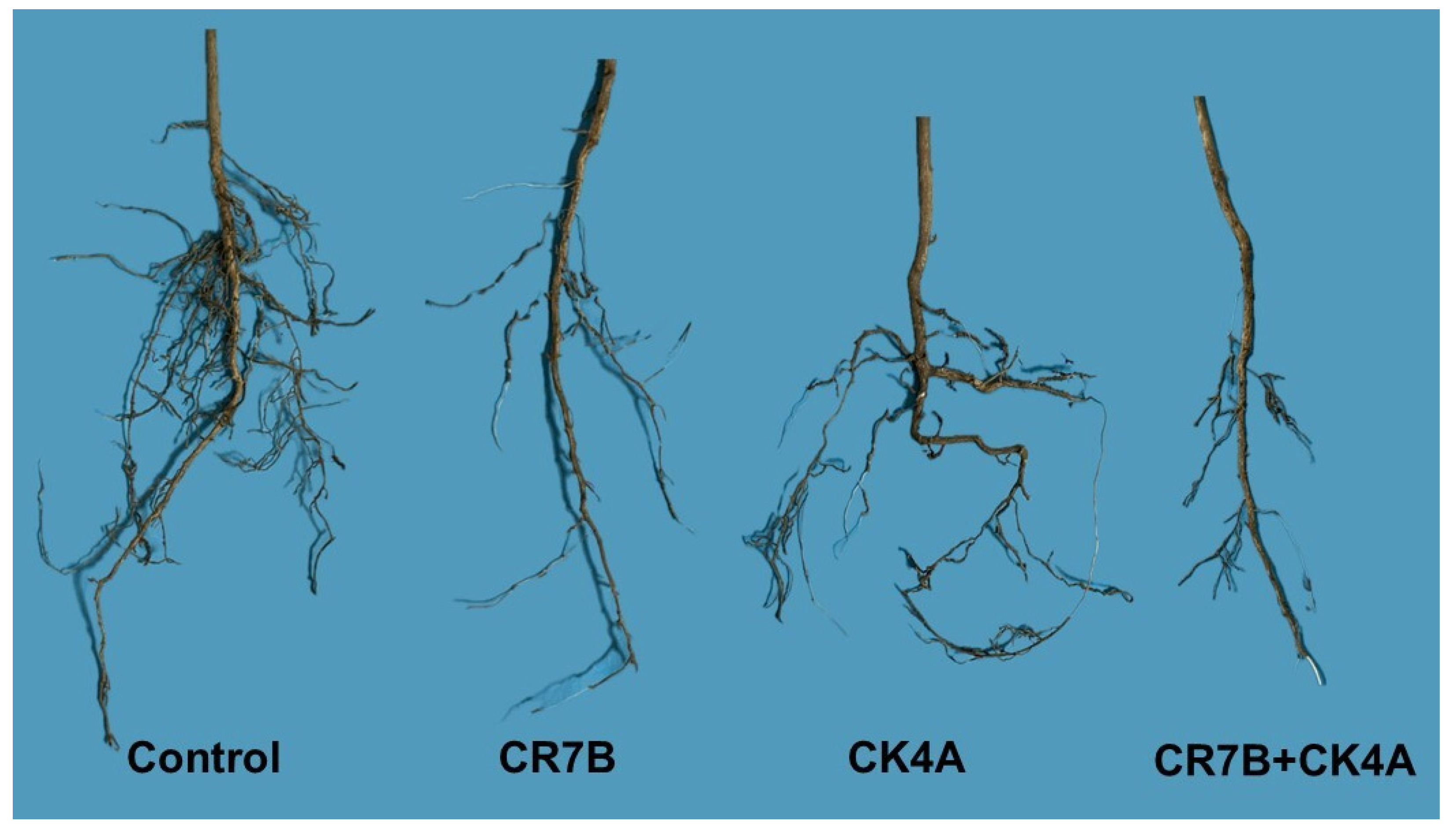
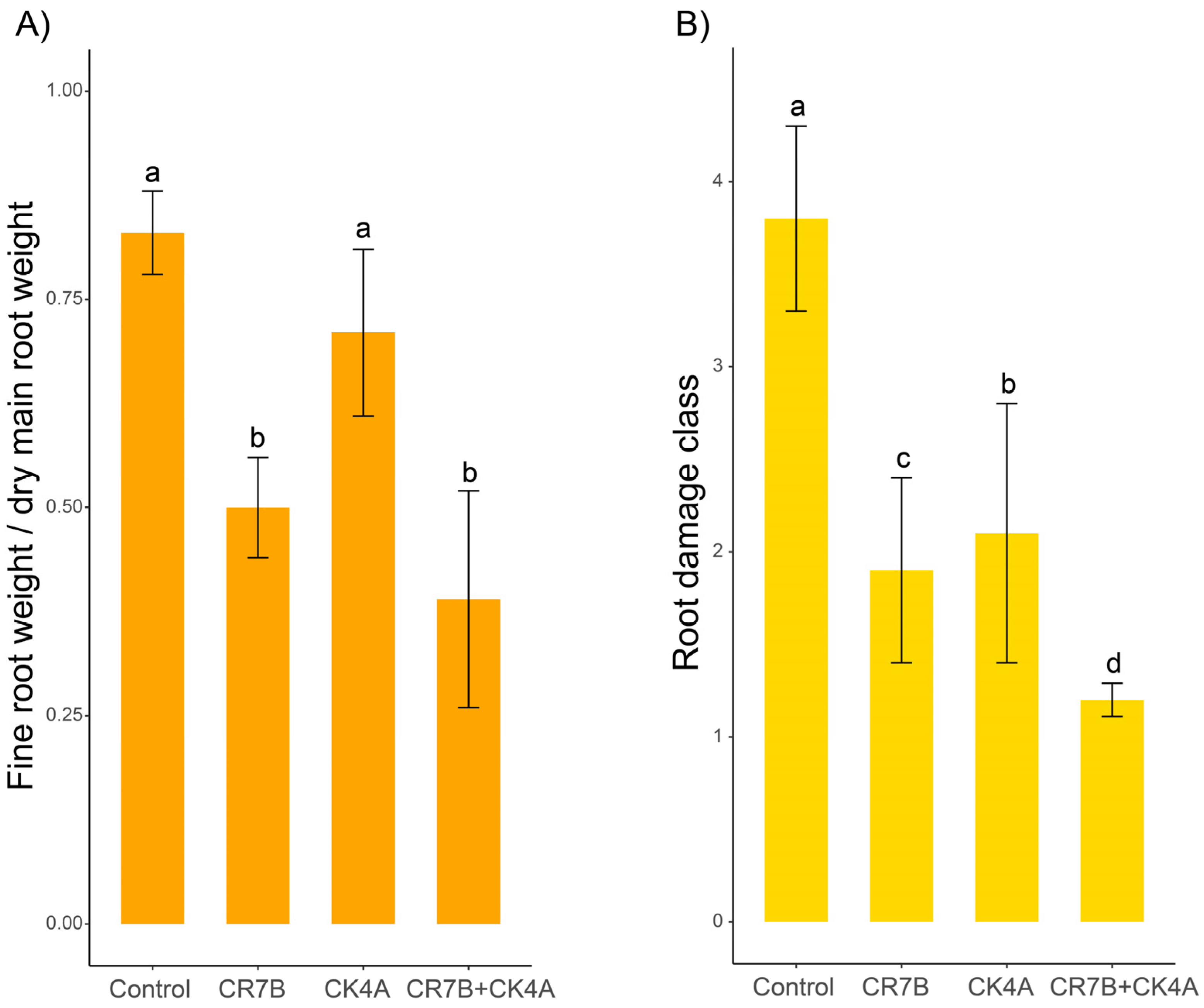
2.3. Pathogenicity Test

Since the results of the two separate pathogenicity tests did not differ significantly, as revealed by the analysis of the variance homogeneity, they were pooled and analyzed all together like it was a single experiment. On saplings inoculated with either a single Phtophthora species or both species simultaneously, above-ground symptoms at 40 dpi consisted in the chlorosis of main leaf vein and leaf shedding, while control saplings showed no symptoms. No difference was noticed in the severity of above-ground symptoms among saplings inoculated singularly with either P. inundata or P. nicotianae and those inoculated with both species simultaneously. By contrast, the severity of root damage and the fine root/main root weight ratio varied significantly among the different treatments (Figure 3). Saplings inoculated with P. nicotianae (isolate CR7B) showed a severe rot of primary roots and a rarefaction of secondary roots. P. inundata (isolate CK4A) was less aggressive. Saplings inoculated with this species showed a denser root apparatus than saplings inoculated with P. nicotianae (CR7B) but were sparser than the control saplings. The most severe symptoms of root rot were observed in saplings inoculated with both P. inundata and P. nicotianae (CR7B+CK4A).
In saplings inoculated with P. inundata, the mean ratio between fine root and main root weight was lower than in control saplings (0.71 ± 0.1) (Figure 4A). However, the difference was not significant, indicating P. inundata was weakly pathogenic. By contrast, mean values of the ratio between fine root and main root weight in saplings inoculated with P. nicotianae, alone (0.50 ± 0.06) and together with P. inundata (0.39 ± 0.13), were significantly lower than in control saplings, indicating a marked detrimental effect on root apparatus. However, the difference between saplings inoculated with P. nicotianae alone and with both P. nicotianae and P. inundata was not significant. As for the root damage class, a similar, albeit more marked, trend was observed (Figure 4B). The mean root damage class of saplings inoculated with P. inundata (2.1 ± 0.7) was significantly lower than the control (3.8 ± 0.5). In turn, the mean root damage class of saplings inoculated with P. nicotianae (1.9 ± 0.5) was even lower. However, the lowest value of mean root damage class was observed in saplings co-inoculated with P. nicotianae and P, inundata (1.2 ± 0.09), suggesting an additive effect between these two pathogens. Differences between mean values of root damage class were significant for p ≤ 0.05.
Overall, symptoms on inoculated saplings were consistent with those observed in the commercial citrus orchard from which isolates of P. inundata and P. nicotianae used in pathogenicity tests had been recovered. Both Phytophthora species were successfully re-isolated from necrotic roots of inoculated saplings, thereby fulfilling Koch’s postulates. No Phytophthora species were obtained from the roots of asymptomatic control seedlings.

3. Discussion

Phytophthora inundata is a species in Phytophthora phylogenetic subclade 6a and, like other species in this clade, it is a common inhabitant of riparian habitats in Europe. Since its formal description [35], this Phytophthora species has also been reported from Northern and Southern America, Asia and Australia on various hosts and diverse environments [16,31,36,37,38,39,40]. Recently, Nawza et al. [41] documented a case of crown rot on olive in Pakistan, confirming the ability of this oomycete to infect this host under favorable environmental conditions. In several regions of Italy, including Calabria, Friuli-Venezia Giulia, Sicily, Sardinia and Veneto, P. inundata has been recorded in soil of forest ecosystems and as an occasional pathogen of horticultural and ornamental crops, such as olive, peach, walnut and dwarf banana, almost exclusively in flooded or waterlogged soils and mostly associated with other more aggressive Phytophthora species [35,42,43,44,45]. Overall, these reports would suggest that P. inundata can be regarded as an opportunistic plant pathogen with a broad host range. Moreover, they indicate that this Phytophthora species has a wide distribution in Italy. By contrast, in Korea, P. inundata has not yet been reported and is regarded as a potentially invasive species; therefore, the possibility of including it in the list of quarantine pathogens is being considered [46]. This study is the first report of P. inundata associated with citrus root rot in Europe and the Mediterranean region. Previously, the only documented cases of P. inundata affecting citrus were from Chile and India [21,23]. In India, P. inundata was identified as the Phytophthora species responsible for a decline of Kinnow mandarin trees on rough lemon (Citrus jambhiri) as rootstock in waterlogged soils [23]. In Chile, P. inundata was recovered from citrus trees affected by root and foot rot in association with P. citrophthora [21] but was considered a secondary pathogen; in pathogenicity tests, it proved to be weakly pathogenic which was different to P. citrophthora. Both reports are consistent with the findings of the present study. The former highlights that soil waterlogging and root asphyxiation are conducive to infections by P. inundata, while the latter suggests that this species, although a weak pathogen on its own, when it is present in multiple infections, may contribute to the decline of citrus trees incited primarily by more aggressive pathogens such as P. citrophthora and P. nicotianae. As a matter of fact, in the present study on citrus saplings inoculated with both P. inundata and P. nicotianae, symptoms of root rot were more severe than on saplings inoculated only with the latter species, indicating an additive detrimental effect of the two oomycetes. Although in this study the optimum and high upper limit of temperature for the mycelium growth of P. inundata (25 °C and less than 35 °C, respectively) were lower than the optimum and high upper limit of temperature reported in the original description of the species (28–30 °C and around 35–37 °C, respectively) [35], it can be assumed that both P. inundata and P. nicotianae are mesophilic and prefer a warm climate. It is likely that in the Mediterranean region late spring is the most favorable season for the co-infection of fibrous roots of citrus trees by these two species. In recent decades, the study of complex interactions between plants and multiple microorganisms and the mechanisms by which pathogens interact with each other and affect the phenotypic and genetic response of plants have been gaining special attention [47,48]. The synergistic or additive effect of diverse pathogens and their interactions with both the plant and environmental conditions are crucial to understand the etiology and mechanisms of pathogenesis of complex diseases, such as Phytophthora root rot (PRR) and Dry root rot (DRR), another complex soil-borne disease of citrus trees supposed to be caused by Fusarium spp. [5]. PRR and DRR in citrus have some traits in common. Diverse species of Phytophthora may be responsible for PRR, as in the case documented in this study, and several Fusarium and Fusarium-like species differing in virulence are associated with DRR [49]. In both diseases, soil asphyxiation is crucial as predisposing factor and consequently both diseases are particularly damaging in poorly drained or flooded soils. Infections of multiple Phytophthora species differing in lifestyle and ecology may have implications for the management of PRR, as even opportunistic pathogens, like P. inundata, might impair the resilience of commercial citrus rootstocks in heavy soil, which are more prone to waterlogging. In a recent study of the genetic response of Monterey pine (Pinus radiata) seedlings that were inoculated simultaneously with Fusarium circinatum, the causal agent of pine pitch canker disease, and the oomycetes P. × cambivora and P. parvispora, it was hypothesized that co-infection might repress the expression of defense-related genes, thus exacerbating the severity of the disease [48]. It is noteworthy that in the Mediterranean region, as a consequence of climate change, during recent years, the frequency of soil water saturation due to more intense precipitations has been increasing. Moreover, warmer temperatures might expand the period of the year favorable to multiple infections of diverse mesophilic Phytophthora species associated with the citrus tree rhizosphere.

4. Material and Methods

4.1. Isolation and Morphological Identification of Isolates

At the end of May 2024, in a 20-year-old commercial orchard in the province of Siracusa, eastern Sicily (geographic coordinates: DATUM WGS 84: 37°18′51.0″ N, 15°02′21.0″ E), soil and fine root samples were collected from five declining mature ‘Tarocco Lempso’ citrus trees grafted on citrange ‘Carrizo’ (Citrus sinensis ‘Washington Navel’ × Poncirus trifoliata) as rootstock, showing symptoms of root rot and leaf chlorosis starting from the main veins, which are suggestive of Phytophthora root rot. During the same month, meteorological data recorded by the Sicilian Agrometeorological Information Service (SIAS, http://www.sias.regione.sicilia.it/, accessed on 15 December 2024) at the Lentini station indicated a mean daily minimum air temperature of 14.6 °C, a mean daily maximum temperature of 27.7 °C, and a total monthly precipitation of 47.6 mm.
The soil in the orchard was characterized by a clay loam texture and in 2018 had been subjected to flooding. Isolation was performed from the soil using leaf baits in accordance with the protocol described by Riolo et al. [14] as well as directly from fine roots by plating root segments (3–5 mm) on PARPNH agar medium in Petri dishes (9 cm in diameter) and incubating at 22 ± 2 °C in the dark. Subsamples of approximately 400 mL of soil were tested by leaf baiting in a walk-in growth chamber with 12 h of natural daylight at 20 °C. Young leaves of carob (Ceratonia siliqua) and oak (Quercus spp.) were used as baits. Necrotic pieces (2 × 2 mm) from symptomatic leaves were plated onto selective PARPNH agar medium. Petri dishes were incubated at 22 ± 2 °C, in the dark. Outgrowing hyphae from leaf pieces or root segments were transferred onto V8-juice agar (V8A) under a stereomicroscope. Purified cultures were finally obtained by single hyphal culture on V8-agar. Colony morphology and morphological features of isolates, including the morphology and dimensions of reproductive structures, were determined on colonies grown on V8A at 22 ± 2 °C in the dark according to standard procedures [19]. Sporangia production was stimulated following the method described by Jung et al. [50]. Small fragments (size 2 mm) were cut from the growing edge of 5- to 7-day-old cultures grown in Petri dishes on V8A at 20 °C in the dark. Fragments were placed in 5 cm diameter Petri dishes and flooded with non-sterile soil extract water (prepared by 200 g soil suspended in 1 L of de-ionized water for 24 h at room temperature and then filtered). After incubation at 20 °C in the dark for 24–72 h, dimensions and morphological features of 50 mature sporangia of each isolate were determined at ×400 magnification. All isolates were maintained on V8A and stored at 6 °C in the dark. Other Phytophthora isolates preserved in the Molecular Plant Pathology Laboratory collection at the University of Catania were included in this study for comparison.

4.2. Assessment of the Effect of Temperature-On Mycelial Growth

The effect of temperature on mycelial growth was evaluated following the methodology described by Scanu et al. [34] with slight modifications. Agar plugs (5 mm in diameter) were excised from actively growing cultures and centrally placed in 90 mm Petri dishes containing V8 agar. Each isolate was incubated in triplicate at temperatures of 5, 15, 20, 25, 30 and 35 °C in the dark.
Radial mycelial growth was monitored daily. Once colonies reached their maximum expansion at the optimal temperature, growth measurements were recorded. To quantify radial growth, two perpendicular lines were drawn on the underside of each Petri dish, intersecting at the center of the inoculum. The radial growth of the colony was recorded along both axes, excluding the initial 5 mm agar plug. The average of these two measurements was then used to determine the growth rate.

4.3. Molecular Identification

DNA-based identification of 13 Phytophthora isolates that are representative of the two distinct morphotypes obtained from citrus trees in Sicily and randomly selected on the basis of colony morphology (Table 1) was performed via the sequence analysis of the internal transcribed spacer region of ribosomal DNA (ITS-rDNA), β-tubulin (β-tub) and cytochrome c oxidase subunit 1 (COI). DNA was extracted from 7-day-old cultures grown on V8A at 20 °C using the PowerPlant Pro DNA Isolation Kit (MO BIO Laboratories, Inc., Carlsbad, CA, USA), following the manufacturer’s instructions. ITS-rDNA was amplified using the ITS6 and ITS4 primers [29]. All PCR reactions were performed in a 25 µL reaction mix containing PCR buffer (1×), dNTP mix (0.2 mM), MgCl2 (1.5 mM), forward and reverse primers (0.5 mM each), Taq DNA Polymerase (1 U) and 100 ng of template DNA. The amplification conditions were as follows: initial denaturation at 94 °C for 3 min, followed by 35 cycles of 94 °C for 30 s, 55 °C for 30 s and 72 °C for 30 s, with a final extension at 72 °C for 10 min [51]. The amplification of β-tubulin and COI regions was performed using the primer pairs TUBUF2/TUBUR1 and COXF4N/COXR4N, respectively [29,52]. Thermocycling conditions included an initial denaturation at 94 °C for 2 min, followed by 35 cycles of 94 °C for 30 s, annealing for 30 s and extension at 72 °C for 60 s, with a final extension at 72 °C for 10 min. Annealing temperatures were set at 60 °C for β-tubulin and 52 °C for COI. Amplicons were visualized on 1% agarose gel and sequenced bidirectionally using an external sequencing service (Macrogen, Amsterdam, The Netherlands). Chromatograms were verified using FinchTV v.1.4.0 [53]. Consensus sequences were initially compared using the BLAST program (https://blast.ncbi.nlm.nih.gov/Blast.cgi, accessed on 4 July 2024) against gene sequences in the NCBI databases and further compared with those of ex-type and other authenticated Phytophthora specimens. Phylogenetic analysis was performed using sequences from the isolates in this study alongside validated reference sequences from GenBank (Table 1). Prior to analysis, duplicate reference sequences were removed using Elim Dupes software (https://www.hiv.lanl.gov/content/sequence/elimdupesv2/elimdupes.html, accessed on 4 July 2024)). Sequence alignment was conducted with MUSCLE, and phylogenetic trees, using a combined dataset of all sequenced markers (ITS, β-tubulin and COI), were generated using MEGA 11 [54] with the maximum-likelihood method with the Tamura–Nei model. Bootstrap analysis with 1000 replications was conducted.

4.4. Pathogenicity Tests

The pathogenicity of Phytophthora isolates was evaluated in greenhouse experiments using the soil infestation method [29,45,55,56]. Two isolates, one of Phytophthora inundata (CK4A) and one of P. nicotianae (CR7B), were tested. They were randomly selected among the isolates recovered directly from fine roots. Saplings (12 months old) of ‘Tarocco Lempso’ sweet orange on ‘Carrizo’ citrange rootstock were used as test plants. ‘Carrizo’ citrange is regarded as a rootstock tolerant of Phytophthora root rot [57]. The experiment included four treatments: (I) soil infested with P. nicotianae alone, (II) soil infested with P. inundata alone, (III) soil infested with both P. nicotianae and P. inundata and (IV) control, with an equal volume of potting soil and non-infested wheat seeds. It was organized in a randomized block with four blocks and five replicate saplings for each treatment, with a total of 20 saplings per block. For the soil infestation assays, the citrus saplings were transplanted into free-draining pots (12 cm in diameter) containing a 1:1 mixture of autoclaved universal potting soil (Cifo Srl, Giorgio di Piano, Bologna, Italy) and inoculum. The inoculum consisted of a 21-day-old culture grown in the dark at 25 °C in 750 mL flasks containing 50 mL of wheat seeds and 50 mL of V8 juice broth. The inoculum was incorporated into the potting mix at a rate of 20 cm3 per 1000 cm3. For the combined inoculation treatment, equal volumes of inoculum of P. inundata and P. nicotianae were mixed together before being incorporated into the potting mix at a rate of 20 cm3 per 1000 cm3. Control saplings were transplanted into pots with the same sterilized potting mixture without inoculum. After transplanting, all saplings were maintained in saturated soil for 48 h and then transferred to a growth chamber set at 23 °C, with an 80% relative humidity and a photoperiod of 16 h light and 8 h dark. Symptoms were assessed visually at 40 dpi. The effect of inoculation on the above-ground part of saplings was evaluated on the basis of the type and severity of symptoms, while the effect on the roots was evaluated using a root damage scale [56], which allowed a more precise measurement of the virulence of the Phytophthora species tested. The root damage scale was as follows: a score of 4 indicated a healthy root system with a dense fine root system and well-developed taproots; a score of 3 indicated less than 25% fine root loss with well-developed taproots; a score of 2 indicated 26–50% fine root loss, initial taproot decay and small necrotic lesions on woody roots or the collar; a score of 1 indicated 51–75% fine root loss, advanced taproot decay and large necrotic lesions on taproots and/or the collar; and a score of 0 indicated 76–100% fine root loss, extensive taproot decay and girdling necrotic lesions on taproots and/or the collar. Moreover, the roots were collected, dried for 72 h at 65 °C and weighed. The ratio between the dry weights of fine roots (diameter < 2 mm) and main roots (diameter 2–10 mm) was recorded for each sapling. Re-isolations of Phytophthora from necrotic fresh roots were performed using the selective PARPNH agar medium [56] and the identity of isolates recovered from artificially inoculated symptomatic saplings was confirmed on the basis of morphological characters and molecular analyses. The pathogenicity test was repeated once in the same experimental conditions and with the same experimental design.

4.5. Statistical Analysis

The normality of data relative to the effect of inoculation on the sapling roots was preliminarily assessed using the Shapiro–Wilk test while the homogeneity of variance of data from the two separate pathogenicity tests was assessed using the Levene’s test at p < 0.05. Then, one-way ANOVA was applied and the differences of means were evaluated with Tukey’s HSD post hoc test for multiple comparisons. Differences were considered statistically significant at p < 0.05. All analyses were performed using R software version 4.3.1 (https://www.R-project.org/, accessed on 9 January 2025).

5. Conclusions

Phytophthora inundata is reported as a pathogen of citrus trees for the first time in Europe and the Mediterranean region. To determine whether this is a one-time occurrence or, by contrast, P. inundata is a potential threat for the citrus industry, a more extended survey should be carried out in commercial citrus orchards across Sicily and other citrus producing areas of the Mediterranean macroregion. Preliminary evidence from this study and data from the literature would suggest P. inundata, differently from other invasive species such as P. citrophthora and P. nicotianae, is a weak opportunistic pathogen that favors water-saturated soil. An interesting finding of this study is the additive effect of P. inundata infection on the root decay of citrus tree when this species is associated with a more aggressive pathogen, such as P. nicotianae. The co-infection of citrus with two oomycetes differing in virulence, such as P. inundata and P. nicotianae, could be a model system to investigate the complex interactions among host plant and diverse pathogens acting simultaneously and could elucidate the plant defense mechanisms in multiple infections.

Author Contributions

Conceptualization, M.R. and S.O.C.; methodology, C.B., M.C.T., M.R. and S.C.T.; software, M.C.T., C.B., S.C.T. and M.R.; validation, M.R., A.P. and S.O.C.; formal analysis, M.C.T. and C.B.; investigation, C.B., M.C.T. and S.C.T.; resources, S.O.C. and A.V.; data curation, M.R. and S.C.T.; writing—original draft preparation, M.R., S.C.T. and C.B.; writing—review and editing, M.R, S.C.T. and S.O.C.; visualization, M.R. and S.O.C.; supervision, M.R., A.P. and S.O.C.; project administration, S.O.C. and A.V.; funding acquisition, M.R. All authors have read and agreed to the published version of the manuscript.

Funding

This research received no external funding.

Data Availability Statement

Data are contained within the article.

Acknowledgments

The authors wish to thank the project “S.A.I.A.—Soluzioni Automatizzate per l’Irrigazione degli Agrumi” (Programma di Sviluppo Rurale della Regione Sicilia 2014–2022, Call n. 62662 GAL NATIBLEI–19.2-Operazione sottomisura 16.2 “Progetti pilota o introduzioni di prodotti, pratiche, processi o tecnologie nuove”—Ambito 2.1.2 “Progetto Scale-Up Agrumicolo Zootecnico” CUP: G38G23000030009) and the University of Catania, Italy for providing support with the projects “Nuove soluzioni tecnologiche per la filiera degli agrumi: trasferimento di in-novazione per il miglioramento degli standard qualitativi delle produzioni agrumicole e per fronteggiare la minaccia di malattie infettive di origine fungina degli agrumi nel comprensorio del GAL Eloro—NewCitrusTech” (cod. 1.3.1”—Misura 19 PSR 2014/2022 Sostegno allo sviluppo locale leader, sottomisura 19.2 ‘‘Sostegno all’ esecuzione degli interventi nell’ ambito della strategia di Sviluppo locale di tipo partecipativo—CLLD) and “Azioni Innovative per la Produttività del Distretto dell’Ortofrutta di Qualità—INNOVAPROD” (cod. 1.3.2”—Misura 19 PSR 2014/2022 Sostegno allo sviluppo locale leader, sottomisura 19.2 ‘‘Sostegno all’ esecuzione degli interventi nell’ ambito della strategia di Sviluppo locale di tipo partecipativo—CLLD). C.B. has been granted a Ph.D. fellowship “Agricultural, Food and Environmental Science—XL cycle. This study is part of his Ph.D. activity. M.C.T. has been supported by a fellowship funded by the project “S.A.I.A”.

Conflicts of Interest

The authors declare no conflicts of interest. The funders had no role in the design of the study; in the collection, analyses, or interpretation of data; in the writing of the manuscript; or in the decision to publish the results.

References

  1. FAO. Citrus Fruit Statistical Compendium Rome 2021. 2020. Available online: https://www.fao.org/publications/card/fr/c/cB6492en/ (accessed on 28 October 2023).
  2. Rovetto, E.I.; Luz, C.; La Spada, F.; Meca, G.; Riolo, M.; Cacciola, S.O. Diversity of mycotoxins and other secondary metabolites recovered from blood oranges infected by Colletotrichum, Alternaria, and Penicillium species. Toxins 2023, 15, 407. [Google Scholar] [CrossRef] [PubMed]
  3. Barbieri, H.B.; Fernandes, L.S.; Pontes, J.G.d.M.; Pereira, A.K.; Fill, T.P. An overview of the most threating diseases that affect worldwide citriculture: Main features, diagnose, and current control strategies. Front. Nat. Prod. 2023, 2, 1045364. [Google Scholar] [CrossRef]
  4. Rovetto, E.I.; La Spada, F.; Aloi, F.; Riolo, M.; Pane, A.; Garbelotto, M.; Cacciola, S.O. Green solutions and new technologies for sustainable management of fungus and oomycete diseases in the citrus fruit supply chain. J. Plant Pathol. 2024, 106, 411–437. [Google Scholar] [CrossRef]
  5. Ezrari, S.; Radouane, N.; Tahiri, A.; El Housni, Z.; Mokrini, F.; Özer, G.; Lazraq, A.; Belabess, Z.; Amiri, S.; Lahlali, R. Dry Root rot disease, an emerging threat to citrus industry worldwide under climate change: A review. Physiol. Mol. Plant Pathol. 2022, 117, 101753. [Google Scholar] [CrossRef]
  6. Alvarez, L.A.; Vicent, A.; De La Roca, E.; Bascón, J.; Abad-Campos, P.; Armengol, J.; García-Jiménez, J. Branch cankers on citrus trees in Spain caused by Phytophthora citrophthora. Plant Pathol. 2008, 57, 84–91. [Google Scholar] [CrossRef]
  7. Graham, J.; Feichtenberger, E. Citrus Phytophthora Diseases: Management Challenges and Successes. J. Citrus Pathol. 2015, 2, 1. [Google Scholar] [CrossRef]
  8. La Spada, F.; Bua, C.; Pane, A.; Tuccitto, N.; Riolo, M.; Cacciola, S.O. Exploring eco-friendly solutions for Phytophthora disease management: Harnessing the anti-oomycete potential of a fermented lemon waste formulation. J. Agric. Food Res. 2024, 17, 101227. [Google Scholar] [CrossRef]
  9. Gaikwad, P.N.; Sharma, V.; Singh, J.; Singh Sidhu, G.; ìSingh, H.; Omar, A.A. Biotechnological advancements in Phytophthora disease diagnosis, interaction and management in citrus. Sci. Hortic. 2023, 310, 111739. [Google Scholar] [CrossRef]
  10. Cacciola, S.O.; Magnano di San Lio, G. Management of citrus diseases caused by Phytophthora spp. In Integrated Management of Diseases Caused by Fungi, Phytoplasma and Bacteria; Integrated management of Plant Pests and Diseases; Ciancio, A., Mukerji, K.G., Eds.; Springer: Dordrecht, The Netherlands, 2008; Volume 3, pp. 61–82. [Google Scholar] [CrossRef]
  11. Graham, J.H.; Menge, J. Root Diseases. In Citrus Health Management; Timmer, L.W., Duncan, L.W., Eds.; American Phytopathological Society: St. Paul, MN, USA, 1999; pp. 126–135. [Google Scholar]
  12. Graham, J.H.; Menge, J.A. Phytophthora-Induced Diseases. In Compendium of Citrus Diseases; Timmer, L.W., Garnsey, S.M., Graham, J.H., Eds.; American Phytopathological Society: St. Paul, MN, USA, 2000; pp. 12–15. [Google Scholar]
  13. Aparicio-Durán, L.; Arjona-López, J.M.; Hervalejo, A.; Calero-Velázquez, R.; Arenas-Arenas, F.J. Preliminary findings of new citrus rootstocks potentially tolerant to foot rot caused by Phytophthora. Horticulturae 2021, 7, 389. [Google Scholar] [CrossRef]
  14. Riolo, M.; Aloi, F.; La Spada, F.; Sciandrello, S.; Moricca, S.; Santilli, E.; Pane, A.; Cacciola, S.O. Diversity of Phytophthora communities across different types of Mediterranean vegetation in a nature reserve area. Forests 2020, 11, 853. [Google Scholar] [CrossRef]
  15. Riolo, M.; Aloi, F.; Conti Taguali, S.; Pane, A.; Franco, M.; Cacciola, S.O. Phytophthora × cambivora as a major factor inciting the decline of European beech in a stand within the southernmost limit of its natural range in Europe. J. Fungi 2022, 8, 973. [Google Scholar] [CrossRef] [PubMed]
  16. Abad, Z.G.; Burgess, T.I.; Bourret, T.; Bensch, K.; Cacciola, S.O.; Scanu, B.; Mathew, R.; Kasiborski, B.; Srivastava, S.; Kageyama, K.; et al. Phytophthora: Taxonomic and phylogenetic revision of the genus. Stud. Mycol. 2023, 106, 259–348. [Google Scholar] [CrossRef]
  17. Jung, T.; Milenković, I.; Balci, Y.; Janoušek, J.; Kudláček, T.; Nagy, Z.; Baharuddin, B.; Bakonyi, J.; Broders, K.D.; Cacciola, S.O.; et al. Worldwide forest surveys reveal forty-three new species in Phytophthora major clade 2 with fundamental implications for the evolution and biogeography of the genus and global plant biosecurity. Stud. Mycol. 2024, 107, 251–388. [Google Scholar] [CrossRef] [PubMed]
  18. Conti Taguali, S.; Bua, C.; Rovetto, E.I.; Pane, A.; La Spada, F.; Cacciola, S.O. Bleeding stem cankers and root rot caused by Phytophthora multivora in Morus alba, Pistacia atlantica and Sterculia diversifolia trees in Eastern Sicily. J. Plant Pathol. 2024, 106, 291. [Google Scholar] [CrossRef]
  19. Erwin, D.C.; Ribeiro, O.K. Phytophthora Diseases Worldwide; American Phytopathological Society: St. Paul, MN, USA, 1996; p. 562. ISBN 0-89054-212-0. [Google Scholar]
  20. Naqvi, S.A.M.H. Diagnosis and management of pre and post-harvest diseases of citrus fruit. In Diagnosis and Management. Diseases of Fruits and Vegetables; Naqvi, S.A.M.H., Ed.; Springer: Dordrecht, The Netherlands, 2004; Volume 1, pp. 339–359. [Google Scholar] [CrossRef]
  21. Vial, A.; Latorre, B.A.; Ortúzar, J. Characterization of Phytophthora citrophthora and P. inundata associated to foot and root rot of citrus trees in Chile. Cienc. Investig. Agrar. 2006, 33, 173–184. [Google Scholar] [CrossRef]
  22. Crous, P.W.; Wingfield, M.J.; Burgess, T.I.; Hardy, G.E.S.J.; Barber, P.A.; Alvarado, P.; Barnes, C.W.; Buchanan, P.K.; Heykoop, M.; Moreno, G.; et al. Fungal planet description sheets: 558–624. Persoonia Mol. Phylogeny Evol. Fungi 2017, 38, 240–384. [Google Scholar] [CrossRef] [PubMed]
  23. Das, A.K.; Nerkar, S.; Thakre, N.; Kumar, A. First report of an atypical strain of Phytophthora inundata causing Kinnow mandarin decline in India. Can. J. Plant Pathol. 2017, 39, 365–372. [Google Scholar] [CrossRef]
  24. Khanchouch, K.; Pane, A.; Chriki, A.; Cacciola, S.O. Major and emerging fungal diseases of citrus in the Mediterranean region. In Citrus Pathology; Gill, H., Ed.; IntechOpen, 2017. [Google Scholar] [CrossRef]
  25. Puglisi, I.; De Patrizio, A.; Schena, L.; Jung, T.; Evoli, M.; Pane, A.; Van Hoa, N.; Van Tri, M.; Wright, S.; Ramstedt, M.; et al. Two previously unknown Phytophthora species associated with brown rot of pomelo (Citrus grandis) fruits in Vietnam. PLoS ONE 2017, 12, e0172085. [Google Scholar] [CrossRef]
  26. Chi, N.M.; Thu, P.Q.; Nam, H.B.; Quang, D.Q.; Phong, L.V.; Van, N.D.; Trang, T.T.; Kien, T.T.; Tam, T.T.T.; Dell, B. Management of Phytophthora palmivora disease in Citrus reticulata with chemical fungicides. J. Gen. Plant Pathol. 2020, 86, 494–502. [Google Scholar] [CrossRef]
  27. La Spada, F.; Cock, P.J.A.; Randall, E.; Pane, A.; Cooke, D.E.L.; Cacciola, S.O. DNA Metabarcoding and isolation by baiting complement each other in revealing Phytophthora diversity in anthropized and natural ecosystems. J. Fungi 2022, 8, 330. [Google Scholar] [CrossRef]
  28. Madhu, G.S.; Rani, A.T.; Muralidhara, B.M.; Deepak, G.N.; Rajendiran, S.; Rakshith, V.; Venkataravanappa, V. Phylogenetic and pathogenic characterization of Phytophthora species associated with decline of horticultural crops in high humid tropic region of Western Ghats, India. Physiol. Mol. Plant Pathol. 2024, 133, 102355. [Google Scholar] [CrossRef]
  29. Aloi, F.; Parlascino, R.; Conti Taguali, S.; Faedda, R.; Pane, A.; Cacciola, S.O. Phytophthora pseudocryptogea, P. nicotianae and P. multivora associated to Cycas revoluta: First report worldwide. Plants 2023, 12, 1197. [Google Scholar] [CrossRef] [PubMed]
  30. Scanu, B.; Jung, T.; Masigol, H.; Linaldeddu, B.T.; Jung, M.H.; Brandano, A.; Mostowfizadeh-Ghalamfarsa, R.; Janoušek, J.; Riolo, M.; Cacciola, S.O. Phytophthora heterospora sp. nov., a new pseudoconidia-producing sister species of P. palmivora. J. Fungi 2021, 7, 870. [Google Scholar] [CrossRef]
  31. Safaiefarahani, B.; Mostowfizadeh-Ghalamfarsa, R.; Cooke, D.E.L. Characterisation of Phytophthora inundata according to host range, morphological variation and multigene molecular phylogeny. Phytopathol. Mediterr. 2013, 52, 46–65. [Google Scholar]
  32. Aghighi, S.; Hardy, G.E.S.J.; Scott, J.K.; Burgess, T.I. Phytophthora bilorbang sp. nov., a new species associated with the decline of Rubus anglocandicans (European blackberry) in Western Australia. Eur. J. Plant Pathol. 2012, 133, 841–855. [Google Scholar] [CrossRef]
  33. Scanu, B.; Linaldeddu, B.T.; Deidda, A.; Jung, T. Diversity of Phytophthora species from declining Mediterranean maquis vegetation, including two new species, Phytophthora crassamura and P. ornamentata sp. nov. PLoS ONE 2015, 10, e0143234. [Google Scholar] [CrossRef]
  34. Scanu, B.; Hunter, G.C.; Linaldeddu, B.T.; Franceschini, A.; Maddau, L.; Jung, T.; Denman, S. A Taxonomic re-evaluation reveals that Phytophthora cinnamomi and P. cinnamomi var. parvispora are separate species. For. Pathol. 2014, 44, 1–20. [Google Scholar] [CrossRef]
  35. Brasier, C.M.; Sanchez-Hernandez, E.; Kirk, S.A. Phytophthora inundata sp. nov., a part heterothallic pathogen of trees and shrubs in wet or flooded soils. Mycol. Res. 2003, 107, 477–484. [Google Scholar] [CrossRef]
  36. Cunnington, J.H.; Jones, R.H.; De Alwis, S.; Minchinton, E.J. Two new Phytophthora records for Australia. Australas. Plant Pathol. 2006, 35, 383–384. [Google Scholar] [CrossRef]
  37. Ho, H.H.; Hong, C.X.; Erwin, D.C. Phytophthora inundata isolated from diseased alfalfa roots in Southern California. Mycotaxon 2006, 97, 349–358. [Google Scholar]
  38. Stukely, M.J.C.; Webster, J.L.; Ciampini, J.A.; Brown, E.; Dunstan, W.A.; Hardy, G.E.S.J.; Woodman, G.J.; Davison, E.M.; Tay, F.C.S. Phytophthora inundata from native vegetation in Western Australia. Australas. Plant Pathol. 2007, 36, 606–608. [Google Scholar] [CrossRef]
  39. Parkunan, V.; Johnson, C.S.; Bowman, B.C.; Hong, C.X. First report of Phytophthora inundata associated with a latent infection of tobacco (Nicotiana tabacum) in Virginia. Plant Pathol. 2010, 59, 1164. [Google Scholar] [CrossRef]
  40. Kurbetli, İ.; Sülü, G.; Taştekin, E.; Polat, İ. First report of Phytophthora inundata causing olive tree decline in Turkey. Can. J. Plant Pathol. 2016, 38, 254–257. [Google Scholar] [CrossRef]
  41. Nawaz, H.H.; Ali, M.; Rehman, A.; Iqbal, M.A.; Nawaz, K.; Amjad, M. First report of Phytophthora inundata causing crown rot on Olea europaea (olive) in Pakistan. Plant Dis. 2025. [Google Scholar] [CrossRef] [PubMed]
  42. Cacciola, S.; Faeddda, R.; Pane, A.; Scarito, G. Root and crown rot of olive caused by Phytophthora spp. In Olive Diseases and Disorders; Schena, L., Agosteo, G.E., Cacciola, S.O., Eds.; Transworld Research Network: Trivandrum, Kerala, India, 2011; pp. 305–327. ISBN 9788178955391. [Google Scholar]
  43. Haegi, A.; Luongo, L.; Garaguso, I.; Petrucci, M.; Vitale, S. First report of Phytophthora inundata associated with decline and death of walnut (Juglans regia) in Italy. Plant Dis. 2023, 107, 2267. [Google Scholar] [CrossRef] [PubMed]
  44. Linaldeddu, B.T.; Rossetto, G.; Maddau, L.; Vatrano, T.; Bregant, C. Diversity and pathogenicity of Botryosphaeriaceae and Phytophthora species associated with emerging olive diseases in Italy. Agriculture 2023, 13, 1575. [Google Scholar] [CrossRef]
  45. Deidda, A.; Satta, G.G.A.; Brandano, A.; Morittu, C.; Mureddu, D.; Scanu, B. Multiple Phytophthora species associated with declining wild olive trees in Sardinia, Italy. Plant Pathol. 2024, 74, 465–475. [Google Scholar] [CrossRef]
  46. Hyun, I.H.; Choi, W. Phytophthora species, new threats to the plant health in Korea. Plant Pathol. J. 2014, 30, 331–342. [Google Scholar] [CrossRef]
  47. Lamichhane, J.R.; Venturi, V. Synergisms between microbial pathogens in plant disease complexes: A growing trend. Front. Plant Sci. 2015, 6, 385. [Google Scholar] [CrossRef]
  48. Aloi, F.; Zamora-Ballesteros, C.; Martín-García, J.; Diez, J.J.; Cacciola, S.O.; Ospina-Giraldo, M.; Castillo, P. Co-Infections by Fusarium circinatum and Phytophthora spp. on Pinus radiata: Complex phenotypic and molecular interactions. Plants 2021, 10, 1976. [Google Scholar] [CrossRef]
  49. Sandoval-Denis, M.; Guarnaccia, V.; Polizzi, G.; Crous, P.W. Symptomatic citrus trees reveal a new pathogenic lineage in Fusarium and two new Neocosmospora species. Persoonia Mol. Phylogeny Evol. Fungi 2018, 40, 1–25. [Google Scholar] [CrossRef]
  50. Jung, T.; Cooke, D.E.L.; Blaschke, H.; Duncan, J.M.; Oßwald, W. Phytophthora quercina sp. nov., causing root rot of european oaks. Mycol. Res. 1999, 103, 785–798. [Google Scholar] [CrossRef]
  51. Cooke, D.E.L.; Drenth, A.; Duncan, J.M.; Wagels, G.; Brasier, C.M. A Molecular phylogeny of Phytophthora and related oomycetes. Fungal Genet. Biol. 2000, 30, 17–32. [Google Scholar] [CrossRef]
  52. Kroon, L.P.N.M.; Bakker, F.T.; Van Den Bosch, G.B.M.; Bonants, P.J.M.; Flier, W.G. Phylogenetic analysis of Phytophthora species based on mitochondrial and nuclear DNA sequences. Fungal Genet. Biol. 2004, 41, 766–782. [Google Scholar] [CrossRef] [PubMed]
  53. FinchTV, v.1.4.0. Available online: https://digitalworldbiology.com/FinchTV (accessed on 18 May 2020).
  54. MEGA—Molecular Evolutionary Genetics Analysis. Available online: https://www.megasoftware.net/ (accessed on 10 March 2024).
  55. Santilli, E.; Riolo, M.; La Spada, F.; Pane, A.; Cacciola, S.O. First report of root rot caused by Phytophthora bilorbang on Olea europaea in Italy. Plants 2020, 9, 826. [Google Scholar] [CrossRef] [PubMed]
  56. Jung, T.; Jung, M.H.; Cacciola, S.O.; Cech, T.; Bakonyi, J.; Seress, D.; Mosca, S.; Schena, L.; Seddaiu, S.; Pane, A.; et al. Multiple new cryptic pathogenic Phytophthora species from Fagaceae forests in Austria, Italy and Portugal. IMA Fungus 2017, 8, 219–244. [Google Scholar] [CrossRef]
  57. Maserti, B.; Michelozzi, M.; Cencetti, G.; Riolo, M.; La Spada, F.; Aloi, F.; Pane, A.; Bartolini, P.; Pacori, F.; de Andrade Silva, E.M.; et al. Leaf volatile organic compounds profile from two citrus genotypes differing in susceptibility to Phytophthora citrophthora infection. Physiol. Mol. Plant Pathol. 2024, 133, 192319. [Google Scholar] [CrossRef]
Figure 1. Multilocus phylogenetic tree based on internal transcribed spacer (ITS), β-tubulin (β-tub) and cytochrome c oxidase subunit 1 (COI) sequences, developed using the maximum likelihood method, in accordance with the Tamura–Nei model. The tree with the greatest log likelihood (−4792.34) is shown. Relationships between the 13 Phytophthora isolates characterized in this study (highlighted in red), the Phytophthora nicotianae and P. inundata isolates and other isolates of Phytophthora species from Clades 1, 4, 6 and 7 were used as references. Phytopythium vexans (isolate STE-U6737) was used as the outgroup.
Plants 14 01333 g001
Figure 2. Temperature-dependent radial growth of Phytophthora inundata (CK4A) and Phytophthora nicotianae (CR7B). (A) Growth rate (mm day−1) of P. inundata and P. nicotianae on V8A, measured at six different temperatures, 5, 15, 20, 25, 30 and 35 °C, after 6 and 8 days’ incubation in the dark, respectively. Data points represent mean values and error bars indicate standard deviations. (B) Representative colony morphology of P. inundata and P. nicotianae grown on V8A plates at different temperatures after an incubation period of 6 and 8 days, respectively.
Plants 14 01333 g002
Figure 3. Symptoms of root rot on citrus saplings, 40 days after transplant in soil infested with P. nicotianae (CR7B), Phytophthora inundata (CK4A) and both Phytophthora species (CK4A+CR7B). No symptoms of root rot were observed in control plants.
Plants 14 01333 g003
Figure 4. (A) The mean fine root/main root weight ratio of saplings of ‘Tarocco Lempso‘ sweet orange on Carrizo’ citrange rootstock, 40 months after transplanting into infested soil, with non-infested soil as control, soil infested with P. nicotianae (CR7B), soil infested with P. inundata (CK4A) and soil infested with both Phytophthora species (CR7B+CK4A). Bars represent standard deviations. Values sharing a common letter are not statistically different according to Tukey’s honestly significant difference (HSD) test (p ≤ 0.05). (B) The mean root damage class of ‘Tarocco Lempso’ saplings 40 months after transplanting into infested soil, with non-infested soil as control, soil infested with P. nicotianae (CR7B), soil infested with P. inundata (CK4A) and soil infested with both Phytophthora species (CR7B+CK4A). Bars represent standard deviations. Values sharing a common letter are not statistically different according to Tukey’s honestly significant difference (HSD) test (p ≤ 0.05).
Plants 14 01333 g004
Table 1. GenBank accession numbers of sequences of the Phytophthora spp. isolates used as references and Phytopythium vexans used as the outgroup in phylogenetic analyses.
SpeciesIsolate CodeCladeGenbank Accession No.Reference
ITSCOIβ-Tub
Phytophthora iranicaCPHST BL401bMG865519MH136913MH493961[28]
P. infestansCPHST BL 1431cMG865513MH136907MH493955[28]
P. nicotianaeCPHST BL 441dMG865550MH136943MH493985[28]
P. nicotianaeCPHST BL 1611dMG865551MH477751MH493987[28]
P. nicotianaeCycC12R1dOP557998OP629938OP563139[29]
P. nicotianaeCycC11R1dOP557997 OP563138OP629937OP629937[29]
P. heterosporaCBS 1480354MT232394MZ782830MZ782809[30]
P. heterosporaPH0574MZ927097MZ782829MZ782808[30]
P. palmivoraCBS 179.264MT232400 MZ782817MZ782838[30]
P. inundataSCRP6456aEF210201EF210207EF210203[31]
P. inundataSCRP6446aEF210200EF210206EF210202[31]
P. inundataCPHST BL 206aMG865516MH136910MH493958[16]
P. gibbosaVHS219986bHQ012933HQ012846JN547596[32]
P. gonapodydesSLPA726bHQ012937HQ012850JN547598[32]
P. megaspermaIMI3897416bAF266794JN935959JN935977[33]
P. bilorbangSA926cJN547621JN547643JN547582[32]
P. cambivoraCPHST BL1557aMG783387MH136860MN207270[28]
P. europaeaCPHST BL37G7aMG865488MH136884MH493935[28]
P. cinnamomiCBS 144.227cKC478663KC609419KC609408[34]
Phytopythium vexansSTE-U6737-GU133616GU133509GU133454[28]
Table 2. Phytophthora spp. isolates obtained from the rhizosphere of declining citrus trees (‘Tarocco Lempso’ grafted on citrange ‘Carrizo’) sampled in a commercial orchard in eastern Sicily. For each isolate, species, isolate code, phylogenetic clade, and GenBank accession numbers for internal transcribed spacer (ITS), cytochrome c oxidase subunit I (COI), and β-tubulin (β-Tub) gene sequences are reported.
SpeciesIsolate CodeCladeGenbank Accession No.
ITSCOIβ-Tub
Phytophthora nicotianaeCR1A1bPQ838813PQ855579PQ855571
P. nicotianaeCR1F1bPQ838814PQ855580PQ855572
P. nicotianaeCR6A1bPQ838815PQ855581PQ855573
P. nicotianaeCR5B1bPQ838816PQ855582PQ855574
P. nicotianaeCR2C1bPQ838817PQ855583PQ855575
P. nicotianaeCR10A1bPQ838818PQ855584PQ855576
P. nicotianaeCR11A1bPQ838819PQ855585PQ855577
P. nicotianaeCR7B1bPQ838820PQ855586PQ855578
Phytophthora inundataCK1A6aPQ838648PQ855587PQ855566
P. inundataCK4A6aPQ838649PQ855588PQ855567
P. inundataCK6A6aPQ838650PQ855589PQ855568
P. inundataCK8A6aPQ838651PQ855590PQ855569
P. inundataCK14B6aPQ838652PQ855591PQ855570
Disclaimer/Publisher’s Note: The statements, opinions and data contained in all publications are solely those of the individual author(s) and contributor(s) and not of MDPI and/or the editor(s). MDPI and/or the editor(s) disclaim responsibility for any injury to people or property resulting from any ideas, methods, instructions or products referred to in the content.

