Article

A Multidrug and Toxic Compound Extrusion Transporter, RgMATE6, Facilitates Vacuolar Transport of Acteoside in Rehmannia glutinosa

1
School of Bioengineering, Henan University of Technology, Lianhua Street 100, Zhengzhou High-Technology Zone, Zhengzhou 450001, China
2
College of Bee Science and Biomedicine, Fujian Agriculture and Forestry University, Jinshan Road, Cangshan District, Fuzhou 350002, China
*
Author to whom correspondence should be addressed.
Plants 2025, 14(23), 3608; https://doi.org/10.3390/plants14233608
Submission received: 5 October 2025 / Revised: 16 November 2025 / Accepted: 22 November 2025 / Published: 26 November 2025

Abstract

Acteoside (ACT), a prominent compound of the hydroxytyrosol-type phenylethanol glycoside (HPG) class, is present in plants and holds significant potential for food and pharmaceutical applications. However, the limited production of ACT in plants restricts its broader utilization. Although the biosynthetic pathways of ACT are increasingly understood, its transport mechanisms within plants remain unclear. RgMATE6, a vacuolar-type Multidrug and Toxic Compound Extrusion (MATE) transporter identified in Rehmannia glutinosa (a plant known for ACT p roduction), was selected for investigation. This study aims to elucidate the role of RgMATE6 in ACT transport and its impact on ACT biosynthesis. Our study utilized a multidisciplinary approach, including in silico analysis to predict substrate specificity, quantitative real-time PCR (qRT-PCR) to quantify gene expression, HPLC to measure HPG levels, vacuolar membrane vesicle uptake assays to validate RgMATE6 transport activity in vitro, and genetic transformation in R. glutinosa to assess its functional roles in vivo. In silico analysis identified RgMATE6 as a phenolic compound transporter, and correlation analysis revealed a strong positive association between the HPG accumulation and RgMATE6 expression in R. glutinosa. Functional validation through vacuolar membrane vesicle uptake assays in Nicotiana benthamiana confirmed RgMATE6’s role as an HPG transporter, demonstrating a significant preference for ACT. Overexpression and repression experiments in R. glutinosa further demonstrated that RgMATE6 facilitates ACT import into vacuoles and enhances its production. Additionally, tissue-specific expression analysis revealed the coordinated expression patterns between RgMATE6 and six ACT biosynthetic genes in the transgenic plants. RgMATE6 facilitates the transport and accumulation of ACT within vacuoles, and its expression might synergize with ACT biosynthesis. These findings establish a framework for improving ACT and other HPG production through targeted manipulation of plant MATE transporters.

1. Introduction

Hydroxytyrosol-type phenylethanol glycosides (HPGs) constitute a significant group of naturally occurring phenolic compounds. Encompassing hundreds of distinct molecules, HPGs are found in a wide range of plants and herbs [1,2]. Research has documented their diverse biological effects, which include antioxidant, antimicrobial, and neuroprotective activities [3,4,5]. Structurally, HPGs are characterized by a hydroxyphenylethyl core linked to a β-glucopyranose unit, often decorated with aromatic acids or various sugars via ester or glycosidic bonds [6,7]. Among HPGs, acteoside (ACT) stands out as a well-known bioactive compound with a hydroxysalidroside moiety conjugated to caffeoyl and rhamnose groups. This compound is a major pharmacological constituent in several medicinal plants, including Rehmannia glutinosa [8,9], Cistanche tubulosa [10], and Sesamum indicum [11]. Renowned for its multifunctional health benefits, ACT exhibits broad-spectrum therapeutic potential against inflammatory, neoplastic, and infectious pathologies [12,13,14,15]. However, the limited production of ACT in plants presents a major bottleneck for its industrial applications, necessitating the development of effective strategies to enhance its biosynthesis and accumulation [9,16].
The content of ACT in plants, like other plant metabolites, depends on its biosynthesis, transport, and accumulation processes [9,16]. Its biosynthesis route starts from phenylalanine and tyrosine, precursors generated by the cytosolic shikimate pathway [8,9,17]. While significant progress has been made in elucidating the shared upstream biosynthetic pathways of HPGs [18,19,20,21], recent research has also revealed the downstream, specific branch metabolic pathways involved in ACT biosynthesis in plants [7,16,22]. However, the mechanisms governing the transport and accumulation of ACT within plant cells remain largely unknown.
Plant vacuoles function as dynamic reservoirs for secondary metabolites, the accumulation and distribution of which are tightly controlled by transmembrane transporters [23,24,25]. Among these, the Multidrug and Toxic Compound Extrusion (MATE) family of transporters has emerged as a major player in the vacuolar import of specialized metabolites, including phenolic compounds and glycosides [25,26,27]. MATE proteins, typically 400–550 amino acids in length and containing 9–12 transmembrane helices, mediate H+/Na+-coupled antiport activity to facilitate metabolite sequestration into vacuoles [28,29,30]. Functional characterization of MATE transporters in species such as Arabidopsis thaliana [31], Medicago truncatula [32], and Glycine max [33] has revealed their roles in the transport of phenolic compounds. However, despite the pharmacological importance of HPGs, including ACT, the transporters responsible for their import into vacuoles remain largely uncharacterized.
R. glutinosa, a medicinal plant belonging to the family Orobanchaceae, is renowned for producing high-value HPGs, with ACT serving as a critical quality control marker due to its potent pharmacological activities [8,9]. Transcriptome-wide analyses have identified 66 MATE family transporters in R. glutinosa, which are phylogenetically categorized into distinct subgroups associated with diverse metabolite transport activities, including phenolics and alkaloids [34]. Among these, only two potential vacuolar-localized MATE transporters, RgMATE6 and RgMATE12, were identified. Both contain dual MatE domains characteristic of this transporter family [35] but exhibit critical structural and functional divergences: RgMATE6 comprises 524 amino acids with 12 transmembrane helices, whereas RgMATE12 consists of 512 residues and 9 transmembrane segments [34]. To prioritize candidate transporters for HPGs, this study employed in silico analyses of RgMATE protein features and correlation analysis between HPG accumulation and RgMATE expression. These approaches highlighted RgMATE6 as the most promising candidate for mediating vacuolar HPG transport. Given the established role of ACT as a key phenolic-derived HPGs component [8,9], we proposed that RgMATE6 facilitates both ACT transport and its vacuolar accumulation, potentially influencing biosynthesis. To test this hypothesis, a comprehensive functional characterization of RgMATE6 was conducted using integrated in vitro and in vivo assays. By deciphering the specific function underlying RgMATE6-mediated ACT transport, this study establishes a foundational framework for engineering HPG production in medicinal plants.

2. Results

2.1. Sequence Analysis of the RgMATE Proteins

To analyze the potential functions of the RgMATEs, we reconstructed a phylogenetic tree that included the two RgMATEs and 36 reported vacuolar MATE transporters from diverse plant species. This analysis revealed two evolutionarily distinct clades (Group I and Group II), which correlated with substrate specificity (Figure 1). Group I contained RgMATE6, which clustered closely with phenolic-associated transporters such as VvAM1/VvAM3 from Vitis vinifera, AtTT12 from A. thaliana, and MtMATE2 from Medicago truncatula. Group II included RgMATE12, which grouped with transporters specialized in alkaloid and terpenoid sequestration, including NtJAT2 from Nicotiana tabacum, CjMATE1 from Coptis japonica, and GmMATE2 from Glycine max. Additionally, a multiple sequence alignment of RgMATE6 and RgMATE12 with four vacuolar MATE transporters involved in phenolic compound transport showed that RgMATE6 shared approximately 50% sequence identity with these phenolic transporters, whereas RgMATE12 exhibited only ~30% identity to the reference sequences (Figure S1). These results suggest that RgMATE6 likely retains ancestral activity in vacuolar phenolic transport, consistent with the roles of its Group I homologs, whereas RgMATE12 may have evolved distinct substrate preferences, potentially transporting alkaloids and terpenoids, in line with the functional specialization observed in Group II transporters.

2.2. Correlation Analysis Between the HPG Accumulation and RgMATE Expression in R. glutinosa

To investigate the correlation between the HPG accumulation and RgMATE expression in R. glutinosa, the contents of four major HPGs (ACT, isoacteoside (ISO), salidroside (SAL), and hydroxytyrosol-O-glucoside (HT-Glc)) were quantified, and the tissue-specific expression profiling of RgMATE6 and RgMATE12 (in roots, stems, and leaves) was analyzed across cultivation stages. The quantification of the four HPGs relied on external calibration curves (Figure S2), which showed excellent linear relationships with coefficients of determination (R2) greater than 0.995. As shown in Figure 2, the HPG accumulation exhibited significant variation among tissues: leaves consistently demonstrated the highest levels, followed by roots, while stems showed minimal accumulation. For example, at 20 days of cultivation, the HPG contents in leaves were 5.81-fold (ACT), 2.66-fold (ISO), 3.29-fold (SAL), and 3.54-fold (HT-Glc) higher than those in stems. Representative HPLC chromatograms confirming the separation and detection of these four HPGs in different tissues are provided in Figure S3. Notably, ACT was the most predominant HPG in leaves during the entire cultivation period. At 20 days, for instance, ACT levels in leaves were 4.85-, 13.89-, and 7.38-fold higher than those of ISO, SAL, and HT-Glc, respectively. The expression of RgMATE6 exhibited a similar tissue-specific pattern to the HPG accumulation, with leaves showing the highest expression and stems showing the lowest. At 20 days of cultivation, RgMATE6 expression levels in leaves were 1.54- and 3.64-fold higher than those in roots and stems, respectively. In contrast, RgMATE12 exhibited a divergent pattern, with both leaves and stems displaying higher expression levels than roots, and no significant difference was observed between leaves and stems. For example, at 20 days of cultivation, the RgMATE12 expression levels in leaves and stems were 2.23-fold and 2.02-fold higher than those in roots, respectively.
These results indicated a strong positive Pearson correlation coefficient (r > 0.83) between the HPG contents and RgMATE6 expression levels across tissues and cultivation stages (Table 1), as visually represented in the scatter plot of Figure S4, with ACT content demonstrating the strongest correlation (r = 0.861). Conversely, HPG contents exhibited a weaker correlation (r < 0.60) with RgMATE12 expression levels (Table 1). These findings suggest that RgMATE6 is the superior candidate for involvement in HPG transport, particularly for ACT.
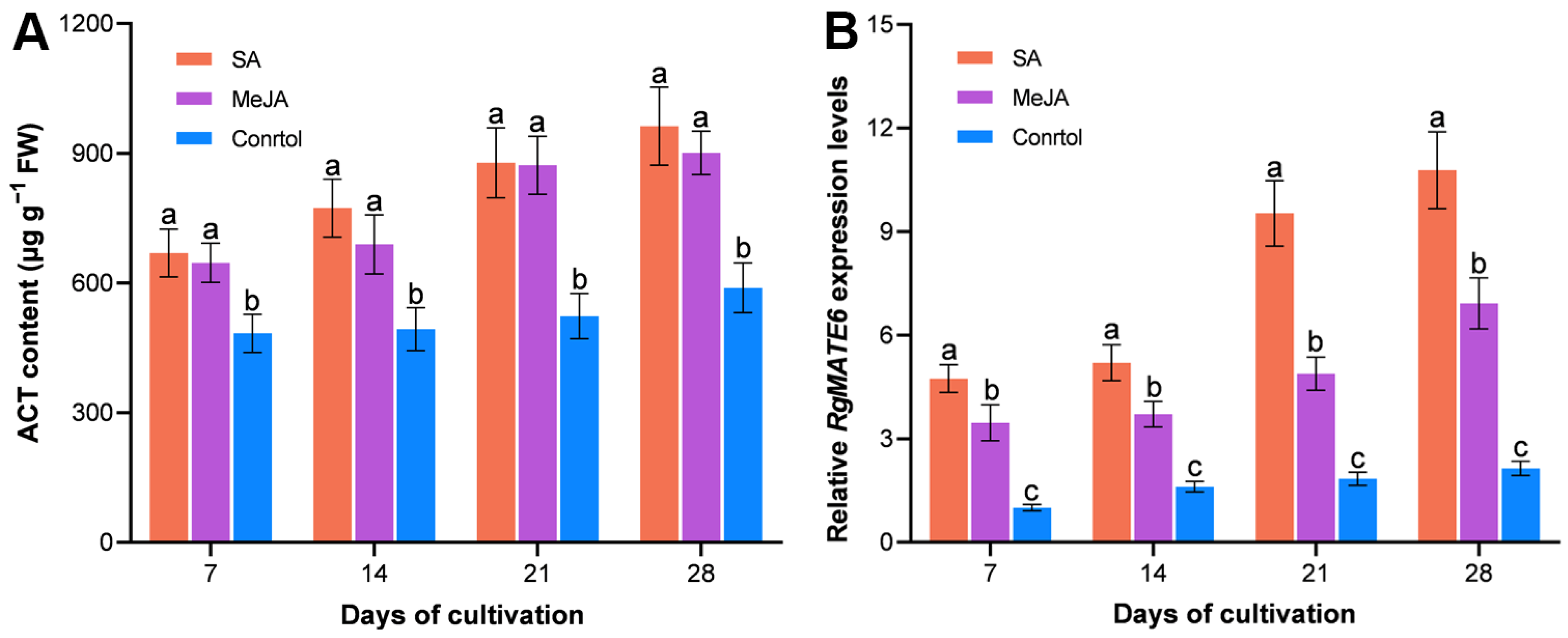
Given the evidence from previous studies that salicylic acid (SA) and methyl jasmonate (MeJA) as elicitors enhance ACT production [8,36], further investigations were conducted to study the effect of SA or MeJA treatment on the content of ACT and RgMATE6 expression levels in leaves. The treatment of leaves resulted in significant differences in ACT accumulation compared to controls, with SA-treated leaves having the highest ACT levels, followed by MeJA-treated leaves. Following the 28-day treatment period, the content of ACT in SA- and MeJA-treated leaves was found to be 1.64- and 1.53-fold higher than in the control group, respectively. A similar trend was observed in RgMATE6 expression levels, with SA- and MeJA-treated leaves exhibiting approximately 5- and 3-fold higher expression levels compared to the control group after 28 days of treatment (Figure 3 and Figure S5). The results indicated a strong correlation between ACT accumulation and RgMATE6 expression levels (r > 0.86 across treatments) (Table S1).
Collectively, these findings suggest that RgMATE6 could be a key transporter of HPGs in R. glutinosa, with its expression level strongly coupled to both tissue-specific and phytohormone-induced ACT accumulation.

2.3. RgMATE6 Transports ACT More Efficiently than Other HPGs in the Nicotiana benthamiana System

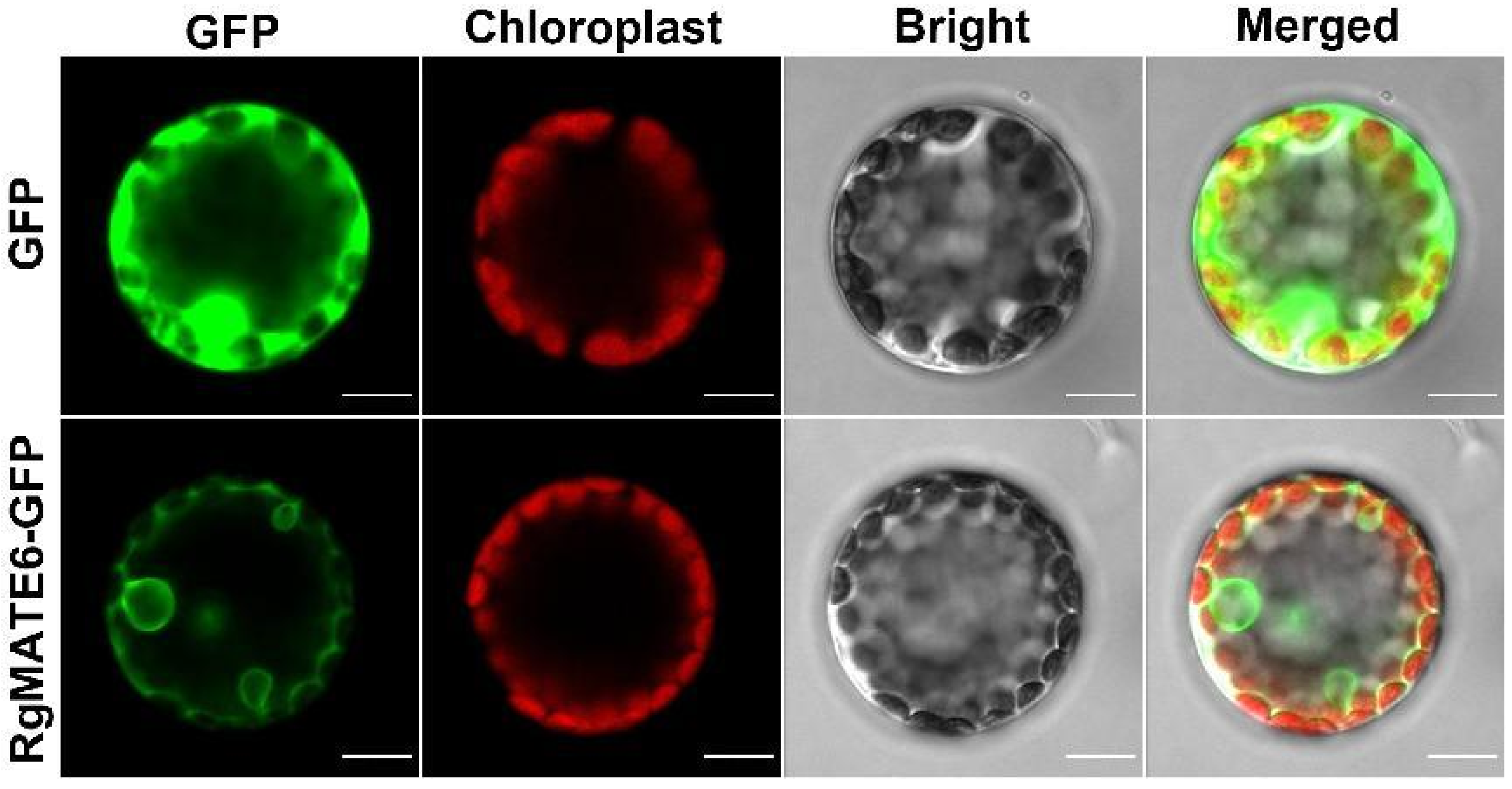
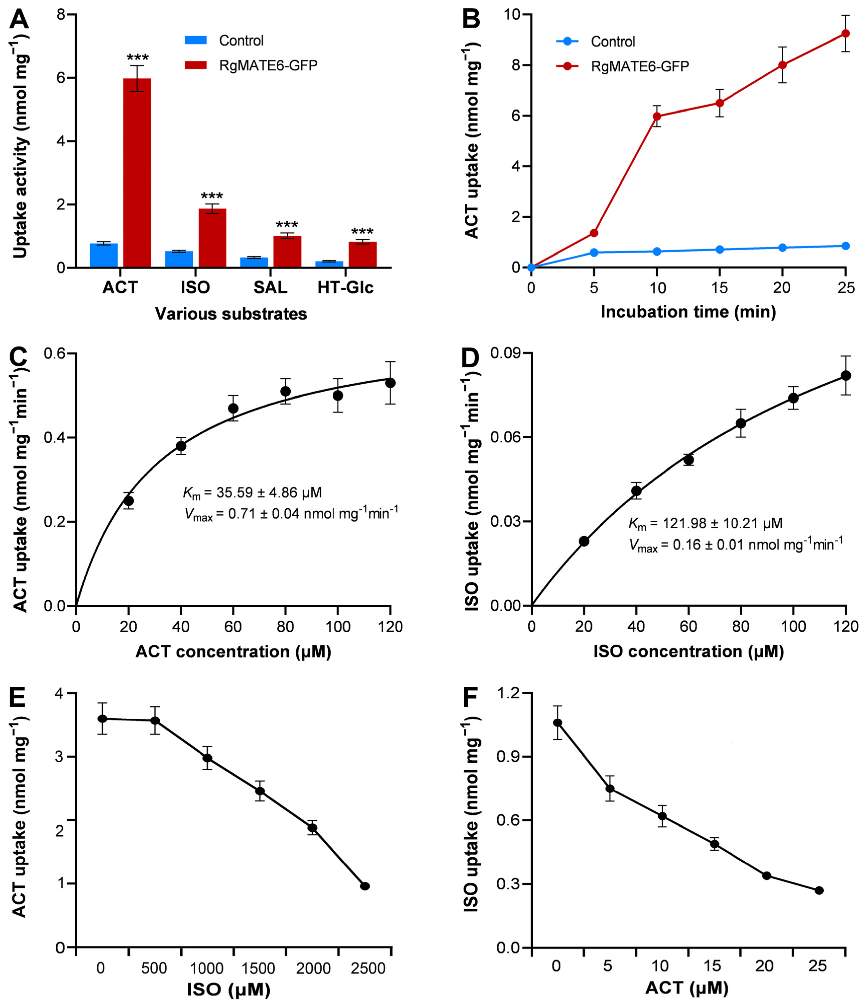
To deepen our understanding of the substrate specificity and preferred substrates of RgMATE6, the constructs pBI121-RgMATE6-GFP (Figure S6A) and the control vector pBI121-GFP (control) were transiently expressed in protoplasts isolated from the mesophyll cells of N. benthamiana. A distinct GFP fluorescence signal was observed in protoplasts expressing RgMATE6-GFP, localizing to the vacuolar membrane. In contrast, the control GFP signal was predominantly detected within the cytosol (Figure 4). These findings provide unequivocal confirmation of the successful expression and precise localization of the RgMATE6-GFP protein at the vacuolar membrane in plant cells, as previously mentioned [34]. Subsequently, the uptake activities from the vacuole membrane vesicles expressing RgMATE6-GFP and GFP (control) were analyzed using the four HPG compounds after incubation for 20 min. A notable enhancement in uptake activity was observed for all four HPGs in vacuole membrane vesicles expressing RgMATE6-GFP, in comparison to the control (Figure 5A). Specifically, ACT displayed the highest uptake activity, followed by ISO and SAL, whereas the uptake of HT-Glc by RgMATE6-GFP exhibited the lowest activity. The time-course analysis showed a significant increase in the HPG uptake amounts from the vacuole membrane vesicles expressing RgMATE6-GFP compared to the control (Figure 5B). Kinetic analyses further revealed a Km of 35.59 μM for ACT uptake, alongside a Vmax of 0.71 nmol mg−1 min−1. Additionally, the uptake of ISO by RgMATE6-GFP exhibited a Km of 121.98 μM and a Vmax of 0.16 nmol mg−1 min−1 (Figure 5C,D). These results indicate that RgMATE6 possesses distinct transport activities towards the tested HPGs, with optimal efficiency observed for ACT.
To further explore the substrate preferences of RgMATE6, a competition assay was performed. These data revealed that ISO inhibited the uptake of ACT only at the higher concentrations (Figure 5E), whereas ACT impeded the uptake of ISO at the lower concentrations (Figure 5F). These results suggested a notable competitive interaction between the import of ISO and ACT. These findings provide compelling evidence that RgMATE6 transports ACT more efficiently than the other three HPGs tested.

2.4. RgMATE6 Facilitates the Transport of ACT into Vacuoles in R. glutinosa

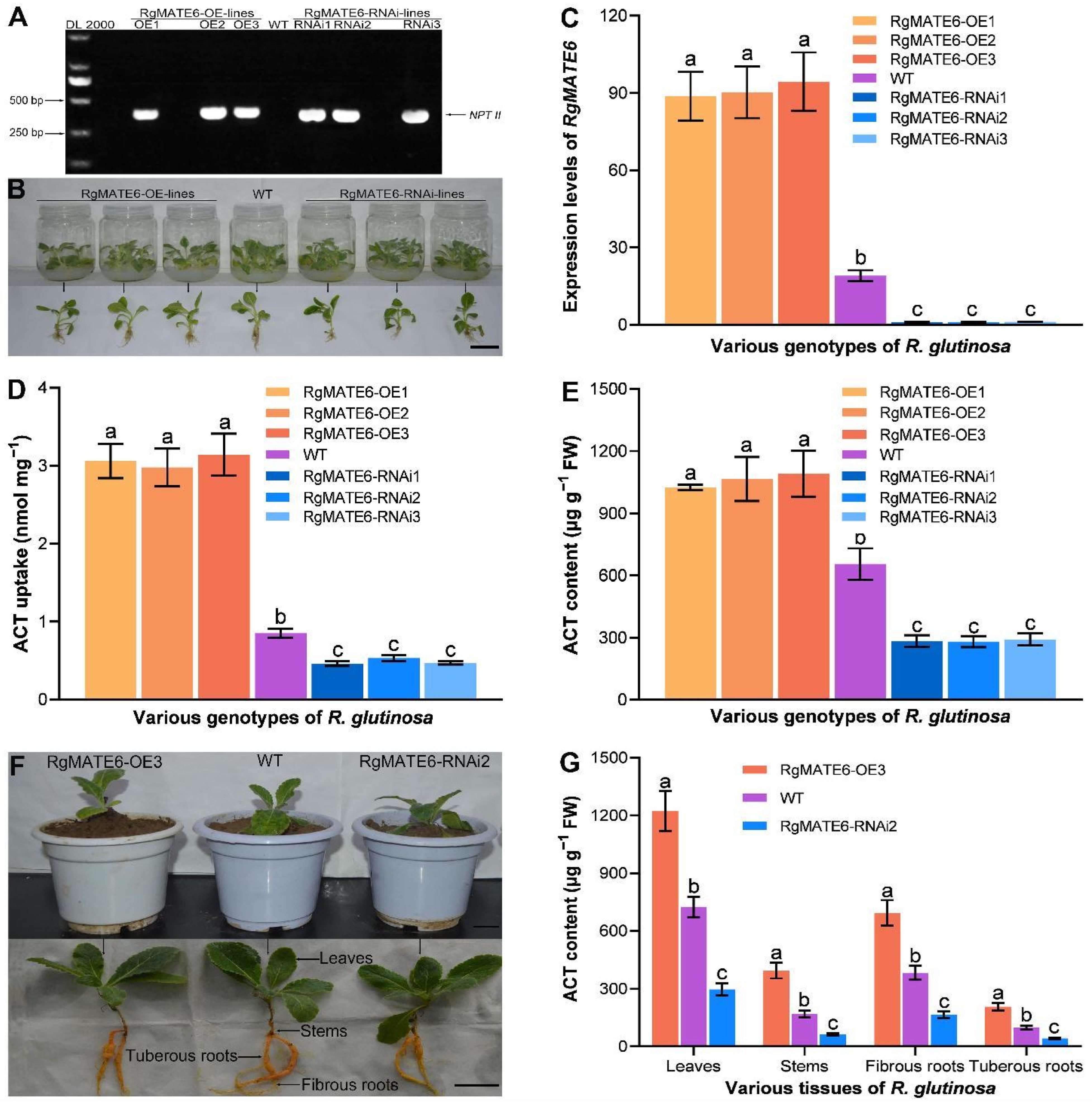
To investigate the molecular function of RgMATE6 in R. glutinosa, we generated RgMATE6-overexpressing (RgMATE6-OE) and RgMATE6-RNAi transgenic lines using the CaMV35S promoter for gene overexpression and repression, respectively (Figure S6B,C). Successful generation of these transgenic lines was confirmed (Figure 6 and Figure S7A–H). PCR analysis of transgenic shoots and wild-type (WT) plants detected a 385 bp fragment of the NPTII gene (conferring kanamycin resistance) exclusively in the transgenic lines, confirming their genetic modification (Figure 6A). These lines were then propagated clonally via shoot culture. After 30 days of sterile cultivation, we performed qRT-PCR analysis on the leaves (Figure 6B). The results showed significantly higher RgMATE6 expression in RgMATE6-OE lines and reduced expression in RgMATE6-RNAi lines compared to the WT control (Figure 6C).
To validate the transport activity of RgMATE6 in R. glutinosa, vacuolar membrane vesicles isolated from the transgenic and WT seedling leaves were incubated with ACT for 20 min. As shown in Figure 6D, the ACT uptake amount in vesicles from RgMATE6-OE lines was significantly higher than that in WT controls. For example, RgMATE6-OE3 exhibited a 3.7-fold increase in ACT uptake amount compared to WT. Conversely, the vesicles from RgMATE6-RNAi lines showed markedly reduced uptake activity. For example, RgMATE6-RNAi2 displayed only 62% of the uptake levels of WT. These results demonstrate that RgMATE6 overexpression enhances ACT vacuolar sequestration, while its repression suppresses transport efficiency in R. glutinosa.

2.5. Genetic Manipulation of RgMATE6 Modifies ACT Production in R. glutinosa by Facilitating Vacuolar Import

To assess the function of RgMATE6 in vivo, the ACT content was quantified in the leaves of transgenic R. glutinosa seedlings (Figure 6E). The RgMATE6-OE lines showed a significant increase in ACT content compared to the WT control. For example, the ACT content in the leaves of RgMATE6-OE3 was approximately 1.7-fold higher than that of the WT. Conversely, the RgMATE6-RNAi lines exhibited a significant decrease in ACT content compared to the WT. For instance, the ACT content in the seedlings of RgMATE6-RNAi2 lines was approximately 50% of that in the WT. These results demonstrate that overexpression of RgMATE6 enhances ACT accumulation, while its repression diminishes it.
To validate these findings, we cultivated the RgMATE6-OE3 and RgMATE6-RNAi2 lines in pots filled with soil for 30 days, followed by ACT quantification across various tissues. Overall, the RgMATE6-OE3 plants showed significantly higher ACT levels in all tissues compared to the WT plants (Figure 6F,G). For example, the leaves of RgMATE6-OE3 exhibited approximately 1.7-fold higher ACT content, while the tuberous roots showed a 2.1-fold increase relative to the WT. In contrast, the RgMATE6-RNAi2 plants exhibited significantly lower ACT content. The leaves and tuberous roots exhibited approximately 40% of the WT levels. These data confirm RgMATE6’s role in ACT accumulation and reveal the tissue-specific regulatory variation.

2.6. RgMATE6 Shows Positive Coordination with the Expression of ACT Biosynthetic Genes in R. glutinosa

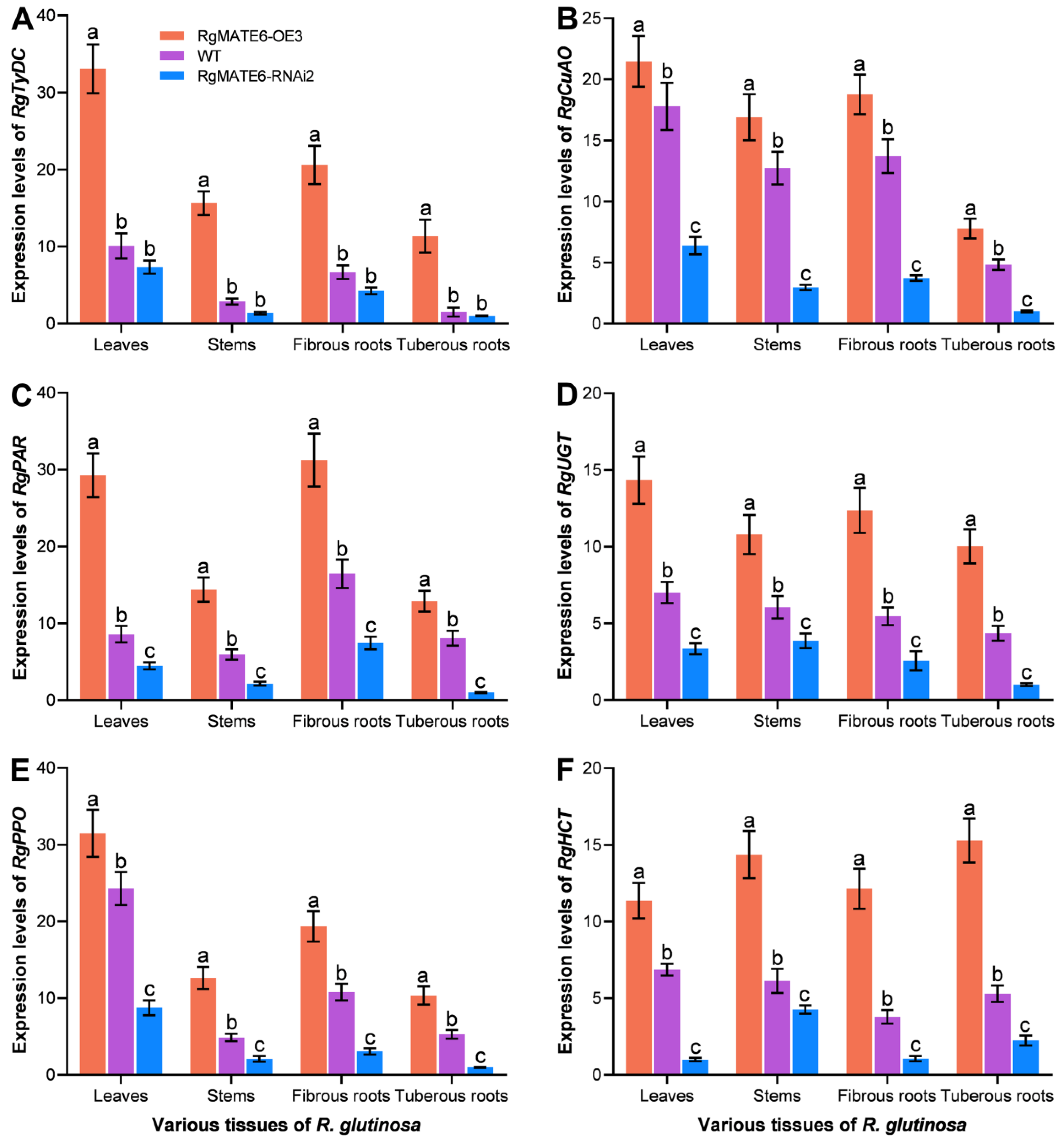
To examine potential relationships between RgMATE6 and ACT biosynthesis, we compared tissue-specific expression profiles of six key ACT biosynthetic genes (RgTyDC encoding tyrosine decarboxylase, RgCuAO copper amine oxidase, RgPAR phenylacetaldehyde reductase, RgPPO polyphenol oxidase, RgUGT UDP-glucose glucosyltransferase, and RgHCT shikimate O-hydroxycinnamoyltransferase) [8,20] across three R. glutinosa genotypes at 30 days of cultivation. RgMATE6-OE3 plants showed significantly elevated expression of all six genes across tissues compared to WT controls, while RgMATE6-RNAi2 plants exhibited substantially reduced expression levels in corresponding tissues (Figure 7). For example, RgPAR expression in RgMATE6-OE3 leaves and fibrous roots increased by 3.5-fold and 1.9-fold, respectively, relative to WT. RgHCT expression displayed a similar pattern with approximately 1.7-fold upregulation in RgMATE6-OE3 plants. In contrast, RgPAR expression in RgMATE6-RNAi2 leaves and fibrous roots decreased to 50% of WT levels, with RgHCT expression reduced to 70% of WT levels. These results reveal coordinated expression patterns between RgMATE6 and ACT biosynthetic genes in R. glutinosa.

3. Discussion

In R. glutinosa, ACT, a pivotal HPG, manifests well-documented bioactivities [8,9]. Consequently, enhancing ACT biosynthesis in this medicinal plant holds significant agricultural and pharmacological value. Building on a previous study that identified RgMATE6 and RgMATE12 as potential vacuolar transporters in R. glutinosa [34], our phylogenetic and homology analyses revealed that RgMATE6 shares closer evolutionary relationships and higher sequence identity with VvAM3 (an anthocyanin transporter from V. vinifera) [37] and MtMATE2 (a flavonoid glycoside transporter from M. truncatula) [32] compared to RgMATE12. These findings suggest that RgMATE6 may function in phenolic transport. In contrast, RgMATE12 shows homology to transporters of non-phenolic metabolites, such as NtJAT2 (an alkaloid transporter from N. tabacum) [38], CjMATE1 (an alkaloid transporter from Coptis japonica) [39], and GmMATE1 (a terpenoid transporter from Glycine max) [33], implying potential roles in alkaloid or terpenoid transport. Previous studies have reported that the production of plant-specialized metabolites shows a positive correlation with transcript levels of genes encoding MATE transporter proteins responsible for their vacuolar sequestration [25,31]. In this work, tissue-specific correlation analysis demonstrated a stronger positive association between the HPG accumulation and RgMATE6 expression than with RgMATE12 expression. Furthermore, treatment with metabolic elicitors SA and MeJA [40] produced a pronounced elevation in ACT production and the upregulation of RgMATE6 expression. Collectively, these results imply that RgMATE6 may contribute to ACT transport and vacuolar accumulation in R. glutinosa, though direct functional validation remains required.
The transformation of mesophyll cells from N. benthamiana has been commonly used as a functional expression system for plant vacuolar transporters in previous studies [23,41]. To characterize RgMATE6’s substrate specificity/preference, RgMATE6-GFP was expressed in N. benthamiana cells, which confirmed vacuolar membrane localization. Transport assays using vacuolar membrane vesicles demonstrated RgMATE6’s capacity to mediate HPG transport, with the highest efficiency for ACT. Competitive uptake experiments between ISO and ACT further confirmed RgMATE6’s preferential affinity for ACT. Most importantly, ACT uptake assays using vacuolar membrane vesicles from transgenic R. glutinosa plants verified RgMATE6’s essential role in ACT transport. Collectively, these results from N. benthamiana, and R. glutinosa systems consistently demonstrate that RgMATE6 functions to efficiently transport ACT into vacuoles at the molecular level.
Plant vacuoles are well-known as central storage compartments for crucial secondary metabolites within plant cells [23,24,25]. Having demonstrated through in vitro assays that RgMATE6 facilitates the import of ACT into the vacuoles, we next sought to elucidate its molecular function in vivo. We found that overexpression of RgMATE6 enhanced ACT accumulation, while suppression of RgMATE6 reduced accumulation in R. glutinosa seedlings and various tissues. These in vivo results not only confirmed our in vitro findings but also revealed tissue-specific accumulation patterns. This demonstrates that RgMATE6 positively regulates ACT production in R. glutinosa. MATE transporters typically mediate metabolite transport across the tonoplast into the vacuolar lumen [29,31]. Our results provide compelling evidence that RgMATE6 serves as a principal transporter governing ACT accumulation in R. glutinosa.
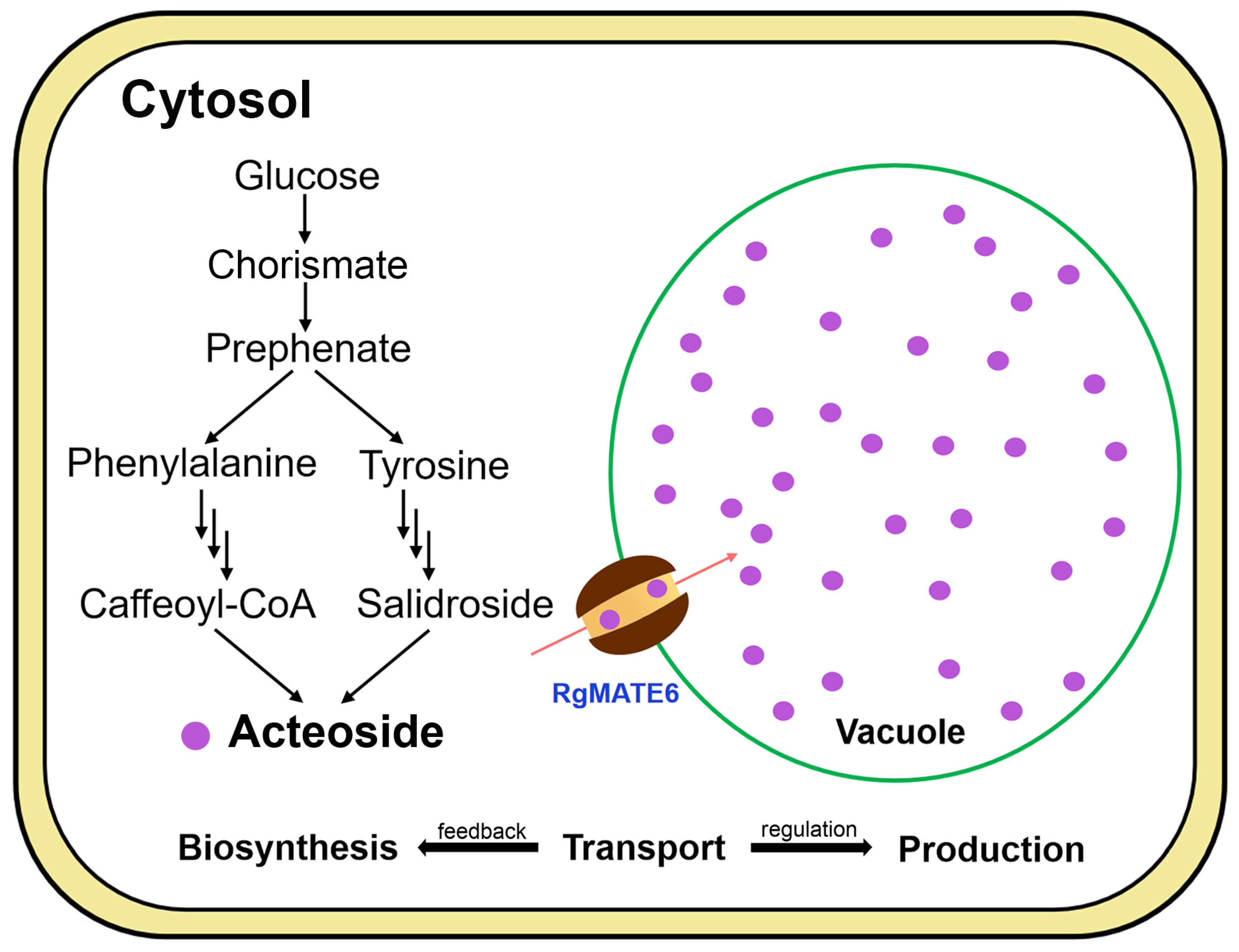
MATE transporters have been widely demonstrated to act as key regulators of secondary metabolite biosynthesis in plants [29,30,32]. Our study demonstrated that RgMATE6 overexpression upregulated ACT biosynthetic genes, whereas its repression downregulated their expression, implying its potential association with the ACT biosynthetic pathway. The coordinated processes of secondary metabolite biosynthesis, transport, and accumulation involve MATE transporters in feedback regulation mechanisms in plants [24,31,42]. In this study, RgMATE6 was shown to positively coordinate the expression of the ACT biosynthetic genes, speculating its critical role in tuning a positive feedback that may enhance ACT biosynthesis. Furthermore, RgMATE6-mediated transport of ACT into the vacuole highlights the integrated regulation of these processes for optimal metabolite production, as previously reported [24,25,27,33]. Thus, RgMATE6 enhances ACT production by both promoting vacuolar accumulation and possibly synergizing with the biosynthetic pathway (Figure 8). These findings provide novel insights into the ACT production mechanisms and potential strategies for enhancing valuable HPG production through genetic or biotechnological approaches.
ACT represents a key example among the diverse HPGs produced by plants. However, the functional characteristics of other MATE transporters involved in HPGs transport remain incompletely understood. Therefore, additional studies are needed to identify other transporters mediating HPG import, as this would significantly advance our understanding of the regulatory networks controlling the HPG biosynthesis in plants. Investigating alternative transport systems may lead to more efficient approaches for utilizing plant transporter resources, potentially enabling the development of novel methods and targeted strategies to enhance the production of both ACT and other valuable HPGs in plants.

4. Materials and Methods

4.1. In Silico Analysis

For phylogenetic analysis, RgMATE6 (GenBank ID: QBM79436.1) and RgMATE12 (QBM79442.1), along with other reported plant MATE proteins, were retrieved through BLASTP searches against the NCBI database (https://blast.ncbi.nlm.nih.gov/Blast.cgi, accessed on 16 November 2025). Redundant sequences were removed using CD-HIT [43] with a 90% identity cutoff. From the retained unique sequences, functionally characterized plant vacuolar MATE proteins involved in metabolite transport were selected through manual curation. Sequence alignment was performed using ClustalW in MEGA software package, version 12.0 [44,45], with gap opening and extension penalties set to 10.00 and 0.10 for both pairwise and multiple alignments. Gappy or poorly aligned regions were manually inspected and trimmed to improve alignment quality. Phylogenetic reconstruction was conducted using the maximum likelihood method in MEGA12 with the Jones–Taylor–Thornton (JTT) model and 1000 bootstrap replicates. Additionally, multiple sequence alignment for RgMATE protein identity analysis was performed using DNAMAN 7.0 software.

4.2. Cultivation of R. glutinosa, RNA Extraction, and cDNA Synthesis

The seedlings of R. glutinosa cultivar ‘Wen 85-5’ were aseptically propagated in glass tubes containing 50 mL of solidified Murashige and Skoog (MS) medium (Solarbio, Beijing, China), with 3% sucrose supplementation. The seedlings were grown in a controlled-environment greenhouse (26 °C, 14 h light/10 h dark) photoperiod until reaching the six-leaf developmental stage. Subsequently, the young plants were moved to pots containing organic nutrient-rich soil and cultivated in the greenhouse at 65% relative humidity. For the purpose of conducting a tissue-specific analysis, roots, stems and leaves of the plants were harvested at 5, 10, 15, and 20 days of cultivation, respectively. For elicitor treatments, the plants at 10 days post-transplantation were irrigated every four days with 100 mL of water (control), 20 μM SA, or 20 μM MeJA, respectively. Leaf samples from each treatment group were collected at 7, 14, 21, and 28 days after treatment initiation (Figure S5). Each experimental group comprised three biological replicates.
To ensure high-quality RNA extraction, all samples were immediately flash-frozen in liquid nitrogen. The total RNA was subsequently isolated employing TRIzol reagent (Invitrogen, Carlsbad, CA, USA), following the manufacturer’s instructions. The RNA yield and quality were measured with a NanoDrop 2000 system (Thermo Scientific, Wilmington, DE, USA). RNA integrity was checked by 1% agarose gel electrophoresis. cDNA was generated from 1 μg RNA using HiScript III Reverse Transcriptase (Vazyme, Nanjing, China) and oligo-dT primers. Subsequent to this, the synthesized cDNA was diluted 1:5 (v/v) for downstream applications.

4.3. Quantitative Real-Time PCR Analysis

Gene-specific primers were computationally designed (Beacon Designer), with sequences provided in Table S2. Gene expression was quantified by quantitative real-time PCR (qRT-PCR) with SYBR Green Master Mix (Fermentas) using a Bio-Rad iQ5 system, in accordance with established protocols [46]. Relative gene expression levels were calculated using RgActin as the internal control. The 2−ΔΔCT algorithm [47] was employed for gene expression analysis, incorporating triplicate biological and technical replicates.

4.4. Measurement of the HPGs in R. glutinosa

The contents of the HPGs, including ACT, ISO, SAL, and HT-Glc (MedChemExpress, Shanghai, China; HPLC-determined purity >98%), were quantified using the following protocol: 0.5 g of fresh sample was ground into a fine powder and then extracted with 10 mL of methanol for 5 h at room temperature. To enhance the extraction process, sonication in an ice bath was employed for a cumulative time of 5 min, with intermittent sonication cycles of 10 s on and 5 s off. Following 12,000× g centrifugation, the collected pellet was lyophilized and redissolved in 5 mL of 30% (v/v) aqueous acetonitrile. The sample was passed through a 0.22-μm sterile filter for purification before HPLC measurement. HPG content was determined using an Agilent 1260 series HPLC instrument (Agilent Technologies, Santa Clara, CA, USA). A series of standard solutions at seven different concentrations was prepared by diluting the certified reference materials for each analyte (ACT, ISO, SAL, and HT-Glc). The concentration ranges were 0–32 mg L−1 for ACT and ISO and 0–15 mg L−1 for SAL and HT-Glc. Each calibration curve was constructed by plotting the peak area against the corresponding concentration, and all showed excellent linearity with a coefficient of determination greater than 0.995. The mobile phase for ACT consisted of 30% acetonitrile and 70% water with 0.1% phosphoric acid. For both ISO and SAL analyses, the mobile phase contained 20% pure acetonitrile mixed with 80% water with 0.1% phosphoric acid. HT-Glc separation employed a mobile phase formulation of 33% acetonitrile combined with 67% water with 0.1% phosphoric acid. The chromatographic separation was performed at a constant flow rate of 0.8 mL per minute. For analyte detection, different wavelengths were optimized for each compound: 330 nm for ACT, 334 nm for ISO, 275 nm for SAL, and 280 nm for HT-Glc, while keeping the analytical column at 30 °C throughout the analysis. Quantitative determination of the HPG levels, expressed in micrograms per gram of fresh weight, was achieved through external calibration with certified reference materials. Each experimental group included triplicate biological samples.

4.5. Construction of Various Expression Vectors

For RgMATE6 constructs, the gene fragments, including complete and truncated open reading frames (ORFs), were PCR-amplified employing Phanta MaX Super-Fidelity DNA Polymerase (Vazyme, Nanjing, China) with specific primer pairs detailed in Table S3. For transient expression in N. benthamiana, the full-length RgMATE6 ORF was ligated into the XhoI site of the pBI121-GFP vector (Miaoling Biology, Wuhan, China), producing a CaMV35S promoter-driven C-terminal GFP fusion construct (pBI121-RgMATE6-GFP; Figure S6A). The full-length RgMATE6 coding sequence was directionally cloned into pBI121-GFP (Miaoling, Wuhan, China) using KpnI/XhoI restriction sites, generating a CaMV35S-driven overexpression vector containing the NPTII kanamycin resistance gene. (RgMATE6-OE; Figure S6B). For RNAi vector construction, a partial RgMATE6 ORF fragment was cloned into the BsaI site of pRNAi-GG (Biovector Science Lab, Beijing, China), generating the RgMATE6-RNAi construct with CaMV35S promoter and NPTII selection marker (Figure S6C). All recombinant plasmids were introduced into Escherichia coli DH5α, with correct clones identified by DNA sequencing (Sangon, Shanghai, China).

4.6. N. benthamiana Transformation and Vacuolar Membrane Vesicle Preparation

Leaf mesophyll protoplasts were extracted from six-leaf stage N. benthamiana plants following an adapted version of the Arabidopsis protoplast isolation method [48]. The pBI121-RgMATE6-GFP and pBI121-GFP (control) constructs were transiently transformed into protoplasts via polyethylene glycol-mediated transfection. After 36–48 h incubation at 25 °C, RgMATE6 subcellular localization was analyzed by confocal microscopy (Olympus Corporation, Tokyo, Japan), as aforementioned.
Subsequently, vacuolar membrane vesicles were prepared from the transformed protoplasts following a modified protocol described by Yamanishi and Kasamo [49]. Briefly, the protoplast suspension was prepared in homogenization buffer composed of 0.1 M mannitol, 1.5 mM MES at pH 5.6, 154 mM NaCl, 125 mM CaCl2, 5 mM KCl, and 5 mM glucose. Following centrifugation (50,000× g, 30 min, 4 °C), the microsomal pellet was resuspended in 36 mL sucrose buffer (0.6 M sucrose, 10 mM KCl, 1 mM EDTA, 2 mM DTT) and layered with 3.5 mL sorbitol buffer (0.25 M sorbitol, 5 mM HEPES-Tris pH 7.5, 1 mM EDTA, 2 mM DTT). After high-speed centrifugation (100,000× g, 40 min), the vacuolar membrane interface was collected and preserved in storage solution (0.3 M sorbitol, 0.1 M KCl, 5 mM Tris-MES pH 7.5, 1 mM DTT with protease inhibitors) at −80 °C.

4.7. Generation of RgMATE6 Transgenic Lines of R. glutinosa and the Isolation of Vacuolar Membrane Vesicles

Both RgMATE6-OE and RgMATE6-RNAi vectors were delivered into Agrobacterium tumefaciens GV3101 through freeze–thaw transformation [50]. For the genetic modulation of RgMATE6 expression, surface-sterilized leaf explants from R. glutinosa seedlings were infected with the engineered Agrobacterium strains following established protocols [51]. To verify transgenic integration, putative transgenic shoots (originating from distinct explants subjected to independent Agrobacterium co-cultivation steps) were generated on the MS basal medium (Solarbio, Beijing, China) supplemented with additions of 30 g L−1 sucrose, 0.5 mg L−1 NAA, 3 mg L−1 6-BA, 100 mg L−1 kanamycin, 200 mg L−1 timentin, and 7.8 g L−1 agar. The cultures were maintained under the controlled greenhouse conditions as mentioned above. Genomic DNA was extracted from both transgenic shoots and WT controls employing the CTAB isolation protocol [52]. Transgenic plants were validated by NPTII marker amplification. For further analysis, each positive transgenic line was propagated aseptically through shoot culture to generate multiple clonal plants. Leaves from these transgenic plants after 30 days of culture were used to analyze RgMATE6 expression levels by qRT-PCR, measure ACT uptake in vacuolar membrane vesicles as described above, and quantify ACT content by HPLC analysis.

4.8. Transport Activity Assays

The transport capacity of vacuolar membrane vesicles was assessed at 25 °C according to an optimized protocol [53]. The reaction system, with a total volume of 500 μL, chilled buffer containing 0.4 M sorbitol and 25 mM Tris-MES (pH 8.0) and 0.1% (w/v) BSA, and four substrates (each at 100 μM): ACT, ISO, SAL and HT-Glc. Subsequent to the incorporation of vesicle addition (approximately 50 μg of membrane protein) to initiate transport, the mixture was vortexed for 10 s. At predetermined intervals (5, 10, 15, 20, and 25 min), 100 μL aliquots were withdrawn and quenched with 1.0 mL of ice-cold termination buffer (0.4 M sorbitol and 25 mM Tris-MES (pH 8.0)). Reaction suspensions were vacuum-filtered via a 0.22 μm filter (pore size). Thereafter, membrane-trapped analytes were extracted in 1 mL of methanol–water mixture (1:1, v/v) using orbital shaking (180 rpm, 2 h, 25 °C). The resulting eluates were then analyzed by HPLC, following our established chromatographic parameters. All experimental procedures included three independent replicates.
To determine the transport kinetic parameters, transport assays were conducted in a 500 μL reaction system: substrate at gradient concentrations (20–120 μM), pre-cooled buffer as described above, and vesicle preparation were sequentially added to initiate the reaction. Following an incubation period at 25 °C for 20 min, the reaction mixture was vacuum-filtered through a 0.22 μm membrane filter, followed by quantitative analysis of the HPG transport activity using the aforementioned HPLC method. Kinetic parameters, including Km and Vmax, were derived from Lineweaver–Burk plots, with all assays performed in triplicate.
For competition assays using vacuolar membrane vesicles from N. benthamiana, ACT or ISO was utilized as the uptake substrate at a concentration of 100 µM, with various concentrations of their corresponding competition substrates. Following an incubation of 20 min at 25 °C, the transported amounts of ACT or ISO were measured using HPLC.

4.9. Statistical Analysis

All experimental data were statistically analyzed using the statistical package SPSS v. 20.0 (IBM, NY, USA). Gene expression and HPG content differences in R. glutinosa were evaluated by one-way ANOVA. For post hoc analysis, we employed both Fisher’s least significant difference test and Duncan’s multiple range test with a 0.05 significance level to detect group differences. Variable correlations were assessed by Pearson’s method, and both the correlation coefficient and the coefficient of determination are reported (p < 0.05). We employed Student’s independent t-tests to compare transport activities between experimental treatments and controls (p < 0.01 or 0.001). The error bars depicted in all figures represent standard deviations (SD) calculated from a minimum of three biological or technical replicates (n = 3).

5. Conclusions

This study provides a comprehensive understanding of the role of RgMATE6 in the transport and biosynthesis of ACT. In silico analysis identified RgMATE6 as a putative phenolic compound transporter, which was further supported by correlation analyses linking its expression to HPG accumulation in R. glutinosa. In vivo assays confirmed RgMATE6 as an HPG transporter with marked specificity for ACT. Functional characterization revealed that RgMATE6 not only mediates ACT vacuolar import but might also synergize with its biosynthetic pathway. Additionally, tissue-specific expression profiling of ACT biosynthesis-related genes in transgenic plants suggested that RgMATE6 positively regulates ACT biosynthesis. Collectively, these findings demonstrate that RgMATE6 enhances ACT production through vacuolar transport/accumulation and potential coordination with biosynthesis via a feedback loop. This work advances the understanding of HPG regulation in plants and highlights biotechnological opportunities for targeted metabolite production.

Supplementary Materials

The following supporting information can be downloaded at: https://www.mdpi.com/article/10.3390/plants14233608/s1, Figure S1: Multiple sequence alignment of RgMATE6 and RgMATE12 with homologs from Arabidopsis thaliana, Vitis vinifera, Vaccinium corymbosum and Medicago truncatula; Figure S2: The curves for ACT, ISO, SAL and HT-Glc were established by plotting the peak area against the concentration of the analytical standards. The regression equation, coefficient of determination (R2), and linear range for each compound are provided in the graph; Figure S3: HPLC chromatogram samples of HPG contents in the leaves of R. glutinosa at 20 days of cultivation; Figure S4: Scatter plot analysis of the correlation between the content of four HPGs and the expression of RgMATE6; Figure S5: R. glutinosa seedlings at various days after SA and MeJA treatments (scale bar = 4 cm); Figure S6: The constructs for various RgMATE6 vectors. These include the CaMV35S: RgMATE6-GFP (A), RgMATE6-OE (B) and RgMATE6-RNAi (C) constructs; Figure S7: Generation of transgenic RgMATE6 R. glutinosa plants. (A) Co-cultivation of the explants and Agrobacterium tumefaciens; (B) and (C) the callus induction/selection medium from the explants-transferred; (D) the adventitious shoots induction/selection medium from the callus; (E) and (F) the transgenic seedlings were generated in the selection medium; (G) and (H) acclimatization to organic and field soils (scale bars = 1 cm); Table S1: The correlation between the RgMATE6 expression levels and the ACT content of R. glutinosa under various treatments was quantified by the Pearson correlation coefficient (r). Note: “*” represent significantly different levels of 0.05, respectively; Table S2: Primer sequences used for qRT-PCR analysis; Table S3: The primer sequences of RgMATE6 used to constructs, and the primer sequences of positive screened gene in R. glutinosa. Note: the lowercases represent the sequences of these vector adapters.

Author Contributions

Formal analysis, Y.Y.; Funding acquisition, Y.Y. and M.L.; Project administration, Y.Y.; Resources, Y.Y. and Z.Z.; Software, Y.L., Y.W., W.W., F.S. and M.Y.; Writing—Review and editing, Y.Y., Y.L. and R.L.; All authors have read and agreed to the published version of the manuscript.

Funding

This work was supported by grants from the National Natural Science Foundation of China (No. 82274049 and 81973417) and the Key Research and Development and Promotion Special Project of Henan Province (No. 242102311169).

Data Availability Statement

The data sets generated and analyzed in this study are available from the corresponding author upon reasonable request.

Conflicts of Interest

There are no conflicts of interest.

Abbreviations

The following abbreviations are used in this manuscript:
HPGHydroxytyrosol-type phenylethanol glycoside
R. glutinosaRehmannia glutinosa
N. benthamianaNicotiana benthamiana
ORFsOpen reading frames
ACTActeoside
ISOIsoacteoside
SALSalacteoside
HT-GlcHydroxytyrosol-O-glucoside
SASalicylic acid
MeJAMethyl jasmonate
A. thalianaArabidopsis thaliana
RgTyDCTyrosine decarboxylase
RgCuAOCopper amine oxidase
RgPARPhenylacetaldehyde reductase
RgPPOPolyphenol oxidase
RgUGTUDP-glucose glucosyltransferase
RgHCTShikimate O-hydroxycinnamoyltransferase

References

  1. Liu, Y.; Yang, L.; Wang, J.; Chen, D. New lignans and phenylethanoid with antioxidant activity from aerial parts of Forsythia suspensa (Thunb.) Vahl. Nat. Prod. Res. 2023, 37, 725–733. [Google Scholar] [CrossRef]
  2. Chen, R.; Fan, J.; Wu, Y.; Huang, X.; Zhang, W.; Xu, Y.; Zhang, Y.; Li, L.; Wang, C.; Yu, M.; et al. Strobilanthes sarcorrhiza root phenolic extract prevent diabetic nephropathy in mice by regulating NF-κB/IL-1β signaling and glycerophospholipid metabolism. J. Pharmaceut. Biomed. 2025, 253, 116534. [Google Scholar] [CrossRef]
  3. Wu, L.; Georgiev, M.I.; Cao, H.; Nahar, L.; El-Seedi, H.R.; Sarker, S.D.; Xiao, J.; Lu, B. Therapeutic potential of phenylethanoid glycosides: A systematic review. Med. Res. Rev. 2020, 40, 2605–2649. [Google Scholar] [CrossRef] [PubMed]
  4. Pruccoli, L.; Nicolini, B.; Lianza, M.; Teti, G.; Falconi, M.; Tarozzi, A.; Antognoni, F. Antioxidant and anti-melanogenesis effects of Teucrium chamaedrys L. cell suspension extract and its main phenylethanoid glycoside in B16–F10 cells. Plants 2024, 13, 808. [Google Scholar] [CrossRef]
  5. Kurt-Celep, I.; Zheleva-Dimitrova, D.; Sinan, K.I.; Uba, A.I.; Nilofar; Mahomoodally, M.F.; Aumeeruddy, M.Z.; Cakilcioglu, U.; Dall’Acqua, S.; Zengin, G. Uncovering chemical profiles biological potentials and protection effect against ECM destruction in H2O2-treated HDF cells of the extracts of Stachys tundjeliensis. Arch. Pharm. 2024, 357, e2300528. [Google Scholar] [CrossRef] [PubMed]
  6. Tian, X.Y.; Li, M.X.; Lin, T.; Qiu, Y.; Zhu, Y.T.; Li, X.L.; Tao, W.D.; Wang, P.; Ren, X.X.; Chen, L.P. A review on the structure and pharmacological activity of phenylethanoid glycosides. Eur. J. Med. Chem. 2021, 209, 112563. [Google Scholar] [CrossRef]
  7. Yang, Y.; Xi, D.; Wu, Y.; Liu, T. Complete biosynthesis of the phenylethanoid glycoside verbascoside. Plant Commun. 2023, 4, 100592. [Google Scholar] [CrossRef]
  8. Wang, F.; Zhi, J.; Zhang, Z.; Wang, L.; Suo, Y.; Xie, C.; Li, M.; Zhang, B.; Du, J.; Gu, L.; et al. Transcriptome analysis of salicylic acid treatment in Rehmannia glutinosa hairy roots using RNA-seq technique for identification of genes involved in acteoside biosynthesis. Front. Plant Sci. 2017, 8, 787. [Google Scholar] [CrossRef]
  9. Yang, Y.H.; Yang, M.R.; Zhu, J.Y.; Dong, K.W.; Yi, Y.J.; Li, R.F.; Zeng, L.; Zhang, C.F. Functional characterization of tyrosine decarboxylase genes that contribute to acteoside biosynthesis in Rehmannia glutinosa. Planta 2022, 255, 64. [Google Scholar] [CrossRef]
  10. Cui, Q.; Pan, Y.; Xu, X.; Zhang, W.; Wu, X.; Qu, S.; Liu, X. The metabolic profile of acteoside produced by human or rat intestinal bacteria or intestinal enzyme in vitro employed UPLC-Q-TOF-MS. Fitoterapia 2016, 109, 67–74. [Google Scholar] [CrossRef]
  11. Fuji, Y.; Ohtsuki, T.; Matsufuji, H. Accumulation and subcellular localization of acteoside in sesame plants (Sesamum indicum L.). Acs Omega 2018, 3, 17287–17294. [Google Scholar] [CrossRef]
  12. Alipieva, K.; Korkina, L.; Orhan, I.E.; Georgiev, M.I. Verbascoside—A review of its occurrence (bio)synthesis and pharmacological significance. Biotechnol. Adv. 2014, 32, 1065–1076. [Google Scholar] [CrossRef] [PubMed]
  13. Sciandra, F.; Bottoni, P.; De Leo, M.; Braca, A.; Brancaccio, A.; Bozzi, M. Verbascoside elicits its beneficial effects by enhancing mitochondrial spare respiratory capacity and the Nrf2/HO-1 mediated antioxidant system in a murine skeletal muscle cell line. Int. J. Mol. Sci. 2023, 24, 15276. [Google Scholar] [CrossRef]
  14. Pongkitwitoon, B.; Putalun, W.; Triwitayakorn, K.; Kitisripanya, T.; Kanchanapoom, T.; Boonsnongcheep, P. Anti-inflammatory activity of verbascoside- and isoverbascoside-rich Lamiales medicinal plants. Heliyon 2023, 10, e23644. [Google Scholar] [CrossRef] [PubMed]
  15. Gao, W.; Gao, S.; Zhang, Y.; Wang, M.; Liu, Y.; Li, T.; Gao, C.; Zhou, Y.; Bian, B.; Wang, H.; et al. Altered metabolic profiles and targets relevant to the protective effect of acteoside on diabetic nephropathy in db/db mice based on metabolomics and network pharmacology studies. J. Ethnopharmacol. 2024, 318, 117073. [Google Scholar] [CrossRef]
  16. Bai, P.; Yang, Y.; Tang, J.; Xi, D.; Hao, Y.; Jiang, L.; Yin, H.; Liu, T. High-level sustainable production of complex phenylethanoid glycosides from glucose through engineered yeast cell factories. Metab. Eng. 2025, 87, 95–108. [Google Scholar] [CrossRef]
  17. Zhang, X.; Li, C.; Wang, L.; Fei, Y.; Qin, W. Analysis of Centranthera grandiflora Benth transcriptome explores genes of catalpol acteoside and azafrin biosynthesis. Int. J. Mol. Sci. 2019, 20, 6034. [Google Scholar] [CrossRef]
  18. Saimaru, H.; Orihara, Y. Biosynthesis of acteoside in cultured cells of Olea europaea. J. Nat. Med. 2010, 64, 139–145. [Google Scholar] [CrossRef]
  19. Zhou, Y.; Zhu, J.; Shao, L.; Guo, M. Current advances in acteoside biosynthesis pathway elucidation and biosynthesis. Fitoterapia 2020, 142, 104495. [Google Scholar] [CrossRef]
  20. Yang, Y.; Cao, Y.; Zhu, C.; Jin, Y.; Sun, H.; Wang, R.; Li, M.; Zhang, Z. Functional activities of three Rehmannia glutinosa enzymes: Elucidation of the Rehmannia glutinosa salidroside biosynthesis pathway in Saccharomyces cerevisiae. Gene 2024, 928, 148815. [Google Scholar] [CrossRef]
  21. Luaces, P.; Sánche, R.; Expósito, J.; Pérez-Pulido, A.J.; Pérez, A.G.; Sanz, C. Functional and physiological characterization of tyrosine decarboxylases from Olea europaea L. involved in the synthesis of the main phenolics in Olive fruit and Virgin Olive Oil. Int. J. Mol. Sci. 2024, 25, 10892. [Google Scholar] [CrossRef] [PubMed]
  22. Liu, Y.; Han, X.; Zhao, M.; Liu, L.; Deng, Z.; Zhao, Q.; Yu, Y. Functional characterization of polyphenol oxidase of PPO2 supports its involvement in parallel biosynthetic pathways of acteoside. Plant J. 2024, 119, 927–941. [Google Scholar] [CrossRef] [PubMed]
  23. Zhang, H.; Zhao, F.G.; Tang, R.J.; Yu, Y.; Song, J.; Wang, Y.; Li, L.; Luan, S. Two tonoplast MATE proteins function as turgor-regulating chloride channels in Arabidopsis. Proc. Natl. Acad. Sci. USA 2017, 114, E2036–E2045. [Google Scholar] [CrossRef]
  24. Shao, D.; Li, Y.; Zhu, Q.; Zhang, X.; Liu, F.; Xue, F.; Sun, J. GhGSTF12 a glutathione S-transferase gene is essential for anthocyanin accumulation in cotton (Gossypium hirsutum L.). Plant Sci. 2021, 305, 110827. [Google Scholar] [CrossRef] [PubMed]
  25. Ma, L.; Yuan, J.; Qin, H.; Zhang, M.; Zhang, F.; Yu, F.; Tian, Z.; Wang, G. GmMATE100 is involved in the import of Soyasaponins A and B into vacuoles in soybean plants (Glycine max L.). J. Agric. Food Chem. 2024, 72, 9994–10004. [Google Scholar] [CrossRef]
  26. Gani, U.; Nautiyal, A.K.; Kundan, M.; Rout, B.; Pandey, A.; Misra, P. Two homeologous MATE transporter genes NtMATE21 and NtMATE22 are involved in the modulation of plant growth and flavonol transport in Nicotiana tabacum. J. Exp. Bot. 2022, 73, 6186–6206. [Google Scholar] [CrossRef]
  27. Li, F.; Shahsavarani, M.; Handy-Hart, C.J.; Côté, A.; Brasseur-Trottier, X.; Montgomery, V.; Beech, R.N.; Liu, L.; Bayen, S.; Qu, Y. Characterization of a vacuolar importer of secologanin in Catharanthus roseus. Commu. Biol. 2024, 7, 939. [Google Scholar] [CrossRef]
  28. Gani, U.; Vishwakarma, R.A.; Misra, P. Membrane transporters: The key drivers of transport of secondary metabolites in plants. Plant Cell Rep. 2021, 40, 1–18. [Google Scholar] [CrossRef]
  29. Ma, Y.; Li, D.; Zhong, Y.; Wang, X.; Li, L.; Osbourn, A.; Lucas, W.J.; Huang, S.; Shang, Y. Vacuolar MATE/DTX protein-mediated cucurbitacin C transport is co-regulated with bitterness biosynthesis in cucumber. New Phyto. 2023, 238, 995–1003. [Google Scholar] [CrossRef]
  30. Saad, K.R.; Kumar, G.; Puthusseri, B.; Srinivasa, S.M.; Giridhar, P.; Shetty, N.P. Genome-wide identification of MATE functional analysis and molecular dynamics of DcMATE21 involved in anthocyanin accumulation in Daucus carota. Phytochemistry 2023, 210, 113676. [Google Scholar] [CrossRef]
  31. Zhao, J.; Dixon, R.A. MATE transporters facilitate vacuolar uptake of epicatechin 3’-O-glucoside for proanthocyanidin biosynthesis in Medicago truncatula and Arabidopsis. Plant Cell 2009, 21, 2323–2340. [Google Scholar] [CrossRef] [PubMed]
  32. Zhao, J.; Huhman, D.; Shadle, G.; He, X.Z.; Sumner, L.W.; Tang, Y.; Dixon, R.A. MATE2 mediates vacuolar sequestration of flavonoid glycosides and glycoside malonates in Medicago truncatula. Plant Cell 2011, 23, 1536–1555. [Google Scholar] [CrossRef] [PubMed]
  33. Ng, M.S.; Ku, Y.S.; Yung, W.S.; Cheng, S.S.; Man, C.K.; Yang, L.; Song, S.; Chung, G.; Lam, H.M. MATE-Type Proteins are responsible for isoflavone transportation and accumulation in soybean seeds. Int. J. Mol. Sci. 2021, 22, 12017. [Google Scholar] [CrossRef] [PubMed]
  34. Yang, Y.H.; Yang, H.; Li, M.J.; Zhang, Z.Y.; Yi, Y.J.; Li, R.F.; Hong, C.; Wang, C.J. Identification subcellular localization and expression analysis of the RgMATEs potentially involved in phenolics release in Rehmannia glutinosa. Int. J. Agric. Biol. 2020, 25, 1196–1206. [Google Scholar]
  35. Upadhyay, N.; Kar, D.; Deepak Mahajan, B.; Nanda, S.; Rahiman, R.; Panchakshari, N.; Bhagavatula, L.; Datta, S. The multitasking abilities of MATE transporters in plants. J. Exp. Bot. 2019, 70, 4643–4656. [Google Scholar] [CrossRef]
  36. Zhu, Y.; Hu, X.; Dong, L.; Yang, H.; Zhou, D.; Liu, X.; Dong, C.; Yue, X.; Zhao, L. Green synthesized silver nanoparticles induced accumulation of biomass and secondary metabolites in hairy roots of Rehmannia glutinosa. Int. J. Mol. Sci. 2024, 25, 13088. [Google Scholar] [CrossRef]
  37. Gomez, C.; Terrier, N.; Torregrosa, L.; Vialet, S.; Fournier-Level, A.; Verriès, C.; Souquet, J.M.; Mazauric, J.P.; Klein, M.; Cheynier, V.; et al. Grapevine MATE-type proteins act as vacuolar H+-dependent acylated anthocyanin transporters. Plant Physiol. 2009, 150, 402–415. [Google Scholar] [CrossRef]
  38. Shitan, N.; Minami, S.; Morita, M.; Hayashida, M.; Ito, S.; Takanashi, K.; Omote, H.; Moriyama, Y.; Sugiyama, A.; Goossens, A.; et al. Involvement of the leaf-specific multidrug and toxic compound extrusion (MATE) transporter Nt-JAT2 in vacuolar sequestration of nicotine in Nicotiana tabacum. PLoS ONE 2014, 9, e108789. [Google Scholar] [CrossRef]
  39. Takanashi, K.; Yamada, Y.; Sasaki, T.; Yamamoto, Y.; Sato, F.; Yazaki, K. A multidrug and toxic compound extrusion transporter mediates berberine accumulation into vacuoles in Coptis japonica. Phytochemistry 2017, 138, 76–82. [Google Scholar] [CrossRef]
  40. Kikowska, M.; Kędziora, I.; Krawczyk, A.; Thiem, B. Methyl jasmonate yeast extract and sucrose stimulate phenolic acids accumulation in Eryngium planum L. shoot cultures. Acta Biochim. Pol. 2015, 62, 197–200. [Google Scholar] [CrossRef]
  41. Liu, J.; Yang, L.; Luan, M.; Wang, Y.; Zhang, C.; Zhang, B.; Shi, J.; Zhao, F.G.; Lan, W.; Luan, S. A vacuolar phosphate transporter essential for phosphate homeostasis in Arabidopsis. Proc. Natl. Acad. Sci. USA 2015, 112, E6571–E6578. [Google Scholar] [CrossRef]
  42. Selwal, N.; Rahayu, F.; Herwati, A.; Latifah, E.; Supriyono; Suhara, C.; Kade Suastika, I.B.; Mahayu, W.M.; Wani, A.K. Enhancing secondary metabolite production in plants: Exploring traditional and modern strategies. J. Agric. Food Res. 2023, 14, 100702. [Google Scholar] [CrossRef]
  43. Fu, L.; Niu, B.; Zhu, Z.; Wu, S.; Li, W. CD-HIT: Accelerated for clustering the next-generation sequencing data. Bioinformatics 2012, 28, 3150–3152. [Google Scholar] [CrossRef] [PubMed]
  44. Kumar, S.; Stecher, G.; Suleski, M.; Sanderford, M.; Sharma, S.; Tamura, K. MEGA12: Molecular evolutionary genetic analysis version 12 for adaptive and green computing. Mol. Biol. Evol. 2024, 41, msae263. [Google Scholar] [CrossRef] [PubMed]
  45. Sharma, S.; Kumar, S. Discovering Fragile Clades and Causal Sequences in Phylogenomics by Evolutionary Sparse Learning. Mol. Biol. Evol. 2024, 41, msae131. [Google Scholar] [CrossRef]
  46. Yang, Y.; Guo, B.; Jin, Y.; Li, M.; Wang, Z.; Zhao, J.; Ma, H.; Wu, T.; Zhang, Z. Rehmannia glutinosa RgMATE35 participates in the root secretion of phenolic acids and modulates the development of plant replant disease. Plants 2024, 13, 3007. [Google Scholar] [CrossRef]
  47. Livak, K.J.; Schmittgen, T.D. Analysis of relative gene expression data using Real-Time Quantitative PCR and the 2−ΔΔCt method. Methods 2001, 25, 402–408. [Google Scholar] [CrossRef]
  48. Yoo, S.D.; Cho, Y.H.; Sheen, J. Arabidopsis mesophyll protoplasts: A versatile cell system for transient gene expression analysis. Nat. Protoc. 2007, 2, 1565–1572. [Google Scholar] [CrossRef]
  49. Yamanishi, H.; Kasamo, K. Binding of 7-Chloro-4-nitrobenzo-2-oxa-13-diazole to an essential Cysteine Residue(s) in the tonoplast H-ATPase from Mung Bean (Vigna radiata L.) hypocotyls. Plant Physiol. 1992, 99, 652–658. [Google Scholar] [CrossRef]
  50. Wise, A.A.; Liu, Z.; Binns, A.N. Three methods for the introduction of foreign DNA into Agrobacterium. Methods Mol. Biol. 2006, 343, 43–53. [Google Scholar] [CrossRef]
  51. Yang, Y.; Zhang, Z.; Li, R.; Yi, Y.; Yang, H.; Wang, C.; Wang, Z.; Liu, Y. RgC3H involves in the biosynthesis of allelopathic phenolic acids and alters their release amount in Rehmannia glutinosa Roots. Plants 2020, 9, 567. [Google Scholar] [CrossRef]
  52. Murray, M.G.; Thompson, W.F. Rapid isolation of high molecular weight plant DNA. Nucleic Acids Res. 1980, 8, 4321–4325. [Google Scholar] [CrossRef]
  53. Bartholomew, D.M.; Van Dyk, D.E.; Lau, S.M.; O’Keefe, D.P.; Rea, P.A.; Viitanen, P.V. Alternate energy-dependent pathways for the vacuolar uptake of glucose and glutathione conjugates. Plant Physiol. 2002, 130, 1562–1572. [Google Scholar] [CrossRef]
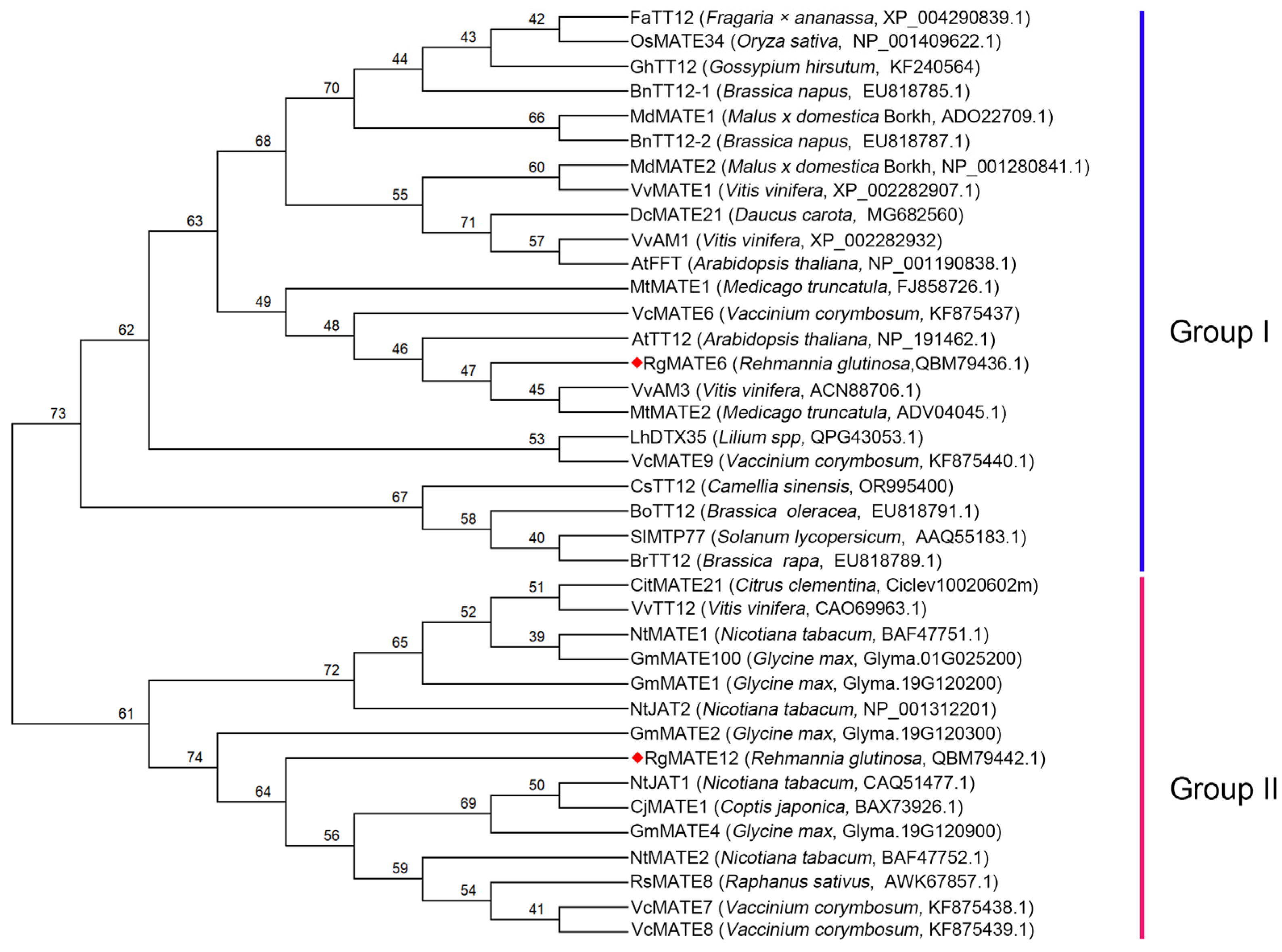
Figure 1. Phylogenetic tree for RgMATE6 and RgMATE12 with other reported plant homologs. The red diamond highlights the RgMATEs investigated in this study.
Plants 14 03608 g001
Figure 2. The HPG contents (AD) and the expression levels of RgMATE6 (E) and RgMATE12 (F) in different tissues of R. glutinosa across cultivation stages. The error bars represent the standard deviations of the mean (n = 3, p < 0.05). Lowercase letters indicate significant differences at the 0.05 level among different groups (the same convention applies below).
Plants 14 03608 g002
Figure 3. The ACT contents (A) and the RgMATE6 expression levels (B) in R. glutinosa under SA and MeJA treatments across cultivation stages. Lowercase letters indicate significant differences at the 0.05 level among different groups.
Plants 14 03608 g003
Figure 4. Localization and uptake assays of RgMATE6 in N. benthamiana. Subcellular localization of RgMATE6 in N. benthamiana leaf mesophyll protoplasts (left to right): GFP signal, chloroplast autofluorescence, bright-field view, and merged image. The green fluorescence represents the GFP-tagged RgMATE6 protein, and the red indicates chlorophyll autofluorescence (scale bars = 10 μm).
Plants 14 03608 g004
Figure 5. Uptake assays of ACT and the other three HPG by vacuolar membrane vesicles from N. benthamiana expressing RgMATE6-GFP or control. (A) Uptake activities of RgMATE6-GFP toward various substrates; (B) time-dependent uptake of ACT into vesicles from N. benthamiana expressing RgMATE6-GFP; (C) concentration-dependent uptake of ACT into vesicles from N. benthamiana expressing RgMATE6-GFP; (D) concentration-dependent uptake of ISO into the vesicles from N. benthamiana expressing RgMATE6-GFP; (E) inhibition of ACT uptake into vesicles from N. benthamiana expressing RgMATE6-GFP by ISO; (F) inhibition of ISO uptake into the vesicles from N. benthamiana expressing RgMATE6-GFP by ACT. Asterisks “***” indicate statistically significant differences at the 0.001 level, respectively.
Plants 14 03608 g005
Figure 6. Confirmation of positive RgMATE6 transgenic R. glutinosa and assay of ACT content in these transgenic lines. (A) Agarose gel electrophoresis images showing NPTII PCR products amplified from transgenic RgMATE6 and WT plants; (B) photographs of the sterile seedlings from the transgenic and WT R. glutinosa after 30 days of cultivation (scale bar = 4 cm); (C) the relative expression levels of RgMATE6 in the transgenic and WT R. glutinosa; (D) uptake assay of ACT into vacuole membrane vesicles from different genotype R. glutinosa; (E) ACT content assay from different genotype R. glutinosa; (F) photographs of various genotype plants cultured in pots filled with soil for 30 days (scale bar = 4 cm); (G) ACT content assay in various tissues from these plants. Lowercase letters indicate significant differences at the 0.05 level among different groups.
Plants 14 03608 g006
Figure 7. Expression profiles of six genes involved in ACT biosynthesis in various tissues of the transgenic and WT R. glutinosa. (A) RgTyDC; (B) RgCuAO; (C) RgPAR; (D) RgUGT; (E) RgPPO; (F) RgHCT. Lowercase letters indicate significant differences at the 0.05 level among different samples.
Plants 14 03608 g007
Figure 8. Function model of RgMATE6 in the transport and production of ACT in R. glutinosa. The multiple arrows indicate the metabolism of cinnamic acid, p-coumaric acid, and caffeic acid in the phenylpropanoid pathway, as well as tyramine and tyrosol in the tyrosine metabolism pathway.
Plants 14 03608 g008
Table 1. The correlation between RgMATE6 expression levels and ACT content across various treatments was quantified by the Pearson correlation coefficient (r). Note: “*” indicates significance at the 0.05 level.
Expression LevelsRgMATE6RgMATE12
Content of HPGs
ACT0.861 *0.459
ISO0.859 *0.508
SAL0.8540.575
HT-Glc0.8390.546
Disclaimer/Publisher’s Note: The statements, opinions and data contained in all publications are solely those of the individual author(s) and contributor(s) and not of MDPI and/or the editor(s). MDPI and/or the editor(s) disclaim responsibility for any injury to people or property resulting from any ideas, methods, instructions or products referred to in the content.