Article

Genome-Wide Identification and Expression Analysis of Mitochondrial Dicarboxylate Carriers (DICs) in Medicago Under Aluminum Stress

College of Grassland Science, Nanjing Agricultural University, Nanjing 210095, China
*
Author to whom correspondence should be addressed.
These authors contributed equally to this work.
Plants 2025, 14(21), 3250; https://doi.org/10.3390/plants14213250
Submission received: 5 August 2025 / Revised: 9 October 2025 / Accepted: 20 October 2025 / Published: 23 October 2025
(This article belongs to the Section Plant Response to Abiotic Stress and Climate Change)

Abstract

Aluminum (Al) is solubilized as phytotoxic Al3+ in acidic soils, rapidly inhibiting root elongation. To detoxify Al, plant roots secrete organic acids that chelate the ion. The transcription factor SENSITIVE-TO-PROTON-RHIZOTOXICITY1 (STOP1) regulates the export, distribution and metabolism of organic acids, which is crucial for Al resistance. Plant DICARBOXYLATE-CARRIERs (DICs) located in the inner mitochondrial membrane are presumed to exchange the dicarboxylates. However, whether Al or STOP1 modulates DIC expression to coordinate the organic acid shuttle remains unclear. Here, in the model legume Medicago truncatula, we identified three DIC genes and twelve in tetraploid Medicago sativa. Phylogenetic analysis places all Medicago DICs in a clade with Arabidopsis AtDIC1 and AtDIC2, whereas AtDIC3 lacks an ortholog in M. truncatula. Mining RNA-seq datasets followed by qRT-PCR validation showed that MtDIC2 is upregulated by Al in roots in a MtSTOP1-dependent manner. Consistently, STOP1-binding motifs exist in the MtDIC2 promoter, and MtSTOP1 binds to the MtDIC2 promoter in yeast. Furthermore, MsDIC2.4 shows an increase under Al treatment. Our study provides a genome-wide characterization of Medicago DICs and identifies MtDIC2 as a candidate target of MtSTOP1, whose Al-responsive induction may enhance organic acid flux across the mitochondrial membrane.

1. Introduction

In addition to respiration and ATP generation, plant mitochondria also participate in photorespiration, supply carbon skeletons for anabolism, and modulate cellular redox balance [1,2]. Because these interconnected pathways span several cell compartments, hydrophilic metabolites must cross the inner mitochondrial membrane via members of the nuclear-encoded mitochondrial carrier family (MCF) [3,4,5]. Among them, DICARBOXYLATE-CARRIERs (DICs) mediate the transport of dicarboxylates [3,6,7]. In Arabidopsis, three of the six originally annotated UNCOUPLING-PROTEINs (AtUCP4–6) were renamed AtDIC1–3 after liposome assays revealed their capacity to transport malate, oxaloacetate, succinate, phosphate, thiosulfate, sulfate, and sulfite [7]. Subsequent analyses showed that AtDIC2 preferentially imports malate in exchange for citrate export [8]. The reduced growth of Atdic2 mutants cannot be restored by compensatory mechanisms such as accelerated sucrose catabolism, increased citrate supply from fatty-acid β-oxidation, alternative carrier activities, or remodeling of the TCA cycle and amino acid pathways. Therefore, despite the existence of redundant transport routes such as DICARBOXYLATE/TRICARBOXYLATE-CARRIER (DTC) and SUCCINATE/FUMARATE-CARRIER (SFC) [5,9], AtDIC2 remains essential for maintaining energy, metabolic, and redox homeostasis.
Environmental stress rapidly disrupts mitochondrial metabolism in plants, underscoring the critical role of MCFs in stress tolerance [10,11]. During prolonged darkness, AtDIC2 expression rises steadily; without it, compromised organic acid mobilization and NAD redox homeostasis accelerate leaf senescence [8]. Transcript profiling reveals that AtDIC1 and AtDIC2 are upregulated by several environmental stresses, including exposure to cold, salt, osmotic, drought, phosphate starvation, UV-B irradiation, and wounding [5]. Biotic stresses likewise trigger the coordinated upregulation of all three AtDICs, while submergence rapidly induces AtDIC1, AtDIC2, and their Eucalyptus grandis orthologs EgDICs [12]. This hypoxic induction is regulated by GROUP-VII-ETHYLENE-RESPONSE-FACTOR (ERFVIIs), whose oxygen-dependent turnover is controlled by the N-end rule pathway [12,13,14]. Accordingly, AtDIC2 overexpression markedly enhances submergence tolerance, establishing its critical role in metabolic resilience under hypoxia [12].
Aluminum (Al) is the most abundant metal in the Earth’s crust. Under acidic conditions, it dissolves as toxic Al3+ ions, which rapidly inhibits root tips and reduces crop yields [15,16,17]. Within transient exposure, Al represses mitochondrial respiration, triggering ATP depletion, reactive oxygen species (ROS) formation, dissipation of the inner mitochondrial membrane potential, and an almost simultaneous arrest of root elongation [18,19]. Plants counter this toxicity by secreting organic acids—primarily malate and citrate—that chelate Al in the rhizosphere [20,21,22,23]. The Al-resistance transcription factor SENSITIVE-TO-PROTON-RHIZOTOXICITY1 (STOP1) transcriptionally induces AL-ACTIVATED-MALATE-TRANSPORTER (ALMT) and MULTI-DRUG-AND-TOXINCOMPOUNDS-EXTRUSION (MATE) expression to mediate malate and citrate secretion, respectively [24,25,26]. STOP1 also improves the ATP-binding cassette (ABC) transporter complex STAR1/ALS3 (STAR2) expression or protein abundance at the tonoplast to sequester Al into the vacuole, thereby restricting cytosolic toxicity [27,28,29,30,31]. In addition, STOP1 fine-tunes intracellular redox and metabolic balance [32,33]. SlSTOP1 confers Al tolerance by upregulating mitochondrial formate dehydrogenase SlFDH, which regenerates NAD+ from NADH [33]. In the Atstop1 mutant, transcripts of the vacuolar malate importer AtTDT, the GABA-shunt enzymes AtGDH1, AtGDH2 and AtGAD1, and the pH-stat enzymes AtME1 and AtME2 are all reduced [32]. Consistently, metabolite profiling reveals that sucrose accumulates, while succinate, pyruvate and GABA decline. AtSTOP1 binds to the AtGDH1 and AtGDH2 promoters, and mutation of these genes exacerbates Al sensitivity [34]. Thus, STOP1-mediated control of organic acid distribution and metabolism is important for Al tolerance. However, it remains unknown whether Al or STOP1 modulate DIC expression to coordinate organic acid exchange between mitochondria and cytosol.
Al-sensitive legume crops such as alfalfa (Medicago sativa, 2n = 4x = 32) and the model species barrel medic (Medicago truncatula, 2n = 2x = 16) suffer severe yield losses in acidic soils [35,36]. Recent work has begun to unravel the molecular mechanisms of their Al resistance. In M. sativa, Al-inducible genes including MsMATE66, MsALS3, MsSTAR1, and MsGDH1 have been characterized [37]. Their M. truncatula orthologs are upregulated primarily through MtSTOP1 and its homolog MtSTOP4 [38]. Citrate efflux from Medicago roots is mediated by MtMATE66 [39], while the dehydrin MsDHN1 activates two aquaporins to promote oxalate secretion [40]. Additionally, the transcription factor MsMYB741 enhances flavonoid biosynthesis and exudation, further chelating external Al [41]. Furthermore, overexpression of MALATE-DEHYDROGENASE (MDH) or CITRATE-SYNTHASE (CS) markedly elevates Al resistance in M. sativa [42,43]. Hence, delineating the Medicago DIC family and determining their Al responsiveness might identify new targets for improving Al resistance by modulating mitochondrial-to-cytosolic organic acid flux.
In this study, we identified three DIC genes (MtDIC1–3) in M. truncatula and twelve in M. sativa. The phylogenetic tree, gene expansion patterns, sequence structures, and cis-regulatory elements of the DIC family were characterized. The response of DIC family genes to Al was further investigated using our previous RNA-seq datasets [37,38]. qRT-PCR revealed that MtSTOP1 drives Al-inducible MtDIC2 expression in roots, while MtDIC3 is transcriptionally repressed by Al but in a MtSTOP1-independent manner. Consistent with this, STOP1-binding motifs are present in the MtDIC2 promoter. The MsDIC2.4 expression also exhibits a significant increase under Al treatment. Collectively, our work establishes the Medicago DIC repertoire and pinpoints MtDIC2 and MsDIC2.4 as promising targets for enhancing adaptation of Medicago to acidic soils.

2. Results

2.1. Identification and Phylogenetic Analysis of the Dicarboxylate Carriers in Medicago

Using the three dicarboxylate carriers from Arabidopsis (AtDIC1–3) as query sequences, we searched for DIC homologs from 11 plant species. In the diploid Medicago truncatula, we identified three genes (MtDIC13), whereas the autotetraploid Medicago sativa contains 12 genes that were named based on their chromosomal locations (MsDIC1.13.4) (Figure S1 and Table S1). At the haploid genome level, M. sativa has the same number of DICs as M. truncatula. All Medicago DIC proteins range from 313 to 322 amino acids, exhibit isoelectric points of 9.85–9.96, and possess molecular weights of 33.4–34.5 kDa, which are highly similar to AtDIC1 and AtDIC2 (Table S2).
Subsequently, phylogenetic analysis was performed using DICs from Arabidopsis, M. truncatula, M. sativa, Lotus japonicus, soybean (Glycine max), chickpea (Cicer arietinum), tomato (Solanum lycopersicum), cotton (Gossypium hirsutum), sorghum (Sorghum bicolor), rice (Oryza sativa), and the moss Physcomitrium patens (Figure 1 and Figure S2, and Table S1). Consistent with their shared ancestry, each MtDIC grouped tightly with its four corresponding MsDIC members. MtDIC2 and MtDIC3 clustered more closely with each other than with MtDIC1. All MtDIC and MsDIC proteins clustered near AtDIC1 and AtDIC2 but were clearly separated from AtDIC3. AtDIC3, together with moss DICs, is phylogenetically distant from the other DICs. In addition, MtDICs and MsDICs clustered closely around DICs from other legumes, such as L. japonicus, soybean, and chickpea, indicating a shared evolutionary history and conserved molecular function.

2.2. Synteny Analysis of DICs in the Genome of Medicago

To further examine the evolutionary relationships of the DICs, we performed inter-species synteny analyses among M. truncatula, M. sativa, and Arabidopsis. Three orthologous pairs were identified between M. truncatula and Arabidopsis, while 25 orthologous pairs were detected between M. truncatula and M. sativa (Figure 2 and Table S3). Consistent with the phylogenetic results (Figure 1), no MtDIC was found orthologous to AtDIC3 (Figure 2 and Table S3). We next examined the synteny within each species. Three paralogous pairs were observed among the MtDICs within M. truncatula (Figure S3a and Table S4). In M. sativa, 17 paralogous pairs were discovered (Figure S3b). Notably, none of the MsDIC1 members showed paralogous to MsDIC2 or MsDIC3 members.

2.3. Protein Motifs, Gene Structures and Cis-Elements in DIC Promoters

Next, we aligned these DIC protein sequences from M. truncatula, M. sativa, and Arabidopsis with human UCP1 (HsUCP1) of known cryo-electron microscopy structure [44] (Figure S4). It showed that MtDICs and MsDICs are more closely related to AtDIC1/AtDIC2 than to AtDIC3. All DICs contain three tandem repeats, each comprising two hydrophobic transmembrane α-helices linked by a long hydrophilic matrix loop [4,45,46]. Within the nine predicted α-helices (Hs), transmembrane segments H1, H4, and H7 carry the conserved PX[DE]XX[RK] motif, whose charged residues form the matrix salt bridge network. Adjacent to the transmembrane segments H3, H6, and H9, there are the conserved [YF][DE]XX[KR] motif that can form another salt bridge called the cytoplasmic salt bridge network. Furthermore, MEME motif discovery identified eight conserved motifs in all these DICs (Figure 3a,b and Figure S5). Motifs 5, 3, 6, 1, and 2 are shared by all Medicago and Arabidopsis DICs. Motifs 7 and 4 are additionally present in AtDIC1 and all Medicago DICs, whereas Motif 8 is restricted to MtDIC2/MsDIC2s and MtDIC3/MsDIC3s.
Gene structure analysis revealed that all Medicago DIC genes, like AtDIC1 and AtDIC2, comprise a single exon (Figure 3c). AtDIC3 uniquely contains one intron and two exons. MsDIC genes displayed no annotated 5′- and 3′-UTRs, which may be due to incomplete gene annotation rather than biological loss.
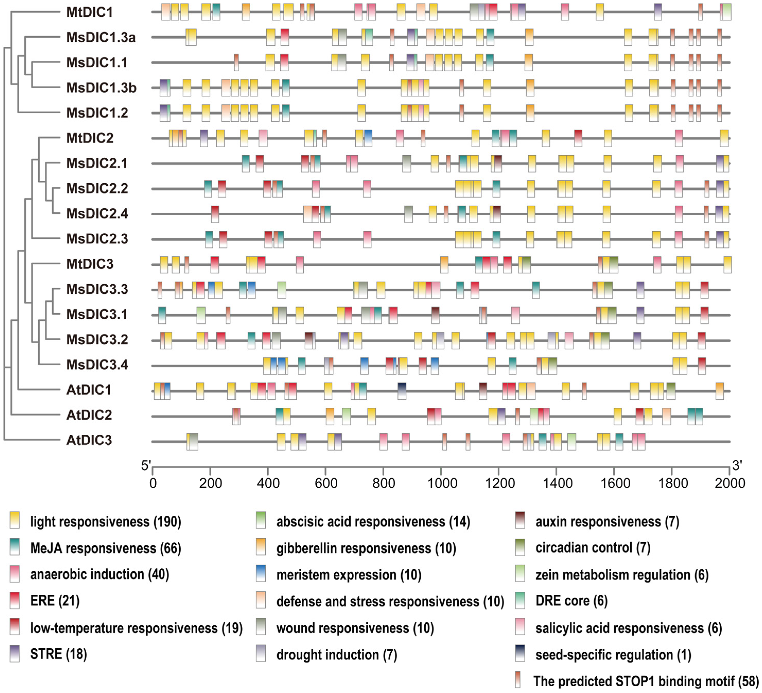
To explore the transcriptional regulatory mechanisms, we scanned the 2 kb promoter regions of Medicago and Arabidopsis DIC genes using the PlantCARE database (Figure 4 and Figure S6). Light-responsive elements (3-AF1 binding site, AE-box, Box 4, chs-CMA1a/2a, G-box, GT1-motif, I-box, MRE, Sp1, TCCC-motif, and TCT-motif) are the most abundant class, with Box 4 present in every Medicago DIC promoter. Hormone-responsive motifs for MeJA, ethylene, ABA, gibberellin, auxin and salicylic acid, together with stress- and development-associated elements, are also widespread. The TGACG-motif, MeJA-responsiveness element, is present in all DIC genes of Medicago and Arabidopsis. Significantly, the Al-resistance transcription factor STOP1 binding motif, identified from AtSTOP1 DAP-seq data [47], is found in the promoters of all Medicago and Arabidopsis DIC genes (Figure 4 and Table S5), suggesting the DIC family in Al signaling.

2.4. Expression Analysis of DIC Genes in Medicago Under Al Treatment

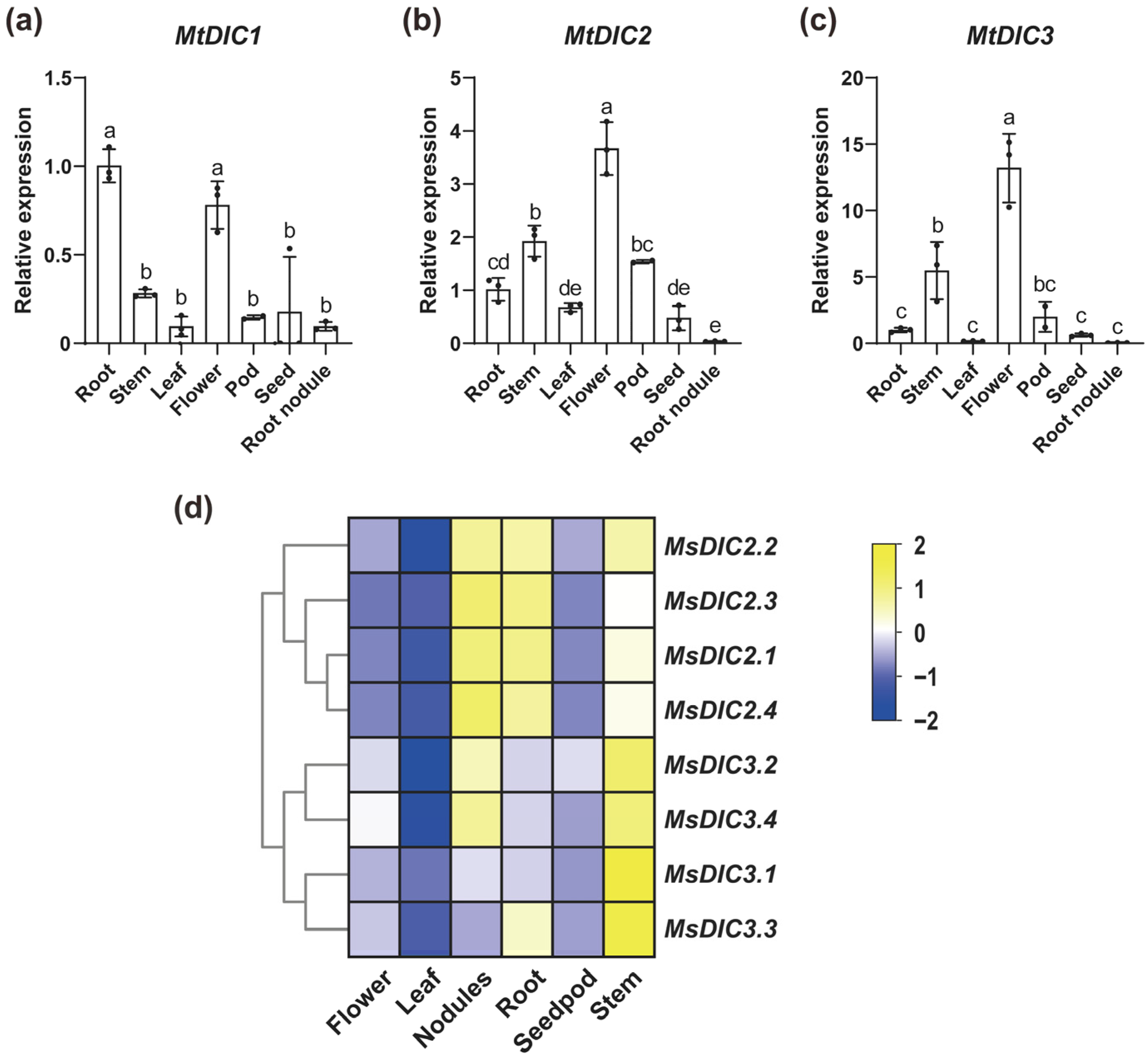
We performed qRT-PCR analysis to determine the tissue-specific expression of DICs in M. truncatula. MtDIC1 transcripts accumulated predominantly in roots and flowers, while MtDIC2 and MtDIC3 were most highly expressed in flowers (Figure 5a–c). In roots, MtDIC2 transcripts were the most abundant (mean 2−ΔCt = 0.0691), followed by MtDIC3 (0.0042), whereas MtDIC1 showed the lowest (0.0014). Analyzing public RNA-seq data from M. sativa [48] further revealed that MsDIC2 members are highly expressed in roots, nodules and stems, with MsDIC2.4 showing the highest FPKM, while MsDIC3 transcripts are most abundant in stems (Figure 5d). Moreover, transcripts of MsDIC1s were too low to be detected, indicating that MtDIC1 and MsDIC1s are the least expressed in these tissues.
To further investigate whether Medicago DICs respond to Al, we analyzed our previous RNA-seq datasets in M. truncatula and M. sativa [37,38]. In WT (R108), we observed that MtDIC2 was significantly upregulated by Al among the differentially expressed genes (DEGs) (fold change, FC = 2.39; false discovery rate, FDR < 0.05) (Figure 6a). In contrast, MtDIC3 was slightly downregulated by Al (FC = 0.60, FDR = 0.136, p = 0.034) but was not identified as a DEG. In a previous stress time-course [49], MtDIC2 expression was also induced by cold and drought, while MtDIC3 responded to salt (Figure S7). In M. sativa, MsDIC2.4 showed a modest induction under Al at pH 5.7 (FC = 1.37, FDR = 0.718, p = 0.012) relative to pH 5.7 without Al. However, this induction did not reach the threshold for DEGs. Both MsDIC2.4 and MsDIC2.1 were slightly induced by low pH alone (pH 4.6 vs. pH 5.7), with FC of 1.39 and 1.52, FDR of 0.482 and 0.419, and p values of 0.004 and 0.003, respectively (Figure 6b). qRT-PCR analysis confirmed that MsDIC2.4 expression can be significantly induced by Al, whereas the expression of MsDIC1.1 and MsDIC3.4 was not (Figure 6c–e). Then, we analyzed RNA-seq data in Arabidopsis [50]. AtDIC2 was slightly induced by Al and excess Fe at pH 5 (with +Pi pH 6 as control), although it did not reach the threshold for DEGs (Figure 6f). This induction was attenuated in the Atstop1 mutant. qRT-PCR analysis revealed that both AtDIC1 and AtDIC2 exhibited slightly reduced expression levels in the Atstop1-2 compared to WT (Col-0) under Al treatment (Figure 6g,h). Furthermore, in previous DAP-seq data [47], we found that the upstream and downstream regions of AtDIC2 were enriched with AtSTOP1, which contains the predicted AtSTOP1 binding motif (Figure S8), indicating that AtSTOP1 directly targets AtDIC2 to regulate its expression.

2.5. MtSTOP1 Upregulates MtDIC2 Expression in Roots Under Al Stress

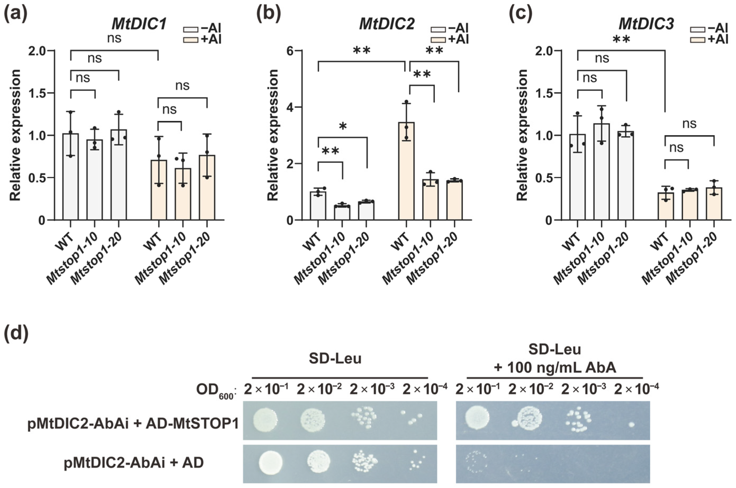
The RNA-seq data showed that among DEGs between WT (R108) and the knockout mutant Mtstop1-10, the expression of MtDIC2 was significantly suppressed in the knockout mutant Mtstop1-10 under both control (FC = 0.45, FDR < 0.05) and Al-treated (FC = 0.22, FDR < 0.05) conditions (Figure 6a). qRT-PCR analysis using root tip confirmed that Al-inducible MtDIC2 expression in WT, whereas induction was abolished in two independent knockout lines, Mtstop1-10 and Mtstop1-20 (Figure 7a–c). Conversely, Al repressed MtDIC3, yet mutation of MtSTOP1 unaffected its expression (Figure 7c). Interestingly, MtSTOP1-regulated expression of MtDIC2 was absent in leaves (Figure S9), and MtDIC1 transcripts were below detection in seedling leaves. Subsequently, we performed a yeast one-hybrid assay to determine whether MtSTOP1 could bind to the MtDIC2 promoter. The yeast strain, whose genome had previously been integrated with the linearized pMtDIC2-AbAi, was then transformed with either an empty vector (AD alone) or AD- MtSTOP1. All transformed yeast strains grew well on SD-Leu medium (Figure 7d). However, only the yeast transformed with the AD-MtSTOP1 fusion grew robustly on SD-Leu medium supplemented with Aureobasidin A (AbA). This result indicates that MtSTOP1 can activate the expression of the reporter gene AbAr, suggesting that MtSTOP1 can bind to the MtDIC2 promoter in yeast.
Since plant DICs share high homology with UCPs [4,51], we also examined MtUCP expression under Al stress, yet none of them responded appreciably (Figure S10). In addition, DTC and SFC are single-copy genes in Arabidopsis and M. truncatula that mediate mitochondrial organic acid transport [9]. Exposure to Al caused a modest induction of MtDTC expression (FC = 1.79, FDR = 0.073, p = 0.013) in a MtSTOP1-independent manner, whereas MtSFC1 expression was unaffected.

3. Discussion

In Arabidopsis, 58 members of MCF have been identified [6,9,45], among which the three DIC proteins (AtDIC1–3) facilitate dicarboxylate transport [4,7,8]. AtDIC1 and AtDIC2 are 70% identical, while AtDIC3 is only 55–60% identical to AtDIC1 and AtDIC2 [7]. A mitochondrial proteomic survey revealed that AtDIC3 is expressed at significantly lower levels than AtDIC1 and AtDIC2 [4], and root RNA-seq likewise failed to detect AtDIC3 transcripts (Figure 6f). We identified three MtDIC genes in M. trucatula and twelve MsDIC genes in M. sativa. All legume DICs form a single clade near to AtDIC1 and AtDIC2 but clearly distinct from AtDIC3 (Figure 1 and Figure S2). The Medicago DIC proteins are 313–322 residues long, closely matching AtDIC1 and AtDIC2 (both 313 aa) and markedly shorter than AtDIC3 (337 aa) (Table S2). Synteny analysis revealed no ortholog of AtDIC3 in M. trucatula (Figure 2 and Table S3). Furthermore, every Medicago DIC, like AtDIC1 and AtDIC2, is encoded by a single exon, whereas AtDIC3 uniquely contains two exons separated by one intron (Figure 3c). Collectively, these data suggest that Medicago DICs are functionally aligned with AtDIC1 and AtDIC2 rather than with the divergent AtDIC3. Functional assays showed that AtDIC1 and AtDIC2 transport dicarboxylates, phosphate and arsenate far more efficiently than AtDIC3 does [7]. Moreover, both in isolated mitochondria and in proteoliposome studies further revealed that AtDIC2 operates as a malate-importer/citrate-exporter antiporter [8]. However, whether the Medicago DIC exhibit the same substrate preferences and transport directionality remains to be determined.
All MCFs share an architecture of three tandem repeats, each repeat comprising two hydrophobic transmembrane helices linked by a characteristic motif, PX[D/E]XX[K/R]X[K/R] (20–30 residues) [D/E]GXXXX[W/Y/F][K/R]G [3,46,52,53]. Consistent with this, MtDIC and MsDIC sequences fold into nine predicted α-helices that contain the three repeated motifs (Figure S4). Strikingly, MEME Motif 8 appears exclusively in MtDIC2/MsDIC2s and MtDIC3/MsDIC3s (Figure 3a,b), and phylogenetic analysis places these sequences in a single clade separate from MtDIC1/MsDIC1s (Figure 1 and Figure S2). In addition, no MsDIC1 member is paralogous to any MsDIC2 or MsDIC3 member (Figure S3b). These suggest that MtDIC2/MsDIC2s and MtDIC3/MsDIC3s constitute a subgroup that has diverged independently from the MtDIC1/MsDIC1s. Expression profiling mirrors this divergence. MtDIC2 and MtDIC3 transcripts accumulate to markedly higher levels than MtDIC1, and are both most abundant in flowers (Figure 5a–c); moreover, transcripts of MsDIC1 in tissues and MtDIC1 in seedling leaves are virtually undetectable (Figure 5d and Figure S9). Three paralogous pairs were observed among the MtDICs (Figure S3a and Table S4), while 17 paralogous pairs were discovered in M. sativa, confirming the duplication of DIC genes in Medicago, which is likely caused by whole-genome duplication (WGD) [54].
Al toxicity is tightly linked to mitochondrial dysfunction [19,55]. Plants detoxify the rhizosphere Al by secreting organic acids, a process that consumes carbon and energy [20,23]. As MCF transporters control metabolite and energy homeostasis, they are likely pivotal to Al resistance [11,18,56]. Among them, DIC, DTC, and SFC proteins are candidates for shuttling organic acids within mitochondrial metabolism [8,9]. In this study, we observed that MtDIC2 expression is significantly up-regulated within 6 h of Al treatment (Figure 6a and Figure 7b), MtDTC shows a modest Al induction, and MtSFC1 remains unchanged (Figure S10). Likewise, MsDIC2.4 expression exhibits an increase under Al (Figure 6b,d). AtDIC1–3, originally designated AtUCP4–6, share high homology with AtUCP1–3 [4,51]. Previous work has shown that AtUCP1 localizes to mitochondria and displays uncoupling activity [10,57]. AtUCP1 and AtUCP2 also transport aspartate, glutamate, and other dicarboxylates [51]. However, profiling the three MtUCP genes under Al stress revealed no significant response (Figure S10), implying they do not participate in Al-triggered shuttling of organic acids in mitochondria.
AtSTOP1 confers Al resistance by transcriptionally regulating AtALMT1, AtMATE, and AtSAUR55 for organic acid secretion [24,26,58] and by inducing AtALS3, AtGDH1, and AtGDH2 for internal detoxification [34]. Additionally, STOP1 adjusts intracellular redox, metabolite, and pH balance by regulating FDH, TDT, GABA-shunt enzymes, and the pH-stat enzymes [32,33]. In M. truncatula, MtSTOP1 primarily modulates Al resistance by inducing MtMATE66, MtALS3, MtSTAR1, and MtGDH1 expression [38]. Here, we found that AtDIC2 expression is slightly reduced in Atstop1 under Al treatment (Figure 6f,h), while the induction of MtDIC2 expression by Al is significantly abolished in Mtstop1 mutants (Figure 6a and Figure 7b). Supporting these observations, AtSTOP1 occupancy is enriched across both upstream and downstream regions of AtDIC2, as identified by AtSTOP1 DAP-seq [47]. Each contains the canonical STOP1-binding motif (Figure S8), suggesting that AtDIC2 is transcriptionally controlled by AtSTOP1. The STOP1-binding motif is also present in the MtDIC2 promoter (Figure 4 and Table S5). Additionally, MtSTOP1 has been shown to bind to the MtDIC2 promoter in yeast (Figure 7d). These findings collectively suggest that MtDIC2 is a candidate target of MtSTOP1.
STOP1 accumulation under Al stress is tightly associated with downstream gene expression [59]. On the one hand, AtSTOP1 stability is negatively regulated by ubiquitin-mediated proteolysis through REGULATIONOF-ATALMT1-EXPRESSION1 (AtRAE1) and its homolog [60,61], while the oxidative modification accelerates this degradation [55]. On the other hand, the THO/TREX complex positively regulates AtSTOP1 abundance [62,63], and phosphorylation further promotes its accumulation [64,65,66]. Additionally, SUMOylation fine-tunes AtSTOP1 accumulation and transcriptional activity [67]. In M. truncatula, although MtSTOP1 transcripts show a slight increase in the very apex of roots, the protein might also be modulated by Al at the post-translational level through MtRAE1-mediated degradation [38]. In this study, we observed that MtSTOP1-regulated expression of MtDIC2 is completely absent in leaves (Figure S9). This likely reflects insufficient MtSTOP1 levels in leaves to activate MtDIC2 expression. Low pH also triggers STOP1 accumulation [68]. In M. sativa, the expression of MtSTOP1-regulated orthologous genes is induced by Al, and their expression is also responsive to acidic pH [37,38]. Here, we found that MsDIC2.4 shows an increase under Al at both pH 5.0 and pH 5.7, despite the different Al speciation present at these pH levels (Figure 6b,d and Figure S11), while both MsDIC2.4 and MsDIC2.1 are induced by low pH.
In addition to Al and low pH, AtSTOP1 accumulates under Fe excess caused by Pi deficiency [50,69]. Here, we found a modest induction of AtDIC2 under excess Fe (Figure 6f). AtDICs are transcriptionally activated by a range of abiotic and biotic stresses [5,10]. Similarly, the expression of MtDIC2 is rapidly upregulated by cold and drought within 2–6 h, followed by a decrease at 12 h, whereas MtDIC3 is induced by salt only after 12 h of treatment (Figure S7). Submergence rapidly induces AtDIC1 and AtDIC2 expression via the hypoxia-responsive ERFVIIs [12]. Hypoxia induces STOP1 expression, and cellular redox perturbations influence STOP1 protein levels [55,70]. Additionally, STOP1 itself is implicated in tolerance to salt, drought, hypoxia, and a spectrum of other stresses [70,71,72]. Thus, DICs might be the targets of STOP1 under these diverse stresses. Al, Fe, heavy metals, and other stresses often cause oxidative damage [55,73,74]. The proposed function of DICs is associated with the transport of organic acids into or out of the mitochondria, thereby maintaining redox homeostasis [8]. Nevertheless, cis-element profiling reveals light-, hormone-, stress-, and development-associated motifs across all Medicago and Arabidopsis DIC promoters (Figure 4), indicating that additional transcription factors might also contribute to their regulation.
In summary, our research has characterized the Medicago DIC gene family and identified MtDIC2 and MsDIC2.4 as potential targets for improving Medicago’s tolerance to acidic soils. Further investigation is required to explore the effect of increasing its expression.

4. Materials and Methods

4.1. Identification of DIC Gene Family in Medicago

Protein sequences of AtDIC1, 2, and 3 were obtained from the Arabidopsis Araport11 database and used as BLASTP queries against Medicago truncatula A17 r5.0 genome [75], the Medicago sativa cv. XinJiangDaYe genome [76] or against eight additional plant genomes (Lotus japonicus Lj1.0v1, Glycine max Wm82.a4.v1, Cicer arietinum v1.0, Solanum lycopersicum ITAG2.4, Gossypium hirsutum v2.1, Sorghum bicolor v3.1.1, Oryza sativa v7.0, and Physcomitrium patens v3.3). The chromosomal locations of Medicago DICs were extracted from the corresponding assemblies and plotted with the TBtools-II software [77].

4.2. Phylogenetic and Synteny Analysis

DIC proteins were aligned using the MUSCLE method (default settings). The trimmed alignment was used to construct a neighbor-joining (NL) or maximum likelihood (ML) phylogenetic tree (1000 bootstrap replicates) in MEGA X [78]. MCScanX was used to infer gene synteny in TBtools-II.

4.3. Protein Motif Prediction and Gene Structure Survey

Physicochemical properties (molecular weight and isoelectric point) of Medicago and Arabidopsis DICs were calculated with ExPASy ProtParam (https://web.expasy.org/protparam/ (accessed on 21 October 2025)). They were aligned to human HsUCP1 and visualized with ESPript 3.0 (https://espript.ibcp.fr/ESPript/ESPript/index.php (accessed on 21 October 2025)). MEME v5.5.8 (http://meme-suite.org/ (accessed on 2 July 2025)) was used to predict the conserved motifs (maximum motifs = 8; otherwise default). The gene structures were extracted from the M. truncatula A17 r5.0, M. sativa XinJiangDaYe, and Arabidopsis Araport11 annotations. The results of protein motif and gene structure analysis were plotted using TBtools-II.

4.4. Cis-Element Profiling

Two-kb upstream regions (from the start codon) of Medicago and Arabidopsis DICs were scanned for cis-regulatory elements using the PlantCARE database (https://bioinformatics.psb.ugent.be/webtools/plantcare/html/ (accessed on 21 October 2025)). Results were visualized with TBtools-II.

4.5. Plant Materials and Growth Conditions

M. truncatula ecotype R108, M. sativa cultivar XinJiangDaYe, and Arabidopsis ecotype Col-0 were employed as the wild-type (WT) controls in this study. The Mtstop1-10 and Mtstop1-20 mutants were previously generated using the CRISPR/Cas9 system [38]. The T-DNA insertion line SALK_114108 (Atstop1-2) was procured from the Arabidopsis Biological Resource Center. Seedlings were grown in a growth chamber set at 22 °C, with a light cycle of 14 h of illumination and 10 h of darkness.

4.6. RNA-Seq Data Analysis

Our previous RNA-Seq data were analyzed to study gene expression under Al stress in M. truncatula [38] and M. sativa [37]. Differential expression analysis was performed using DESeq2 with a significance threshold of false discovery rate (FDR) < 0.05 and a fold change (FC) > 2 or <0.5. Additional transcriptome datasets were downloaded from NCBI GEO to examine expression profiles in different M. sativa tissues [48], Arabidopsis Al responses [50], and M. truncatula stress responses [49]. Expression values were calculated as log2 (FPKM/TPM + 1) and heatmaps were generated using the z-score normalization with TBtools-II.

4.7. Real-Time Quantitative Reverse-Transcription PCR (qRT-PCR) Analysis

To profile the tissue-specific expression of MtDICs, total RNA was extracted from M. truncatula R108 tissues, including roots of 5-day-old seedlings, stems and leaves of 2-week-old seedlings, and flowers, pods, seeds, and root nodules of 12-week-old plants, using a plant RNA isolation kit (Vazyme, Nanjing, China). For Al-stress assays, Medicago species were cultivated in a 0.5 mM CaCl2 solution before Al treatment, while Arabidopsis were grown on 1/2 MS medium with 1.2% agar and 1% sucrose at pH 5.7. Subsequently, 4-day-old WT (R108), Mtstop1-10, and Mtstop1-20 seedlings, as well as 7-day-old XinJiangDaYe, WT (Col), and Atstop1-2 mutants were pretreated with 0.5 mM CaCl2 at pH 5.0 for 6 h and then exposed to 0 or 5 μM AlCl3 in the same solution for 6 h. RNA was also extracted from root segments (0–1 cm) for Medicago species and from total roots for Arabidopsis. To determine MtSTOP1-dependent regulation in shoots, leaves of 2-week-old WT (R108) and Mtstop1-10 were collected. RNA was reverse-transcribed with a 1st strand cDNA synthesis kit (Vazyme). qRT-PCR was performed using the SYBR Green reagent (Vazyme). MtGAPDH (MtrunA17Chr4g0057861) [38] was identified as the optimal internal reference gene for M. truncatula using the Normfinder (Table S6). MsGAPDH (MS.gene033342) and AtUBQ10 (AT4G05320) [60] were used as internal reference genes for M. sativa and Arabidopsis, respectively. Primers are listed in Table S7. Relative expression levels were calculated using the 2−ΔΔCt method.

4.8. Yeast One-Hybrid Assay

The promoter of MtDIC2 was cloned into the pAbAi vector. The coding sequence (CDS) of MtSTOP1 was in-frame fused with the GAL4 activation domain of the yeast expression vector pGADT7. The primers used for these constructions are listed in Table S7. The yeast one-hybrid assay was performed according to the manufacturer’s instructions (Coolaber, Beijing, China). The transformed yeast cells were plated onto synthetic defined (SD) medium lacking leucine (Leu) and supplemented with either 0 or 100 ng/mL Aureobasidin A (AbA) to test the interaction for 3 days.

4.9. Aluminum Speciation Analysis

The speciation of Al was simulated using MINEQL+ 5.0 with default settings. Simulations were conducted for Al treatments at pH levels of 4.6, 5.0, and 5.7. The model input included the complete chemical composition of the solution, which contained 5 μM AlCl3 and 0.5 mM CaCl2.

4.10. Statistical Analysis

qRT-PCR was performed on three independent plant pools. Values are means ± SD. Statistical significance was determined by one-way ANOVA followed by Tukey’s test for more than two groups or a two-tailed t-test for two groups.

Supplementary Materials

The following supporting information can be downloaded at: https://www.mdpi.com/article/10.3390/plants14213250/s1, Figure S1: Chromosomal locations of MtDIC and MsDIC genes; Figure S2: The maximum likelihood phylogenetic tree of the DIC gene family; Figure S3: Syntenic analysis of the DIC gene family in the genomes of M. truncatula and M. sativa, respectively; Figure S4: Multiple sequence alignment of DIC proteins; Figure S5: Sequence logos of the eight conserved motifs identified in Medicago and Arabidopsis DIC proteins; Figure S6: Heatmap showing the quantity of cis-acting elements in Medicago and Arabidopsis DIC promoters; Figure S7: Heatmap of MtDIC expression under cold, drought, and salt stresses; Figure S8: AtSTOP1-binding peaks detected by DAP-seq in upstream and downstream regions of AtDIC2; Figure S9: The expression of MtDIC2 and MtDIC3 in Mtstop1 leaves remained unchanged relative to WT; Figure S10: Heatmap of MtUCP, MtDTC, and MtSFC1 expression under Al stress in WT and Mtstop1-10; Figure S11: Effects of pH on Al speciation; Table S1: The DIC gene family members used for phylogenetic tree construction; Table S2: The protein lengths, molecular weights, and isoelectric points of the DIC gene family in M. truncatula, M. sativa and Arabidopsis; Table S3: The ortholog pairs of DIC family genes in the genomes of M. trucatula, M. sativa, and Arabidopsis; Table S4: The paralog pairs of DIC family genes in the genomes of M. trucatula and M. sativa; Table S5: The predicted STOP1 binding motifs in the promoters of DIC gene family members in M. trucatula, M. sativa, and Arabidopsis; Table S6: Identification of the optimal internal reference gene by Normfinder; Table S7: Primers used in this study.

Author Contributions

Conceptualization, formal analysis, and visualization: C.Y., X.L., Z.L. and Y.Z.; investigation: C.Y., X.L., Z.L. and Y.L.; resources: Z.G. and Y.Z.; funding acquisition: Z.G. and Y.Z.; supervision: Y.Z.; writing—original draft, review and editing: Y.Z. All authors have read and agreed to the published version of the manuscript.

Funding

This research was funded by the National Natural Science Foundation of China (grant number 32101440 to Y.Z.) and the Fundamental Research Funds for the Central Universities (grant YDZX2025002 to Y.Z.).

Data Availability Statement

Data are contained within the article and Supplementary Materials.

Acknowledgments

The authors appreciate the constructive suggestions provided by the anonymous reviewers, which have significantly improved the quality of this paper.

Conflicts of Interest

The authors declare no conflicts of interest.

Abbreviations

The following abbreviations are used in this manuscript:
chrChromosome
RNA-seqRNA sequencing
qRT-PCRReal-time quantitative reverse transcription PCR
FCFold change
FDRFalse discovery rate
DAP-seqDNA affinity purification sequencing

References

  1. Giegé, P.; Heazlewood, J.L.; Roessner-Tunali, U.; Millar, A.H.; Fernie, A.R.; Leaver, C.J.; Sweetlove, L.J. Enzymes of Glycolysis Are Functionally Associated with the Mitochondrion in Arabidopsis Cells. Plant Cell 2003, 15, 2140–2151. [Google Scholar] [CrossRef]
  2. Fernie, A.R.; Carrari, F.; Sweetlove, L.J. Respiratory Metabolism: Glycolysis, the TCA Cycle and Mitochondrial Electron Transport. Curr. Opin. Plant Biol. 2004, 7, 254–261. [Google Scholar] [CrossRef]
  3. Palmieri, F. The Mitochondrial Transporter Family SLC25: Identification, Properties and Physiopathology. Mol. Asp. Med. 2013, 34, 465–484. [Google Scholar] [CrossRef]
  4. Fernie, A.R.; Cavalcanti, J.H.F.; Nunes-Nesi, A. Metabolic Roles of Plant Mitochondrial Carriers. Biomolecules 2020, 10, 1013. [Google Scholar] [CrossRef]
  5. Nunes-Nesi, A.; Cavalcanti, J.H.F.; Fernie, A.R. Characterization of In Vivo Function(s) of Members of the Plant Mitochondrial Carrier Family. Biomolecules 2020, 10, 1226. [Google Scholar] [CrossRef]
  6. Picault, N.; Hodges, M.; Palmieri, L.; Palmieri, F. The Growing Family of Mitochondrial Carriers in Arabidopsis. Trends Plant Sci. 2004, 9, 138–146. [Google Scholar] [CrossRef]
  7. Palmieri, L.; Picault, N.; Arrigoni, R.; Besin, E.; Palmieri, F.; Hodges, M. Molecular Identification of Three Arabidopsis thaliana Mitochondrial Dicarboxylate Carrier Isoforms: Organ Distribution, Bacterial Expression, Reconstitution into Liposomes and Functional Characterization. Biochem. J. 2008, 410, 621–629. [Google Scholar] [CrossRef] [PubMed]
  8. Lee, C.P.; Elsässer, M.; Fuchs, P.; Fenske, R.; Schwarzländer, M.; Millar, A.H. The Versatility of Plant Organic Acid Metabolism in Leaves Is Underpinned by Mitochondrial Malate–Citrate Exchange. Plant Cell 2021, 33, 3700–3720. [Google Scholar] [CrossRef] [PubMed]
  9. Toleco, M.R.; Naake, T.; Zhang, Y.; Heazlewood, J.L.; Fernie, A.R. Plant Mitochondrial Carriers: Molecular Gatekeepers That Help to Regulate Plant Central Carbon Metabolism. Plants 2020, 9, 117. [Google Scholar] [CrossRef]
  10. Barreto, P.; Koltun, A.; Nonato, J.; Yassitepe, J.; Maia, I.d.G.; Arruda, P. Metabolism and Signaling of Plant Mitochondria in Adaptation to Environmental Stresses. Int. J. Mol. Sci. 2022, 23, 11176. [Google Scholar] [CrossRef] [PubMed]
  11. Monteiro-Batista, R.d.C.; Siqueira, J.A.; da Fonseca-Pereira, P.; Barreto, P.; Feitosa-Araujo, E.; Araújo, W.L.; Nunes-Nesi, A. Potential Roles of Mitochondrial Carrier Proteins in Plant Responses to Abiotic Stress. J. Exp. Bot. 2025, eraf032. [Google Scholar] [CrossRef]
  12. Barreto, P.; Arcuri, M.L.C.; Lima, R.P.M.; Marino, C.L.; Maia, I.G. Comprehensive In Silico Analysis and Transcriptional Profiles Highlight the Importance of Mitochondrial Dicarboxylate Carriers (DICs) on Hypoxia Response in Both Arabidopsis thaliana and Eucalyptus grandis. Plants 2022, 11, 181. [Google Scholar] [CrossRef]
  13. van Dongen, J.T.; Licausi, F. Oxygen Sensing and Signaling. Annu. Rev. Plant Biol. 2015, 66, 345–367. [Google Scholar] [CrossRef] [PubMed]
  14. Schmidt, R.R.; Weits, D.A.; Feulner, C.F.J.; van Dongen, J.T. Oxygen Sensing and Integrative Stress Signaling in Plants. Plant Physiol. 2018, 176, 1131–1142. [Google Scholar] [CrossRef]
  15. Kochian, L.V. Cellular Mechanisms of Aluminum Toxicity and Resistance in Plants. Annu. Rev. Plant Biol. 1995, 46, 237–260. [Google Scholar] [CrossRef]
  16. Ma, J.F. Syndrome of Aluminum Toxicity and Diversity of Aluminum Resistance in Higher Plants. In International Review of Cytology; Academic Press: Cambridge, MA, USA, 2007; Volume 264, pp. 225–252. [Google Scholar]
  17. Poschenrieder, C.; Gunsé, B.; Corrales, I.; Barceló, J. A Glance into Aluminum Toxicity and Resistance in Plants. Sci. Total Environ. 2008, 400, 356–368. [Google Scholar] [CrossRef] [PubMed]
  18. Hamilton, C.A.; Good, A.G.; Taylor, G.J. Induction of Vacuolar ATPase and Mitochondrial ATP Synthase by Aluminum in an Aluminum-Resistant Cultivar of Wheat. Plant Physiol. 2001, 125, 2068–2077. [Google Scholar] [CrossRef]
  19. Yamamoto, Y.; Kobayashi, Y.; Devi, S.R.; Rikiishi, S.; Matsumoto, H. Aluminum Toxicity Is Associated with Mitochondrial Dysfunction and the Production of Reactive Oxygen Species in Plant Cells. Plant Physiol. 2002, 128, 63–72. [Google Scholar] [CrossRef] [PubMed]
  20. Ma, J.F.; Ryan, P.R.; Delhaize, E. Aluminium Tolerance in Plants and the Complexing Role of Organic Acids. Trends Plant Sci. 2001, 6, 273–278. [Google Scholar] [CrossRef]
  21. Sasaki, T.; Yamamoto, Y.; Ezaki, B.; Katsuhara, M.; Ahn, S.J.; Ryan, P.R.; Delhaize, E.; Matsumoto, H. A Wheat Gene Encoding an Aluminum-activated Malate Transporter. Plant J. 2004, 37, 645–653. [Google Scholar] [CrossRef]
  22. Magalhaes, J.V.; Liu, J.; Guimarães, C.T.; Lana, U.G.P.; Alves, V.M.C.; Wang, Y.-H.; Schaffert, R.E.; Hoekenga, O.A.; Piñeros, M.A.; Shaff, J.E.; et al. A Gene in the Multidrug and Toxic Compound Extrusion (MATE) Family Confers Aluminum Tolerance in Sorghum. Nat. Genet. 2007, 39, 1156–1161. [Google Scholar] [CrossRef] [PubMed]
  23. Kochian, L.V.; Piñeros, M.A.; Liu, J.; Magalhaes, J.V. Plant Adaptation to Acid Soils: The Molecular Basis for Crop Aluminum Resistance. Annu. Rev. Plant Biol. 2015, 66, 571–598. [Google Scholar] [CrossRef]
  24. Hoekenga, O.A.; Maron, L.G.; Piñeros, M.A.; Cançado, G.M.A.; Shaff, J.; Kobayashi, Y.; Ryan, P.R.; Dong, B.; Delhaize, E.; Sasaki, T.; et al. AtALMT1, Which Encodes a Malate Transporter, Is Identified as One of Several Genes Critical for Aluminum Tolerance in Arabidopsis. Proc. Natl. Acad. Sci. USA 2006, 103, 9738–9743. [Google Scholar] [CrossRef]
  25. Iuchi, S.; Koyama, H.; Iuchi, A.; Kobayashi, Y.; Kitabayashi, S.; Kobayashi, Y.; Ikka, T.; Hirayama, T.; Shinozaki, K.; Kobayashi, M. Zinc Finger Protein STOP1 Is Critical for Proton Tolerance in Arabidopsis and Coregulates a Key Gene in Aluminum Tolerance. Proc. Natl. Acad. Sci. USA 2007, 104, 9900–9905. [Google Scholar] [CrossRef]
  26. Liu, J.; Magalhaes, J.V.; Shaff, J.; Kochian, L.V. Aluminum-activated Citrate and Malate Transporters from the MATE and ALMT Families Function Independently to Confer Arabidopsis Aluminum Tolerance. Plant J. 2009, 57, 389–399. [Google Scholar] [CrossRef]
  27. Larsen, P.B.; Geisler, M.J.B.; Jones, C.A.; Williams, K.M.; Cancel, J.D. ALS3 Encodes a Phloem-Localized ABC Transporter-like Protein That Is Required for Aluminum Tolerance in Arabidopsis. Plant J. 2005, 41, 353–363. [Google Scholar] [CrossRef] [PubMed]
  28. Huang, C.F.; Yamaji, N.; Mitani, N.; Yano, M.; Nagamura, Y.; Ma, J.F. A Bacterial-Type ABC Transporter Is Involved in Aluminum Tolerance in Rice. Plant Cell 2009, 21, 655–667. [Google Scholar] [CrossRef] [PubMed]
  29. Dong, J.; Piñeros, M.A.; Li, X.; Yang, H.; Liu, Y.; Murphy, A.S.; Kochian, L.V.; Liu, D. An Arabidopsis ABC Transporter Mediates Phosphate Deficiency-Induced Remodeling of Root Architecture by Modulating Iron Homeostasis in Roots. Mol. Plant 2017, 10, 244–259. [Google Scholar] [CrossRef]
  30. Zhang, Y.; Guo, J.; Chen, M.; Li, L.; Wang, L.; Huang, C.F. The Cell Cycle Checkpoint Regulator ATR Is Required for Internal Aluminum Toxicity-Mediated Root Growth Inhibition in Arabidopsis. Front. Plant Sci. 2018, 9, 118. [Google Scholar] [CrossRef]
  31. Fan, N.; Li, X.; Xie, W.; Wei, X.; Fang, Q.; Xu, J.; Huang, C.F. Modulation of External and Internal Aluminum Resistance by ALS3-Dependent STAR1-Mediated Promotion of STOP1 Degradation. New Phytol. 2024, 244, 511–527. [Google Scholar] [CrossRef]
  32. Sawaki, Y.; Iuchi, S.; Kobayashi, Y.; Kobayashi, Y.; Ikka, T.; Sakurai, N.; Fujita, M.; Shinozaki, K.; Shibata, D.; Kobayashi, M.; et al. STOP1 Regulates Multiple Genes That Protect Arabidopsis from Proton and Aluminum Toxicities. Plant Physiol. 2009, 150, 281–294. [Google Scholar] [CrossRef]
  33. He, Q.; Jin, J.; Li, P.; Zhu, H.; Wang, Z.; Fan, W.; Yang, J.L. Involvement of SlSTOP1 Regulated SlFDH Expression in Aluminum Tolerance by Reducing NAD+ to NADH in the Tomato Root Apex. Plant J. 2023, 113, 387–401. [Google Scholar] [CrossRef] [PubMed]
  34. Tokizawa, M.; Enomoto, T.; Ito, H.; Wu, L.; Kobayashi, Y.; Mora-Macías, J.; Armenta-Medina, D.; Iuchi, S.; Kobayashi, M.; Nomoto, M.; et al. High Affinity Promoter Binding of STOP1 Is Essential for Early Expression of Novel Aluminum-Induced Resistance Genes GDH1 and GDH2 in Arabidopsis. J. Exp. Bot. 2021, 72, 2769–2789. [Google Scholar] [CrossRef]
  35. Chandran, D.; Sharopova, N.; VandenBosch, K.A.; Garvin, D.F.; Samac, D.A. Physiological and Molecular Characterization of Aluminum Resistance in Medicago truncatula. BMC Plant Biol. 2008, 8, 89. [Google Scholar] [CrossRef] [PubMed]
  36. Cui, W.; Zhang, J.; Xuan, W.; Xie, Y. Up-Regulation of Heme Oxygenase-1 Contributes to the Amelioration of Aluminum-Induced Oxidative Stress in Medicago sativa. J. Plant Physiol. 2013, 170, 1328–1336. [Google Scholar] [CrossRef]
  37. Jin, D.; Chen, J.; Kang, Y.; Yang, F.; Yu, D.; Liu, X.; Yan, C.; Guo, Z.; Zhang, Y. Genome-Wide Characterization, Transcriptome Profiling, and Functional Analysis of the ALMT Gene Family in Medicago for Aluminum Resistance. J. Plant Physiol. 2024, 297, 154262. [Google Scholar] [CrossRef]
  38. Jin, D.; Chen, J.; Yan, C.; Liu, X.; Lin, Y.; Li, Z.; Guo, Z.; Zhang, Y. The Zinc Finger Transcription Factor MtSTOP1 Modulates Aluminum Resistance and Low pH Tolerance in Medicago truncatula. J. Exp. Bot. 2025, 76, eraf112. [Google Scholar] [CrossRef]
  39. Wang, J.; Hou, Q.; Li, P.; Yang, L.; Sun, X.; Benedito, V.A.; Wen, J.; Chen, B.; Mysore, K.S.; Zhao, J. Diverse Functions of Multidrug and Toxin Extrusion (MATE) Transporters in Citric Acid Efflux and Metal Homeostasis in Medicago truncatula. Plant J. 2017, 90, 79–95. [Google Scholar] [CrossRef] [PubMed]
  40. Lv, A.; Wen, W.; Fan, N.; Su, L.; Zhou, P.; An, Y. Dehydrin MsDHN1 Improves Aluminum Tolerance of Alfalfa (Medicago sativa L.) by Affecting Oxalate Exudation from Root Tips. Plant J. 2021, 108, 441–458. [Google Scholar] [CrossRef]
  41. Su, L.; Lv, A.; Wen, W.; Fan, N.; Li, J.; Gao, L.; Zhou, P.; An, Y. MsMYB741 Is Involved in Alfalfa Resistance to Aluminum Stress by Regulating Flavonoid Biosynthesis. Plant J. 2022, 112, 756–771. [Google Scholar] [CrossRef]
  42. Tesfaye, M.; Temple, S.J.; Allan, D.L.; Vance, C.P.; Samac, D.A. Overexpression of Malate Dehydrogenase in Transgenic Alfalfa Enhances Organic Acid Synthesis and Confers Tolerance to Aluminum. Plant Physiol. 2001, 127, 1836–1844. [Google Scholar] [CrossRef] [PubMed]
  43. Barone, P.; Rosellini, D.; LaFayette, P.; Bouton, J.; Veronesi, F.; Parrott, W. Bacterial Citrate Synthase Expression and Soil Aluminum Tolerance in Transgenic Alfalfa. Plant Cell Rep. 2008, 27, 893–901. [Google Scholar] [CrossRef]
  44. Kang, Y.; Chen, L. Structural Basis for the Binding of DNP and Purine Nucleotides onto UCP1. Nature 2023, 620, 226–231. [Google Scholar] [CrossRef]
  45. Palmieri, F.; Pierri, C.L.; De Grassi, A.; Nunes-Nesi, A.; Fernie, A.R. Evolution, Structure and Function of Mitochondrial Carriers: A Review with New Insights. Plant J. 2011, 66, 161–181. [Google Scholar] [CrossRef] [PubMed]
  46. Monné, M.; Vozza, A.; Lasorsa, F.M.; Porcelli, V.; Palmieri, F. Mitochondrial Carriers for Aspartate, Glutamate and Other Amino Acids: A Review. Int. J. Mol. Sci. 2019, 20, 4456. [Google Scholar] [CrossRef]
  47. O’Malley, R.C.; Huang, S.C.; Song, L.; Lewsey, M.G.; Bartlett, A.; Nery, J.R.; Galli, M.; Gallavotti, A.; Ecker, J.R. Cistrome and Epicistrome Features Shape the Regulatory DNA Landscape. Cell 2016, 165, 1280–1292. [Google Scholar] [CrossRef]
  48. Miller, S.S.; Dornbusch, M.R.; Farmer, A.D.; Huertas, R.; Gutierrez-Gonzalez, J.J.; Young, N.D.; Samac, D.A.; Curtin, S.J. Alfalfa (Medicago sativa L.) Pho2 Mutant Plants Hyperaccumulate Phosphate. G3 Genes|Genomes|Genet. 2022, 12, jkac096. [Google Scholar] [CrossRef]
  49. Song, J.; Mo, X.; Yang, H.; Yue, L.; Song, J.; Mo, B. The U-Box Family Genes in Medicago truncatula: Key Elements in Response to Salt, Cold, and Drought Stresses. PLoS ONE 2017, 12, e0182402. [Google Scholar] [CrossRef]
  50. Ojeda-Rivera, J.O.; Oropeza-Aburto, A.; Herrera-Estrella, L. Dissection of Root Transcriptional Responses to Low pH, Aluminum Toxicity and Iron Excess Under Pi-Limiting Conditions in Arabidopsis Wild-Type and Stop1 Seedlings. Front. Plant Sci. 2020, 11, 01200. [Google Scholar] [CrossRef]
  51. Monné, M.; Daddabbo, L.; Gagneul, D.; Obata, T.; Hielscher, B.; Palmieri, L.; Miniero, D.V.; Fernie, A.R.; Weber, A.P.M.; Palmieri, F. Uncoupling Proteins 1 and 2 (UCP1 and UCP2) from Arabidopsis thaliana are Mitochondrial Transporters of Aspartate, Glutamate, and Dicarboxylates. J. Biol. Chem. 2018, 293, 4213–4227. [Google Scholar] [CrossRef] [PubMed]
  52. Palmieri, F. Mitochondrial Carrier Proteins. FEBS Lett. 1994, 346, 48–54. [Google Scholar] [CrossRef]
  53. Pebay-Peyroula, E.; Dahout-Gonzalez, C.; Kahn, R.; Trézéguet, V.; Lauquin, G.J.-M.; Brandolin, G. Structure of Mitochondrial ADP/ATP Carrier in Complex with Carboxyatractyloside. Nature 2003, 426, 39–44. [Google Scholar] [CrossRef] [PubMed]
  54. Wu, S.; Han, B.; Jiao, Y. Genetic Contribution of Paleopolyploidy to Adaptive Evolution in Angiosperms. Mol. Plant 2020, 13, 59–71. [Google Scholar] [CrossRef] [PubMed]
  55. Wei, X.; Zhu, Y.; Xie, W.; Ren, W.; Zhang, Y.; Zhang, H.; Dai, S.; Huang, C.F. H2O2 Negatively Regulates Aluminum Resistance via Oxidation and Degradation of the Transcription Factor STOP1. Plant Cell 2024, 36, 688–708. [Google Scholar] [CrossRef]
  56. Koyama, H.; Kawamura, A.; Kihara, T.; Hara, T.; Takita, E.; Shibata, D. Overexpression of Mitochondrial Citrate Synthase in Arabidopsis thaliana Improved Growth on a Phosphorus-Limited Soil. Plant Cell Physiol. 2000, 41, 1030–1037. [Google Scholar] [CrossRef]
  57. Sweetlove, L.J.; Lytovchenko, A.; Morgan, M.; Nunes-Nesi, A.; Taylor, N.L.; Baxter, C.J.; Eickmeier, I.; Fernie, A.R. Mitochondrial Uncoupling Protein Is Required for Efficient Photosynthesis. Proc. Natl. Acad. Sci. USA 2006, 103, 19587–19592. [Google Scholar] [CrossRef]
  58. Agrahari, R.K.; Kobayashi, Y.; Enomoto, T.; Miyachi, T.; Sakuma, M.; Fujita, M.; Ogata, T.; Fujita, Y.; Iuchi, S.; Kobayashi, M.; et al. STOP1-Regulated SMALL AUXIN UP RNA55 (SAUR55) Is Involved in Proton/Malate Co-Secretion for Al Tolerance in Arabidopsis. Plant Direct 2024, 8, e557. [Google Scholar] [CrossRef]
  59. Huang, C.F.; Ma, Y. Aluminum Resistance in Plants: A Critical Review Focusing on STOP1. Plant Commun. 2024, 101200. [Google Scholar] [CrossRef]
  60. Zhang, Y.; Zhang, J.; Guo, J.; Zhou, F.; Singh, S.; Xu, X.; Xie, Q.; Yang, Z.; Huang, C.F. F-Box Protein RAE1 Regulates the Stability of the Aluminum-Resistance Transcription Factor STOP1 in Arabidopsis. Proc. Natl. Acad. Sci. USA 2019, 116, 319–327. [Google Scholar] [CrossRef]
  61. Fang, Q.; Zhou, F.; Zhang, Y.; Singh, S.; Huang, C.F. Degradation of STOP1 Mediated by the F-box Proteins RAH1 and RAE1 Balances Aluminum Resistance and Plant Growth in Arabidopsis thaliana. Plant J. 2021, 106, 493–506. [Google Scholar] [CrossRef] [PubMed]
  62. Guo, J.; Zhang, Y.; Gao, H.; Li, S.; Wang, Z.; Huang, C.F. Mutation of HPR1 Encoding a Component of the THO/TREX Complex Reduces STOP1 Accumulation and Aluminium Resistance in Arabidopsis thaliana. New Phytol. 2020, 228, 179–193. [Google Scholar] [CrossRef]
  63. Zhu, Y.-F.; Guo, J.; Zhang, Y.; Huang, C.F. The THO/TREX Complex Component RAE2/TEX1 Is Involved in the Regulation of Aluminum Resistance and Low Phosphate Response in Arabidopsis. Front. Plant Sci. 2021, 12, 698443. [Google Scholar] [CrossRef]
  64. Zhou, F.; Singh, S.; Zhang, J.; Fang, Q.; Li, C.; Wang, J.; Zhao, C.; Wang, P.; Huang, C.F. The MEKK1-MKK1/2-MPK4 Cascade Phosphorylates and Stabilizes STOP1 to Confer Aluminum Resistance in Arabidopsis. Mol. Plant 2023, 16, 337–353. [Google Scholar] [CrossRef]
  65. Cao, H.; Cui, R.; Guo, H.; Xia, Q.; Zhang, J.; Liu, W.; Yang, Z.-B. Ca2+-Dependent Cytoplasmic and Nuclear Phosphorylation of STOP1 by CPK21 and CPK23 Confers ALMT1-Dependent Aluminum Resistance. Nat. Commun. 2025, 16, 5225. [Google Scholar] [CrossRef]
  66. Ma, Y.; Zheng, H.; Schmitz-Thom, I.; Wang, J.; Zhou, F.; Li, C.; Zhang, Y.; Cheng, Y.; Miki, D.; Kudla, J.; et al. CPK28-Mediated Ca2+ Signaling Regulates STOP1 Localization and Accumulation to Facilitate Plant Aluminum Resistance. Nat. Commun. 2025, 16, 5224. [Google Scholar] [CrossRef] [PubMed]
  67. Fang, Q.; Zhang, J.; Zhang, Y.; Fan, N.; Van Den Burg, H.A.; Huang, C.F. Regulation of Aluminum Resistance in Arabidopsis Involves the SUMOylation of the Zinc Finger Transcription Factor STOP1. Plant Cell 2020, 32, 3921–3938. [Google Scholar] [CrossRef] [PubMed]
  68. Le Poder, L.; Mercier, C.; Février, L.; Duong, N.; David, P.; Pluchon, S.; Nussaume, L.; Desnos, T. Uncoupling Aluminum Toxicity from Aluminum Signals in the STOP1 Pathway. Front. Plant Sci. 2022, 13, 785791. [Google Scholar] [CrossRef] [PubMed]
  69. Godon, C.; Mercier, C.; Wang, X.; David, P.; Richaud, P.; Nussaume, L.; Liu, D.; Desnos, T. Under Phosphate Starvation Conditions, Fe and Al Trigger Accumulation of the Transcription Factor STOP1 in the Nucleus of Arabidopsis Root Cells. Plant J. 2019, 99, 937–949. [Google Scholar] [CrossRef]
  70. Enomoto, T.; Tokizawa, M.; Ito, H.; Iuchi, S.; Kobayashi, M.; Yamamoto, Y.Y.; Kobayashi, Y.; Koyama, H. STOP1 Regulates the Expression of HsfA2 and GDHs That Are Critical for Low-Oxygen Tolerance in Arabidopsis. J. Exp. Bot. 2019, 70, 3297–3311. [Google Scholar] [CrossRef]
  71. Sadhukhan, A.; Enomoto, T.; Kobayashi, Y.; Watanabe, T.; Iuchi, S.; Kobayashi, M.; Sahoo, L.; Yamamoto, Y.Y.; Koyama, H. Sensitive to Proton Rhizotoxicity1 Regulates Salt and Drought Tolerance of Arabidopsis thaliana through Transcriptional Regulation of CIPK23. Plant Cell Physiol. 2019, 60, 2113–2126. [Google Scholar] [CrossRef]
  72. Li, X.; Tian, Y. STOP1 and STOP1-like Proteins, Key Transcription Factors to Cope with Acid Soil Syndrome. Front. Plant Sci. 2023, 14, 1200139. [Google Scholar] [CrossRef]
  73. Balzergue, C.; Dartevelle, T.; Godon, C.; Laugier, E.; Meisrimler, C.; Teulon, J.-M.; Creff, A.; Bissler, M.; Brouchoud, C.; Hagège, A.; et al. Low Phosphate Activates STOP1-ALMT1 to Rapidly Inhibit Root Cell Elongation. Nat. Commun. 2017, 8, 15300. [Google Scholar] [CrossRef]
  74. Ullah, F.; Saqib, S.; Zaman, W.; Khan, W.; Zhao, L.; Khan, A.; Khan, W.; Xiong, Y.-C. Mitigating Drought and Heavy Metal Stress in Maize Using Melatonin and Sodium Nitroprusside. Plant Soil 2025, 511, 1597–1619. [Google Scholar] [CrossRef]
  75. Pecrix, Y.; Staton, S.E.; Sallet, E.; Lelandais-Brière, C.; Moreau, S.; Carrère, S.; Blein, T.; Jardinaud, M.-F.; Latrasse, D.; Zouine, M.; et al. Whole-Genome Landscape of Medicago truncatula Symbiotic Genes. Nat. Plants 2018, 4, 1017–1025. [Google Scholar] [CrossRef] [PubMed]
  76. Chen, H.; Zeng, Y.; Yang, Y.; Huang, L.; Tang, B.; Zhang, H.; Hao, F.; Liu, W.; Li, Y.; Liu, Y.; et al. Allele-Aware Chromosome-Level Genome Assembly and Efficient Transgene-Free Genome Editing for the Autotetraploid Cultivated Alfalfa. Nat. Commun. 2020, 11, 2494. [Google Scholar] [CrossRef] [PubMed]
  77. Chen, C.; Wu, Y.; Li, J.; Wang, X.; Zeng, Z.; Xu, J.; Liu, Y.; Feng, J.; Chen, H.; He, Y.; et al. TBtools-II: A “One for All, All for One” Bioinformatics Platform for Biological Big-Data Mining. Mol. Plant 2023, 16, 1733–1742. [Google Scholar] [CrossRef]
  78. Kumar, S.; Stecher, G.; Li, M.; Knyaz, C.; Tamura, K. MEGA X: Molecular Evolutionary Genetics Analysis across Computing Platforms. Mol. Biol. Evol. 2018, 35, 1547–1549. [Google Scholar] [CrossRef]
Figure 1. Phylogenetic tree of the DIC gene family. DIC protein sequences from M. truncatula, M. sativa, Arabidopsis, L. japonicus, soybean (G. max), chickpea (C. arietinum), tomato (S. lycopersicum), cotton (G. hirsutum), sorghum (S. bicolor), rice (O. sativa), and P. patens represented are marked by colored circles. The neighbor-joining tree was built with 1000 bootstrap replicates.
Plants 14 03250 g001
Figure 2. Syntenic analysis of DIC genes among M. sativa, M. trucatula, and Arabidopsis. Red lines connect orthologous DIC pairs. Grey lines connect other orthologous pairs.
Plants 14 03250 g002
Figure 3. Protein motifs and gene structures of the DIC family in Medicago and Arabidopsis: (a) neighbor-joining tree (1000 bootstraps); (b) distribution of conserved motifs; (c) gene structures.
Plants 14 03250 g003
Figure 4. Cis-element landscape of DIC promoters in Medicago and Arabidopsis. PlantCare database was used to scan the 2 kb upstream regions of Medicago and Arabidopsis DIC genes.
Plants 14 03250 g004
Figure 5. Tissue-specific expression of Medicago DIC genes: (ac) qRT-PCR quantification of MtDIC1, MtDIC2 and MtDIC3 in R108 (WT) tissues (n = 3 independent pools). Expression is normalized to roots. Means ± SD. Different letters indicate significant differences (one-way ANOVA, Tukey’s test, p < 0.05). (d) Z-score heatmap of M. sativa DIC expression across tissues.
Plants 14 03250 g005
Figure 6. Al-responsive expression profiles of Medicago and Arabidopsis DIC genes. Heatmaps depicting the normalized gene expression (z-score) of MtDICs (a), MsDICs (b), and AtDICs (f) under Al stress. qRT-PCR analysis of MsDIC1.1 (c), MsDIC2.4 (d), and MsDIC3.4 (e) in root tips of WT (XinJiangDaYe), as well as AtDIC1 (g) and AtDIC2 (h) in total roots of WT (Col-0) and Atstop1-2 (n = 3 independent pools). Seven-day-old seedlings treated ± 5 µM AlCl3 in 0.5 mM CaCl2 (pH 5.0, 6 h). Expression is relative to WT under 0 µM AlCl3 conditions. Means ± SD; * p < 0.05 (two-tailed t-test); ns, not significant.
Plants 14 03250 g006
Figure 7. MtSTOP1 is required for Al-induced MtDIC2 expression in roots. qRT-PCR of MtDICs in root tips (ac) of WT (R108), Mtstop1-10, and Mtstop1-20 (n = 3 independent pools): (ac) Four-day-old seedlings treated ± 5 µM AlCl3 in 0.5 mM CaCl2 (pH 5.0, 6 h). Expression is relative to WT under 0 µM AlCl3 conditions. Means ± SD; * p < 0.05, ** p < 0.01 (two-tailed t-test); ns, not significant. (d) Interaction between MtSTOP1 and the MtDIC2 promoter in the yeast one-hybrid assay. Y1H Gold yeast strains carrying the pMtDIC2-AbAi were transformed with either AD-MtSTOP1 or the empty vector (AD alone). These transformed yeast strains were then subjected to a 10-fold serial dilution and cultured on SD-Leu medium or SD-Leu medium supplemented with Aureobasidin A (AbA). Growth on SD-Leu medium plus AbA indicates a positive interaction.
Plants 14 03250 g007
Disclaimer/Publisher’s Note: The statements, opinions and data contained in all publications are solely those of the individual author(s) and contributor(s) and not of MDPI and/or the editor(s). MDPI and/or the editor(s) disclaim responsibility for any injury to people or property resulting from any ideas, methods, instructions or products referred to in the content.