Article

High Basal Expression and Dual Stress Responsiveness of Soybean (Glycine max) Resistance Gene SRC4

by
Zikai Zhou
,
Zhuo Bao
,
Di Miao
,
Yuxi Zhou
,
Niu Niu
and
Hada Wuriyanghan
*
Key Laboratory of Forage and Endemic Crop Biotechnology, Ministry of Education, School of Life Sciences, Inner Mongolia University, Hohhot 010070, China
*
Author to whom correspondence should be addressed.
These authors contributed equally to this work.
Plants 2025, 14(18), 2820; https://doi.org/10.3390/plants14182820
Submission received: 17 August 2025 / Revised: 5 September 2025 / Accepted: 6 September 2025 / Published: 9 September 2025

Abstract

Genes involved in disease resistance are crucial for plant immune systems, yet their transcriptional regulatory mechanisms remain poorly understood. SRC4, a key member of the soybean mosaic virus resistance cluster (SRC), encodes a Ca2+-binding EF-hand domain and possesses antiviral activity, but its expression regulation is unclear. Here, we systematically analyzed 4085 soybean (Glycine max) transcriptome datasets and conducted SMV inoculation experiments to characterize SRC4 expression patterns. Cis-acting element analysis identified 12 regulatory elements in the SRC4 promoter, including salicylic acid (SA)-responsive elements. Furthermore, a ProSRC4::GUS reporter vector was constructed and functional analysis was performed in tobacco (Nicotiana benthamiana) and transgenic Arabidopsis thaliana. SRC4 exhibited significantly higher basal expression than typical resistance genes (R genes) and was induced by SMV infection, SA treatment, and Ca2+ supplementation, with peak expression at 2–5 h post-treatment (hpi). In transgenic tobacco overexpressing NahG, neither SMV nor Ca2+ could induce ProSRC4::GUS expression, demonstrating that SRC4 transcriptional regulation is mediated through SA signaling pathways. SRC4 showed predominant expression in roots and leaves and responded to temperature stress. Transgenic plants overexpressing SRC4 exhibited enhanced tolerance to both 12 °C and 37 °C temperature stress. This study elucidates the molecular mechanisms underlying SRC4 transcriptional regulation through Ca2+ and SA signaling pathways, revealing its dual role in both biotic and abiotic stress responses, especially in temperature stress.

1. Introduction

Plant diseases constitute one of the primary threats to global food security, causing annual crop yield losses of 20–40% and posing severe challenges to feeding the world’s growing population [1,2]. The intensification of climate change has further exacerbated this challenge through the emergence and expanding geographical distribution of novel pathogenic strains, making the development of sustainable crop disease resistance strategies increasingly urgent [3,4]. Among various disease management approaches, the utilization of resistance genes (R genes) for breeding disease-resistant cultivars is widely recognized as the most economical, environmentally sustainable, and effective strategy, circumventing excessive reliance on chemical pesticides and their associated environmental and human health risks [5,6].
Plant R genes encode immune receptor proteins capable of recognizing pathogen-associated molecular patterns (PAMPs) or effector molecules, subsequently activating downstream defense responses to combat pathogen invasion [7,8]. Among these, nucleotide binding site-leucine rich repeat (NBS-LRR) genes constitute the largest family of plant R genes, accounting for over 70% of cloned R genes [9,10]. Proteins encoded by this gene family possess a conserved tripartite domain organization: a variable N-terminal domain (containing TIR or CC domains), a central NBS domain, and a C-terminal LRR domain, enabling recognition of effector molecules from diverse pathogens including bacteria, fungi, viruses, and nematodes [11,12]. Throughout evolutionary processes, plant R genes have developed sophisticated expression regulatory systems, with the most notable characteristic being that most R genes maintain relatively low basal expression levels under pathogen-free conditions [13,14]. This “low expression-high responsiveness” regulatory pattern is considered an evolutionary strategy allowing plants to achieve balance between defense efficacy and fitness costs. Transcriptomic studies reveal that approximately 72% of NBS-LRR genes in Arabidopsis thaliana remain in low expression states under normal conditions, becoming significantly activated only upon pathogen invasion or other stress signal stimulation [15,16].
This stringent expression regulatory strategy primarily stems from the significant fitness costs associated with R gene overexpression. Extensive research confirms that constitutive activation of R genes often leads to severe growth and developmental defects, including plant dwarfism, leaf necrosis, flowering delays, and yield reductions [17,18]. For example, gain-of-function mutations in the SNC1 gene in Arabidopsis result in constitutive defense response activation accompanied by significant growth inhibition and biomass reduction [19]. Similarly, overexpression of the Prf1 gene in tomato (Solanum lycopersicum) causes constitutive defense activation and growth defects [20]. These fitness costs are more pronounced under field conditions, where plants carrying active R genes exhibit up to 10% fitness loss in pathogen-free environments [21].
The evolutionary advantage of inducible expression patterns lies in achieving precise balance between pathogen threats and metabolic costs. When plants perceive PAMPs or effector molecules, R genes rapidly upregulate expression, activating multilayered defense mechanisms including reactive oxygen species (ROS) burst, callose deposition, antimicrobial compound synthesis, and hypersensitive responses (HR) [7,22]. This spatiotemporally specific activation strategy ensures that plants can initiate effective defense when facing genuine threats while avoiding unnecessary resource consumption in safe environments [17,23].
Transcriptional regulation of R genes involves complex molecular networks encompassing multilevel positive and negative regulatory mechanisms. At the promoter level, R genes contain various cis-regulatory elements such as pathogen-responsive elements, hormone-responsive elements, and stress-responsive elements, which bind to corresponding transcription factors for precise gene expression regulation [24,25]. Epigenetic modifications also play important roles, with DNA methylation and histone modifications participating in maintaining the basal expression suppression state of R genes [26,27]. Environmental factors significantly influence R gene expression regulation, reflecting the environmental adaptability of plant immune systems. Almost all environmental changes, whether biotic or abiotic stresses, lead to alterations in R gene expression levels, a response pattern distinctly different from other stress-responsive genes [14].
Calcium ions (Ca2+) and salicylic acid (SA) serve as early signaling molecules and core defense hormones in plant immune responses, respectively, playing crucial roles in plant disease resistance [28,29,30]. Ca2+-SA signal transduction does not operate as two independent pathways but forms a highly integrated signaling cascade through sophisticated spatial molecular networks [31,32]. However, whether these signaling pathways function cooperatively within the same cell types remains poorly understood, and the cell-type-specific interactions between Ca2+ and SA signaling represent a critical research direction requiring further investigation.
When plants recognize PAMPs or effector molecules, they rapidly activate plasma membrane and intracellular Ca2+ channels, leading to transient elevation of cytoplasmic Ca2+ concentrations [33,34]. These Ca2+ signals possess specific spatiotemporal patterns that can be precisely recognized and decoded by intracellular Ca2+-sensing proteins. In SA biosynthesis regulation, CBP60g serves as a key Ca2+-responsive transcription factor, sensing Ca2+ signal changes through its conserved calmodulin-binding domain [35,36]. In sard1 cbp60g double mutants, pathogen-induced ICS1 upregulation and SA accumulation are almost completely blocked, resulting in basal resistance defects and loss of systemic acquired resistance (SAR) [31].
The molecular mechanisms of Ca2+-SA signaling cascades involve multilayer regulatory networks. Calmodulin-binding transcriptional activator (CAMTA) family proteins serve as important negative regulatory factors, playing key roles in Ca2+ signal transduction [32]. CAMTA1, CAMTA2, and CAMTA3 negatively regulate SA biosynthesis by directly suppressing CBP60g and SARD1 gene expression, while pathogen invasion-induced Ca2+ signal changes lead to alterations in CAMTA protein activity, thereby relieving suppression of SA biosynthesis genes [37,38]. This dual regulatory mechanism ensures that SA biosynthesis is strictly suppressed under normal conditions while being rapidly activated under pathogen stress. Similarly, abiotic stresses such as drought and salt stress also regulate plant disease resistance capacity by affecting Ca2+-SA signal transduction [39].
Recent research has revealed close cooperative relationships between Ca2+ signals and NLR-mediated immune responses in immune signal transduction, cell death induction, and inflammatory regulation [40]. NLR proteins can directly function as non-selective cation channels, thereby promoting Ca2+ influx into cells [41]. In plant immune systems, ZAR1 resistosomes have been discovered to form Ca2+ channels, thereby triggering programmed cell death (PCD) to prevent pathogen spread [42,43]. Additionally, NLRP3 inflammasome activation also depends on Ca2+ signal regulation, with Ca2+ elevation promoting NLRP3-ASC binding and further triggering inflammatory responses [44].
The effectiveness of plant immune systems depends not only on pathogen invasion but is also significantly regulated by various environmental factors such as temperature, humidity, and light [45]. Among these, temperature, as one of the most critical environmental factors, exerts profound influences on plant–pathogen interaction processes, with regulatory mechanisms primarily manifested in the following aspects.
First, at the transcriptional level, temperature changes can regulate the expression intensity and spatiotemporal patterns of R genes through multiple mechanisms [45,46]. Studies demonstrate that many NBS-LRR resistance genes exhibit upregulated expression at the transcriptional level under low-temperature conditions, which may represent an adaptive strategy for plants responding to increased pathogen invasion risks in low-temperature environments [47]. Conversely, high-temperature stress often suppresses the expression of certain R genes at the transcriptional level, leading to increased plant susceptibility to pathogens, a phenomenon particularly concerning in the current context of global warming [48].
Second, plant immune system signaling networks exhibit extensive molecular interactions with environmental sensing systems. Temperature changes are often accompanied by fluctuations in intracellular calcium ion concentrations, and Ca2+ signals, serving as common mediators for temperature sensing and immune activation, can significantly influence the expression of immune-related genes, potentially representing a key link connecting abiotic and biotic stress responses [45,49]. However, important gaps remain in current research. Studies on transcriptional regulatory mechanisms of most known R genes under different temperature conditions are relatively limited, particularly the molecular mechanisms of how high basal expression R genes respond to temperature changes at the transcriptional level remain insufficiently elucidated [50]. This research deficiency is particularly prominent in the context of climate change. For example, rice (Oryza sativa) has specific temperature requirements at different developmental stages: the optimal temperature for seed germination is 28–32 °C, while the optimal temperature for heading stage is 25–35 °C, with supra-optimal temperature stress leading to significant losses in yield and quality [51,52]. In the current environment of frequent extreme temperature events, plants face compound pressures from both biotic and abiotic stresses, requiring plant immune systems to possess stronger environmental adaptability and signal integration capabilities.
Therefore, in-depth analysis of how R genes respond to temperature and other environmental factors at the transcriptional level holds important theoretical and practical significance for breeding climate-adaptive disease-resistant varieties. This not only contributes to our deeper understanding of the environmental adaptability of plant immune systems but also provides scientific foundation for sustainable agricultural development under the challenges of climate change.
In earlier work, we reported a soybean mosaic virus (SMV) resistance gene cluster SRC (SMV resistance cluster) in soybean (Glycine max) cultivar Williams, located on chromosome 16 and containing tandemly arranged NBS-LRR genes (SRC1-SRC13) [53]. Functional validation demonstrates that the SRC gene cluster confers broad-spectrum resistance to multiple SMV strains in soybean. Among these, the SRC4 gene exhibits unique functional characteristics: unlike other SRC genes, SRC4 not only participates in SMV resistance but also encodes a Ca2+-binding EF-hand domain. Genome-wide association studies (GWAS) of soybean powdery mildew disease (PMD) also confirmed that SRC4 shows strong association with PMD resistance [54]. More importantly, SRC4 maintains relatively high basal expression levels without affecting normal plant growth and development, suggesting it may possess unique transcriptional regulatory mechanisms and biological functions.
Based on this research background, this study aims to address key questions surrounding SRC4: Does SRC4 truly possess high basal expression characteristics that distinguish it from typical R genes? What are the special features of its transcriptional regulatory patterns? Can SRC4 respond to abiotic stresses such as temperature? What is the tissue-specific expression pattern of SRC4? How does this expression characteristics affect plant growth, development, and stress tolerance? Through integrating multiple technical approaches including transcriptomic analysis, promoter functional validation, signal transduction analysis, and transgenic functional studies, this research is expected to provide new theoretical perspectives for understanding the diversity of R gene expression regulation, supply important genetic resources and theoretical foundations for breeding soybean varieties with broad-spectrum resistance and environmental adaptability under climate change contexts, and contribute new scientific insights to the study of co-evolutionary mechanisms between plant immune systems and environmental adaptability.

2. Results

2.1. Expression Pattern of SRC4

Previously, we reported that the SRC4 gene in the SRC gene cluster exhibits antiviral activity [53]. To further investigate the expression characteristics of SRC4 during soybean growth, we systematically analyzed its expression patterns using the PlantRNAdb (Plant Public RNA-seq Database) transcriptome database (https://plantrnadb.com/, accessed on 15 November 2023). We analyzed 4085 soybean transcriptome datasets, including various treatment conditions, and the results showed that SRC4 exhibited high basal expression levels across all transcriptome samples. As shown in Figure 1A, SRC4 FPKM (fragments per kilobase of transcript per million mapped reads) values were mainly distributed in the 0–20 range. This expression level was significantly higher than the typical expression patterns of other R genes. To validate this observation, we also examined the expression levels of several other well-characterized soybean R genes, including soybean mosaic virus (SMV) R genes such as the Rsv1 candidate gene 3gG2, the RSMV-11 candidate gene GmMATE68, and Rsv3, as well as the Phytophthora sojae R genes RpsUN2. Consistent with typical R gene expression patterns, all of these genes, along with the R genes SRC7 reported in our previous work, showed extremely low basal expression levels in the same transcriptome datasets, with FPKM values generally being below 2. This comparative analysis further highlighted the unique high-expression characteristic of SRC4 among soybean R genes.
To validate the results from transcriptome data analysis, we further compared the expression pattern of SRC4 and SRC7 through promoter activity analysis. First, we obtained SRC4 promoter sequence information from the soybean genome database Phytozome (ID: 275, Organism: Glycine max Wm82.a2.v1) and cloned the SRC4 promoter sequence (ProSRC4) from the soybean Williams cultivar genome. Subsequently, we used Nimble Cloning (NC) cloning technology to clone the promoter sequence to the binary expression vector pNC-121-Pro to obtain the ProSRC4::GUS expression vector, while the ProSRC7::GUS vector was reported in the previous work [53]. After these promoter-GUS vectors were transiently expressed in tobacco (Nicotiana benthamiana), GUS enzyme activity assay was performed. As shown in Figure 1B, compared to ProSRC7, ProSRC4 exhibited much stronger GUS activity. At the mRNA level, ProSRC4::GUS expression was also nine times higher than that of ProSRC7 (Figure 1C). We further constructed ProSRC4 and ProSRC7 into the binary expression vector pNC-Green-LUC to obtain ProSRC4::LUC and ProSRC7::LUC vectors, and performed luciferase assay LCA for reporter gene. As shown in Figure 1D, ProSRC4 induced stronger bioluminescence signals as comparison to ProSRC4. At the mRNA level, ProSRC4::LUC also showed 6.5 times higher accumulation than that by ProSRC7::LUC (Figure 1E).

2.2. Transcriptional Regulatory Analysis of SRC4

Given that SRC4 exhibited high basal expression levels in soybean, which contrasts sharply with the low expression patterns of typical disease R genes, this suggests that SRC4 may possess unique transcriptional regulatory mechanisms. To elucidate the expression regulatory characteristics of SRC4 gene, we first used the PlantCARE database (http://bioinformatics.psb.ugent.be/webtools/plantcare/html/, accessed on 25 December 2023) to predict cis-acting elements in the 2000 bp promoter region (ProSRC4) upstream of its transcription start site. The results showed that this region contains 12 different types of cis-acting elements, including one salicylic acid (SA)-responsive elements in −1761~−1731 bp region (Figure 2A, Table 1).
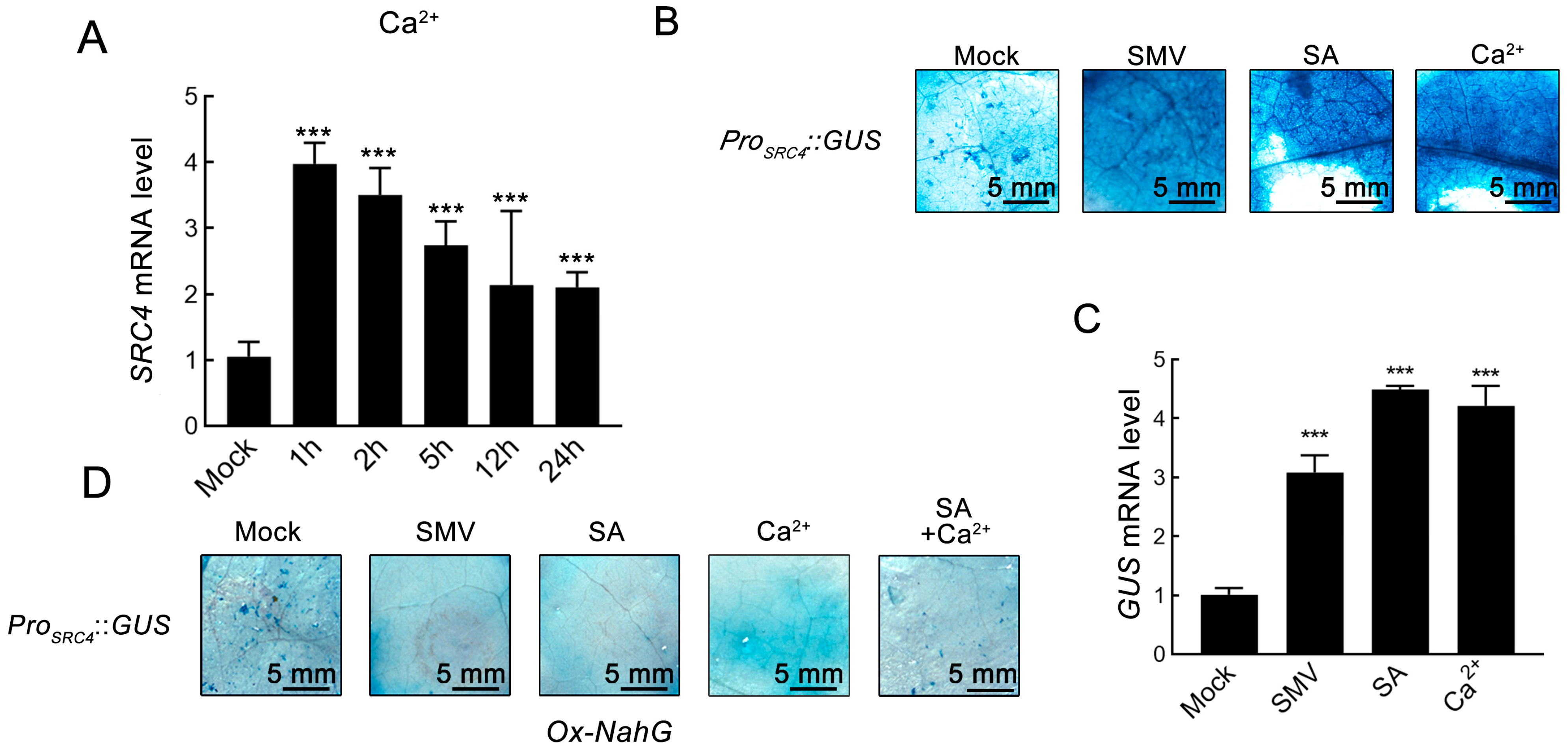
SA serves as an important defense hormone in plants, playing a key role in soybean immune responses against SMV. Based on the discovery of SA-responsive elements in the promoter analysis, we treated V3 stage soybean Williams seedlings with a foliar spray of 0.5 mM SA solution. Quantitative RT-qPCR analysis showed that SRC4 expression was significantly upregulated within 1–5 h after SA treatment, followed by gradual decline (Figure 2B). This result confirms SRC4’s responsiveness to SA signals.
Considering the important role of SA in plant antiviral responses, we further analyzed the expression patterns of SRC4 under viral infection conditions. By mining transcriptome data from three soybean cultivars in the NCBI database—Williams (SRR5466715–SRR5466722), L29 (SRR10098755–SRR10098764), and 5031 (SRR6376246–SRR6376249)—under SMV infection for local analysis, we found that SRC4 showed significant upregulation in all cultivars upon SMV infection (Figure 2C).
To precisely capture the dynamic expression changes of SRC4 during early SMV infection, we conducted time-course sampling at 1, 2, 5, 12, and 24 h post-infection, using SMV-N1 strain rub-inoculation on V3 stage soybean leaves. The results showed that SRC4 responded rapidly at transcription level after viral infection, with peak expression occurring at 2–5 h, exhibiting typical early response gene expression patterns (Figure 2D).

2.3. Ca2+ Signal-Dependent Regulatory Mechanism of SRC4 Expression

SRC4 protein possesses Ca2+-binding EF-hand domain [53]. Tobacco mosaic virus (TMV) infection of tobacco rapidly activates plasma membrane Ca2+ channels leading to increased cytoplasmic Ca2+ concentrations [55,56], and potato virus X (PVX) infection similarly triggers Ca2+ signal transduction [57]. Based on above observations, we hypothesized that transcriptional regulation of SRC4 may also be regulated by Ca2+ signals.
To test this assumption, we applied exogenous Ca2+ treatment to evaluate its effect on SRC4 expression. Three-week-old Williams soybean seedlings were treated with foliar spray of 10 mM CaCl2 solution, with time-course sampling at 1, 2, 5, 12, and 24 hpi. RT-qPCR analysis showed that exogenous Ca2+ treatment could significantly induce SRC4 expression, with peak expression occurring at 1–2 hpi by 4.1 folds of the control (Figure 3A). This result indicates that SRC4 transcriptional levels are indeed subject to positive regulation by Ca2+ signals.
To systematically verify the responsiveness of the SRC4 promoter to multiple signaling molecules, we generated a ProSRC4::GUS reporter gene construct in the tobacco transient expression system. After transforming ProSRC4::GUS fusion vectors into tobacco leaves, the following treatments were applied: (1) SMV-GFP infection (OD600 = 1.0) to simulate viral stress; (2) 0.5 mM SA spray to simulate defense hormone signals; and (3) 10 mM CaCl2 spray to simulate Ca2+ signals. GUS staining detection was performed 72 hpi. The results showed that all three treatments significantly enhanced GUS signal intensity, compared to the basal expression activity of the Mock control group (Figure 3B). RT-qPCR further confirmed that all three treatments significantly upregulated SRC4 expression at the transcriptional level (Figure 3C), confirming the multiple responsiveness of the ProSRC4 promoter to viral infection, SA, and Ca2+ signals.
Extensive research indicates that Ca2+ influx triggered by plant viral infection is a key early event in activating downstream defense responses. Ca2+ signals further activate Ca2+/calmodulin-dependent protein kinases, phosphorylating and activating key enzymes in SA biosynthesis such as phenylalanine ammonia-lyase (PAL) and isochorismate synthase (ICS), thereby promoting massive SA accumulation. Subsequently, SA, as an important defense signaling molecule, induces expression of numerous disease resistance-related genes including pathogenesis-related (PR) genes. Based on the theoretical framework of Ca2+-SA signaling cascade regulation, we hypothesized that Ca2+ might regulate SRC4 transcription through modulating SA accumulation. To test this hypothesis, we conducted functional validation experiments using transgenic tobacco overexpressing the NahG gene. NahG encodes salicylate hydroxylase from Pseudomonas putida, which can specifically catalyze the conversion of SA to catechol, thereby blocking SA accumulation in plants, making it a classic tool for studying SA function.
In Ox-NahG transgenic tobacco, ProSRC4::GUS fusion vectors were transiently expressed, and the same SMV infection, SA treatment, and Ca2+ treatment were applied. GUS staining results showed that under conditions where SA accumulation was blocked, all three treatments failed to induce reporter GUS expression, with expression levels showing no significant difference from the Mock control group (Figure 3D). This result provides strong evidence that the inductive effects of SMV infection, SA treatment, and Ca2+ treatment on SRC4 expression all depend on SA accumulation.

2.4. Tissue-Specific Expression Pattern of SRC4

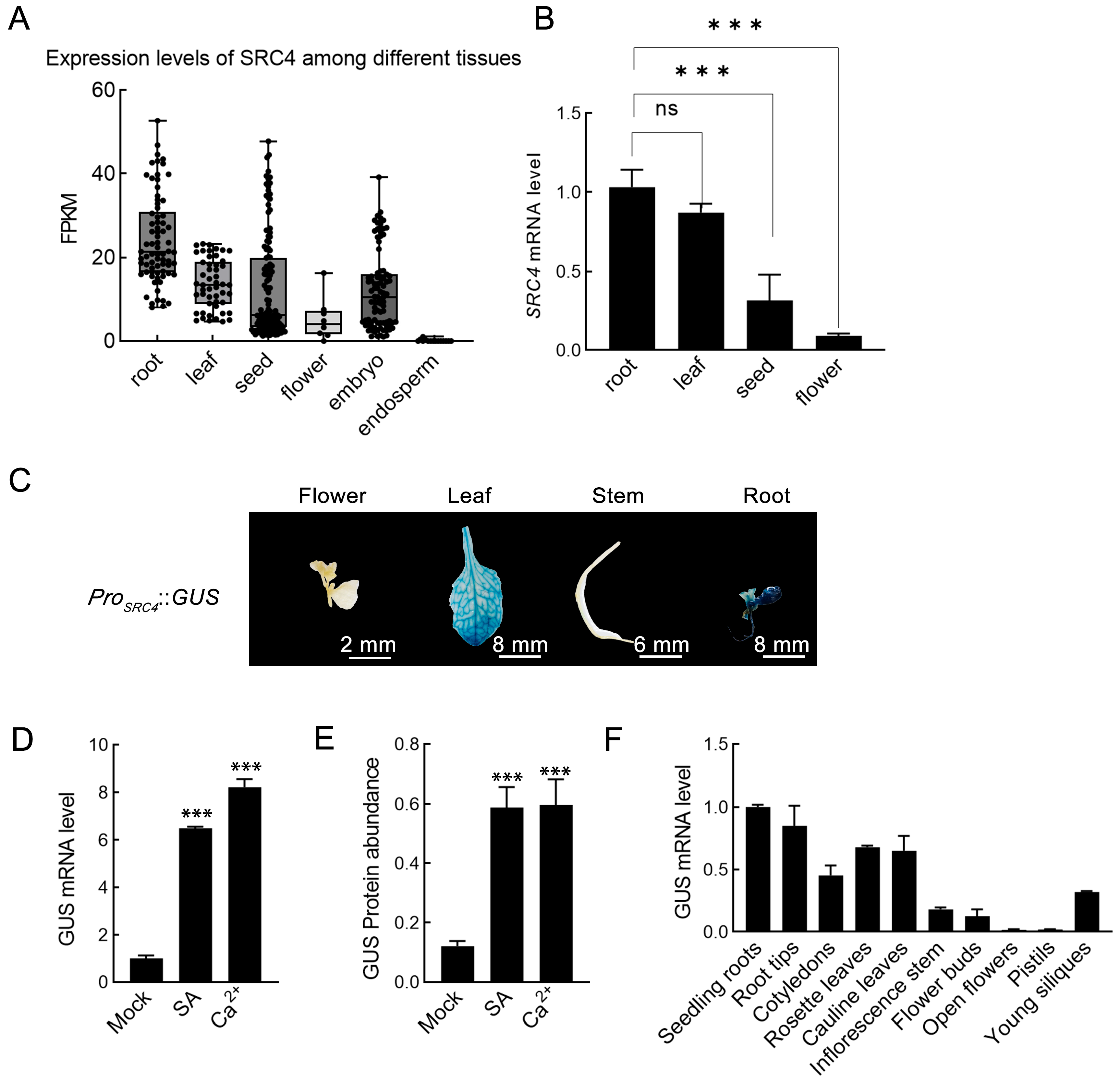
To comprehensively analyze the tissue-specific expression pattern of the SRC4 gene, we systematically analyzed tissue distribution information from 4085 soybean transcriptome datasets in the PlantRNAdb database. Through statistical analysis of expression levels in six major tissue organs, such as roots, leaves, seeds, flowers, embryos, and endosperm, we found that SRC4 exhibited obvious tissue expression preferences (Figure 4A). SRC4 showed the highest expression levels in roots, with median FPKM values reaching 20–25, suggesting that this gene may play important functions in root development, nutrient absorption, or root-specific defense responses. Expression levels in leaf tissues ranked second, which is consistent with its functional characteristics as an R gene primarily exerting defense functions in above-ground parts. Expression levels in seeds, flowers, and embryo tissues were relatively low, with FPKM values mainly distributed in the 0–15 range. In contrast, SRC4 mRNA was barely detectable in endosperm, with FPKM values close to 0, indicating extremely low transcriptional activity of this gene in seed storage tissues.
To validate transcriptome data analysis, we further performed RT-qPCR detection on four tissues—roots, leaves, seeds, and flowers—of soybean Williams cultivar. The results were highly consistent with database analysis, confirming that SRC4 is mainly highly expressed in roots and leaves, while expression levels in seeds and flowers are relatively low (Figure 4B).
To verify the functional characteristics of the ProSRC4 promoter and the conservation of its tissue expression patterns in model plants, we constructed ProSRC4::GUS transgenic Arabidopsis lines. Twenty-five independent transgenic lines were obtained through floral dip transformation, and after kanamycin resistance screening and genomic PCR verification, positive lines were selected and propagated to T3 generation for subsequent analysis. GUS histochemical staining of flowering T3 transgenic plants showed that GUS activity was mainly distributed in leaves, stems, roots, and flowers, which was basically consistent with expression patterns in soybean (Figure 4C), demonstrating the cross-species conservation of ProSRC4 promoter function.
Furthermore, we verified the responsiveness of ProSRC4 to SA and Ca2+ signals in transgenic Arabidopsis. T3 transgenic plants were treated with a foliar spray of 0.5 mM salicylic acid (SA) and 10 mM CaCl2, respectively, and GUS transcriptional levels were detected by RT-qPCR at 48 hpi. The results showed that both SA and Ca2+ treatments significantly upregulated GUS mRNA expression levels (Figure 4D). Meanwhile, we used quantitative GUS protein activity detection to further verify protein-level changes. Total leaf proteins were extracted 72 hpi, protein concentrations were determined by the Bradford method and adjusted to uniform concentration (10 μg/mL), and protein abundance was detected using GUS quantitative detection kit on a FilterMax F5 microplate reader (Molecular Devices, San Jose, CA, USA). The results showed that GUS protein accumulation in both SA and Ca2+ treatment groups was significantly higher than the Mock control group (Figure 4E). To comprehensively characterize the tissue-specific expression pattern of the SRC4 promoter, we performed RT-qPCR analysis of GUS expression across ten distinct tissues from T3 ProSRC4::GUS transgenic Arabidopsis plants These tissues included seedling roots, root tips, cotyledons, rosette leaves, cauline leaves, inflorescence stem, flower buds, open flowers, pistils, and young siliques. The results revealed differential GUS expression levels among various tissues, with the highest expression observed in root tissues (seedling roots and root tips), moderate expression in vegetative organs (cotyledons, rosette leaves, and cauline leaves), and relatively lower expression in reproductive tissues (flower buds, open flowers, pistils, and young siliques) and inflorescence stem (Figure 4F).
These results indicate that SRC4 has clear tissue expression preferences, primarily functioning in roots and leaves, and its promoter maintains responsiveness to SA and Ca2+ signals in heterologous systems.

2.5. Functional Analysis of SRC4 Response to Abiotic Stress

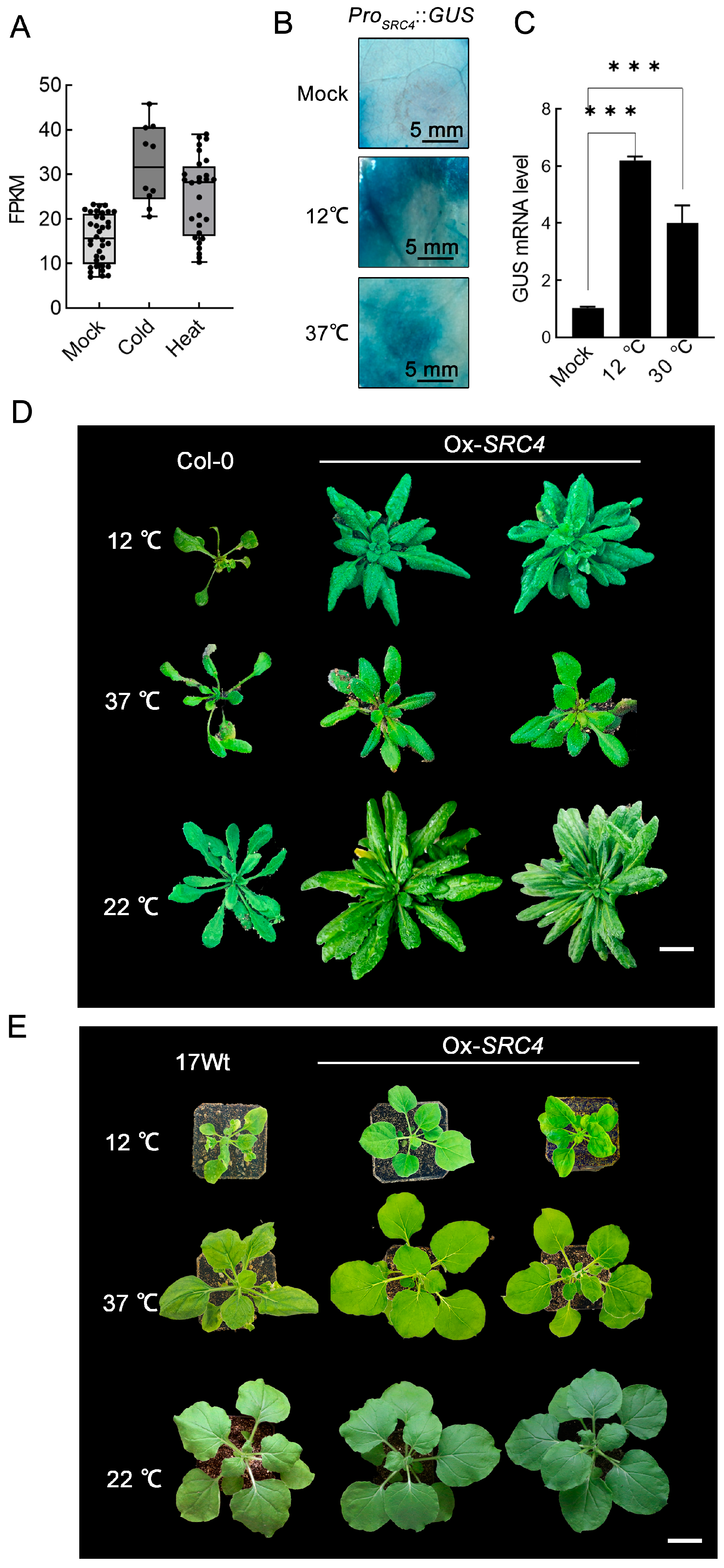
Based on previous studies of SRC4 expression characteristics under biotic stress conditions, we further explored the expression patterns of this gene under abiotic stress. Through analysis of soybean transcriptome data in the PlantRNAdb database, we found that SRC4 not only showed induced expression under pathogen stress but also exhibited significant expression upregulation under low-temperature and high-temperature stress conditions (Figure 5A). Compared to normal conditions, both cold stress and heat stress treatments significantly increased SRC4 transcriptional levels.
To verify the responsiveness of the ProSRC4 promoter to temperature stress, we conducted functional validation in the tobacco transient expression system. After transforming ProSRC4::GUS reporter vectors into tobacco leaves, 12 °C low-temperature treatment and 37 °C high-temperature treatment were applied, respectively, and GUS staining detection was performed 7 days post-treatment. The results showed that both low-temperature and high-temperature stress significantly induced enhanced GUS activity, compared to the 22 °C control group (Figure 5B), confirming the responsiveness of the ProSRC4 promoter to temperature changes.
Furthermore, we validated the above findings via the analysis in ProSRC4::GUS transgenic Arabidopsis. T3 transgenic plants were subjected to the same temperature stress treatments, and GUS mRNA levels were detected by RT-qPCR at 25 days post-treatment. The results showed that both 12 °C and 37 °C treatments significantly upregulated GUS expression, which was highly consistent with tobacco transient expression results (Figure 5C).
To evaluate the effect of SRC4 overexpression on plant temperature stress tolerance, we constructed transgenic Arabidopsis lines overexpressing SRC4. The full-length SRC4 sequence was cloned into the pNC-Cam3304-MCS35S vector. Through herbicide resistance screening, genomic PCR verification, and T2 generation RT-qPCR analysis, we successfully obtained Ox-SRC4 transgenic Arabidopsis lines stably expressing SRC4, which were propagated to T3 generation for phenotypic analysis (Supplementary Figure S1).
In temperature stress tolerance tests, T3 Ox-SRC4 transgenic plants and wild-type Col-0 simultaneously received 12 °C low-temperature stress and 37 °C high-temperature stress treatments for 25 days, and plant growth status was observed. The results showed that Ox-SRC4 transgenic lines exhibited significantly better growth performance than wild-type under both temperature stress conditions. Under low-temperature conditions, Ox-SRC4 transgenic plants maintained normal leaf expansion and growth status; under high-temperature conditions, Ox-SRC4 transgenic plants also showed stronger tolerance, with significantly reduced leaf wilting (Figure 5D). Similar results were also validated in Ox-SRC4 transgenic tobacco, which shows higher tolerance to both low and high-temperature treatment compared with 17Wt tobacco (Figure 5E). Notably, the growth of Ox-SRC4 transgenic tobacco was not inhibited under normal conditions or Ca2+ treatment, demonstrating the absence of fitness penalties typically associated with resistance gene overexpression (Supplementary Figure S2).
These results indicate that the SRC4 gene not only participates in plant biotic stress responses but also plays important roles in abiotic stress adaptation, significantly enhancing plant tolerance to temperature stress.

3. Discussion

High expression of R genes can enhance plant tolerance to biotic stresses (such as pathogen invasion) or abiotic stresses (such as drought, high salinity, and low temperature). However, prolonged overexpression of such genes often imposes metabolic burdens on plant growth and development, thereby affecting plant growth rates or biomass accumulation. Therefore, to maintain normal growth and development, typical R genes usually remain in low expression states under non-stressed conditions, and they are induced and activated only under external environmental stimuli to exert their resistance functions.
Our research represents the first discovery that the SRC4 gene exhibits a high basal expression pattern significantly different from typical NBS-LRR genes, challenging the classical “low expression-high responsiveness” regulatory paradigm of plant R genes. Approximately 72% of NBS-LRR genes maintain constitutive expression at low levels, with most R genes exhibiting strictly controlled basal expression to minimize fitness costs [9,15]. Classical R genes such as tobacco N gene, Arabidopsis RPS2 and RPM1 maintain low expression or transcriptionally silenced states under non-stressed conditions [58,59]. Through systematic analysis of 4085 soybean transcriptome datasets, we found that SRC4 FPKM values were significantly higher than those of the typical R gene SRC7 (FPKM < 2) in the same gene cluster. This fundamental difference in expression patterns suggests that SRC4 may represent a novel class of R genes with unique regulatory mechanisms and biological functions.
Traditional perspectives hold that constitutive expression of R genes leads to severe fitness costs, including negative effects such as plant dwarfism, biomass reduction, and the modification of hormonal balance, with subsequent epigenetic re-programming and constitutive activation of defense responses [17,21]. Overexpression of the R gene Prf1 causes constitutive activation of defense responses that severely affect normal tomato growth [20]. Under field conditions, this can cause fitness losses of up to 10% in plant yield. However, our previous research indicated that the high basal expression of SRC4 does not cause obvious growth and developmental defects, a phenomenon that may be closely related to its unique functional mechanisms. An important explanation is that SRC4 protein can alter its disease resistance function through binding with Ca2+, and this ligand-dependent activation mechanism may enable plants to avoid constitutive activation of defense responses while maintaining high protein abundance. Similar mechanisms have been reported in some calmodulin-dependent kinases, which remain inactive without Ca2+ binding and function only under specific signal stimulation [60]. In rice, there is a mechanism through competitive inhibition of PigmR homodimerization by PigmS, cleverly achieving fine regulation of disease resistance intensity while avoiding adverse effects of excessive immune responses on growth [61].
The high expression of SRC4 reflects adaptive evolutionary strategies of plants when facing complex and variable environments. Unlike inducible expression that requires de novo protein synthesis, high expression ensures rapid availability of defense proteins, enabling plants to quickly initiate defense responses in the very early stages of pathogen invasion. This “pre-storage” strategy is particularly important in viral infections, as viral replication is extremely rapid and traditional transcriptional induction may not be timely enough to prevent early viral establishment [62]. Additionally, we observed high expression of SRC4 in roots and leaves, suggesting it may possess multiple biological roles beyond disease resistance functions, and this functional diversity may be an important reason for maintaining its high expression.
The expression pattern of SRC4 may represent adaptive innovation of plant immune systems in specific ecological niches. In environments with high incidence of RNA viruses such as SMV, maintaining high levels of disease resistance proteins may confer greater selective advantages than conserving metabolic costs. In natural populations with high pathogen pressure, certain R genes tend to maintain higher basal expression levels [63]. Meanwhile, the high expression pattern of SRC4 is also consistent with plant “priming” defense strategies, which balance defense efficacy with metabolic costs by maintaining moderate defensive preparedness [64].
Salicylic acid (SA), as a key signaling molecule in plant defense responses, plays a central regulatory role in inducing R gene expression [29]. Extensive research confirms that SA can significantly induce expression of pathogenesis-related genes, which are widely used as molecular markers of SAR [65]. In Arabidopsis and tobacco, SA treatment can rapidly activate expression of PR-1, PR-2, and PR-5 genes, with PR-1 gene expression being considered the primary marker of SAR establishment [66]. In rice, the R gene OsNPR3.3 was upregulated 4-fold at 24 h after 2 mM SA treatment [67], which is consistent with our findings of SA-induced upregulation of SRC4 expression. Salicylic acid also functions as a multifunctional endogenous substance for treating plant diseases. It plays important roles in alleviating abiotic stresses including high temperature, low temperature, drought, UV radiation, heavy metals, and osmotic shock [68,69].
Upregulation of R gene expression during pathogen invasion is an important phenomenon in plant immune responses. Many NBS-LRR resistance genes exhibit low-level sustained expression in healthy, unchallenged tissues but are upregulated under bacterial flagellin stimulation, consistent with basal resistance induction, indicating the need for rapid responses to pathogen attacks [9]. In maize (Zea mays), the ZmNBS25 gene shows relatively low expression levels in uninfected maize leaves but is significantly upregulated 24 h after inoculation with Bipolaris maydis [70]. From an evolutionary perspective, inducible expression may be an adaptive strategy for plants facing pathogen pressure, providing effective defense during pathogen attacks while avoiding fitness costs that might result from overexpression of R genes under normal conditions [21]. In our study, SRC4 similarly showed upregulated expression after SMV infection, which is highly consistent with the expression patterns of the above R genes.
As an important second messenger in cells, Ca2+ can also induce gene expression upregulation. Early research by Braam (1992) showed that elevated extracellular Ca2+ increased expression of genes encoding calcium sensors, and heat shock or cold shock induction of some genes depends on exogenous Ca2+ signals [71]. SRC4 mRNA levels were upregulated by Ca2+ addition. Through time-course expression analysis, we found that Ca2+ treatment-induced peak SRC4 expression occurred at 1–2 h, while SMV infection-induced expression peaks occurred at 2–5 h. This temporal difference is consistent with the sequence of Ca2+-SA signaling. Ca2+, as an early signaling molecule in plant immune responses, can rapidly activate downstream transcriptional regulatory networks, while SA biosynthesis and accumulation as a defense hormone requires longer time [31,32].
The Ca2+ signaling pathway is a key regulatory mechanism for initiating salicylic acid biosynthesis, with transcriptional regulation of the ICS1 gene playing a decisive role [72]. When plant cells recognize pathogens, plasma membrane Ca2+ channels rapidly open, causing transient elevation of cytoplasmic Ca2+ concentrations. This Ca2+ signal subsequently coordinately activates ICS1 gene transcriptional expression through two functionally complementary parallel pathways—the calmodulin-dependent CBP60g pathway and the calmodulin-independent SARD1 pathway—thereby initiating the salicylic acid biosynthesis process. Notably, CBP60g family transcription factors exhibit high evolutionary conservation across plant lineages, suggesting that the Ca2+-SA regulatory network may have ancient evolutionary origins, reflecting the fundamental status of this signaling pathway in plant immune systems [31].
To verify whether the SRC4 gene is similarly regulated by SA signals, we conducted functional validation experiments using the NahG transgenic tobacco system. NahG-encoded salicylate hydroxylase can specifically degrade SA, thereby blocking SA accumulation. Experimental results clearly showed that under conditions where SA accumulation was effectively blocked by NahG enzyme, Ca2+ treatment, exogenous SA treatment, and SMV viral infection all completely lost their inductive effects on SRC4 gene expression. This key finding strongly demonstrates that SA plays an irreplaceable central role in SRC4 transcriptional activation and establishes SRC4’s molecular identity as a downstream response gene in the SA signaling pathway. This strict SA-dependent regulatory pattern is highly similar to the classical CBP60g-SARD1-ICS1 signaling [35,73], further supporting the hypothesis that SRC4 participates in plant SA-mediated immune responses.
Analysis of the SRC4 promoter region provides important molecular evidence for the above regulatory mechanisms. Within the 2000 bp region upstream of the transcription start site, we identified 12 different types of cis-acting elements, among which the presence of SA-responsive elements (located at −1761~−1731 bp) directly explains SRC4’s strong transcriptional response to SA treatment. Plant SA-responsive elements typically contain TGACG or related sequence motifs that can be recognized and bound by NPR1-TGA transcription factor complexes [74]. Compared to classical SA-responsive genes, the upstream region of SA-responsive elements in the SRC4 promoter also contains multiple other regulatory elements, including two MYB binding sites (−1144~−1114 bp and −537~−507 bp), two defense and stress-responsive elements (−1591~−1561 bp and −188~−158 bp), and various hormone-responsive elements. This combinatorial element arrangement enables SRC4 to integrate multiple environmental signals. The dual distribution of defense and stress-responsive elements in the distal and proximal regions of the SRC4 promoter also represents its unique expression pattern. Distal elements typically participate in maintaining basal expression levels, while proximal elements are more involved in rapid activation of inducible expression [75]. This spatial distribution pattern highly matches our observed SRC4 expression characteristics of high basal expression with strong inducibility. Additionally, the presence of ABA-responsive and GA-responsive elements suggests that SRC4 expression is closely related to plant stress states and growth-developmental states, and this integration of growth-defense signals may be an important mechanism for SRC4 to avoid severe fitness costs [76]. Based on these findings, we speculate that transcriptional regulation of SRC4 involves complex signal integration networks. Ca2+ signals may phosphorylate related transcription factors by activating calcium-dependent protein kinases (CDPKs), while accumulated SA directly activates SA-responsive elements in the SRC4 promoter through NPR1-TGA complexes. Meanwhile, MYB transcription factors may participate in coordinating light signals with immune signals, while various hormone-responsive elements ensure adaptive expression of SRC4 under different physiological states.
Our research demonstrates that SRC4 possesses the unique ability to simultaneously respond to both biotic and abiotic stresses. Through systematic analysis of soybean transcriptome data, we found that SRC4 not only shows significantly upregulated expression under SMV infection conditions but also exhibits significant transcriptional activation under low-temperature and high-temperature stress conditions. This multiple stress response capability is extremely rare among R genes. The regulatory mechanisms of temperature stress on SRC4 expression may involve molecular networks at multiple levels. The various stress-responsive elements we discovered in the SRC4 promoter provide a molecular basis for its temperature sensitivity, while Ca2+ signals, serving as common mediators for temperature sensing and immune activation, may be the key link connecting these two stress responses. Temperature changes, particularly low-temperature stress, can rapidly activate mechanosensitive Ca2+ channels on plant cell membranes, leading to elevated cytoplasmic Ca2+ concentrations [77,78]. The spatiotemporal patterns of such Ca2+ signal generation may differ from Ca2+ influx caused by pathogen invasion, but both can be recognized and responded to by SRC4’s regulatory network, thus achieving unified regulation of different types of stress.
More importantly, we confirmed through overexpression plant Ox-SRC4 experiments that SRC4’s multiple stress response capability has important biological functions. Ox-SRC4 transgenic Arabidopsis and tobacco both exhibited significantly better growth performance and stress tolerance than wild-type under 12 °C low-temperature stress and 37 °C high-temperature stress conditions. This result indicates that SRC4 can not only sense temperature changes but also enhance plant temperature adaptability through the functional activities of its protein products. Considering that SRC4 protein possesses Ca2+-binding capability, we speculate that its temperature protection function may be related to maintaining Ca2+ homeostasis. Under temperature stress conditions, drastic fluctuations in intracellular Ca2+ concentrations often lead to cellular dysfunction [79], and SRC4 protein may buffer these fluctuations through its Ca2+-binding capability and Ca2+ regulatory capacity, thereby protecting cells from temperature stress damage.
The interaction between biotic and abiotic stress signaling pathways is an important strategy for plants to adapt to complex environments. Plants in natural environments often face challenges from multiple stresses simultaneously, and complex interactions exist among these stresses [80]. For example, temperature stress can affect plant susceptibility to pathogens, while pathogen invasion can also alter plant temperature tolerance. SRC4’s multiple stress response capability may enable plants to maintain good adaptability in such complex stress networks. When plants simultaneously face viral infection and temperature stress, SRC4 activation can provide both disease resistance protection and enhanced temperature tolerance, thereby maximizing plant survival opportunities.
From an evolutionary biology perspective, SRC4’s multiple stress response characteristics may reflect adaptive evolution of plant immune systems under climate change contexts. With global warming and increasing extreme weather events, plants increasingly need the capability to simultaneously cope with multiple stresses. Traditional single-function R genes may face selective disadvantages in such environments [81], while genes with multifunctional characteristics like SRC4 are more likely to be retained and optimized by natural selection, as maintaining high levels of defense proteins may confer greater selective advantages than conserving metabolic costs in multiple stress environments [82].
Compared to other reported multiple stress response genes, SRC4 possesses unique functional characteristics. For example, the DREB transcription factor family in Arabidopsis can simultaneously regulate drought and cold stress responses but primarily functions through transcriptional regulation [83]. As an immune receptor protein, SRC4’s multiple stress response function is more reflected in direct actions at the protein level. This difference suggests that plants may achieve adaptation to complex environments through regulatory mechanisms at different levels, and SRC4 represents an important example of functional diversification of immune receptor proteins.
Our research reveals that SRC4 has obvious tissue-specific expression patterns, with its high expression levels in roots and leaves suggesting that this gene may possess multiple biological roles beyond traditional disease resistance functions. Most NBS-LRR resistance genes, such as Arabidopsis RPM1 and RPS2, are mainly expressed in leaves with relatively low expression levels in roots [84]. We found that SRC4 exhibits the highest expression levels in roots, with leaf tissue expression ranking second, while expression in reproductive tissues such as seeds, flowers, and embryos is relatively low, with almost no expression in endosperm, suggesting that SRC4 may play important physiological functions in plant vegetative organs. Root systems, as the primary contact interface between plants and soil environments, not only undertake functions of water and nutrient absorption but also face continuous threats from soil-borne pathogens [85]. The rhizosphere environment contains numerous pathogens including bacteria, fungi, and nematodes, which differ significantly from foliar pathogens in invasion strategies and effector molecule composition [86,87]. High expression of SRC4 in roots may enable plants to rapidly recognize and respond to rhizosphere pathogen invasion, providing first-line defense protection. Reports have also shown that SRC4 expression is significantly increased after infection with soybean cyst nematode (SCN), indicating that SRC4 functions in SCN resistance [88]. Additionally, root systems are important organs for plants to perceive environmental changes, and changes in soil temperature, humidity, and ion concentrations are first perceived by root systems. High expression of SRC4 may enable root systems to rapidly respond to these environmental signals and initiate corresponding adaptive responses.
Particularly noteworthy is that the availability of Ca2+ in the root environment provides ideal conditions for SRC4 function. Soil solutions contain abundant Ca2+, and root cell membranes possess various Ca2+ transporters and channels that can precisely regulate intracellular Ca2+ concentrations [60]. Considering that SRC4 protein can bind Ca2+ to alter its resistance intensity, the high Ca2+ environment in roots may provide favorable conditions for conformational activation and functional performance of SRC4 protein. This spatial distribution matching suggests that tissue-specific expression of SRC4 may be an important manifestation of its functional optimization.
High expression of SRC4 in leaves is more directly related to its antiviral functions. Leaves are the primary target tissues for most plant virus infections, with viruses such as SMV being transmitted and infected through vector insects such as aphids feeding on leaves. High expression of SRC4 in leaves ensures sufficient immune receptor proteins are available to recognize viral effector molecules and initiate defense responses in the early stages of viral infection. The rapid upregulation of SRC4 expression within 2–5 h after SMV infection that we observed, combined with its high basal expression in leaves, forms an efficient viral defense system.
Although this study provides important insights into understanding SRC4’s unique regulatory mechanisms, some limitations remain that need to be addressed in future work. First, our research is mainly based on transcriptional level analysis, and we still lack in-depth understanding of SRC4 protein post-translational modifications, subcellular localization dynamics, and interaction networks with other proteins. The molecular details of how Ca2+ binding precisely regulates SRC4 protein conformational changes and functional domain activation, and whether SRC4 has other ligand-dependent regulatory mechanisms, require further biochemical and structural biology studies for elucidation. Second, although we predicted the involvement of multiple transcriptional regulatory factors through promoter analysis, direct binding of these factors to the SRC4 promoter and their interaction relationships have not been experimentally verified.
In terms of functional validation, current research is mainly conducted under laboratory-controlled conditions, and SRC4’s performance when facing complex and variable combinations of biotic and abiotic stresses in real field environments still requires large-scale field trials for verification. Additionally, our cross-species functional validation is mainly limited to Arabidopsis and tobacco model plants, and the functional conservation and application potential of SRC4 in other important crops require broader validation. Epigenetic regulatory mechanisms, such as the roles of DNA methylation and histone modifications in SRC4 expression regulation, are also important directions worthy of in-depth exploration.

3.1. Limitations

While this study provides valuable insights into SRC4’s transcriptional regulation, we acknowledge several areas that warrant further investigation. Our analysis primarily examined transcriptional-level regulation, while post-translational modifications of SRC4 remain to be explored, particularly the precise molecular mechanisms of how Ca2+ binding affects protein conformation and activity. The promoter–transcription factor interactions were computationally predicted through bioinformatic analysis, though direct experimental validation of these binding events, especially for the SA-responsive and MYB binding sites, would strengthen our mechanistic understanding. Additionally, while our functional validation in heterologous model systems (N. benthamiana and Arabidopsis) provided valuable mechanistic insights, these systems may not fully capture the complex regulatory networks present in native soybean or the multi-stress interactions that occur under natural field conditions. Our tissue expression analysis was conducted at the organ level, and higher-resolution cellular localization studies would provide more detailed information about SRC4’s spatial organization and cell-type specificity within different tissues. Our tissue expression analysis was conducted at the organ level, which provides averaged expression data across multiple cell types with distinct mRNA profiles that may respond differently to external signals. Therefore, our organ-level expression data can only provide initial assumptions about SRC4 regulation rather than definitive conclusions about cellular-level mechanisms. Future studies with higher cellular resolution would be essential to achieve true cell-type specificity.

3.2. Future Directions

Building upon these foundations, several research directions would enhance our understanding of SRC4 function: biochemical characterization of SRC4 protein structure and Ca2+-induced conformational changes, experimental validation of predicted promoter-transcription factor interactions through chromatin immunoprecipitation or electrophoretic mobility shift assays, comprehensive field trials in soybean to evaluate dual stress resistance under natural multi-stress conditions, high-resolution cellular localization studies using advanced microscopic techniques, and investigation of SRC4’s broader role in integrating Ca2+-SA signaling networks. These studies will be essential for fully realizing SRC4’s potential in developing climate-resilient crops and deepening our understanding of its unique regulatory mechanisms.

4. Materials and Methods

4.1. Plant Materials and Growth Conditions

Soybean (Glycine max) cultivar Williams was used for gene expression analysis and promoter cloning. Seeds were surface-sterilized with 10% sodium hypochlorite for 10 min, rinsed five times with sterile water, and directly sown in a 1:1 mixture of nutrient soil and vermiculite at 25 °C under a 16 h light/8 h dark photoperiod. V3-stage soybean plants were used for SA treatment, Ca2+ treatment, and SMV inoculation experiments.
Arabidopsis thaliana ecotype Columbia-0 (Col-0) and transgenic tobacco (Nicotiana benthamiana) plants were grown in a controlled growth chamber at 22 °C under a 16 h light/8 h dark photoperiod with 60% relative humidity. NahG-overexpressing tobacco plants were kindly provided by Professor Jun Liu from China Agricultural University and maintained under the same conditions. The plant materials used in this study also included ProSRC4::GUS transgenic Arabidopsis, Ox-SRC4 At transgenic Arabidopsis, wild-type N. benthamiana 17Wt, and Ox-SRC4 Nb transgenic tobacco plants, all maintained under the same growth conditions.

4.2. Transcriptome Data Analysis

Publicly available soybean RNA-seq datasets were downloaded from the NCBI Sequence Read Archive (SRA) database using SRA Toolkit v3.0.7. Raw sequencing data were converted to FASTQ format and subjected to quality control using FastQC v0.11.9. Expression quantification was performed using Kallisto v0.46.2 with the soybean reference transcriptome from Phytozome v13 (Glycine max Wm82.a4.v1). Transcript abundance was calculated as transcripts per million (TPM) values.
SMV infection-responsive expression data were obtained from three soybean cultivars: Williams (SRR5466715–SRR5466722), L29 (SRR10098755–SRR10098764), and 5031 (SRR6376246–SRR6376249). Differential expression analysis was performed comparing mock-inoculated controls with SMV-infected samples at 12 h post-inoculation (hpi).
Comprehensive expression analysis of SRC4 across different tissues, developmental stages, and stress conditions was conducted using the PlantRNAdb platform (http://ipf.sustech.edu.cn/pub/plantrna/, accessed on 11 May 2024). This database systematically integrates publicly available soybean RNA-seq data from major repositories including Gene Expression Omnibus (GEO), the Sequence Read Archive (SRA), the European Nucleotide Archive (ENA), and the DNA Data Bank of Japan (DDBJ). The 4085 soybean RNA-seq samples encompass diverse experimental conditions including various organs (roots, leaves, stems, seeds, flowers, pods), developmental stages (seedling, vegetative, reproductive, senescence), tissue types, and treatment conditions (biotic stress, abiotic stress, hormone treatments, and control conditions) with high coverage and data consistency. Expression levels were extracted as FPKM (fragments per kilobase of transcript per million mapped reads) values for comparative analysis.

4.3. Promoter Analysis and Vector Construction

Cis-acting regulatory elements were predicted for the 2000 bp genomic sequence upstream of the SRC4 translation start site using PlantCARE software (http://bioinformatics.psb.ugent.be/webtools/plantcare/html/, Version: 1, accessed on 25 December 2023) [89]. The promoter sequence was retrieved from the soybean genome database (Phytozome v13, Glycine max Wm82.a4.v1, accessed on 25 December 2023) and analyzed for the presence of various regulatory motifs including hormone-responsive elements, stress-responsive elements, and tissue-specific elements.
Genomic DNA was isolated from young soybean (Williams cultivar) leaves using the CTAB (cetyltrimethylammonium bromide) method according to standard protocols. The SRC4 promoter region (ProSRC4, approximately 2000 bp upstream of ATG) was PCR amplified using high-fidelity DNA polymerase with specific primers containing Nimble Cloning (NC) universal adapter sequences (forward adapter: AGTGGTCTCTGTCCAGTCCT; reverse adapter: GGTCTCAGCAGACCACAAGT) [90].
The amplified ProSRC4 fragment was gel-purified and cloned into the pNC-121-Pro binary vector using the NC cloning system. The cloning reaction was performed in a total volume of 10 μL containing 5 μL Nimble Mix. The resulting ProSRC4::GUS construct was transformed into Escherichia coli DH5α competent cells, and positive clones were verified by colony PCR and Sanger sequencing. Similarly, the ProSRC4::LUC construct was generated by cloning the promoter fragment into the pNC-Green-LUC vector. For overexpression studies, the full-length SRC4 coding sequence was cloned into the pNC-Cam3304-MCS35S vector downstream of the CaMV 35S promoter. All recombinant vectors were confirmed by restriction enzyme digestion and sequencing analysis before transformation into Agrobacterium tumefaciens strain GV3101.

4.4. Plant Transformation and Transgenic Line Generation

Transient expression assays were performed using 4–6 week-old N. benthamiana plants. Agrobacterium cultures containing the desired constructs were grown overnight at 28 °C, harvested by centrifugation, and resuspended in infiltration buffer (10 mM MES-KOH pH 5.6, 10 mM MgCl2, 100 μM acetosyringone) to an OD600 of 1.0. The bacterial suspension was infiltrated into the abaxial surface of fully expanded leaves using a needleless syringe. Plants were maintained under normal growth conditions and sampled at indicated time points.
The promoter-GUS recombinant vectors were infiltrated into N. benthamiana leaves for promoter activity analysis. For stress treatment assays, infiltrated plants were subjected to various treatments including SMV inoculation (OD600 = 1.0), SA treatment (0.5 mM), and Ca2+ treatment (10 mM CaCl2) at 24 h post-infiltration. Leaf discs (1 cm in diameter) were excised from the infiltrated areas at 48–72 h post-treatment and subjected to GUS histochemical staining using the GUS staining kit (G3060-100 mL Solarbio, Beijing, China) according to the manufacturer’s instructions. To validate the SA-dependency of SRC4 promoter activation, the same constructs were also infiltrated into NahG-overexpressing tobacco plants.
Arabidopsis transformation was performed using the floral dip method [53]. Agrobacterium cells harboring the expression constructs were resuspended in 5% (w/v) sucrose solution containing 0.05% (v/v) Silwet L-77. Flowering Arabidopsis plants were inverted and dipped into the bacterial suspension for 30 s with gentle agitation. Treated plants were covered with plastic wrap and kept in darkness for 24 h, then returned to normal growth conditions. A second transformation was performed 7–10 days later to improve efficiency.
T1 seeds were surface-sterilized and plated on 1/2 MS medium containing appropriate selection agents: 50 mg/L kanamycin for ProSRC4::GUS constructs (pNC-121-Pro vector), or 10 mg/L basta (glufosinate ammonium) for Ox-SRC4 constructs (pNC-Cam3304 vector). Resistant seedlings were transferred to soil and grown to maturity. Homozygous T3 lines were identified by antibiotic/herbicide resistance segregation analysis and confirmed by genomic PCR using construct-specific primers. For tobacco transformation, Agrobacterium strains carrying the Ox-SRC4 constructs were used to transform tobacco leaves following standard leaf disc transformation protocols.

4.5. Gene Expression Analysis

Total RNA was extracted from various plant tissues, including soybean root, stem, leaf, and flower tissues, as well as Arabidopsis leaves and whole plants, and the inoculation sites (punctured zones) of N. benthamiana, using TRIzol reagent (Invitrogen, Carlsbad, CA, USA) according to the manufacturer’s instructions. For comprehensive tissue-specific expression analysis, ten distinct tissues were collected from T3 ProSRC4::GUS Arabidopsis plants at different developmental stages, including seedling roots (6-day-old seedlings), root tips (1–2 mm), cotyledons (6-day-old seedlings), rosette leaves (3-week-old plants), cauline leaves (4-week-old flowering plants), inflorescence stem (4-week-old flowering plants), flower buds (stage 9–12), open flowers (stage 13–14), pistils (hand-dissected from mature flowers), and young siliques (3–5 days post-fertilization). RNA quality and concentration were assessed using a NanoDrop spectrophotometer (Thermo Fisher Scientific, Waltham, MA, USA). First-strand cDNA was synthesized from 1 μg of total RNA using the HiScript III RT SuperMix for qPCR kit (Vazyme, Nanjing, China). RT-qPCR was performed using ChamQ Universal SYBR qPCR Master Mix (Vazyme, Q711) on a CFX96 Real-Time System (Bio-Rad, Hercules, CA, USA). Gene-specific primers were designed using Primer3Plus software (http://www.bioinformatics.nl/cgi-bin/primer3plus/primer3plus.cgi, version: 3.3.0, accessed on 1 September 2023). Soybean actin (GmActin), tobacco actin (NbActin), and Arabidopsis actin (AtActin) were used as internal reference genes for normalization. Relative expression levels were calculated using the 2−ΔΔct method. All experiments included three biological replicates with three technical repeats each.

4.6. GUS Activity Analysis

For quantitative GUS analysis in transgenic Arabidopsis, total protein was extracted from leaf tissues using the Plant Protein Extraction Kit (BC3720, Solarbio , Beijing, China) according to the manufacturer’s instructions. Protein concentration was determined using the Bradford assay (SK1060, Coolaber, Beijing, China) and adjusted to 10 μg/mL. GUS activity was measured using a GUS quantitative detection kit (SL7161, Coolaber, Beijing, China) and a FilterMax F5 microplate reader (Molecular Devices, San Jose, CA, USA).
For promoter responsiveness assays in transgenic Arabidopsis, salicylate (SA) solution (0.5 mM) was applied as a whole-plant spray on T3 transgenic plants, and Ca2+ treatment was performed using 10 mM CaCl2 solution. GUS activity was determined by histochemical staining using the GUS staining kit (G3060, Solarbio, Beijing, China) according to the manufacturer’s instructions. Samples were collected at 48 h post-treatment for RT-qPCR analysis and at 72 h for protein activity quantification.

4.7. Stress Treatment Experiments

For salicylic acid (SA) treatment, V3-stage soybean plants or infiltrated tobacco leaves were sprayed with 0.5 mM SA solution. For Ca2+ treatment, V3-stage soybean plants were treated with 10 mM CaCl2 solution by foliar spray. Mock treatments used water. Samples were collected at 1, 2, 5, 12, and 24 h post-treatment for expression analysis.
SMV-N1 strain was mechanically inoculated onto V3-stage soybean leaves. Infected leaves carrying the virus strain were ground with 0.02 × PBS buffer and silica sand to prepare virus suspension, which was then applied by mechanical friction inoculation. Mock inoculations were performed using virus-free buffer. Leaf samples were collected at 1, 2, 5, 12, and 24 hpi for temporal expression analysis.
For temperature stress assays, plants were transferred to growth chambers set at 12 °C (cold stress) or 37 °C (heat stress), with mock controls maintained at 22 °C. Tobacco plants were treated for 7 days, while Arabidopsis plants were grown continuously for 25 days under these conditions. Plant phenotypes were photographed, and tissue samples were collected for gene expression analysis.

4.8. Statistical Analysis

All experiments were conducted with at least three biological replicates. Data are presented as mean ± SD deviation (SD). Statistical significance was determined using one-way ANOVA followed by Dunnett’s post-hoc test or Student’s t-test as appropriate. Significance levels were indicated as follows: * p < 0.05, and *** p < 0.001. p-values < 0.05 were considered statistically significant. Statistical analyses were performed using GraphPad Prism 8.0 software (GraphPad Software, San Diego, CA, USA).

Supplementary Materials

The following supporting information can be downloaded at https://www.mdpi.com/article/10.3390/plants14182820/s1, Figure S1: Screening and verification of SRC4 overexpressing transgenic Arabidopsis lines. Figure S2: Morphological comparison of transgenic tobacco plants stably expressing SRC4 and wild-type controls under different treatment conditions.

Author Contributions

Validation, Y.Z.; Investigation, N.N.; Data curation, Z.B.; Writing—original draft, Z.Z.; Writing—review & editing, H.W.; Visualization, D.M.; Supervision, H.W.; Funding acquisition, H.W. All authors have read and agreed to the published version of the manuscript.

Funding

This study was supported by the Inner Mongolia Autonomous Region Science and Technology Plan Project (2020GG0045), Inner Mongolia Autonomous Region Higher Education Innovation Team Development Plan (NMGIRT2329), Key Project of Inner Mongolia Natural Science Foundation (2024ZD20), and Open Project of Inner Mongolia University Research Platform (HECB20240F04).

Conflicts of Interest

The authors declare no conflict of interest.

References

  1. Zhao, J.; Yang, H.; Weng, Q.; Zhu, C.; Liu, H.; Lu, X.; Jin, X.; Xu, M.; Huang, L.; Zhang, H.; et al. Current Status and Prospects of Wheat Stripe Rust Research. J. Integr. Agric. 2022, 21, 3706–3721. [Google Scholar]
  2. Savary, S.; Willocquet, L.; Pethybridge, S.J.; Esker, P.; McRoberts, N.; Nelson, A. The Global Burden of Pathogens and Pests on Major Food Crops. Nat. Ecol. Evol. 2019, 3, 430–439. [Google Scholar] [CrossRef]
  3. Karmakar, S.; Molla, K.A.; Chanda, P.K.; Sarkar, S.N.; Datta, S.K.; Datta, K. Green Super Rice for Saltwater Agriculture. Nat. Biotechnol. 2024, 42, 59–66. [Google Scholar]
  4. Singh, R.P.; Hodson, D.P.; Huerta-Espino, J.; Jin, Y.; Bhavani, S.; Njau, P.; Herrera-Foessel, S.; Singh, P.K.; Singh, S.; Govindan, V. The Emergence of Ug99 Races of the Stem Rust Fungus Is a Threat to World Wheat Production. Annu. Rev. Phytopathol. 2011, 49, 465–481. [Google Scholar] [CrossRef]
  5. Pandit, M.K.; Pocock, M.J.; Kunin, W.E. Ploidy Influences Rarity and Invasiveness in Plants. J. Ecol. 2011, 99, 1108–1115. [Google Scholar] [CrossRef]
  6. Li, J.; Besseau, S.; Törönen, P.; Sipari, N.; Kollist, H.; Holm, L.; Palva, E.T. Defense-Related Transcription Factors WRKY70 and WRKY54 Modulate Osmotic Stress Tolerance by Regulating Stomatal Aperture in Arabidopsis. New Phytol. 2013, 200, 457–472. [Google Scholar] [CrossRef] [PubMed]
  7. Jones, J.D.; Dangl, J.L. The Plant Immune System. Nature 2006, 444, 323–329. [Google Scholar] [CrossRef] [PubMed]
  8. Zhou, J.M.; Zhang, Y. Plant Immunity: Danger Perception and Signaling. Cell 2020, 181, 978–989. [Google Scholar] [CrossRef]
  9. McHale, L.; Tan, X.; Koehl, P.; Michelmore, R.W. Plant NBS-LRR Proteins: Adaptable Guards. Genome Biol. 2006, 7, 212. [Google Scholar] [CrossRef]
  10. Wang, W.; Feng, B.; Zhou, J.M.; Tang, D. Plant Immune Signaling: Advancing on Two Frontiers. J. Integr. Plant Biol. 2020, 62, 2–24. [Google Scholar] [CrossRef] [PubMed]
  11. Marone, D.; Russo, M.A.; Laidò, G.; De Leonardis, A.M.; Mastrangelo, A.M. Plant Nucleotide Binding Site-Leucine-Rich Repeat (NBS-LRR) Genes: Active Guardians in Host Defense Responses. Int. J. Mol. Sci. 2013, 14, 7302–7326. [Google Scholar] [PubMed]
  12. Cao, Y.; Mo, W.; Li, Y.; Xiong, Y.; Wang, H.; Zhang, Y.; Lin, M.; Zhang, L.; Li, X. Functional Characterization of NBS-LRR Genes Reveals an NBS-LRR Gene That Mediates Resistance against Fusarium Wilt. BMC Biol. 2024, 22, 59. [Google Scholar] [CrossRef]
  13. Baldwin, I.T.; Halitschke, R.; Paschold, A.; von Dahl, C.C.; Preston, C.A. Volatile Signaling in Plant-Plant Interactions: “Talking Trees” in the Genomics Era. Science 2006, 311, 812–815. [Google Scholar] [CrossRef]
  14. MacQueen, A.; Bergelson, J. Modulation of R-Gene Expression across Environments. J. Exp. Bot. 2016, 67, 2093–2105. [Google Scholar] [CrossRef] [PubMed]
  15. Meyers, B.C.; Kozik, A.; Griego, A.; Kuang, H.; Michelmore, R.W. Genome-Wide Analysis of NBS-LRR-Encoding Genes in Arabidopsis. Plant Cell 2003, 15, 809–834. [Google Scholar] [CrossRef] [PubMed]
  16. Grant, M.R.; Godiard, L.; Straube, E.; Ashfield, T.; Lewald, J.; Sattler, A.; Innes, R.W.; Dangl, J.L. Structure of the Arabidopsis RPM1 Gene Enabling Dual Specificity Disease Resistance. Science 1995, 269, 843–846. [Google Scholar] [CrossRef]
  17. Heil, M.; Baldwin, I.T. Fitness Costs of Induced Resistance: Emerging Experimental Support for a Slippery Concept. Trends Plant Sci. 2002, 7, 61–67. [Google Scholar] [CrossRef]
  18. Rate, D.N.; Cuenca, J.V.; Bowman, G.R.; Guttman, D.S.; Greenberg, J.T. The Gain-of-Function Arabidopsis acd6 Mutant Reveals Novel Regulation and Function of the Salicylic Acid Signaling Pathway in Controlling Cell Death, Defenses, and Cell Growth. Plant Cell 1999, 11, 1695–1708. [Google Scholar] [CrossRef]
  19. Stokes, T.L.; Kunkel, B.N.; Richards, E.J. Epigenetic Variation in Arabidopsis Disease Resistance. Genes Dev. 2002, 16, 171–182. [Google Scholar] [CrossRef]
  20. Oldroyd, G.E.; Staskawicz, B.J. Genetically Engineered Broad-Spectrum Disease Resistance in Tomato. Proc. Natl. Acad. Sci. USA 1998, 95, 10300–10305. [Google Scholar]
  21. Tian, D.; Traw, M.B.; Chen, J.Q.; Kreitman, M.; Bergelson, J. Fitness Costs of R-Gene-Mediated Resistance in Arabidopsis thaliana. Nature 2003, 423, 74–77. [Google Scholar] [CrossRef]
  22. Mauch, F.; Mauch-Mani, B.; Gaille, C.; Kull, B.; Haas, D.; Reimmann, C. Manipulation of Salicylate Content in Arabidopsis thaliana by the Expression of an Engineered Bacterial Salicylate Synthase. Plant J. 2001, 25, 67–77. [Google Scholar] [CrossRef]
  23. Heil, M. Ecological Costs of Induced Resistance. Curr. Opin. Plant Biol. 2002, 5, 345–350. [Google Scholar] [CrossRef]
  24. Singh, K.; Foley, R.C.; Oñate-Sánchez, L. Transcription Factors in Plant Defense and Stress Responses. Curr. Opin. Plant Biol. 2002, 5, 430–436. [Google Scholar] [CrossRef] [PubMed]
  25. Eulgem, T.; Somssich, I.E. Networks of WRKY Transcription Factors in Defense Signaling. Curr. Opin. Plant Biol. 2007, 10, 366–371. [Google Scholar] [CrossRef] [PubMed]
  26. López, A.; Ramírez, V.; García-Andrade, J.; Flors, V.; Vera, P. The RNA Silencing Enzyme RNA Polymerase V Is Required for Plant Immunity. PLoS Genet. 2011, 7, e1002434. [Google Scholar] [CrossRef] [PubMed]
  27. Dowen, R.H.; Pelizzola, M.; Schmitz, R.J.; Lister, R.; Dowen, J.M.; Nery, J.R.; Dixon, J.E.; Ecker, J.R. Widespread Dynamic DNA Methylation in Response to Biotic Stress. Proc. Natl. Acad. Sci. USA 2012, 109, E2183–E2191. [Google Scholar] [CrossRef]
  28. Du, L.; Ali, G.S.; Simons, K.A.; Hou, J.; Yang, T.; Reddy, A.S.N.; Poovaiah, B.W. Ca2+/Calmodulin Regulates Salicylic-Acid-Mediated Plant Immunity. Nature 2009, 457, 1154–1158. [Google Scholar] [CrossRef]
  29. Spoel, S.H.; Dong, X. Salicylic Acid in Plant Immunity and Beyond. Plant Cell 2012, 24, 2022–2038. [Google Scholar]
  30. Spoel, S.H.; Dong, X. How Do Plants Achieve Immunity? Defence Mechanisms. Nat. Rev. Immunol. 2012, 12, 89–100. [Google Scholar]
  31. Wang, L.; Tsuda, K.; Truman, W.; Sato, M.; Nguyen, L.V.; Katagiri, F.; Glazebrook, J. CBP60g and SARD1 Play Partially Redundant Critical Roles in Salicylic Acid Signaling. Plant J. 2011, 67, 1029–1041. [Google Scholar] [CrossRef] [PubMed]
  32. Sun, T.; Busta, L.; Zhang, Q.; Ding, P.; Jetter, R.; Zhang, Y. TGACG-BINDING FACTOR 1 (TGA1) and TGA4 Regulate Salicylic Acid and Pipecolic Acid Biosynthesis by Modulating the Expression of SYSTEMIC ACQUIRED RESISTANCE DEFICIENT 1 (SARD1) and CALMODULIN-BINDING PROTEIN 60g (CBP60g). New Phytol. 2018, 217, 344–354. [Google Scholar] [CrossRef]
  33. DeFalco, T.A.; Zipfel, C. Molecular Mechanisms of Early Plant Pattern-Triggered Immune Signaling. Mol. Cell 2021, 81, 3449–3467. [Google Scholar] [CrossRef]
  34. Tian, W.; Hou, C.; Ren, Z.; Pan, Y.; Jia, J.; Zhang, H.; Bai, F.; Zhang, P.; Zhu, H.; He, Y.; et al. A Molecular Pathway for CO2 Response in Arabidopsis Guard Cells. Nat. Commun. 2015, 6, 6057. [Google Scholar] [CrossRef]
  35. Wang, L.; Tsuda, K.; Sato, M.; Cohen, J.D.; Katagiri, F.; Glazebrook, J. Arabidopsis CaM Binding Protein CBP60g Contributes to MAMP-Induced SA Accumulation and Is Involved in Disease Resistance against Pseudomonas syringae. PLoS Pathog. 2009, 5, e1000301. [Google Scholar] [CrossRef]
  36. Roychowdhury, M.; Liang, C.; Bhadra, T.; Ayaz, A.; Edgerton, M.D.; Gurley, W.B.; Kotak, S. HSF1 and HSF2 Spatially and Temporally Regulate Anthranilate Biosynthesis and ER Body Formation in Arabidopsis. J. Exp. Bot. 2013, 64, 7047–7061. [Google Scholar]
  37. O’Malley, R.C.; Huang, S.C.; Song, L.; Lewsey, M.G.; Bartlett, A.; Nery, J.R.; Galli, M.; Gallavotti, A.; Ecker, J.R. Cistrome and Epicistrome Features Shape the Regulatory DNA Landscape. Cell 2016, 165, 1280–1292. [Google Scholar] [CrossRef]
  38. Kim, Y.; Park, S.; Gilmour, S.J.; Thomashow, M.F. Roles of CAMTA Transcription Factors and Salicylic Acid in Configuring the Low-Temperature Transcriptome and Freezing Tolerance of Arabidopsis. Plant J. 2013, 75, 364–376. [Google Scholar] [CrossRef] [PubMed]
  39. Choudhary, A.; Senthil-Kumar, M. Drought Stress Promotes Plant-Pathogen Interactions. Plant Signal. Behav. 2017, 12, e1082719. [Google Scholar]
  40. Köster, P.; DeFalco, T.A.; Zipfel, C. Ca2+ Signals in Plant Immunity. EMBO J. 2022, 41, e110741. [Google Scholar] [CrossRef]
  41. Jacob, P.; Kim, N.H.; Wu, F.; El-Kasmi, F.; Chi, Y.; Walton, W.G.; Furzer, O.J.; Lietzan, A.D.; Sunil, S.; Kempthorn, K.; et al. Plant “Helper” Immune Receptors Are Ca2+-Permeable Nonselective Cation Channels. Science 2021, 373, 420–425. [Google Scholar] [CrossRef]
  42. Chen, J.; Li, M.; Liu, L.; Chen, G.; Fu, Z.Q. ZAR1 Resistosome and Helper NLRs: Bringing in Calcium and Inducing Cell Death. Mol. Plant 2021, 14, 1234–1236. [Google Scholar] [CrossRef]
  43. Bi, G.; Su, M.; Li, N.; Liang, Y.; Dang, S.; Xu, J.; Hu, M.; Wang, J.; Zou, M.; Deng, Y.; et al. The ZAR1 Resistosome Is a Calcium-Permeable Channel Triggering Plant Immune Signaling. Cell 2021, 184, 3528–3541.e12. [Google Scholar] [CrossRef]
  44. Li, C.; Chen, M.; He, X.; Ouyang, D. A Mini-Review on Ion Fluxes That Regulate NLRP3 Inflammasome Activation. Acta Biochim. Biophys. Sin. 2021, 53, 131–139. [Google Scholar] [CrossRef]
  45. Saijo, Y.; Loo, E.P. Plant Immunity in Signal Integration between Biotic and Abiotic Stress Responses. New Phytol. 2020, 225, 87–104. [Google Scholar] [CrossRef]
  46. Huot, B.; Castroverde, C.D.M.; Velásquez, A.C.; Hubbard, E.; Pulman, J.A.; Yao, J.; Childs, K.L.; Tsuda, K.; Montgomery, B.L.; He, S.Y. Dual Impact of Elevated Temperature on Plant Defence and Bacterial Virulence in Arabidopsis. Nat. Commun. 2017, 8, 1808. [Google Scholar] [CrossRef]
  47. Cheng, C.; Yun, K.Y.; Ressom, H.W.; Mohanty, B.; Bajic, V.B.; Jia, Y.; Yun, S.J.; de los Reyes, B.G. An Early Response Regulatory Cluster Induced by Low Temperature and Hydrogen Peroxide in Seedlings of Chilling-Tolerant Japonica Rice. BMC Genom. 2007, 8, 175. [Google Scholar] [CrossRef]
  48. Zhao, C.; Liu, B.; Piao, S.; Wang, X.; Lobell, D.B.; Huang, Y.; Huang, M.; Yao, Y.; Bassu, S.; Ciais, P.; et al. Temperature Increase Reduces Global Yields of Major Crops in Four Independent Estimates. Proc. Natl. Acad. Sci. USA 2017, 114, 9326–9331. [Google Scholar] [PubMed]
  49. Dong, Q.; Wallrad, L.; Almutairi, B.O.; Kudla, J. Ca2+ Signaling in Plant Responses to Abiotic Stresses. J. Integr. Plant Biol. 2022, 64, 287–300. [Google Scholar] [CrossRef] [PubMed]
  50. Noel, K.; Wolf, I.R.; Hughes, D.; Valente, G.T.; Qi, A.; Huang, Y.J.; Fitt, B.D.L.; Stotz, H.U. Transcriptomics of Temperature-Sensitive R Gene-Mediated Resistance Identifies a WAKL10 Protein Interaction Network. Sci. Rep. 2024, 14, 5023. [Google Scholar] [CrossRef] [PubMed]
  51. Arshad, M.S.; Farooq, M.; Asch, F.; Krishna, J.S.V.; Prasad, P.V.V.; Siddique, K.H.M. Thermal Stress Impacts Reproductive Development and Grain Yield in Rice. Plant Physiol. Biochem. 2017, 115, 57–72. [Google Scholar] [CrossRef]
  52. Zhang, G.F.; Wang, S.H.; You, J.; Zhang, Y.X.; Wang, Q.S.; Ding, Y.F. Effects of Relatively High Temperature at Grain-Filling Stage on Rice Grain’s Starch Viscosity Profile and Magnesium and Potassium Contents. Ying Yong Sheng Tai Xue Bao 2008, 19, 1959–1964. [Google Scholar]
  53. Yan, T.; Zhou, Z.; Wang, R.; Bao, D.; Li, S.; Li, A.; Yu, R.; Wuriyanghan, H. A Cluster of Atypical in Soybean Confers Broad-Spectrum Antiviral Activity. Plant Physiol. 2022, 188, 1277–1293. [Google Scholar] [CrossRef]
  54. Liu, G.; Fang, Y.; Liu, X.; Jiang, J.; Ding, G.; Wang, Y.; Zhao, X.; Xu, X.; Liu, M.; Wang, Y.; et al. Genome-Wide Association Study and Haplotype Analysis Reveal Novel Candidate Genes for Resistance to Powdery Mildew in Soybean. Front. Plant Sci. 2024, 15, 1369650. [Google Scholar] [CrossRef]
  55. Galaud, J.P.; Genin, S.; Aldon, D. Pathogen Effectors Hijack Calcium Signaling to Promote Virulence. Trends Plant Sci. 2024, 29, 356–363. [Google Scholar] [CrossRef] [PubMed]
  56. Liu, C.; Nelson, R.S. The Cell Biology of Tobacco Mosaic Virus Replication and Movement. Front. Plant Sci. 2013, 4, 12. [Google Scholar] [CrossRef] [PubMed]
  57. Perraki, A.; Gronnier, J.; Gouguet, P.; Boudsocq, M.; Deroubaix, A.F.; Simon, V.; German-Retana, S.; Legrand, A.; Habenstein, B.; Zipfel, C.; et al. REM1.3’s Phospho-Status Defines Its Plasma Membrane Nanodomain Organization and Activity in Restricting PVX Cell-to-Cell Movement. PLoS Pathog. 2018, 14, e1007378. [Google Scholar] [CrossRef]
  58. Takken, F.L.; Goverse, A. How to Build a Pathogen Detector: Structural Basis of NB-LRR Function. Curr. Opin. Plant Biol. 2012, 15, 375–384. [Google Scholar] [CrossRef] [PubMed]
  59. Lai, Y.; Eulgem, T. Transcript-level Expression Control of Plant NLR Genes. Mol. Plant Pathol. 2018, 19, 1267–1281. [Google Scholar] [CrossRef]
  60. Kudla, J.; Batistič, O.; Hashimoto, K. Calcium Signals: The Lead Currency of Plant Information Processing. Plant Cell 2010, 22, 541–563. [Google Scholar] [CrossRef]
  61. Deng, Y.; Zhai, K.; Xie, Z.; Yang, D.; Zhu, X.; Liu, J.; Wang, X.; Qin, P.; Yang, Y.; Zhang, G.; et al. Epigenetic Regulation of Antagonistic Receptors Confers Rice Blast Resistance with Yield Balance. Science 2017, 355, 962–965. [Google Scholar] [CrossRef] [PubMed]
  62. Pallas, V.; García, J.A. How Do Plant Viruses Induce Disease? Interactions and Interference with Host Components. J. Gen. Virol. 2011, 92, 2691–2705. [Google Scholar] [CrossRef]
  63. Karasov, T.L.; Kniskern, J.M.; Gao, L.; DeYoung, B.J.; Ding, J.; Dubiella, U.; Lastra, R.O.; Nallu, S.; Roux, F.; Innes, R.W.; et al. The Long-Term Maintenance of a Resistance Polymorphism through Diffuse Interactions. Nature 2014, 512, 436–440. [Google Scholar] [CrossRef]
  64. Conrath, U.; Beckers, G.J.; Langenbach, C.J.; Jaskiewicz, M.R. Priming for Enhanced Defense. Annu. Rev. Phytopathol. 2015, 53, 97–119. [Google Scholar] [CrossRef]
  65. Zhang, J.; Du, X.; Wang, Q.; Chen, X.; Lv, D.; Xu, K.; Qu, S.; Zhang, Z. Expression of Pathogenesis Related Genes in Response to Salicylic Acid, Methyl Jasmonate and 1-Aminocyclopropane-1-Carboxylic Acid in Malus hupehensis (Pamp.) Rehd. BMC Res. Notes 2010, 3, 208. [Google Scholar] [CrossRef] [PubMed]
  66. Backer, R.; Naidoo, S.; van den Berg, N. The NONEXPRESSOR OF PATHOGENESIS-RELATED GENES 1 (NPR1) and Related Family: Mechanistic Insights in Plant Disease Resistance. Front. Plant Sci. 2019, 10, 102. [Google Scholar] [CrossRef] [PubMed]
  67. Jiang, G.; Yin, D.; Shi, Y.; Zhou, Z.; Li, C.; Liu, P.; Jia, Y.; Wang, Y.; Liu, Z.; Yu, M.; et al. OsNPR3.3-Dependent Salicylic Acid Signaling Is Involved in Recessive Gene Xa5-Mediated Immunity to Rice Bacterial Blight. Sci. Rep. 2020, 10, 6313. [Google Scholar] [CrossRef]
  68. Vicente, M.R.S.; Plasencia, J. Salicylic Acid beyond Defence: Its Role in Plant Growth and Development. J. Exp. Bot. 2011, 62, 3321–3338. [Google Scholar] [CrossRef] [PubMed]
  69. Song, W.; Shao, H.; Zheng, A.; Zhao, L.; Xu, Y. Advances in Roles of Salicylic Acid in Plant Tolerance Responses to Biotic and Abiotic Stresses. Plants 2023, 12, 3475. [Google Scholar] [CrossRef]
  70. Xu, Y.; Liu, F.; Zhu, S.; Li, X. The Maize NBS-LRR Gene ZmNBS25 Enhances Disease Resistance in Rice and Arabidopsis. Front. Plant Sci. 2018, 9, 1033. [Google Scholar] [CrossRef]
  71. Braam, J. Regulated Expression of the Calmodulin-Related TCH Genes in Cultured Arabidopsis Cells: Induction by Calcium and Heat Shock. Proc. Natl. Acad. Sci. USA 1992, 89, 3213–3216. [Google Scholar] [CrossRef]
  72. Seyfferth, C.; Tsuda, K. Salicylic Acid Signal Transduction: The Initiation of Biosynthesis, Perception and Transcriptional Reprogramming. Front. Plant Sci. 2014, 5, 697. [Google Scholar] [CrossRef]
  73. Zhang, Y.; Xu, S.; Ding, P.; Wang, D.; Cheng, Y.T.; He, J.; Gao, M.; Xu, F.; Li, Y.; Zhu, Z.; et al. Control of Salicylic Acid Synthesis and Systemic Acquired Resistance by Two Members of a Plant-Specific Family of Transcription Factors. Proc. Natl. Acad. Sci. USA 2010, 107, 18220–18225. [Google Scholar] [CrossRef]
  74. Johnson, C.; Boden, E.; Arias, J. Salicylic Acid and NPR1 Induce the Recruitment of Trans-Activating TGA Factors to a Defense Gene Promoter in Arabidopsis. Plant Cell 2003, 15, 1846–1858. [Google Scholar] [CrossRef] [PubMed]
  75. Hernandez-Garcia, C.M.; Finer, J.J. Identification and Validation of Promoters and Cis-Acting Regulatory Elements. Plant Sci. 2014, 217–218, 109–119. [Google Scholar] [CrossRef]
  76. Huot, B.; Yao, J.; Montgomery, B.L.; He, S.Y. Growth-Defense Tradeoffs in Plants: A Balancing Act to Optimize Fitness. Mol. Plant 2014, 7, 1267–1287. [Google Scholar] [PubMed]
  77. Choi, W.G.; Toyota, M.; Kim, S.H.; Hilleary, R.; Gilroy, S. Salt Stress-Induced Ca2+ Waves Are Associated with Rapid, Long-Distance Root-to-Shoot Signaling in Plants. Proc. Natl. Acad. Sci. USA 2014, 111, 6497–6502. [Google Scholar] [CrossRef]
  78. Park, C.J.; Shin, R. Calcium Channels and Transporters: Roles in Response to Biotic and Abiotic Stresses. Front. Plant Sci. 2022, 13, 964059. [Google Scholar] [CrossRef] [PubMed]
  79. Ranty, B.; Aldon, D.; Cotelle, V.; Galaud, J.P.; Thuleau, P.; Mazars, C. Calcium Sensors as Key Hubs in Plant Responses to Biotic and Abiotic Stresses. Front. Plant Sci. 2016, 7, 327. [Google Scholar] [CrossRef]
  80. Suzuki, N.; Rivero, R.M.; Shulaev, V.; Blumwald, E.; Mittler, R. Abiotic and Biotic Stress Combinations. New Phytol. 2014, 203, 32–43. [Google Scholar] [CrossRef]
  81. Atkinson, N.J.; Urwin, P.E. The Interaction of Plant Biotic and Abiotic Stresses: From Genes to the Field. J. Exp. Bot. 2012, 63, 3523–3543. [Google Scholar] [CrossRef]
  82. Kamiya, T.; Oña, L.; Wertheim, B.; van Doorn, G.S. Coevolutionary Feedback Elevates Constitutive Immune Defence: A Protein Network Model. BMC Ecol. Evol. 2016, 16, 92. [Google Scholar] [CrossRef] [PubMed]
  83. Shinozaki, K.; Yamaguchi-Shinozaki, K. Gene Networks Involved in Drought Stress Response and Tolerance. J. Exp. Bot. 2007, 58, 221–227. [Google Scholar] [CrossRef]
  84. Boller, T.; Felix, G. A Renaissance of Elicitors: Perception of Microbe-Associated Molecular Patterns and Danger Signals by Pattern-Recognition Receptors. Annu. Rev. Plant Biol. 2009, 60, 379–406. [Google Scholar] [CrossRef] [PubMed]
  85. Berendsen, R.L.; Pieterse, C.M.; Bakker, P.A. The Rhizosphere Microbiome and Plant Health. Trends Plant Sci. 2012, 17, 478–486. [Google Scholar] [CrossRef]
  86. Saeed, Q.; Xiukang, W.; Haider, F.U.; Kučerik, J.; Mumtaz, M.Z.; Holatko, J.; Naseem, M.; Kintl, A.; Ejaz, M.; Naveed, M.; et al. Rhizosphere Bacteria in Plant Growth Promotion, Biocontrol, and Bioremediation of Contaminated Sites: A Comprehensive Review of Effects and Mechanisms. Int. J. Mol. Sci. 2021, 22, 10529. [Google Scholar] [CrossRef]
  87. Kim, J.S.; Yoon, S.J.; Park, Y.J.; Kim, S.Y.; Ryu, C.M. Crossing the Kingdom Border: Human Diseases Caused by Plant Pathogens. Environ. Microbiol. 2013, 15, 2485–2495. [Google Scholar] [CrossRef]
  88. Torabi, S.; Seifi, S.; Geddes-McAlister, J.; Tenuta, A.; Wally, O.; Torkamaneh, D.; Eskandari, M. Soybean–SCN Battle: Novel Insight into Soybean’s Defense Strategies against Heterodera glycines. Int. J. Mol. Sci. 2023, 24, 16232. [Google Scholar] [CrossRef]
  89. Zhang, X.; Xu, G.; Cheng, C.; Lei, L.; Sun, J.; Xu, Y.; Deng, K.; Han, L.; Ma, Q.; Wei, X.; et al. A Comprehensive Online Database for Exploring ∼20,000 Public Arabidopsis RNA-Seq Libraries. Mol. Plant 2020, 13, 1231–1233. [Google Scholar] [CrossRef] [PubMed]
  90. Yan, P.; Zeng, Y.; Shen, W.; Tuo, D.; Li, X.; Zhou, P. Nimble Cloning: A Simple, Versatile, and Efficient System for Standardized Molecular Cloning. Front. Bioeng. Biotechnol. 2019, 7, 460. [Google Scholar] [CrossRef]
Figure 1. Expression analysis of SRC4 promoter activities. (A) FPKM distribution analysis of SRC4 and SRC7 expression across soybean transcriptome databases. Expression data from 4085 soybean RNA-seq samples were retrieved from PlantRNAdb. Violin plots show the distribution of FPKM values for SRC4 (left) and SRC7 (right) across all libraries. (B) GUS staining assay for promoter activity comparison. Promoter sequences of SRC7 and SRC4 were cloned from soybean cultivar Williams and fused to GUS reporter gene in pNC-121-Pro binary vector. Constructs were transiently expressed in N. benthamiana leaves via Agrobacterium-mediated infiltration. GUS activity was visualized by X-Gluc staining at 48 h post-infiltration (hpi). Scale bar = 5 mm. (C) Quantitative RT-qPCR analysis of GUS mRNA levels in transiently expressed N. benthamiana leaves. Total RNA was extracted at 48 hpi and GUS expression levels were normalized to N. benthamiana actin. Data represent mean ± standard deviation (SD) of three biological replicates. (D) Luciferase complementation assay (LCA) for promoter activity. ProSRC7::LUC and ProSRC4::LUC constructs were infiltrated into N. benthamiana leaves and bioluminescence signals were captured at 48 hpi. Pseudocolor scale indicates signal intensity from low (blue) to high (red). (E) Quantitative RT-qPCR analysis of LUC mRNA levels. Expression levels were normalized to N. benthamiana actin gene. Data represent mean ± SD of three biological replicates, each with three technical repeats. Triple asterisks (***) indicate significant differences at p < 0.001 (Student’s t-test).
Plants 14 02820 g001
Figure 2. Transcriptional regulatory analysis of SRC4. (A) Schematic diagram of ProSRC4 promoter structure and cis-acting element prediction. The 2000 bp region upstream of SRC4 coding sequence was obtained from soybean genome database, and cis-acting elements were predicted using PlantCARE database. Colored boxes indicate the positions and types of predicted regulatory elements in the promoter region. ProSRC4::GUS fusion expression vector was constructed by cloning the promoter sequence into pNC-121-Pro binary vector. (B) Expression analysis of SRC4 under salicylic acid (SA) treatment. Three-week-old soybean Williams seedlings were treated with foliar spray of 0.5 mM SA, and leaf samples were collected at 1, 2, 5, 12, and 24 h post-treatment (hpi). SRC4 expression levels were detected by quantitative RT-qPCR and normalized to soybean actin gene (GmActin). (C) Expression response analysis of SRC4 to SMV infection in different soybean cultivars. RNA-seq data from three soybean cultivars in NCBI database were analyzed: Williams (SRR5466715–SRR5466722), L29 (SRR10098755–SRR10098764), and 5031 (SRR6376246–SRR6376249). Expression levels are presented as transcripts per million (TPM) values, comparing expression differences between mock-inoculated plants and SMV-infected plants at 12 h post-infection (hpi). (D) Time-course analysis of SRC4 expression after SMV-N1 infection. V3 stage soybean Williams leaves were rub-inoculated with SMV-N1, and samples were collected at 1, 2, 5, 12, and 24 h. SRC4 expression levels were detected by quantitative RT-qPCR and normalized to GmActin. Data represent mean ± SD deviation (SD) of three biological replicates, each with three technical repeats. Statistical analysis was performed using one-way ANOVA post-hoc test. Triple asterisks (***) indicate extremely significant differences compared to mock control group (p < 0.001).
Plants 14 02820 g002
Figure 3. Analysis of Ca2+ signal-dependent regulatory mechanism of SRC4 expression. (A) Time-course effects of exogenous Ca2+ treatment on SRC4 expression. Three-week-old soybean Williams seedlings were treated with a foliar spray of 10 mM CaCl2 solution, and leaf samples were collected at 1, 2, 5, 12, and 24 hpi. SRC4 relative expression levels were detected by quantitative RT-qPCR and normalized to soybean actin gene (GmActin). Data represent mean ± SD of three biological replicates, each with three technical repeats. Statistical analysis was performed using one-way ANOVA. Triple asterisks (***) indicate extremely significant differences compared to Mock control group (p < 0.001). (B) Response characteristics of ProSRC4 promoter to multiple signaling molecules. ProSRC4::GUS fusion vectors were transiently transformed into tobacco (N. benthamiana) leaves via Agrobacterium-mediated method, with the following treatments: Mock (control group), SMV infection (OD600 = 1.0), SA treatment (0.5 mM salicylic acid foliar spray), and Ca2+ treatment (10 mM CaCl2 foliar spray). GUS activity was detected by X-Gluc staining 72 hpi. Scale bar = 5 mm. (C) Quantitative analysis of GUS expression under different treatment conditions. Quantitative RT-qPCR was used to detect relative expression levels of GUS mRNA in the above treatments, normalized to tobacco actin gene. Data represent mean ± SD deviation of three biological replicates. One-way ANOVA was used, with asterisks indicating significant differences compared to Mock group (*** p < 0.001). (D) Transient expression of ProSRC4::GUS fusion vectors in transgenic tobacco overexpressing NahG gene (Ox-NahG), with the same treatments as wild-type: Mock, SMV infection, SA treatment, Ca2+ treatment, and combined SA and Ca2+ treatment (SA + Ca2+). GUS staining was detected 72 hpi. NahG gene encodes salicylate hydroxylase, which can degrade endogenous SA, used to verify SA-dependency of signal transduction. Scale bar = 5 mm.
Plants 14 02820 g003
Figure 4. Tissue-specific expression pattern analysis and promoter functional verification of SRC4. (A) Expression level distribution of SRC4 in different soybean tissues. Based on 4085 soybean transcriptome datasets from PlantRNAdb database, expression levels (FPKM) of SRC4 in six tissues—root, leaf, seed, flower, embryo, and endosperm—were analyzed. Box plots show median, interquartile range, and outlier distribution of expression levels in each tissue. (B) Quantitative RT-qPCR verification of SRC4 expression levels in different tissues of soybean Williams cultivar. SRC4 relative expression in roots, leaves, seeds, and flowers was detected using soybean actin gene (GmActin) as internal reference. Data represent mean ± SD deviation of three biological replicates. Triple asterisks (***) indicate extremely significant differences compared to Mock group (p < 0.001). ns indicates no significant difference. (C) Tissue-specific expression analysis of ProSRC4::GUS transgenic Arabidopsis. T3 transgenic Arabidopsis plants at flowering stage were stained with X-Gluc to show GUS activity distribution in different tissues. Scale bars: flower = 2 mm, leaf = 8 mm, stem = 6 mm, root = 8 mm. (D) Responsiveness detection of ProSRC4 to signaling molecules in transgenic Arabidopsis. T3 transgenic plants received SA treatment (0.5 mM SA foliar spray) and Ca2+ treatment (10 mM CaCl2 foliar spray), and GUS mRNA levels were detected by quantitative RT-qPCR 48 hpi, normalized to Arabidopsis actin gene. Data represent mean ± SD deviation of three biological replicates. Triple asterisks (***) indicate extremely significant differences compared to Mock group (p < 0.001). (E) Quantitative detection of GUS protein activity. Total leaf proteins from transgenic Arabidopsis were extracted 72 hpi, protein concentrations were adjusted to 10 μg/mL by Bradford method, and protein activity was measured using GUS quantitative detection kit. Data represent mean ± SD deviation of three biological replicates. Triple asterisks (***) indicate extremely significant differences compared to Mock group (p < 0.001) (F) Quantitative RT-qPCR analysis of GUS expression across different tissues in ProSRC4::GUS Arabidopsis. T3 transgenic Arabidopsis plants were collected at different developmental stages, and GUS mRNA levels were detected in ten distinct tissues: seedling roots, root tips, cotyledons, rosette leaves, cauline leaves, inflorescence stem, flower buds, open flowers, pistils, and young siliques. GUS relative expression was normalized to Arabidopsis actin gene with seedling roots as calibrator. Data represent mean ± SD of three biological replicates.
Plants 14 02820 g004
Figure 5. Response characteristics and functional analysis of SRC4 to abiotic stress. (A) Expression level distribution of SRC4 under different abiotic stress conditions in soybean. Based on 4085 soybean transcriptome datasets from PlantRNAdb database, expression levels (FPKM) of SRC4 under normal conditions (Mock), cold stress (Cold), and heat stress (Heat) treatments were analyzed. Box plots show median, interquartile range, and outlier distribution of expression levels in each treatment group, with scatter points representing specific FPKM values in different samples. (B) Verification of ProSRC4 promoter responsiveness to temperature stress. ProSRC4::GUS fusion vectors were transformed in a tobacco transient expression system, with 22 °C control (Mock), 12 °C low-temperature, and 37 °C high-temperature treatments, and GUS activity was detected by X-Gluc staining 7 days post-treatment. Scale bar = 5 mm. (C) Transcriptional response of ProSRC4 to temperature stress in transgenic Arabidopsis. T3 ProSRC4::GUS transgenic Arabidopsis plants received 12 °C and 37 °C temperature treatments for 25 days, and GUS mRNA levels were detected by quantitative RT-qPCR, normalized to Arabidopsis actin gene. Data represent mean ± SD deviation of three biological replicates. Triple asterisks (***) indicate extremely significant differences compared to Mock group (p < 0.001). (D) Temperature stress tolerance analysis of Ox-SRC4 transgenic Arabidopsis. T3 wild-type Col-0 and Ox-SRC4 transgenic plants simultaneously received 12 °C low-temperature stress, 37 °C high-temperature stress, and 22 °C control treatments for 25 days, and plant phenotypes were photographed. Scale bar = 1 cm. (E) Temperature stress tolerance analysis of Ox-SRC4 transgenic tobacco. T3 wild-type 17Wt and three independent Ox-SRC4 transgenic tobacco lines received 12 °C low-temperature stress, 37 °C high-temperature stress, and 22 °C control treatments for 21 days, and plant phenotypes were photographed. Scale bar = 2 cm.
Plants 14 02820 g005
Table 1. Major regulatory elements and positions of the SRC4 promoter.
Element Position (bp)Function DescriptionElement Position (bp)Function Description
−1906~−1876Light-responsive element−1144~−1114MYB binding site, involved in light response
−1905~−1875Abscisic acid-responsive element−950~−920Light-responsive element
−1898~−1868Light-responsive element−586~−556Light-responsive element
−1761~−1731Salicylic acid-responsive element−537~−507MYB binding site, involved in light response
−1694~−1664Zein metabolism regulatory element−457~−427Light-responsive element
−1645~−1615Conserved DNA module (CMA3)−450~−420Light-responsive element
−1627~−1597Zein metabolism regulatory element−237~−207Conserved DNA module, involved in light response
−1591~−1561Defense and stress-responsive element−188~−158Defense and stress-responsive element
−1571~−1541Gibberellin-responsive element−139~−109Light-responsive element
−1490~−1460Anaerobic induction essential element−129~−99Inducible activation element
−1314~−1284Anaerobic induction essential element−1906~−1876Light-responsive element
Disclaimer/Publisher’s Note: The statements, opinions and data contained in all publications are solely those of the individual author(s) and contributor(s) and not of MDPI and/or the editor(s). MDPI and/or the editor(s) disclaim responsibility for any injury to people or property resulting from any ideas, methods, instructions or products referred to in the content.