Article

Unveiling the Petunia hybrida Virome: Metatranscriptomic Profiling from the Bulgarian Market and In Vitro Cultures

1
Department of Molecular Biology 24, University of Plovdiv, Tsar Assen Str., 4000 Plovdiv, Bulgaria
2
Fruit Growing Institute, Agricultural Academy, 12 Ostromila Str., 4004 Plovdiv, Bulgaria
3
Department of Biology, Faculty of Science, University of Zagreb, Marulićev trg 9a, 10000 Zagreb, Croatia
*
Author to whom correspondence should be addressed.
Plants 2025, 14(16), 2597; https://doi.org/10.3390/plants14162597
Submission received: 17 July 2025 / Revised: 15 August 2025 / Accepted: 19 August 2025 / Published: 21 August 2025
(This article belongs to the Special Issue Virus-Induced Diseases in Horticultural Plants)

Abstract

RNA sequencing is a high-throughput sequencing method essential for unbiased detection and characterization of known and emerging plant viruses. Its high sensitivity makes it particularly well-suited for identifying low-abundance viral sequences, even in asymptomatic plants or those affected by complex, mixed infections. Here, we conducted a metatranscriptomic survey of Petunia hybrida plants from the Bulgarian market, both symptomatic and asymptomatic, and their corresponding in vitro plantlets. Viruses were detected in all tested samples demonstrating that visual symptoms are not a reliable indicator of infection. The viromes were dominated by petunia vein clearing virus (PVCV, Petuvirus venapetuniae), cucumber mosaic virus (CMV, Cucumovirus CMV), and tomato aspermy virus (TAV, Cucumovirus TAV), along with bacteriophages and fungus-associated viruses. However, the PVCV and CMV abundance was elevated in in vitro samples, possibly due to cutting-induced activation and/or prolonged cultivation. Phylogenetic analysis of the Bulgarian CMV, TAV, and PVCV isolates highlights their genetic links to strains from a wide geographic range and diverse hosts, emphasizing the potential for virus movement and genetic exchange among plant viruses across regions and species. It also suggests that petunias may contribute to the transmission dynamics of viruses within ornamental trade networks. These findings also emphasize the phytosanitary risks to horticulture and establish a basis for further investigation into plant virus ecology.

1. Introduction

The genus Petunia, a member of the Solanaceae family, comprises herbaceous perennial plants that are cultivated as annuals for ornamental purposes. Petunia hybrida, the most widely grown species, is a complex hybrid derived primarily from P. axillaris and species or subspecies of P. integrifolia [1,2]. This hybridization, followed by nearly two centuries of intensive breeding, has resulted in a broad range of cultivars exhibiting remarkable variation in flower color, shape, size, and growth habit [3,4,5,6]. Petunias hold significant commercial value in global horticulture with an estimated market worth of $141.7 million in the United States [7]. The global economic importance of petunias is further supported by high seed trade.
Cultivating petunias across varying environmental conditions elevates the risk of infection by a broad array of pathogens. The viral pathogens comprise members of the Alfamovirus, Cucumovirus, Potyvirus, Tobamovirus, Orthotospovirus, Fabavirus, and Nepovirus genera, together with viroid representatives of the Pospiviroidae family, which are transmitted through mechanical means, insect vectors (aphids, thrips, nematodes), or contaminated seeds [8,9,10,11]. Tomato mosaic virus (ToMV, Tobamovirus tomatotessellati) and tobacco mosaic virus (TMV, Tobamovirus tabaci) are predominantly mechanically transmitted, while petunia vein clearing virus (PVCV, Petuvirus venapetuniae), a member of the Petuvirus genus in the family Caulimoviridae, is vertically transmitted through the host genome. Notably, cultivated petunias can harbor more than 50 PVCV proviral copies integrated into their genome, likely originating from wild species such as P. axillaris [12].
Viruses are important constituents of the phytobiome, yet our understanding of their interactions with host plants remains limited. Mixed viral infections within individual plants are now recognized as common occurrences, with the potential to influence disease progression, host responses, and the persistence of pathogens [13]. Despite these insights, substantial knowledge gaps persist regarding virome composition, ecological dynamics, and their overall impact on plant health. Plant tissue culture provides a reliable platform for in vitro studies of virus–host interactions by preserving viral and microbial communities, especially when larger stem explants are used [14]. Additionally, it enables the propagation of genetically uniform plantlets under controlled conditions, ensuring reproducibility in viral pathogenesis research. The utility of in vitro systems in virology was first demonstrated by White (1934), who cultured excised tomato roots infected with TMV [15]. Subsequent studies confirmed the capacity of callus and cell suspension cultures of tomato (Solanum lycopersicum) and potato (S. tuberosum) to support persistent viroid replication [16,17,18,19]. These systems have become valuable models for studying viral replication, pathogenesis, and host responses under defined conditions. For virus detection in both greenhouse-grown and tissue-cultured plants, several diagnostic tools such as electron microscopy, serological assays, and nucleic acid-based techniques have been applied. Nevertheless, these methods may fail to detect viruses present at low concentrations or those that are previously uncharacterized. In such cases, high-throughput sequencing (HTS) provides a powerful, unbiased alternative capable of identifying a broad spectrum of viral pathogens without prior sequence information [20,21,22,23,24].
In Bulgaria, petunias are extensively cultivated for ornamental landscaping and garden decoration. Recognizing the growing commercial and ecological significance of ornamental plant viromes, we conducted a metatranscriptomic survey of P. hybrida specimens sourced from commercial markets in Bulgaria, alongside their in vitro-propagated counterparts. Our objectives were to characterize the composition, diversity, and dynamics of petunia-associated viruses, and to evaluate the potential of in vitro systems for virome studies. The findings offer valuable insights into virus–host coexistence, highlight the role of asymptomatic infections in virus transmission, and underscore the phytosanitary risks associated with the petunia trade.

2. Results

2.1. Phenotypic and Photosynthetic Alterations in Symptomatic and Asymptomatic Petunia Plants

P. hybrida plants were collected from the market and categorized into two groups. The first group included ten visually healthy (asymptomatic) plants with no phenotype changes (Figure 1A), while the second consisted of seventeen plants displaying symptoms such as chlorosis, leaf deformation, curling, and vein clearing (Figure 1B), suggesting possible viral infection.
The plant health was assessed by employing the chlorophyll a fluorescence induction parameters (OJIP), which enable early detection of stress-induced photosynthetic disturbances. The symptomatic plants exhibited significantly higher baseline fluorescence (F0 = 334 ± 25) compared to asymptomatic ones (F0 = 241 ± 7), indicating impaired excitation energy transfer at the PSII reaction centers (Figure 2, Table S1). Furthermore, a reduction in the maximum quantum yield of PSII photochemistry (Fv/Fm) was observed in the symptomatic plants (0.761 ± 0.006) relative to the asymptomatic ones (0.828 ± 0.006), suggesting photoinhibition or structural damage to PSII. Other OJIP-derived parameters reported in Table S1 also differed between the groups, further underscoring the physiological stress in symptomatic plants. However, the potential presence of viruses and their delayed effects on asymptomatic individuals warrant further investigation.

2.2. In Vitro Culture of Symptomatic and Asymptomatic Plantlets

In vitro cultures, maintained under aseptic conditions to eliminate the introduction of new pathogens and reduce the influence of abiotic stress through environmental control, provide a stable system for virome analysis. Axillary buds from both symptomatic and asymptomatic plants were surface-sterilized and cultured under aseptic conditions to establish in vitro plantlets (Figure 3). The in vitro plantlets showing virus-like symptoms resembling their parent plants were selected for further analysis.

2.3. Virus Detection by Long Non-Coding RNA Sequencing

Leaves from six asymptomatic and six symptomatic plants from both market and in vitro sources were pooled into four RNA groups: Mas (market asymptomatic), Ms (market symptomatic), Ias (in vitro asymptomatic), and Is (in vitro symptomatic), and subjected to long non-coding RNA sequencing (lncRNA sequencing). We used this sequencing approach because its sensitivity in detecting low-abundance viral sequences makes it particularly valuable for analyzing asymptomatic plants or plants affected by complex, mixed infections. Market samples yielded 47,025,103 (Mas) and 48,897,157 (Ms) raw reads (Table S2). Viral reads represented 0.00531% in Mas and were nearly 8-fold higher in Ms (0.0402%). In vitro samples produced 46,380,965 (Ias) and 48,016,732 (Is) reads, with viral reads comprising 0.171% and 0.155%, respectively. In vitro plantlets demonstrated increased viral abundance compared to market plants, most notably in Ias, where viral reads were approximately 32 times higher than in Mas.

2.4. Taxonomic Composition of the Petunia Virome Across Market and In Vitro Samples

Taxonomic analysis from both market and in vitro samples consistently revealed the presence of Caulimoviridae and Bromoviridae at the family level (Figure 4, Table S3). In Ms, Bromoviridae accounted for 40.4% of the viral community, followed by Mitoviridae at 36.2% and Caulimoviridae at 9.9%. In Mas, Bromoviridae remained dominant (54.2%), while Caulimoviridae and Mitoviridae represented 25.5% and 7.5%, respectively. In Is, Caulimoviridae was more prevalent, comprising 69.4% of the viral population, while Bromoviridae constituted 30.5%. In Ias, Bromoviridae dominated the virome, making up 97% of the viral content, while Caulimoviridae was present at 1.8%. Despite variations in relative abundance across samples, certain viral genera—Petuvirus and Cucumovirus—were consistently detected, indicating a stable association with the petunias studied.
At the species level, the Ms sample harbored a substantial proportion (37%) of Petunia exserta mitovirus 1 (PeexMV1, Duamitovirus peex1), typically associated with the mitochondria of plant-pathogenic fungi, along with tomato aspermy virus (TAV, Cucumovirus TAV) (36.9%) and PVCV (10%) (Figure 4). In Mas, TAV accounted for 45.5%, followed by PVCV at 27%. PeexMV1 was also detected at a relatively low level (3.9%). In Is, PVCV (70.1%) and cucumber mosaic virus (CMV, Cucumovirus CMV) (29.6%) were the dominant species, while TAV was detected at a much lower abundance (0.3%). In Ias, CMV was the most prevalent species, representing 93.5% of the viral reads. TAV (3.2%) and PVCV (1.8%) were also identified. Additionally, erysiphales ourmia-like virus 2 (0.5%) was detected, which may indicate potential plant–fungus interactions.
Since RNA sequencing was performed on pooled samples, PCR or RT-PCR was applied to confirm the presence of the predominant viruses—PVCV, TAV, and CMV in individual plants (Table 1).
The results show that CMV and PVCV were present in petunia samples, regardless of the symptom expression or propagation method. In certain individuals (43, 47 and 31) the two viruses were found only in the in vitro counterparts. This widespread occurrence suggests that CMV and PVCV are persistent even in absence of symptoms in the studied petunias indicating a certain level of tolerance in these plants. TAV was only detected in some of the in vitro and in vivo plants analyzed (41, 42, 48, 33 and 35), suggesting a lower prevalence.
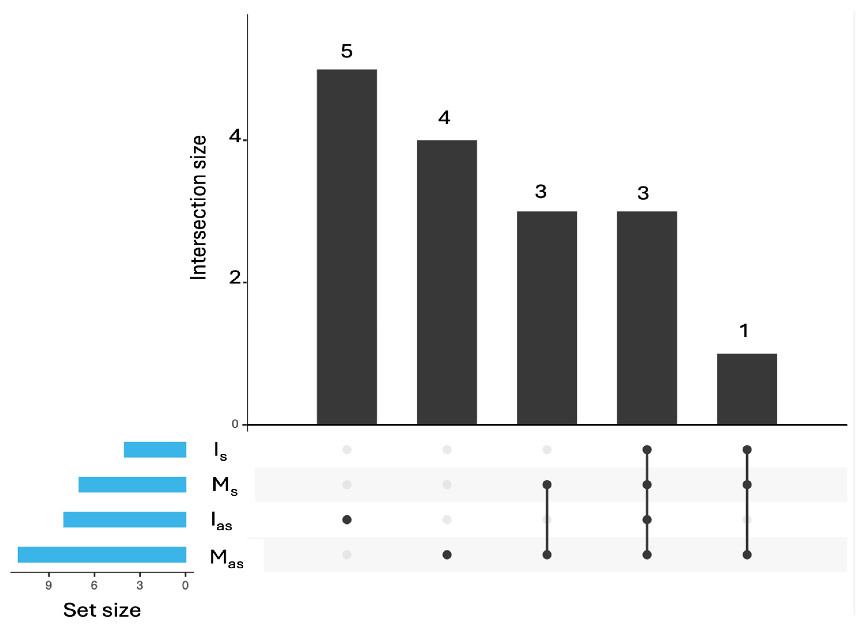
The common and unique viral species identified in the analyzed petunia samples, as revealed by the UpSet diagram (Figure 5) and Table 2.
Three viruses—CMV, TAV, and PVCV—were consistently detected across all sample types, including both symptomatic and asymptomatic plants from market and in vitro sources, highlighting their widespread presence. Beyond these common viruses, species from the families Mitoviridae (such as Duamitovirus peex 1), Partitiviridae, and Secoviridae (such as Nopovirus avii), indicating the presence of Petunia exserta mitovirus 1 and cherry leafroll virus, respectively, were shared between symptomatic and asymptomatic market plants, suggesting a common viral background in these groups. The asymptomatic market plants also harbored a small group of unique viruses, including apple mosaic virus (sequenes belonging to the species Ilarvirus ApMV), Botrytis cinerea alpha-like virus 1 (Betasclernavirus botrytidis), Botrytis cinereal mitovirus 1 (Duamitovirus boci1), and grapevine associated narnavirus-1, indicating a more diverse virome composition. Interestingly, the virome of symptomatic in vitro plants included Bacteriophage sp. shared with market-sourced groups. This suggests that in vitro culture conditions tend to reflect the virome composition of market plants, regardless of symptom development. Regarding asymptomatic in vitro plants, viral sequences corresponding to Erysiphe-associated viruses (likely mycoviruses infecting the fungal pathogen Erysiphe necator) and Caudoviricetes sp. members (bacteriophages) were detected, despite the absence of visible fungal or bacterial contamination. Their presence may be attributed to the high sensitivity of metatranscriptomic sequencing, which enables the detection of low-abundance viral sequences even in the absence of overt infection.

2.5. Phylogenetic Analysis of Plant Viruses in Bulgarian Petunia Isolates

In our study, we detected TAV, CMV, and PVCV co-infecting petunia plants in Bulgaria, providing further evidence of complex viral interactions (Table 1).
For CMV, we analyzed the coat protein (CP) gene sequence of the Bulgarian isolate (GenBank accession no. PV037061.1) in comparison with reference strains representing subgroups IA, IB, and II, as well as with CMV sequences from ornamental plants and additional Bulgarian accessions available in NCBI GenBank (Figure 6, Table S4a). Phylogenetic analysis revealed that isolate PV037061.1 clusters within CMV subgroup IA. Notably, it exhibits high genetic similarity to LN810059.1 (isolated from Citrullus lanatus in Greece, a region geographically proximate to Bulgaria), as well as to JF918966.1 and JF918964.1 (both from Vinca minor in the USA), and KU69526.1 (from Dimorphotheca caulescens in Iran).
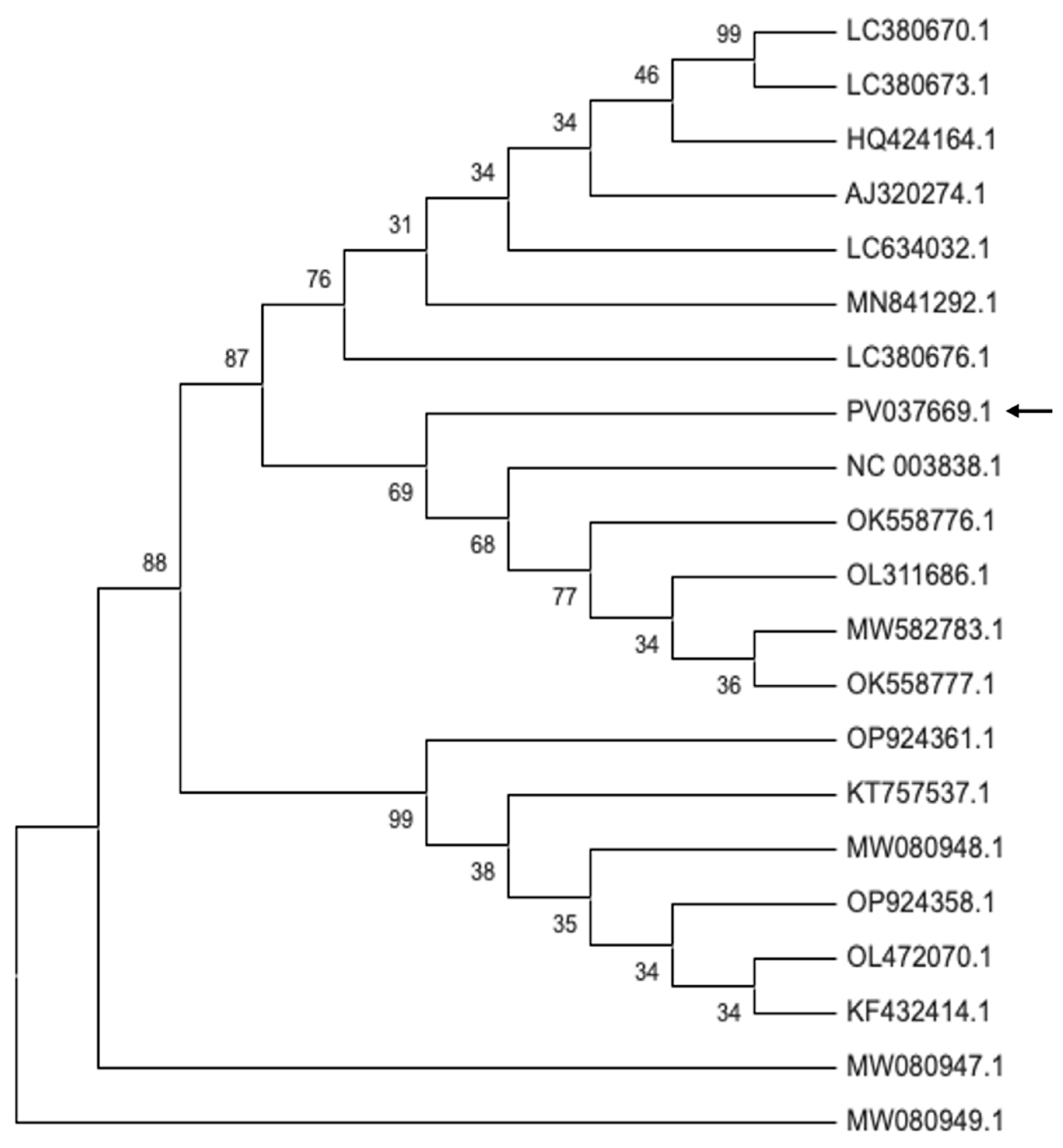
The RNA-dependent RNA polymerase (RdRp) sequence of the Bulgarian TAV isolate PV037669.1 was compared with RdRp sequences from TAV isolates infecting various Solanaceae hosts (Figure 7, Table S4b). Phylogenetic analysis showed that the petunia isolate PV037669.1 clustered within a subgroup that includes an isolate from Spain (NC_003838.1), two isolates from Canada (OK558776.1, OK558777.1), and two TAV isolates from Germany—one from chrysanthemum (OL311686.1) and one from tomato (MW582783.1).
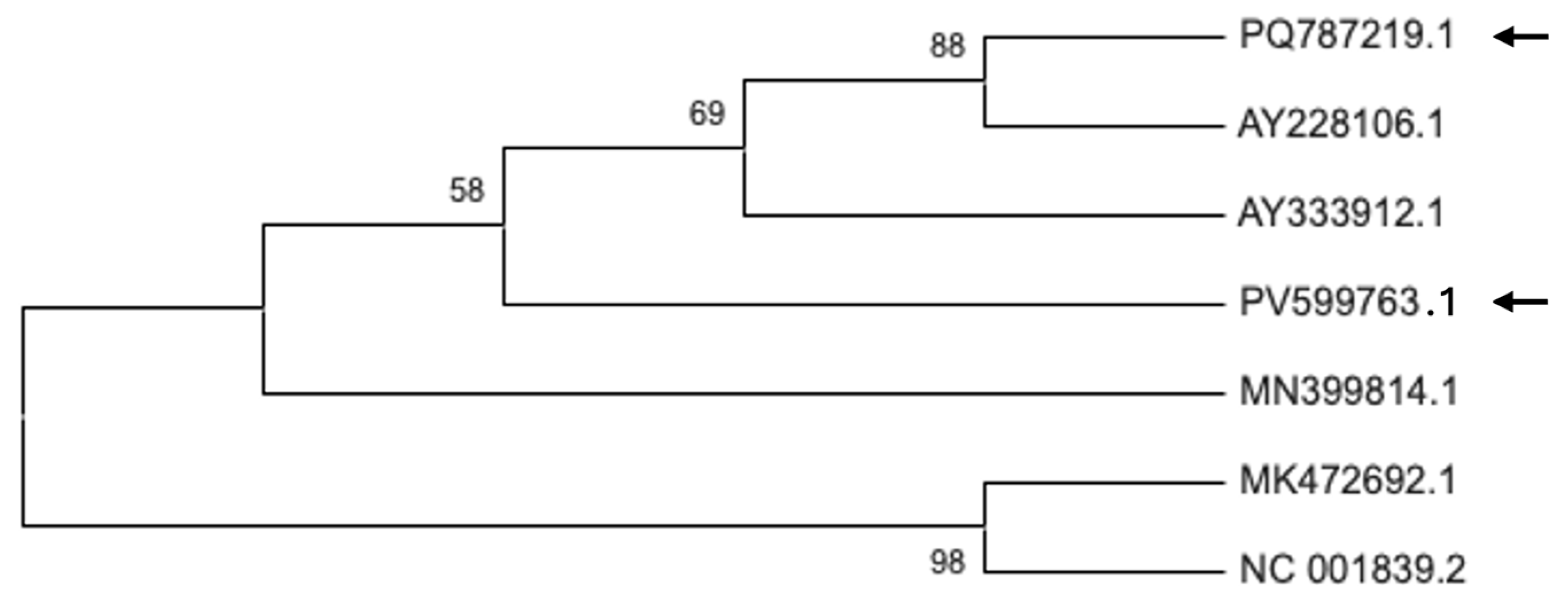
The phylogenetic relationships of the Bulgarian PVCV isolates PV599763.1 and PQ787219.1 were inferred based on the reverse transcriptase domain of the polyprotein sequence (Figure 8, Table S4c). These two isolates clustered with AY228106.1, AY333912.1 (The Netherlands), and MN399814.1 (South Korea), suggesting a shared evolutionary origin.

3. Discussion

Petunia plants are susceptible to a range of pathogens, including bacteria, viruses, fungi, and phytoplasmas; nonetheless, viral pathogens affecting the Bulgarian petunias market remain insufficiently investigated. This study aimed to characterize the virome composition, prevalence and genetic diversity of viruses affecting P. hybrida. Different factors, such as virus strain, environmental conditions, host genotype, and mixed infections, can influence the phenotypic viral appearance, and even some infected plants may remain asymptomatic [8,25]. To objectively evaluate plant physiological status, chlorophyll a fluorescence (OJIP test) was used [26]. Symptomatic plants exhibited increased baseline fluorescence (F0) and reduced maximum quantum efficiency (Fv/Fm), confirming photosynthetic impairment (Figure 2, Table S1). These findings confirm physiological disturbances in the symptomatic plants, while potential latent effects in asymptomatic ones require further investigation. Since visible phenotypic traits alone do not always reliably distinguish healthy from diseased plants, chlorophyll fluorescence provided an objective parameter to assess the physiological state and detect early disruptions in the photosynthetic apparatus.
RNA sequencing, combined with PCR/RT-PCR and Sanger sequencing, enabled the molecular detection of viruses present in the studied petunias. Three prevalent viruses were detected: two RNA viruses from the Bromoviridae family (CMV and TAV) and one DNA virus from the Caulimoviridae family (PVCV). Most market plants were co-infected with multiple viruses, which emphasized complex virus-virus and virus–host interactions (Table 1). Similar mixed infections involving CMV and TAV, in combination with other viruses, have previously been documented in the ornamental plant Hippeastrum hybridum in Bulgaria [27]. Detection of the three viruses (TAV, CMV and PVCV) in the asymptomatic petunias highlights the importance of molecular diagnostics for effective phytosanitary management, as the phenotype alone does not reliably indicate the presence of viral infections. These viruses may persist latently and maintain stable associations with the host regardless of external stressors or phenotypic expression. To our knowledge, the current study is the first on the petunia virome from the Bulgarian market.
In vitro cultivation was reported to propagate virus infection and affect the virome distribution [14,22,28], such changes can be characterized in greater detail using NGS [22]. In our study, we established in vitro cultures from both symptomatic and asymptomatic market-sourced petunia plants and applied NGS to assess changes in virome composition. We found that both CMV and PVCV were more abundant in the in vitro samples compared to the original market plants (Figure 4). This result is not unexpected, as tissue culture procedures involve repeated cutting, a process known to induce DNA demethylation at specific genomic loci—an effect previously linked to the activation of endogenous pararetroviral sequences [12,29]. Recent findings indicate that the infectious status and genetic diversity of grapevine viruses remain stable in long shoot tip cultures even after four subcultures. Notably, in vitro grapevine plants exhibited a much higher viral titer compared to their in vivo counterparts [14]. Conversely, PeexMV1 was absent in in vitro samples, suggesting possible elimination during surface sterilization for culture initiation. The detection of non-plant viruses, such as fungi- and bacteriophage-associated sequences, in asymptomatic in vitro plants highlights the complexity of plant–microbe interactions under sterile conditions. These results emphasize the need for integrated molecular screening in the established in vitro model system, as low-abundance viruses can persist without visible symptoms or overt contamination.
The phylogenetic analysis of the Bulgarian CMV isolate PV037670.1, shows that it belongs to subgroup IA. This isolate demonstrates close genetic similarity to strains from Greece, as well as to isolates from ornamental plants from the USA, the Netherlands, and Iran, indicating a broad genetic similarity among subgroup IA members across different regions and host species. The Bulgarian TAV isolate PV037669.1, clusters with isolates from Spain, Canada, and Germany, reflecting close genetic ties to TAV strains from both neighboring and distant countries, as well as from various hosts within the Solanaceae family and related ornamentals. The Bulgarian PVCV isolates PV599763.1 and PQ787219.1, group with isolates from the Netherlands and South Korea, suggesting genetic connectivity across Europe and Asia. Collectively, these findings highlight that the Bulgarian CMV, TAV, and PVCV isolates are genetically linked to strains from a wide geographic range and diverse hosts, emphasizing the potential for virus movement and genetic exchange among plant viruses across regions and species.

4. Materials and Methods

4.1. Plant Sampling

A total of 27 P. hybrida plants, both symptomatic and asymptomatic, were collected during the blooming stage from commercial nurseries in the Plovdiv region, South-East Bulgaria (42.1354° N, 24.7453° E) between 2023 and 2024. Plants were transferred to the laboratory and maintained under controlled conditions (16/8 h light/dark photoperiod at 24 °C) for one month. Two to three leaves were harvested per plant and rinsed with distilled water (ddH2O) to remove dust and epiphytic organisms. These samples were subsequently used for RNA extraction.

4.2. Chlorophyll a Fluorescence Assay and Chlorophyll Content

Chlorophyll a fluorescence induction (OJIP) curves were measured on the first fully expanded leaf of five studied plants of each group using a Plant Efficiency Analyzer (Handy-PEA, Hansatech Instruments Ltd., Norfolk, UK). Before measurement, leaf areas were dark-adapted for 40 min using leaf clips. Measurements were taken under 1 s of illumination at 3000 µmol m−2 s−1 PPFD. Data were processed using PEA Plus Software (v1.10) and interpreted following the protocols of Strasser and Strasser [30] and Goltsev and co-authors [31].
The chlorophyll content was measured using the portable chlorophyll meter CL-01 (Handy-PEA, Hansatech Instruments Ltd., Norfolk, UK) on the leaf used for chlorophyll fluorescence assessment.

4.3. Establishment of In Vitro Cultures from Axillary Buds

Young stem segments without flowers were rinsed under running tap water to remove surface debris. The large leaves were removed before sterilization, while smaller leaves were excised post-sterilization. Surface sterilization was carried out using 70% ethanol for 10 s, followed by 1% sodium hypochlorite for 30 min. Segments were then rinsed three times with sterile distilled water, dried on sterile filter paper, and cut into 15 mm explants. Explants were placed in test tubes containing sterile half-strength Murashige and Skoog (1/2 MS) medium solidified with agar [32]. Cultures were maintained at 24 ± 1 °C under a 16/8 h light/dark cycle. After four weeks, sterile plantlets were transferred to full-strength MS medium in larger vessels and subcultured every four weeks.

4.4. Long Non-Coding RNA Sequencing and Bioinformatics Analysis

Total RNA was extracted from 0.1 g of leaf tissue using the innuPREP Plant RNA Kit (Analytik Jena, Jena, Germany). RNA concentration and integrity were assessed using the Qubit 4 Fluorometer. Equimolar RNA pools were prepared from symptomatic and asymptomatic plants for sequencing. Ribosomal RNA was depleted, and libraries were prepared by Novogene Ltd., Cambridge, UK. Sequencing was performed on a NovaSeq X Plus Series (PE150) platform. Raw reads were deposited in the SRA database (accession number PRJNA1276299). Quality trimming was performed using TrimGalore (v6.7). Taxonomic classification was executed with Kraken2 (v2.1.3) based on raw read counts and employing the core_nt Database 28 December 2024. Classification outputs were visualized with Pavian (v1.0) to assess taxonomic composition. UpSet analysis was conducted using the E Venn web tool [33,34] to visualize intersections of viral taxa across sample groups. A read count threshold of >10 was applied.

4.5. PCR and RT-PCR Virus Amplification and Phylogenetic Analysis

CMV, TAV and PVCV were detected using a one-step cDNA RT-PCR kit (Genaxxon Bioscience GmbH, Ulm, Germany). Virus-specific primers are listed in Table S5. For Sanger sequencing: first-strand cDNA was synthesized from 500 ng of total RNA using the SCRIPT cDNA synthesis kit (Jena Bioscience, Jena, Germany) with random hexamer primers. PCR amplification was performed with Pfu X polymerase (Jena Bioscience GmbH, Jena, Germany) following the manufacturer’s protocol (Table S6). PCR products were purified from agarose gels using the DNA Gel Extraction Kit and subsequently sequenced by Eurofins Genomics Germany GmbH.
Phylogenetic trees were constructed for the CP of CMV, the RdRp of TAV and the polyprotein gene for PVCV. Viral sequences obtained in this study were aligned with reference sequences from NCBI using GENEIOUS software (v4.8.5, Dottmatics, Boston, MA, USA) (Table S4). Phylogenetic tree construction was performed using the Maximum Likelihood method and Tamura–Nei (1993) model [35] with 1000 bootstrap replicates for statistical support. Evolutionary analyses were conducted in MEGA12 utilizing up to 4 parallel computing threads [36,37].

5. Conclusions

Overall, our analysis revealed that viral distribution and diversity in petunia plants are not always associated with phenotypic expression. This requires integrated virome studies that take into account both the stage of individual development and the physiological state of the host, as well as environmental conditions, in order to fully understand the interactions between the plant and the virus and the dynamics of the diseases. On the other hand, our observations confirm the importance of metatranscriptome analysis for virus transmission in asymptomatic plants as well as in mixed infections. The created in vitro model system preserves the viruses detected in the original source, which allows their propagation and subsequent study.

Supplementary Materials

The following supporting information can be downloaded at: https://www.mdpi.com/article/10.3390/plants14162597/s1, Supplementary Table S1: Parameters of the rapid chlorophyll a fluorescence (OJIP test) and chlorophyll content (Chl) of asymptomatic market collected plants and symptomatic market petunia plants; Supplementary Table S2: Summary of sequencing results and classification by Pavian software (v1.0); Supplementary Table S3: Distribution of viral reads at family (a), genus (b), and species (c) ranks of Petunia hybrida samples belonging to Ia (in vitro asymptomatic); I (in vitro symptomatic); Ma (market asymptomatic); M (market, symptomatic) tested plants; Supplementary Table S4: List of viral isolates used for respective phylogenetic analyses with accession numbers, host species, and country of submission or origin for: (a) cucumber mosaic virus (b) tomato aspermy virus and (c) petunia vein clearing virus; Supplementary Table S5: Primers, used to amplify tomato aspermy virus, cucumber mosaic virus and petunia vein clearing virus in P. hybrida samples; Supplementary Table S6: RT-PCR, one and two step protocols used for the detection of cucumber mosaic virus, tomato aspermy virus and petunia vein clearing virus in P. hybrida samples.

Author Contributions

Conceptualization, M.G., R.V., D.Š., and G.Y.; methodology, R.V., E.A.-K., and L.N.; software, V.B.; validation, R.V., E.A.-K., L.N., and S.J.; data curation, E.A.-K.; writing—original draft preparation, M.G., R.V., and G.Y.; writing—review and editing, D.Š., M.G., R.V., E.A.-K., V.B., L.N., S.J., and G.Y.; visualization, V.B. and S.J.; supervision, D.Š., G.Y. and V.B.; project administration, M.G.; funding acquisition, M.G. All authors have read and agreed to the published version of the manuscript.

Funding

This research was funded by the Bulgarian National Science Fund, grant No. КП-06-H 76/10 from 2023, contract No. BG-175467353-2023-13-0151-C01.

Data Availability Statement

The raw HTS data generated in this study have been deposited in the NCBI Sequence Read Archive (SRA) under the BioProject accession number PRJNA1276299. The data are publicity available.

Conflicts of Interest

The authors declare no conflicts of interest.

References

  1. Bombarely, A.; Moser, M.; Amrad, A.; Bao, M.; Bapaume, L.; Barry, C.S.; Bliek, M.; Boersma, M.R.; Borghi, L.; Bruggmann, R.; et al. Insight into the Evolution of the Solanaceae from the Parental Genomes of P. hybrida. Nat. Plants 2016, 2, 16074. [Google Scholar] [CrossRef] [PubMed]
  2. de Vlaming, P.; Gerats, A.G.M.; Wiering, H.; Wijsman, H.J.W.; Cornu, A.; Farcy, E.; Maizonnier, D.P. hybrida: A Short Description of the Action of 91 Genes, Their Origin and Their Map Location. Plant Mol. Biol. Rep. 1984, 2, 21–42. [Google Scholar] [CrossRef]
  3. Galliot, C.; Hoballah, M.E.; Kuhlemeier, C.; Stuurman, J. Genetics of Flower Size and Nectar Volume in Petunia Pollination Syndromes. Planta 2006, 225, 203–212. [Google Scholar] [CrossRef]
  4. Morita, Y.; Hoshino, A. Recent Advances in Flower Color Variation and Patterning of Japanese Morning Glory and Petunia. Breed. Sci. 2018, 68, 128–138. [Google Scholar] [CrossRef]
  5. Cao, Z.; Guo, Y.; Yang, Q.; He, Y.; Fetouh, M.I.; Warner, R.M.; Deng, Z. Genome-Wide Identification of Quantitative Trait Loci for Important Plant and Flower Traits in Petunia Using a High-Density Linkage Map and an Interspecific Recombinant Inbred Population Derived from Petunia integrifolia and P. axillaris. Hortic. Res. 2019, 6, 27. [Google Scholar] [CrossRef]
  6. Geitmann, A. Petunia. Evolutionary, Developmental and Physiological Genetics. Ann. Bot. 2011, 107, vi–vii. [Google Scholar] [CrossRef]
  7. National Agricultural Statistics Service. Floriculture Crops 2018 Summary; USDA: Washington, DC, USA, 2019.
  8. Lesemann, D.-E. Viruses Recently Detected In Vegetatively Propagated Petunia. Acta Hortic. 1996, 88–95. [Google Scholar] [CrossRef]
  9. Hammond, J.; Huang, Q.; Jordan, R.; Meekes, E.; Fox, A.; Vazquez-Iglesias, I.; Vaira, A.M.; Copetta, A.; Delmiglio, C. International Trade and Local Effects of Viral and Bacterial Diseases in Ornamental Plants. Annu. Rev. Phytopathol. 2023, 61, 73–95. [Google Scholar] [CrossRef]
  10. Boonham, N.; Hims, M.; Barker, I.; Spence, N. Potato Virus Y from Petunia Can Cause Symptoms of Potato Tuber Necrotic Ringspot Disease (PTNRD). Eur. J. Plant Pathol. 1999, 105, 617–621. [Google Scholar] [CrossRef]
  11. Chung, B.-N.; Kim, J.-S.; Cho, J.-D.; Cheong, S.-R.; Jeong, M.-I. Tobacco Mosaic Virus Detected in Vegetatively Propagated Petunia Hybrids “Surfinia”. Plant Pathol. J. 2007, 23, 34–36. [Google Scholar] [CrossRef]
  12. Richert-Poggeler, K.R. Induction of Infectious Petunia Vein Clearing (Pararetro) Virus from Endogenous Provirus in Petunia. EMBO J. 2003, 22, 4836–4845. [Google Scholar] [CrossRef] [PubMed]
  13. Tollenaere, C.; Susi, H.; Laine, A.-L. Evolutionary and Epidemiological Implications of Multiple Infection in Plants. Trends Plant Sci. 2016, 21, 80–90. [Google Scholar] [CrossRef]
  14. Hu, G.; Dong, Y.; Zhang, Z.; Fan, X.; Ren, F. Effect of In vitro Culture of Long Shoot Tip on Variant Structure and Titer of Grapevine Viruses. Plants 2022, 11, 1907. [Google Scholar] [CrossRef] [PubMed]
  15. White, P.R. Multiplication of the Viruses of Tobacco and Aucuba Mosaics in Growing Excised Tomato Root-Tips. Phytopathology 1934, 24, 1003–1011. [Google Scholar]
  16. Marton, L.; Duran-Vila, N.; Lin, J.J.; Semancik, J.S. Properties of Cell Cultures Containing the Citrus Exocortis Viroid. Virology 1982, 122, 229–238. [Google Scholar] [CrossRef]
  17. Lin, J.J.; Semancik, J.S. Coordination between Host Nucleic Acid Metabolism and Citrus Exocortis Viroid Turnover. Virus Res. 1985, 3, 213–230. [Google Scholar] [CrossRef]
  18. Mühlbach, H.-P.; Sänger, H.L. Continous Replication of Potato Spindle Tuber Viroid (PSTV) in Permanent Cell Cultures of Potato and Tomato. Biosci. Rep. 1981, 1, 79–87. [Google Scholar] [CrossRef]
  19. Wang, M.C.; Lin, J.J.; Duran-Vila, N.; Semancik, J.S. Alteration in Cell Wall Composition and Structure in Viroid-Infected Cells. Physiol. Mol. Plant Pathol. 1986, 28, 107–124. [Google Scholar] [CrossRef]
  20. Haegeman, A.; Foucart, Y.; De Jonghe, K.; Goedefroit, T.; Al Rwahnih, M.; Boonham, N.; Candresse, T.; Gaafar, Y.Z.A.; Hurtado-Gonzales, O.P.; Kogej Zwitter, Z.; et al. Looking beyond Virus Detection in RNA Sequencing Data: Lessons Learned from a Community-Based Effort to Detect Cellular Plant Pathogens and Pests. Plants 2023, 12, 2139. [Google Scholar] [CrossRef]
  21. Pecman, A.; Kutnjak, D.; Gutiérrez-Aguirre, I.; Adams, I.; Fox, A.; Boonham, N.; Ravnikar, M. Next Generation Sequencing for Detection and Discovery of Plant Viruses and Viroids: Comparison of Two Approaches. Front. Microbiol. 2017, 8, 1998. [Google Scholar] [CrossRef] [PubMed]
  22. Bömer, M.; Rathnayake, A.I.; Visendi, P.; Sewe, S.O.; Sicat, J.P.A.; Silva, G.; Kumar, P.L.; Seal, S.E. Tissue Culture and Next-Generation Sequencing: A Combined Approach for Detecting Yam (Dioscorea Spp.) Viruses. Physiol. Mol. Plant Pathol. 2019, 105, 54–66. [Google Scholar] [CrossRef] [PubMed]
  23. Maclot, F.; Candresse, T.; Filloux, D.; Malmstrom, C.M.; Roumagnac, P.; van der Vlugt, R.; Massart, S. Illuminating an Ecological Blackbox: Using High Throughput Sequencing to Characterize the Plant Virome Across Scales. Front. Microbiol. 2020, 11, 578064. [Google Scholar] [CrossRef]
  24. Ismajli, B.; Galbács, Z.N.; Takács, A.P.; Várallyay, É. The First High-Throughput Sequencing-Based Study of Viruses Infecting Solanaceous Crops in Kosovo Reveals Multiple Infections in Peppers by Six Plant Viruses. Plants 2025, 14, 1273. [Google Scholar] [CrossRef] [PubMed]
  25. Sanchez-Cuevas, M.-C.; Nameth, S.G.P. Virus-Associated Diseases of Double Petunia: Frequency and Distribution in Ohio Greenhouses. HortScience 2002, 37, 543–546. [Google Scholar] [CrossRef]
  26. Strasser, R.J.; Srivastava, A.; Tsimilli-Michael, M. The Fluorescence Transient as a Tool to Characterize and Screen Photosynthetic Samples. In Probing Photosynthesis: Mechanism, Regulation & Adaptation; Yunus, M., Pathre, U., Mohanty, P., Eds.; Taylor & Francis Group: London, UK, 2000; pp. 443–480. [Google Scholar]
  27. Bakardjieva, N.; Denkova, S.; Hristova, D.; Hristoval, D. Tomato Spotted Wilt Virus on Ornamental Species in Bulgaria. Biotechnol. Biotechnol. Equip. 1998, 12, 49–52. [Google Scholar] [CrossRef]
  28. Shih, S.M.-H.; Doran, P.M. In vitro Propagation of Plant Virus Using Different Forms of Plant Tissue Culture and Modes of Culture Operation. J. Biotechnol. 2009, 143, 198–206. [Google Scholar] [CrossRef]
  29. Noreen, F.; Akbergenov, R.; Hohn, T.; Richert-Pöggeler, K.R. Distinct Expression of Endogenous Petunia Vein Clearing Virus and the DNA Transposon DTph1 in Two P. hybrida Lines Is Correlated with Differences in Histone Modification and SiRNA Production. Plant J. 2007, 50, 219–229. [Google Scholar] [CrossRef]
  30. Strasser, B.J.; Strasser, R.J. Measuring Fast Fluorescence Transients to Address Environmental Questions: The JIP-Test. In Photosynthesis: From Light to Biosphere; Springer: Dordrecht, The Netherlands, 1995; pp. 4869–4872. [Google Scholar]
  31. Goltsev, V.N.; Kalaji, H.M.; Paunov, M.; Bąba, W.; Horaczek, T.; Mojski, J.; Kociel, H.; Allakhverdiev, S.I. Variable Chlorophyll Fluorescence and Its Use for Assessing Physiological Condition of Plant Photosynthetic Apparatus. Russ. J. Plant Physiol. 2016, 63, 869–893. [Google Scholar] [CrossRef]
  32. Murashige, T.; Skoog, F. A Revised Medium for Rapid Growth and Bio Assays with Tobacco Tissue Cultures. Physiol. Plant 1962, 15, 473–497. [Google Scholar] [CrossRef]
  33. Yang, M.; Chen, T.; Liu, Y.-X.; Huang, L. Visualizing Set Relationships: EVenn’s Comprehensive Approach to Venn Diagrams. iMeta 2024, 3, e184. [Google Scholar] [CrossRef]
  34. Khan, A.; Mathelier, A. Intervene: A Tool for Intersection and Visualization of Multiple Gene or Genomic Region Sets. BMC Bioinform. 2017, 18, 287. [Google Scholar] [CrossRef] [PubMed]
  35. Tamura, K.; Nei, M. Estimation of the Number of Nucleotide Substitutions in the Control Region of Mitochondrial DNA in Humans and Chimpanzees. Mol. Biol. Evol. 1993, 10, 512–526. [Google Scholar] [CrossRef] [PubMed]
  36. Stecher, G.; Tamura, K.; Kumar, S. Molecular Evolutionary Genetics Analysis (MEGA) for MacOS. Mol. Biol. Evol. 2020, 37, 1237–1239. [Google Scholar] [CrossRef]
  37. Kumar, S.; Stecher, G.; Suleski, M.; Sanderford, M.; Sharma, S.; Tamura, K. MEGA12: Molecular Evolutionary Genetic Analysis Version 12 for Adaptive and Green Computing. Mol. Biol. Evol. 2024, 41, msae263. [Google Scholar] [CrossRef] [PubMed]
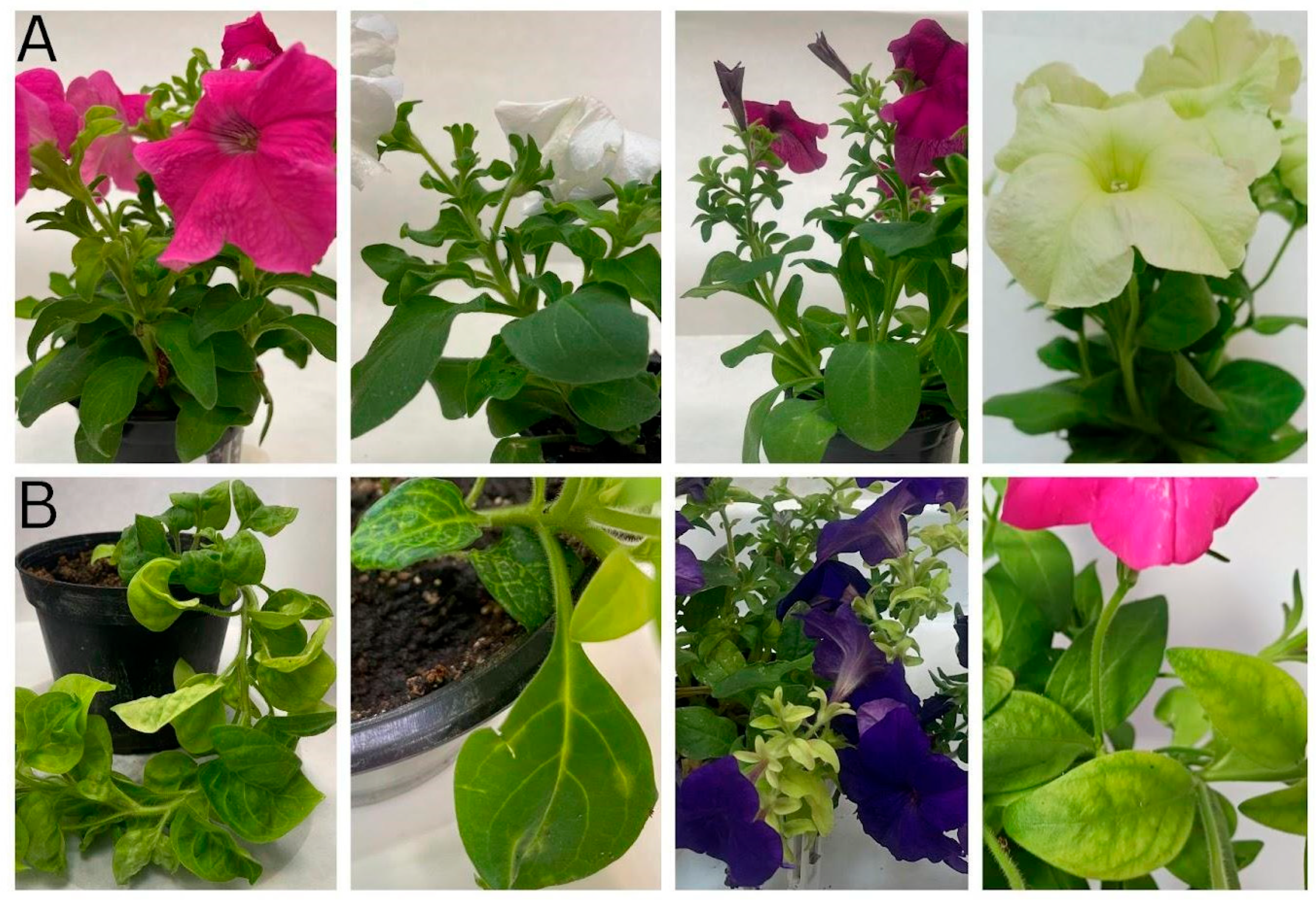
Figure 1. The phenotype of selected asymptomatic (A) and symptomatic (B) petunias collected from the Bulgarian market.
Plants 14 02597 g001
Figure 2. OJIP chlorophyll a fluorescence induction curves. Blue line—asymptomatic market collected plants, red line—symptomatic market plants.
Plants 14 02597 g002
Figure 3. In vitro-derived P. hybrida plantlets: (A) Asymptomatic; (B) Symptomatic plants.
Plants 14 02597 g003
Figure 4. Classification of viral sequences detected in petunia samples illustrated by Sankey diagrams. The classification follows hierarchical taxonomic levels from kingdom to species. Mas = asymptomatic market; Ms = symptomatic market; Ias = asymptomatic in vitro; Is = symptomatic in vitro. In the figure, the virus names were generated automatically by the program according to the Kraken database, so some viruses are represented erroneously by their non-italicized species names; petunia vein clearing virus (PVCV) is represented as Petuvirus venapetuniae.
Plants 14 02597 g004aPlants 14 02597 g004b
Figure 5. UpSet plot showing shared and unique viruses across symptomatic and asymptomatic petunia samples from different sources. Mas = asymptomatic market; Ms = symptomatic market; Ias = asymptomatic in vitro; Is = symptomatic in vitro.
Plants 14 02597 g005
Figure 6. The phylogeny of Bulgarian cucumber mosaic virus isolates from P. hybrida. The phylogenetic tree was constructed using the Maximum Likelihood method and Tamura–Nei model, conducted in MEGA 12. The analytical procedure encompassed 33 nucleotide sequences with 428 positions of the CP gene in the final dataset. The percentage of replicate trees in which the associated taxa clustered together (1000 replicates) is shown above the branches. The arrow indicates the analyzed Bulgarian accession available in NCBI GenBank.
Plants 14 02597 g006
Figure 7. Molecular phylogenetic analysis of Bulgarian tomato aspermy virus isolates from P. hybrida. The phylogenetic tree was constructed using Maximum Likelihood method and Tamura–Nei model based on 21 nucleotide sequences with 947 positions of the RdRp gene in the final dataset. Evolutionary analysis was conducted in MEGA12. The percentage of replicate trees in which the associated taxa clustered together (1000 replicates) is shown above the branches. The arrow indicates the analyzed Bulgarian accession available in NCBI GenBank.
Plants 14 02597 g007
Figure 8. The phylogeny of Bulgarian petunia vein clearing virus isolates from P. hybrida. The phylogenetic tree was constructed using Maximum Likelihood method and Tamura–Nei model based on 7 nucleotide sequences with 654 positions of the polyprotein gene in the final dataset. Evolutionary analyses were conducted in MEGA12. The percentage of replicate trees in which the associated taxa clustered together (1000 replicates) is shown next to the branches. The arrows indicate the analyzed Bulgarian accessions available in NCBI GenBank.
Plants 14 02597 g008
Table 1. Identification of plant viruses in individual petunias by PCR/RT-PCR.
Market In Vitro
Sympt.
plants
41 Ms42 Ms43 Ms47 Ms48 Ms49 Ms41 Is42 Is43 Is47 Is48 Is49 Is
TAV ++++++
CMV ++++++++++
PVCV +++++++++++
MarketIn vitro
Asympt. plants 31 Mas33 Mas35 Mas40 Mas51 Mas53 Mas31 Ias33 Ias35 Ias40 Ias51 Ias53 Ias
TAV ++++
CMV ++++++++++++
PVCV +++++++++++
Mas = asymptomatic market; Ms = symptomatic market; Ias = asymptomatic in vitro; Is = symptomatic in vitro.
Table 2. Intersection of virus distribution between petunia sample groups. Shared virus distributions among different petunia sample groups were analyzed using UpSet plots to visualize the intersections; virus names are presented as listed in the Kraken database.
IntersectionViruses
Ias & Is & Mas & Mscucumber mosaic vírus; tomato aspermy vírus; petunia vein clearing vírus
Mas & MsPetunia exserta mitovirus 1, * Partitiviridae sp.; cherry leaf roll virus
Is & Mas & Ms* Bacteriophage sp.
Masapple mosaic virus; Botrytis cinerea alpha-like virus 1; Botrytis cinerea mitovirus 1; grapevine associated narnavirus-1
IasErysiphe necator associated partitivirus 2; Erysiphales ourmia-like virus 2; Erysiphe necator ourmia-like virus 82; Erysiphe necator associated ourmia-like virus 82; * Caudoviricetes sp.
Mas = asymptomatic market; Ms = symptomatic market; Ias = asymptomatic in vitro; Is = symptomatic in vitro. * Viruses with not only plant hosts (Partitiviridae sp.) and bacteriophages are represented in the virome analyses software outputs as species in a certain genus or a family (Bacteriophage sp., Caudoviricetes sp., respectively) are listed in the table as in the original outputs.
Disclaimer/Publisher’s Note: The statements, opinions and data contained in all publications are solely those of the individual author(s) and contributor(s) and not of MDPI and/or the editor(s). MDPI and/or the editor(s) disclaim responsibility for any injury to people or property resulting from any ideas, methods, instructions or products referred to in the content.